Báo cáo y học: "Importance of lysosomal cysteine proteases in lung disease" ppt
Bạn đang xem bản rút gọn của tài liệu. Xem và tải ngay bản đầy đủ của tài liệu tại đây (228.5 KB, 8 trang )
Review
Importance of lysosomal cysteine proteases in lung disease
Paul J Wolters and Harold A Chapman
Department of Medicine and Cardiovascular Research Institute, University of California,
San Francisco, California, USA
Abstract
The human lysosomal cysteine proteases are a family of 11 proteases whose members
include cathepsins B, C, H, L, and S. The biology of these proteases was largely ignored for
decades because of their lysosomal location and the belief that their function was limited to
the terminal degradation of proteins. In the past 10 years, this view has changed as these
proteases have been found to have specific functions within cells. This review highlights
some of these functions, specifically their roles in matrix remodeling and in regulating the
immune response, and their relationship to lung diseases.
Keywords: asthma, cathepsin, emphysema, extracellular matrix, invariant chain
Received: 10 October 2000
Revisions requested: 7 November 2000
Revisions received: 10 November 2000
Accepted: 10 November 2000
Published: 20 November 2000
Respir Res 2000, 1:170–177
The electronic version of this article can be found online at
/>© Current Science Ltd (Print ISSN 1465-9921; Online ISSN 1465-993X)
APC, antigen-presenting cell; CLIP = class II-associated invariant chain peptide; DPPI = dipeptidyl peptidase I; Ii = invariant chain; IL = interleukin;
LHVS = leucyl-homophenylalanine-vinylsulfone; MHC = major histocompatibility complex; SNARE = soluble N-ethylmaleimide-sensitive factor-
attachment protein receptor; t-SNARE = SNARE on target membrane; v-SNARE = SNARE on vesicle.
/>Introduction
Members of the papain family of cysteine proteases are
found predominantly within the endosomal and lysosomal
compartment of cells. It was initially believed that they
were ‘housekeeping’ genes and that they functioned
exclusively as the cell’s garbage disposals, terminally
degrading unwanted, abnormal, or endocytosed proteins.
Recently this view has evolved as members of the family
have been found to have distinctive patterns of expression
(Table 1), have regulated expression, have important roles
in specific biologic processes [1,2], and have been linked
to inherited genetic diseases [3–5].
The first members of the papain family of cysteine pro-
teases included cathepsins B, C, H, L, and S. During the
past ten years six new members have been added, giving
11 (Table 1). Cathepsins B, C, F, H, O, and Z are constitu-
tively expressed in most tissues. Although widely
expressed, some of these proteases are found in signifi-
cantly greater quantities in specific cells within tissues.
Examples include cathepsin C (better known as dipeptidyl
peptidase I or DPPI) (found in the greatest amounts in
cytotoxic T lymphocytes [6], macrophages [7] and mast
cells [8]), cathepsin K (osteoclasts, airway epithelium)
[9,10], cathepsin S [antigen-presenting cells (APCs)], and
cathepsin W (CD8
+
T cells) [11].
Structurally, members of the papain family of cysteine pro-
teases consist of two domains folded together in a
V-shaped configuration. At the bottom of the V, a cysteine
and a histidine residue form the catalytic diad [12].
Although their overall topographical structure is similar,
each cathepsin has unique features that confer specific
proteolytic activity on the enzyme. Cathepsins B and Z
have a peptide loop overlying their active site that binds
the C-terminus of proteins, making these cathepsins
carboxypeptidases [13,14]. Cathepsin H is an amino-
peptidase because a residual eight amino acids of the
propeptide allows only the amino-terminal amino acid of a
protein to access the active site [15]. The aminodipeptidase
/>commentary
review
reports primary research
DPPI is an oligomer and has a residual propeptide that
probably blocks its active site as well [16]. The endopepti-
dases cathepsins F, K, L, O, S, V, and W each have
unique amino acids near the active site that confer their
substrate specificity [12]. These unique structural features
and patterns of expression suggest that the enzymes have
specific roles in the cells and tissues in which they are
expressed. Two examples, which are the focus of this
review, are their role in matrix remodeling and the regula-
tion of the immune response.
Matrix remodeling by cathepsins
Many of the lysosomal cysteine proteases can degrade
components of the extracellular matrix. An example of how
well these enzymes degrade matrix components is their
ability to hydrolyze elastin, a protein notoriously resistant to
proteolysis. In fact, cathepsins K, L, and S are among the
most potent elastases known [1]. In vitro, cathepsin B
reportedly degrades collagen type IV and X and fibronectin
[17,18]. Cathepsins L, S, and K degrade fibrillar collagens
[19], fibronectin, and laminin, and DPPI cleaves fibronectin
and collagens type I, III, and IV [20]. One basic requirement
for matrix degradation in vivo is that the proteases must
encounter the matrix molecule in a microenvironment in
which the protease maintains its activity. This might occur
intracellularly on phagocytosed matrix molecules, or extra-
cellularly after secretion of the lysosomal cathepsin.
Intracellular matrix degradation by cathepsins
The extracellular matrix of most tissues contains a vast
network of different collagens. This mixture of collagens is
not static; rather they are subjected to continuous degrada-
tion and turnover. For complete degradation, several pro-
teases can act in concert both extracellularly and
intracellularly [18,21]. Extracellularly, collagens can be
degraded by collagenase, gelatinases A and B, stromelysin,
and the cathepsins. Extracellular degradation of collagen
can be incomplete, leaving fragments to be phagocytosed
by cells such as fibroblasts, macrophages and smooth
muscle cells [22]. Within these cells, the collagen-contain-
ing phagosome fuses with lysosomes, in which cathepsins
complete the degradation of the collagen molecules.
This process of collagen phagocytosis and degradation
can be regulated by hormones, cytokines and growth
factors. Studies with periosteal fibroblasts have demon-
strated that interleukin-1α and cortisol decrease the
uptake of fibrillar collagen, whereas transforming growth
factor-β enhances phagocytosis [22,23]. Furthermore, a
decrease in collagen breakdown products is found in the
culture medium when collagen phagocytosis and intracel-
lular collagen digestion are reduced.
A disease that illustrates the importance of intracellular
collagen degradation is pycnodysostosis. Pycnodysosto-
sis is an autosomal recessive disease caused by muta-
tions of the gene encoding cathepsin K and is
characterized by osteosclerosis, short stature, bone
fragility, clavicular dysplasia, and skull deformities [4].
These mutations result in an absence of cathepsin K activ-
ity and inadequate intracellular degradation of the organic
bone matrix. This is demonstrated by ultrastructural exami-
nation of osteoclasts from affected individuals, showing
vacuoles containing undigested collagen fibrils [24].
Using pycnodysostosis as a model, it is reasonable to
propose that a loss of cathepsin activity in resident lung
cells might contribute to pathologic lung diseases, such as
idiopathic pulmonary fibrosis, where decreased collagen
Table 1
Human acidic cathepsins
Cathepsin Tissue expression Chromosome Human disease*/mouse phenotype
†
B Widespread 8
†
Reduced apoptosis
L Widespread 9
†
Defective CD4 selection, hair loss
V Thymic epithelium 9 –
K Osteoclasts, bronchial epithelium 1q21 *Pycnodysostosis
S Antigen-presenting cells 1q21
†
Defective antigen presentation
H Widespread 15 –
W CD8
+
T cells 11q13 –
F Macrophages, ?widespread 11q13 –
C Myeloid cells, ?others 11q14 *Hyperkeratosis, periodontitis
O Widespread 4q31-32 –
Z Widespread 20q13 –
Respiratory Research Vol 1 No 3 Wolters and Chapman
phagocytosis and intracellular digestion lead to the build-
up of collagen fibers in the extracellular space, favoring
tissue fibrosis.
Extracellular matrix degradation by cathepsins
In addition to the intracellular degradation of collagens,
cathepsins can also degrade matrix proteins extracellu-
larly. Before this action, the cathepsins must first be
released into the extracellular space. Cells found to
release cathepsins include macrophages (cathepsins B, L,
S, and K) [25], mast cells (cathepsin L and DPPI) [8],
smooth muscle cells (cathepsins S and K) [26], fibroblasts
(cathepsin B), and tumor cells (cathepsins B, L, and S)
[27]. The two major mechanisms of release are altered
trafficking of newly formed enzyme and regulated release
from endosomes and lysosomes.
Cathepsins are synthesized in the endoplasmic reticulum
as pre-proproteins consisting of a signal peptide, a
propeptide and a catalytic region of the enzyme. The
signal peptide serves to target the cathepsin to the Golgi
apparatus, where it is glycosylated with high-mannose car-
bohydrates. These carbohydrates bind to one of the two
mannose-6-phosphate receptors and the complex is trans-
ported to the prelysosomal compartment, where the acidic
environment causes dissociation of the enzyme–receptor
complex and activation of the enzyme. In some disease
states, a decrease in affinity for, or number of, mannose-6-
phosphate receptors can result in mistrafficking and
secretion of cathepsins. Examples include the observation
that pro-cathepsin B released by some tumor cells has a
different glycosylation pattern from that of control cells
[28], and that a decrease in the number of mannose-6-
phosphate receptors in transformed mouse squamous-cell
carcinoma cells results in the secretion of cathepsin B
[29]. However, there are probably alternative explanations
for the mistrafficking of cathepsins, because recent
reports have suggested that mechanisms independent of
mannose-6-phosphate exist for the targeting of cathepsins
to lysosomes [30].
The regulated release of lysosomal contents is a second
mechanism by which cathepsins can be secreted from
cells. Many cells of hematopoietic origin (T-cells, neu-
trophils, and mast cells) have secretory granules that are
released in a regulated manner when their contents are
needed to destroy target cells (T-cells), or to control a
bacterial (neutrophil) or parasitic (mast cell) infection.
These cells also have granules that can be identified as
lysosomes by the presence of lysosomal markers (for
example, lysosomal-associated membrane protein [LAMP]
and vesicle-associated membrane protein [VAMP]-2 or
cathepsins). In mature hematopoietic cells, many granules
contain both lysosomal and secretory markers and seem
to have dual functions (that is, secretory lysosomes) [31].
Functionally, these doubly labeled granules act as lyso-
somes and secretory granules, and release both their
secretory and lysosomal constituents when the cells are
activated to do so. Examples include the release of DPPI
by natural killer cells and mast cells [8,32], and cathep-
sins B, L, K, and S by macrophages [25].
The secretion of lysosomal contents does not seem to be
limited to ‘secretory lysosomes’ of hematopoietic cells and
might be a feature of lysosomes in other cells. This is sup-
ported by the recent observation that smooth muscle cells
stimulated with interferon-γ synthesize and secrete
cathepsin S [26]. Similarly, the activation of fibroblasts by
calcium ionophore causes the release of lysosomal β-hex-
osaminadase [33]. Thus, the regulated secretion of lyso-
somes is a feature of many cells and might represent a
primitive secretory function in these cells.
The observation that lysosomal contents are released by
increasing the intracellular concentration of Ca
2+
ions is
intriguing and might provide a clue to the mechanism of
how this occurs. An understanding of how secretory gran-
ules are released has been developing for several years
[34]. One of the basic mechanisms involved is the interac-
tion of proteins integrated into the membrane of secretory
vesicles (v-SNAREs; SNARE stands for soluble N-ethyl-
maleimide-sensitive factor-attachment protein receptor)
with proteins integrated into the target cell membrane
(t-SNAREs). The interaction of these proteins seems to
promote membrane fusion by bringing the secretory
vesicle into close apposition with the outer cell membrane.
An example of this phenomenon is the Ca
2+
-dependent
release of neurotransmitters triggered by the interaction of
the v-SNARE synaptotagmin I with the t-SNAREs syntaxin
and SNAP-25 (in which SNAP stands for soluble N-ethyl-
maleimide-sensitive fusion protein attachment protein)
[35]. After depolarization, intracellular concentrations of
Ca
2+
ions increase within the nerve. Ca
2+
ions then bind
to Ca
2+
-binding regions on synaptotagmin I (C2-domains)
[36], giving the molecule a more positive charge and facili-
tating an interaction with the negatively charged
t-SNAREs (for example, syntaxin 1) and phospholipids on
the cell surface.
Of the 11 members of the synaptotagmin family, synapto-
tagmins I, II, III, V, and X are expressed exclusively in the
nervous system [35]. The others are expressed ubiqui-
tously, suggesting that they have functions that are more
general in non-neuronal cells. One possibility is that, simi-
larly to the regulation of neurotransmitter release by synap-
totagmin I, specific synaptotagmins might also regulate
the release of lysosomal constituents from non-neuronal
cells. This is supported by a recent study reporting that
synaptotagmin VII regulates the Ca
2+
-dependent exocyto-
sis of lysosomes from normal rat fibroblasts [37]. Lysoso-
mal constituents, including cathepsins, can therefore be
released from many cells, and this exocytosis might be
regulated by v-SNAREs and t-SNAREs, including the
synaptotagmins.
Most cathepsins have optimal activity at an acidic pH and
lose their activity quickly at a neutral pH (exceptions
include cathepsin S and DPPI) [8,38]. Consequently, to
maintain their activity extracellularly, the cathepsins must
also be released into an acidic environment. This might
occur in pathologic conditions, such as pyogenic infections
or malignancy, which are known to be associated with
acidic extracellular environments. In these disease states,
the acidic environment is due to several factors associated
with the disease process as a whole rather than an individ-
ual group of cells that release the cathepsins.
In other circumstances, cells releasing cathepsins might
promote extracellular proteolysis by directly acidifying the
pericellular space in which the cathepsins are released.
One example, reported by Punturieri et al [25], is the acidi-
fication of the pericellular environment by macrophages
during elastinolysis. In vitro, monocyte-derived macro-
phages adhere tightly to elastin particles and form a
sequestered environment between the cell membrane and
the elastin particle to be degraded. The macrophage then
acidifies this pericellular space by using a vacuolar type
H
+
-ATPase to pump protons from the cytoplasmic space
to the extracellular space. Concurrently, the macrophage
releases elastinolytic cathepsins L, S, and K into the acidic
microenvironment, where they can degrade the elastin.
Furthermore, these tight junctions might also promote
extracellular proteolysis by cathepsins in vivo by prevent-
ing the interaction of secreted cathepsins with cysteine
protease inhibitors, such as cystatin C, that are found in all
tissues and body fluids.
Lung cancer or chronic inflammatory conditions such as
asthma, emphysema, and idiopathic pulmonary fibrosis are
lung diseases in which the regulated secretion of lysoso-
mal cathepsins might be important in disease progression.
In lung cancer, degradation of the stroma surrounding
tumors by cathepsins might promote the growth and
metastasis of lung cancer. This is suggested by findings in
vitro that non-small cell lung carcinomas secrete cathep-
sins B and L [27], that squamous cell carcinomas can
invade matrigel (a surrogate of extracellular matrix), and
that this invasion can be inhibited by heterologous expres-
sion of the cysteine protease inhibitor cystatin C [39].
Although data in vivo supporting a role for cathepsins in
tumor progression are lacking, the study of tumor models
in cathepsin knockout mice should provide more definitive
answers in the future.
Emphysema is characterized by the proteolytic degrada-
tion of lung extracellular matrix, especially lung elastin. The
elastinolytic cysteine proteases cathepsins K, L, and S
might be important in this process [40]. As discussed
above, two highly abundant cell types in the lung,
macrophages and smooth muscle cells, can synthesize
and secrete cathepsins K, L, and S, which might then
degrade lung elastin. Recent studies suggest novel mech-
anisms by which this might occur. Elias and colleagues
have established transgenic murine models of inducible
expression of cytokines along alveolar surfaces (with the
murine CC10 promoter). Remarkably, the induction of
interleukin-13 (IL-13) overexpression in mice six to eight
weeks old results in alveolar space enlargement and a
loss of alveolar attachment sites, morphological hallmarks
of emphysema, on a timescale of weeks to months [41].
Increased levels of both active metalloproteases and cys-
teine proteases develop along the alveolar surfaces and
presumably in lung tissues. Mice given the cysteine pro-
tease inhibitor E64 or leupeptin have markedly attenuated
emphysematous changes, implying an important role for
cysteine proteases in IL-13-induced emphysema. It is
noteworthy that matrix metalloprotease inhibitors also
attenuated the process, indicating the probable involve-
ment of multiple enzyme systems.
The fact that these cytokines affect both mesenchymal
and hematopoietic cells suggests that not only
macrophages but also multiple cells in the cytokine-
exposed lung might contribute to tissue cathepsin activity,
underscoring the complexity of matrix remodeling in this
disorder. Whether IL-13 or other cytokines already shown
to induce the secretion of elastinolytic cathepsins (for
example, interferon-γ) promote emphysema and COPD in
cigarette smokers remains to be established.
Innate immunity
Lysosomal cysteine proteases can be important for the
regulation of innate immunity. An example is the activation
of granule-associated serine proteases (namely, neutrophil
elastase, cathepsin G, granzymes A and B, and mast cell
chymase) by DPPI. These enzymes are synthesized as
proproteins with a two-residue propeptide (or activation
dipeptide) that maintains them in an inactive conformation.
Proteolytic removal of the activation dipeptide induces a
conformational change and activation of the serine pro-
tease. The activation dipeptides of the granule-associated
serine proteases are similar, suggesting that they might be
removed by the same protease or proteases [42].
Because of DPPI’s amino-terminal dipeptidase activity, it
was a logical candidate for an activator of these serine
proteases. By using a DPPI-specific inhibitor, or recombi-
nant proenzymes, it was shown that DPPI could activate
neutrophil elastase, cathepsin G, granzymes A and B, and
mast cell chymase in vitro [42,43]. To test this possibility
in vivo and to determine whether other proteases could
compensate for DPPI’s activity, DPPI knockout mice were
generated [44]. In characterizing the protease activity in
leukocytes of these animals, it was found that DPPI is
essential for the activation of granzymes A and B.
/>commentary
review
reports primary research
This absence of DPPI activity, and consequently serine
protease activity, has been shown to have important bio-
logic consequences in both mice and humans. By activat-
ing granzymes A and B, DPPI might regulate the
lymphocyte-mediated cytotoxicity of virally infected or
malignant cells [45]. Cytotoxic T lymphocytes and natural
killer cells eradicate abnormal cells by the simultaneous
release of granzymes A and B and perforin. Perforin forms
a pore in the target cell through which the granzymes
pass. Once inside the cell, the granzymes trigger apopto-
sis directly by activating the caspase cascade (granzyme
B) or by other less well defined processes (granzyme A).
DPPI might also have a role in defense against Gram-neg-
ative bacterial infections in mice by activating neutrophil
elastase. Neutrophil elastase then destroys Gram-negative
bacteria by hydrolyzing outer-membrane protein A on their
cell walls [46,47]. Therefore, by regulating the activity of
serine proteases DPPI is important in the primary host
defense against both bacterial and viral infections in mice.
DPPI also seems to be important for the primary host
defense in humans. This is suggested by recent findings
that patients with Papillon–Lefèvre syndrome have muta-
tions of the gene encoding DPPI and an absence of DPPI
activity [3,5]. Papillon–Lefèvre syndrome is a disease
characterized by early periodontitis, skin hyperkeratosis
and a predisposition to bacterial infections such as pneu-
monia, liver abscesses, and furuncles. Although exact
explanations for these phenotypic features are currently
unknown, findings in the DPPI knockout mice suggest that
the general susceptibility to bacterial infections may be
due to decreased amounts of neutrophil elastase and
cathepsin G activities [44]. Furthermore, periodontitis
might also be due to a subclinical infection. The pathogen-
esis of the hyperkeratosis is unexplained but suggests that
DPPI might have a role in cell growth or in matrix degradation.
Adaptive immunity
Endosomal proteolysis directs the efficiency and character
of major histocompatibility complex (MHC) class II-depen-
dent antigen presentation by fulfilling two important roles:
generation of antigenic epitopes and degradation of the
invariant chain (Ii), an MHC class II-associated molecular
chaperone [48,49]. Ii binds to the peptide-binding groove
of newly synthesized MHC class II α/β heterodimers, pre-
venting their premature association with endogenous
polypeptides, and promoting, by means of a cytoplasmic
endosomal targeting sequence, Ii/MHC class II trafficking
through the endosomal compartments of APCs. Within
these compartments, the Ii luminal domain undergoes step-
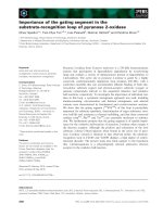
wise proteolytic degradation to smaller fragments (Fig. 1).
The first major intermediate, Iip24, interacts avidly with
MHC class II and is not easily displaced by peptide. Iip24
accumulates in human APCs treated with the cysteine pro-
tease inhibitor E64 [50,51]. The smallest fragment contain-
ing both the retention sequence and the C-terminal
extension through the class II peptide groove has been
termed Iip10. Iip10 is converted subsequently to CLIP (a
roughly 3 kDa class II-associated invariant chain peptide).
CLIP-bearing MHC class II molecules are now mature and
competent to load peptide because CLIP, but not larger
fragments of Ii, rapidly dissociates from MHC class II dimers
in the presence of a second MHC-like molecule, HLA-DM,
within endosomal compartments [52,53]. Thus the endoso-
mal proteolysis of Iip10 to CLIP generates the substrate for
HLA-DM and allows the efficient loading of MHC class II
molecules with peptides generated from endocytosed
protein. Once free from endosomal retention, peptide-
loaded MHC class II dimers move to the cell surface.
As indicated in Table 1, cysteine proteases are of two
general types: exopeptidases (aminopeptidases and car-
boxypeptidases) and endopeptidases. Endocytosed pro-
teins are fragmented by endoproteases and then
repeatedly ‘trimmed’ by the exopeptidases to yield an anti-
genic epitope (see Fig. 2).
That specific cleavages in antigens are crucial to antigen
presentation was recently confirmed by Watts, who
reported that the mutation of a single asparagine residue
in tetanus toxin blocked cleavage by the asparagine-spe-
cific endosomal enzyme legumain and abrogated further
processing to antigenic peptides [54]. Thus, the manner
in which endocytosed proteins are initially fragmented,
Respiratory Research Vol 1 No 3 Wolters and Chapman
Figure 1
Invariant chain (Ii) undergoes stepwise C-terminal degradation to
generate class II-associated invariant chain peptide (CLIP), which
occupies the peptide-binding groove of major histocompatibility
complex (MHC) class II molecules until its exchange with antigen
peptides. The figure depicts distinct intermediates in Ii chain
processing, leading to the formation of CLIP. Ii undergoes progressive
carboxy-terminal processing within endosomes by distinct cysteine
proteases to generate CLIP. The enzymes responsible for the
generation of Iip24 and Iip10 remain to be defined, although in purified
form cathepsin S can generate CLIP from intact Ii. Mice deficient in
cathepsin S accumulate Iip10 but not Ii or Iip24 in their B cells and
dendritic cells, implying that additional important enzymes in this
process remain to be defined.
/>commentary
review
reports primary research
whether by legumain or other endoproteases, is poten-
tially an important determinant of the display of MHC
class II peptides.
One function of antibodies in this process is in directing
the trafficking and degradative process leading to antigen
presentation [55]. Both the efficiency of antigen capture
and the process of antigen degradation are modified by
the presence of specific antibodies. As alluded to above,
antigen presentation is also intimately linked to the timing
of MHC class II maturation, which is itself linked to Ii prote-
olysis, because the peptide groove functions to protect
potential epitopes from terminal degradation [49]. Given
time, most if not all epitopes free in solution will be
destroyed. Thus, efficient antigen presentation requires
mature MHC class II molecules to be in the right place at
the right time. The limited time during which free peptides
can encounter mature class II peptides before degradation
might contribute to the capacity of proteins that can
persist in the lysosomal compartment, such as the mite
cysteine protease Derp1, to be particularly immunogenic.
As both antigen processing and MHC class II maturation
are crucially dependent on specific proteases, perturba-
tion of the activity of specific proteases within the antigen-
presenting compartment could modify antigen
presentation. Conceivably, this could be exploited to
control MHC class II-driven disease processes in the lung,
such as sarcoidosis and asthma.
Recent studies support this notion. In previous work the
availability of a relatively specific inhibitor of cathepsin S,
leucyl-homophenylalanine-vinylsulfone (LHVS), was used
to establish that cathepsin S has an essential role in CLIP
formation by B cells [51]. This was subsequently verified
by targeting the gene encoding cathepsin S [56]. Spleno-
cytes from ‘knockouts’ of cathepsin S fail to process Ii
beyond Iip10 (Fig. 1) and have defective TH-1-dependent
immune responses. However, further analysis of these
mice revealed a surprise. Cathepsin S ‘knockouts’ immu-
nized with ovalbumin developed normal IgE responses
and exhibited the same or greater pulmonary eosinophilia
in response to inhalation of ovalbumin as did wild-type
mice. This result was completely different from that
observed when normal mice were administered relatively
high doses of LHVS. Mice given LHVS during immuniza-
tion with ovalbumin had virtually completely suppressed
IgE responses and pulmonary eosinophilia [57]. LHVS
also abrogated IgE and lung eosinophilia when given to
cathepsin S-deficient mice.
To explore this discrepancy, cathepsin S and L double
‘knockouts’ were generated and MHC class II maturation
was studied in these mice. Surprisingly, lung macrophages
(but not splenocytes) from cathepsin S/L knockouts
hydrolyzed Iip10 and loaded peptide normally. However,
macrophages exposed to relatively high doses of LHVS
(1µM) accumulated Iip10 and failed to load peptides effi-
ciently. This implied that an LHVS-inhibitable cysteine pro-
tease(s) does in fact mediate Iip10 processing
independently of cathepsin S/L and does so preferentially
in macrophages (and potentially myeloid-like dendritic
cells). This finding led to the discovery that another
member of the cathepsin L-like subfamily of endoprote-
olytic cysteine proteases, cathepsin F, is expressed prefer-
entially in macrophages and efficiently mediates the
degradation of Iip10 to CLIP [58]. Thus, different APCs
have distinct pathways of antigen processing and MHC
class II maturation. These observations also suggest that
macrophages could have a larger role in antigen presenta-
tion pertinent to asthma than is currently believed. An
important area for further research will be to determine
whether the selective inhibition of cathepsins S, L, and F or,
alternatively, the exopeptidase group of cathepsins B, H,
and X favorably influence the immune response in the lung.
Conclusion
With the use of specific inhibitors and genetically modified
mice, our understanding of the importance of lysosomal
cysteine proteases has advanced considerably in recent
years. It is now evident that they regulate biologic
processes such as matrix remodeling and the immune
response. Although their exact roles in the pathobiology of
lung diseases are uncertain, continued research in vivo
with animal models and samples from patients with lung
disease should clarify their roles in this area.
Figure 2
Schematic summary of the role of endosomal proteases in antigen
presentation. Both endoproteases (a) and exopeptidases (b, c)
contribute to terminal degradation of internalized protein. The figure
emphasizes that the progressive fragmentation of an internalized
antigen (depicted in gray) is regulated by two separate processes: the
presence of antibody and the ability of mature MHC class II to complex
with free peptides. Antibodies greatly enhance the efficiency of antigen
uptake and alter antigen processing [55]. Mature MHC class II
molecules bind peptides and direct these complexes to the cell
surface [59].
Respiratory Research Vol 1 No 3 Wolters and Chapman
Acknowledgement
Supported in part by grants HL-04055 and HL-48261 from the National
Institutes of Health.
References
1. Chapman HA Jr, Shi GP: Protease injury in the development of
COPD: Thomas A. Neff Lecture. Chest 2000, 117:295S–299S.
2. Riese RJ, Chapman HA: Cathepsins and compartmentalization in
antigen presentation. Curr Opin Immunol 2000, 12:107–113.
3. Hart TC, Hart PS, Bowden DW, Michalec MD, Callison SA, Walker
SJ, Zhang Y, Firatli E: Mutations of the cathepsin C gene are
responsible for Papillon–Lefevre syndrome. J Med Genet 1999,
36:881–887.
4. Gelb BD, Shi GP, Chapman HA, Desnick RJ: Pycnodysostosis, a
lysosomal disease caused by cathepsin K deficiency. Science
1996, 273:1236–1238.
5. Toomes C, James J, Wood AJ, Wu CL, McCormick D, Lench N, Hewitt
C, Moynihan L, Roberts E, Woods CG, Markham A, Wong M, Widmer
R, Ghaffar KA, Pemberton M, Hussein IR, Temtamy SA, Davies R,
Read AP, Sloan P, Dixon MJ, Thakker NS: Loss-of-function muta-
tions in the cathepsin C gene result in periodontal disease and
palmoplantar keratosis. Nat Genet 1999, 23:421–424.
6. Pham CTN, Armstrong RJ, Zimonjic DB, Popescu NC, Payan DG, Ley
TJ: Molecular cloning, chromosomal localization, and expression
of murine dipeptidyl peptidase I. J Biol Chem 1997, 272:10695–
10703.
7. Rao NV, Rao GV, Hoidal JR: Human dipeptidyl-peptidase I. Gene
characterization, localization, and expression. J Biol Chem 1997,
272:10260–10265.
8. Wolters PJ, Raymond WW, Blount JL, Caughey GH: Regulated
expression, processing, and secretion of dog mast cell dipeptidyl
peptidase I. J Biol Chem 1998, 273:15514–15520.
9. Drake FH, Dodds RA, James IE, Connor JR, Debouck C, Richardson
S, Lee-Rykaczewski E, Coleman L, Rieman D, Barthlow R, Hastings G,
Gowen M: Cathepsin K, but not cathepsins B, L, or S, is abundantly
expressed in human osteoclasts. J Biol Chem 1996, 271:12511–
12516.
10. Bühling F, Gerber A, Häckel C, Krüger S, Köhnlein T, Brömme D,
Reinhold D, Ansorge S, Welte T: Expression of cathepsin K in lung
epithelial cells. Am J Resp Cell Mol Biol 1999, 20:612–619.
11. Linnevers C, Smeekens SP, Bromme D: Human cathepsin W, a puta-
tive cysteine protease predominantly expressed in CD8+ T-lym-
phocytes. FEBS Lett 1997, 405:253–259.
12. Turk B, Turk V, Turk D: Structural and functional aspects of papain-
like cysteine proteinases and their protein inhibitors. Biol Chem
1997, 378:141–150.
13. Musil D, Zucic D, Turk D, Engh RA, Mayr I, Huber R, Popovic T, Turk V,
Towatari T, Katunuma N, Bode W: The refined 2.15 Å X-ray crystal
structure of human liver cathepsin B: the structural basis for its
specificity. EMBO J 1991, 10:2321–2330.
14. Guncar G, Klemencic I, Turk B, Turk V, Karaoglanovic-Carmona A,
Juliano L, Turk D: Crystal structure of cathepsin X: a flip-flop of the
ring of His23 allows carboxy-monopeptidase and carboxy-dipepti-
dase activity of the protease. Structure Fold Des 2000, 8:305–313.
15. Guncar G, Podobnik M, Pungercar J, Strukelj B, Turk V, Turk D: Crystal
structure of porcine cathepsin H determined at 2.1 Å resolution:
location of the mini-chain C-terminal carboxyl group defines
cathepsin H aminopeptidase function. Structure 1998, 6:51–61.
16. Dolenc I, Turk B, Pungercic G, Ritonja A, Turk V: Oligomeric struc-
ture and substrate induced inhibition of human cathepsin C. J Biol
Chem 1995, 270:21626–21631.
17. Buck MR, Karustis DG, Day NA, Honn KV, Sloane BF: Degradation of
extracellular-matrix proteins by human cathepsin B from normal
and tumour tissues. Biochem J 1992, 282:273–278.
18. Sires UI, Schmid TM, Fliszar CJ, Wang ZQ, Gluck SL, Welgus HG:
Complete degradation of type X collagen requires the combined
action of interstitial collagenase and osteoclast-derived cathep-
sin-B. J Clin Invest 1995, 95:2089–2095.
19. Garnero P, Borel O, Byrjalsen I, Ferreras M, Drake FH, McQueney MS,
Foged NT, Delmas PD, Delaissé JM: The collagenolytic activity of
cathepsin K is unique among mammalian proteinases. J Biol
Chem 1998, 273:32347–32352.
20. Wolters PJ, Laig-Webster M, Caughey GH: Dipeptidyl peptidase I
cleaves matrix-associated proteins and is expressed mainly by
mast cells in normal dog airways. Am J Respir Cell Mol Biol 2000,
22:183–190.
21. Werb Z, Bainton DF, Jones PA: Degradation of connective tissue
matrices by macrophages. III. Morphological and biochemical
studies on extracellular, pericellular, and intracellular events in
matrix proteolysis by macrophages in culture. J Exp Med 1980,
152:1537–1553.
22. Everts V, van der Zee E, Creemers L, Beertsen W: Phagocytosis and
intracellular digestion of collagen, its role in turnover and remod-
eling. Histochem J 1996, 28:229–245.
23. van der Zee E, Everts V, Hoeben K, Beertsen W: Cytokines modulate
phagocytosis and intracellular digestion of collagen fibrils by
fibroblasts in rabbit periosteal explants. Inverse effects on procol-
lagenase production and collagen phagocytosis. J Cell Sci 1995,
108:3307–3315.
24. Everts V, Aronson DC, Beertsen W: Phagocytosis of bone collagen
by osteoclasts in two cases of pycnodysostosis. Calcif Tissue Int
1985, 37:25–31.
25. Punturieri A, Filippov S, Allen E, Caras I, Murray R, Reddy V, Weiss SJ:
regulation of elastinolytic cysteine proteinase activity in normal
and cathepsin K-deficient human macrophages. J Exp Med 2000,
192:789–800.
26. Shi GP, Sukhova GK, Grubb A, Ducharme A, Rhode LH, Lee RT,
Ridker PM, Libby P, Chapman HA: Cystatin C deficiency in human
atherosclerosis and aortic aneurysms. J Clin Invest 1999, 104:
1191–1197.
27. Heidtmann HH, Salge U, Havemann K, Kirschke H, Wiederanders B:
Secretion of a latent, acid activatable cathepsin L precursor by
human non-small cell lung cancer cell lines. Oncol Res 1993, 5:
441–451.
28. Pagano M, Dalet-Fumeron V, Engler R: The glycosylation state of the
precursors of the cathepsin B-like proteinase from human malig-
nant ascitic fluid: possible implication in the secretory pathway of
these proenzymes. Cancer Lett 1989, 45:13–19.
29. Lorenzo K, Ton P, Clark JL, Coulibaly S, Mach L: Invasive properties
of murine squamous carcinoma cells: secretion of matrix-degrad-
ing cathepsins is attributable to a deficiency in the mannose 6-
phosphate/insulin-like growth factor II receptor. Cancer Res 2000,
60:4070–4076.
30. Dittmer F, Ulbrich EJ, Hafner A, Schmahl W, Meister T, Pohlmann R,
von Figura K: Alternative mechanisms for trafficking of lysosomal
enzymes in mannose 6-phosphate receptor-deficient mice are
cell type-specific. J Cell Sci 1999, 112:1591–1597.
31. Stinchcombe JC, Griffiths GM: Regulated secretion from hemopoi-
etic cells. J Cell Biol 1999, 147:1–6.
32. Brown GR, McGuire MJ, Thiele DL: Dipeptidyl peptidase I is
enriched in granules of in vitro- and in vivo-activated cytotoxic T
lymphocytes. J Immunol 1993, 150:4733–4742.
33. Rodriguez A, Webster P, Ortego J, Andrews NW: Lysosomes
behave as Ca
2+
-regulated exocytic vesicles in fibroblasts and
epithelial cells. J Cell Biol 1997, 137:93–104.
34. Jahn R, Südhof TC: Membrane fusion and exocytosis. Annu Rev
Biochem 1999, 68:863–911.
35. Schiavo G, Osborne SL, Sgouros JG: Synaptotagmins: more isoforms
than functions? Biochem Biophys Res Commun 1998, 248:1–8.
36. Rizo J, Südhof TC: C2-domains, structure and function of a univer-
sal Ca
2+
-binding domain. J Biol Chem 1998, 273:15879–15882.
37. Martinez I, Chakrabarti S, Hellevik T, Morehead J, Fowler K, Andrews
NW: Synaptotagmin VII regulates Ca
2+
-dependent exocytosis of
lysosomes in fibroblasts. J Cell Biol 2000, 148:1141–1149.
38. Petanceska S, Devi L: Sequence analysis, tissue distribution, and
expression of rat cathepsin S. J Biol Chem 1992, 267:26038–26043.
39. Coulibaly S, Schwihla H, Abrahamson M, Albini A, Cerni C, Clark JL,
Ng KM, Katunuma N, Schlappack O, Glossl J, Mach L: Modulation of
invasive properties of murine squamous carcinoma cells by het-
erologous expression of cathepsin B and cystatin C. Int J Cancer
1999, 83:526–531.
40. Takahashi H, Ishidoh K, Muno D, Ohwada A, Nukiwa T, Kominami E,
Kira S: Cathepsin L activity is increased in alveolar macrophages
and bronchoalveolar lavage fluid of smokers. Am Rev Respir Dis
1993, 147:1562–1568.
41. Zheng T, Zhu Z, Wang Z, Homer RJ, Ma B, Riese RJ, Chapman HA,
Shapiro SD, Elias JA: Inducible targeting of IL-13 to the adult lung
causes matrix metalloproteinase- and cathepsin-dependent
emphysema. J Clin Invest 2000, 106:1081–1093.
42. McGuire MJ, Lipsky PE, Thiele DL: Generation of active myeloid and
lymphoid granule serine proteases requires processing by the
granule thiol protease dipeptidyl peptidase I. J Biol Chem 1993,
268:2458–2467.
/>commentary
review
reports primary research
43. Murakami M, Karnik SS, Husain A: Human prochymase activation. A
novel role for heparin in zymogen processing. J Biol Chem 1995,
270:2218–2223.
44. Pham CT, Ley TJ: Dipeptidyl peptidase I is required for the pro-
cessing and activation of granzymes A and B in vivo. Proc Natl
Acad Sci USA 1999, 96:8627–8632.
45. Podack ER: How to induce involuntary suicide: the need for dipep-
tidyl peptidase I. Proc Natl Acad Sci USA 1999, 96:8312–8314.
46. Belaaouaj A, McCarthy R, Baumann M, Gao Z, Ley TJ, Abraham SN,
Shapiro SD: Mice lacking neutrophil elastase reveal impaired host
defense against Gram-negative bacterial sepsis. Nat Med 1998,
4:615–618.
47. Belaaouaj A, Kim KS, Shapiro SD: Degradation of outer membrane
protein A in Escherichia coli killing by neutrophil elastase. Science
2000, 289:1185–1188.
48. Chapman HA: Endosomal proteolysis and MHC class II function.
Curr Opin Immunol 1998, 10:93–102.
49. Wolf PR, Ploegh HL: How MHC class II molecules acquire peptide
cargo: biosynthesis and trafficking through the endocytic
pathway. Annu Rev Cell Dev Biol 1995, 11:267–306.
50. Cresswell P: Antigen presentation. Getting peptides into MHC
class II molecules. Curr Biol 1994, 4:541–543.
51. Riese RJ, Wolf PR, Bromme D, Natkin LR, Villadangos JA, Ploegh HL,
Chapman HA: Essential role for cathepsin S in MHC class II-asso-
ciated invariant chain processing and peptide loading. Immunity
1996, 4:357–366.
52. Sloan VS, Cameron P, Porter G, Gammon M, Amaya M, Mellins E,
Zaller DM: Mediation by HLA-DM of dissociation of peptides from
HLA-DR. Nature 1995, 375:802–806.
53. Sherman MA, Weber DA, Jensen PE: DM enhances peptide binding
to class II MHC by release of invariant chain-derived peptide.
Immunity 1995, 3:197–205.
54. Antoniou AN, Blackwood SL, Mazzeo D, Watts C: Control of antigen
presentation by a single protease cleavage site. Immunity 2000,
12:391–398.
55. Watts C: Capture and processing of exogenous antigens for pre-
sentation on MHC molecules. Annu Rev Immunol 1997, 15:
821–850.
56. Shi GP, Villadangos JA, Dranoff G, Small C, Gu L, Haley KJ, Riese R,
Ploegh HL, Chapman HA: Cathepsin S required for normal MHC
class II peptide loading and germinal center development. Immu-
nity 1999, 10:197–206.
57. Riese RJ, Mitchell RN, Villadangos JA, Shi GP, Palmer JT, Karp ER, De
Sanctis GT, Ploegh HL, Chapman HA: Cathepsin S activity regu-
lates antigen presentation and immunity. J Clin Invest 1998, 101:
2351–2363.
58. Shi G-P BR, Riese, R, Ploegh HL, Chapman HA.: Role for cathepsin
F in invariant chain processing and MHC class II peptide loading
by macrophages. J Exp Med 2000, 191:1177–1185.
59. Ojcius DM, Gapin L, Kanellopoulos JM, Kourilsky P: Is antigen pro-
cessing guided by major histocompatibility complex molecules?
FASEB J 1994, 8:974–978.
Authors’ affiliations: Paul J Wolters and Harold A Chapman
(Department of Medicine and Cardiovascular Research Institute,
University of California, San Francisco, California, USA)
Correspondence: Harold A Chapman, MD, Cardiovascular Research
Institute, University of California, San Francisco, CA 94143-0110,
USA. Tel: +1 415 514 0896; fax: +1 415 476 2283;
e-mail: