Báo cáo y học: "Molecular mechanisms of severe acute respiratory syndrome (SARS)" pot
Bạn đang xem bản rút gọn của tài liệu. Xem và tải ngay bản đầy đủ của tài liệu tại đây (960.75 KB, 16 trang )
BioMed Central
Page 1 of 16
(page number not for citation purposes)
Respiratory Research
Open Access
Review
Molecular mechanisms of severe acute respiratory syndrome
(SARS)
David A Groneberg*
1
, Rolf Hilgenfeld
2
and Peter Zabel
3,4
Address:
1
Pneumology and Immunology, Otto-Heubner-Centre, Charité School of Medicine, Free University and Humboldt-University, D-13353
Berlin, Germany,
2
Institute of Biochemistry, University of Lübeck, D-23538 Lübeck, Germany,
3
Division of Clinical Infectiology and Immunology,
Department of Medicine, Research Center Borstel, D-23845 Borstel, Germany and
4
Division of Thoracic Medicine, Department of Medicine,
University of Lübeck, D-23538 Lübeck, Germany
Email: David A Groneberg* - ; Rolf Hilgenfeld - ; Peter Zabel - pzabel@fz-
borstel.de
* Corresponding author
Severe Acute Respiratory SyndromeSARScoronavirusmolecular mechanismstherapyvaccination
Abstract
Severe acute respiratory syndrome (SARS) is a new infectious disease caused by a novel
coronavirus that leads to deleterious pulmonary pathological features. Due to its high morbidity
and mortality and widespread occurrence, SARS has evolved as an important respiratory disease
which may be encountered everywhere in the world. The virus was identified as the causative agent
of SARS due to the efforts of a WHO-led laboratory network. The potential mutability of the
SARS-CoV genome may lead to new SARS outbreaks and several regions of the viral genomes open
reading frames have been identified which may contribute to the severe virulence of the virus. With
regard to the pathogenesis of SARS, several mechanisms involving both direct effects on target cells
and indirect effects via the immune system may exist. Vaccination would offer the most attractive
approach to prevent new epidemics of SARS, but the development of vaccines is difficult due to
missing data on the role of immune system-virus interactions and the potential mutability of the
virus. Even in a situation of no new infections, SARS remains a major health hazard, as new
epidemics may arise. Therefore, further experimental and clinical research is required to control
the disease.
Introduction
Severe acute respiratory syndrome (SARS) is the first new
infectious disease of this millennium. SARS has originated
from Southern China at the end of 2002 and has a high
mortality and morbidity. Within a period of six months
beginning at the end of 2002, the disease has affected
more than 8,000 people and killed nearly 800 [1]. The dis-
ease poses a new threat for respiratory medicine and rep-
resents a challenge for antiviral drug development and
administration [2,3].
SARS is caused by a novel, SARS-associated coronavirus
(SARS-CoV) [4-6] which has been identified by a World
Health Organization (WHO)-led global laboratory net-
work. The first cases of SARS were reported from a hospital
in Hanoi, Vietnam, by Carlo Urbani, a WHO scientist who
himself died from the disease [7]. After reports from
Published: 20 January 2005
Respiratory Research 2005, 6:8 doi:10.1186/1465-9921-6-8
Received: 10 November 2004
Accepted: 20 January 2005
This article is available from: />© 2005 Groneberg et al; licensee BioMed Central Ltd.
This is an Open Access article distributed under the terms of the Creative Commons Attribution License ( />),
which permits unrestricted use, distribution, and reproduction in any medium, provided the original work is properly cited.
Respiratory Research 2005, 6:8 />Page 2 of 16
(page number not for citation purposes)
health authorities in Hong Kong on the outbreak of a new
form of epidemical atypical pneumonia in public hospi-
tals, the WHO issued a global alert on the disease. During
this period, cases of SARS were also reported from China,
other Asian countries and even other continents including
America (Canada, U.S.A.) and Europe (Germany).
Shortly after the initial global alert, the WHO initiated a
collaborative multi-center research project on SARS diag-
nosis, led by eleven principal laboratories in nine coun-
tries [8]. Using modern communication technologies to
optimize the analysis of SARS tissue samples, it was soon
shown that a novel coronavirus is the causative agent of
SARS (SARS-CoV) [4-6]. Due to the death of Carlo Urbani
who first identified the new disease, the first isolate of the
virus was proposed to be named Urbani strain of SARS-
associated coronavirus, but a final terminology has not
been proposed so far [9]. Since Koch's principles have
been shown to be fulfilled by the new pathogen [10,11],
it is not necessary to call the virus SARS-associated and the
general agreement is now to call it SARS coronavirus
(SARS-CoV).
Parallel to the progress made in the epidemiology and
clinical diagnosis which has recently been demonstrated
by numerous case reports, clinical studies and definitions
[1], scientists have also revealed basic mechanisms of the
underlying causative agent, the SARS coronavirus. As it is
crucial for future strategies that SARS is detected in its ear-
liest stages and that therapeutic options are optimized,
insights into the molecular mechanism of SARS have to be
used to develop new therapeutic strategies and vaccines.
While other reviews have focused on the epidemiology,
clinical presentation and potential treatment of SARS, the
present overview aims to analyze and present the cur-
rently available data on molecular mechanisms of SARS.
In this respect, it is important to underline that in the
present state of no specific drug or vaccine being available,
research on molecular mechanism is crucial to identify
potential treatment targets.
Etiology
Prior to the development of therapeutic regimes based on
molecular mechanisms of the disease, the causative agent
had to be isolated and analysed. Soon after the fast estab-
lishment of the international WHO laboratory network,
rapid progress was made in the identification process of
the causative agent, and it was reported that SARS is most
probably caused by a novel strain of the family of corona-
viruses [4-6]. These viruses are commonly known to cause
respiratory and gastrointestinal diseases of humans and
domestic animals [12,13]. The group of coronaviruses is
classified as a member of the order nidovirales, which rep-
resents a group of enveloped positive-sense RNA viruses
consisting of coronaviridae and arteriviridae [14]. Viruses
of this group are known to synthesize a 3' co-terminal set
of subgenomic mRNAs in the infected cells [15].
Origin of the SARS virus
Soon after the identification of a new coronavirus as the
causative agent of SARS and of a southern Chinese prov-
ince as the first area of occurrence, animal species of this
area have been speculated to be the origin of the SARS-
CoV. As analysis of the SARS-CoV genetic sequence
revealed large differences to any other currently known
coronaviruses in humans or domestic animals [16,17], it
was hypothesized that the new virus might originate from
wild animals. This hypothesis was supported by a search
for coronaviruses in wild animals sold on markets in
southern China, which identified the presence of a coro-
navirus in civet cats. This animal coronavirus was shown
to have a sequence identity of more than 99% to the SARS
coronavirus [18] with only a limited number of deletions
and mutations between both viruses. SARS-CoV has a
deletion of 29 nucleotides relative to the civet cat virus,
indicating that if there was direct transmission, it went
from the animal to man, because deletions occur proba-
bly more easily than insertions. Recent reports indicate
that SARS-CoV is distinct from the civet cat virus and it has
not been answered so far if the civet cat virus is the origin
of the SARS-CoV or if civet cats were also infected from
other species [19]. Therefore, there are no data available
on the possibility of horizontal transmission between ani-
mals, and the question whether the jump of the virus from
an animal to humans was a single accident or may fre-
quently occur in future with the animals as dangerous res-
ervoirs for future SARS epidemics remains unanswered. So
far, the SARS-CoV has been reported to be able to infect
not only humans but also macaque monkeys [11],
domestic cats, and ferrets [20]. However, transmission of
the virus from the domestic cat to man has not been
shown. The ability of the SARS-CoV to infect other animal
species could point to potential natural reservoirs of the
virus. In this respect, coronaviruses are known to relatively
easily jump to other species. I.e., the human coronavirus
OC43 shares a high degree of genetic sequence homology
to bovine coronavirus (BCoV) and it is commonly
assumed that it has jumped from one species to the other
[21,22]. In the same way, BCoV has been reported to be
able to infect humans and cause diarrhea [23]. Whereas
the precise mechanisms of these species jumps remain
unclear, it is most likely that they represent the results of
mutations and epidemiological studies of coronavirus
infections in wild animals will therefore be crucial for
future understanding and control of new SARS outbreaks.
SARS virus taxonomy
Until the identification of the new SARS-CoV, the corona-
viruses have been divided into three subgroups, which
Respiratory Research 2005, 6:8 />Page 3 of 16
(page number not for citation purposes)
differ with respect to their genome [24]. The first group
consists of viruses such as the human coronavirus 229E
(HCoV-229E), porcine respiratory coronavirus (PRCV),
porcine transmissible gastroenteritis virus (TGEV), feline
infectious peritonitis virus (FIPV) and feline enteritis virus
(FEV) or the canine coronavirus (CCoV). The second
group comprises human coronavirus OC43 (HCoV-
OC43), bovine coronavirus (BCoV), and mouse hepatitis
virus (MHV), and the third group mainly consists of avian
species such as the chicken infectious bronchitis virus
(IBV). Whereas the SARS-CoV has been shown to cross-
react with some group I coronavirus antibodies [6], its
genetic sequence does not belong to this group. Within
the nucleic acid or protein sequence phylogenetic trees of
the coronavirus family, the SARS-CoV has first been
located at an equal distance from the second and third
group, irrespective of which SARS-CoV RNA region is used
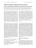
for analysis [6,16,17]. Therefore, the SARS-CoV may rep-
resent the first member of a new group of coronaviruses
(Figure 1). However, the taxonomy is still no clear
[19,25], and recent studies that focused on the N-terminal
domain of the spike protein and on poorly conserved pro-
teins such as Nsp1, matrix protein, or nucleocapsid, have
suggested a relation to group II viruses [26]. A similar con-
clusion can be drawn if the polymerase gene is examined,
pointing to an early split-off from the coronavirus group
2 lineage [27].
Despite the fact that this new virus most likely jumped to
humans from wild animal species, it has remarkably well
adapted to the human organism as shown by its high per-
son-to-person transmissibility.
SARS virus genome structure
The structure of the SARS viral RNA is organized in 13–15
open reading frames (ORF) and contains a total of
approximately 30,000 nucleotides [6,16,17].
Recently, 61 SARS-CoV sequences derived from the early,
middle, and late phases of the SARS epidemic together
with two viral sequences from palm civets were analyzed
[28]. Genotypes characteristic of each phase were discov-
ered, and it was found that the neutral mutation rate of
the viral genome was constant but the amino acid substi-
tution rate of the coding sequences slowed during the
course of the epidemic. The spike protein showed the
strongest initial responses to positive selection pressures
[28].
Only ORFs exceeding fifty amino acids in translational
capacity are considered relevant as they contain the
sequences for the structural and functional properties of
the virus and are therefore of potential interest for the
development for future therapeutic strategies. The com-
parison of the different SARS-CoV ORFs with those of
other coronaviruses reveals a familiar pattern of structural
gene arrangement with replicase and protease genes (gene
1a-1b) and the spike (S), envelope (E), membrane (M)
and nucleocapsid (N) genes in a typical 5'- to 3' order of
appearance [29]. The proteins encoded by these genes
may be targets for novel treatments. Between these well-
known genes, a series of ORFs of unknown function was
found: There are two ORFs situated between the spike and
the envelope genes and three to five ORFs between the
membrane and nucleocapsid genes. Comparison of this
gene organization with other known coronaviruses does
not indicate a closest proximity to group II coronaviruses.
Also, the SARS-CoV genomic sequence does not contain a
gene for hemagglutinin-esterase (HE) protein, which is
present in the majority of group II coronaviruses.
Two-thirds of the SARS RNA is organized in the gene 1a-
1b. The sequence of this gene is highly conserved among
all coronaviruses [17]. ORFs 1a and 1b encode two poly-
proteins, pp1a and pp1ab, the latter through a ribosomal
frameshifting mechanism. These polyproteins are
Coronavirus classificationFigure 1
Coronavirus classification. The family of coronaviruses
belongs to the order of nidovirales and consists of three
groups so far. It is still debatable whether the new SARS-CoV
should be assigned to group II or to a new fourth group.
Group I includes human coronavirus 229E (HCoV-229E),
transmissible gastroenteritis virus (TEGV), porcine epidemic
diarrhea virus (PEDV), canine coronavirus (CCoV), and feline
coronavirus (FIPV). Group II viruses include human coronavi-
rus OC43 (HCoV-OC43), murine hepatitis virus (MHV), and
bovine coronavirus (BCoV), and group III species are turkey
coronavirus (TCoV), and avian infectious bronchitis virus
(IBV).
MHV
HCoV-OC43
BCoV
HCoV-229E
TGEV
PEDV
CCoV
FIPV
Group II
Group I
IBV
TCoV
Group III
SARS-CoV
Group II or IV ?
Respiratory Research 2005, 6:8 />Page 4 of 16
(page number not for citation purposes)
processed by virus-encoded proteinases, to yield 16 indi-
vidual proteins. Most potential gene 1a-1b products are
fairly well conserved between SARS-CoV and other coro-
naviruses [17,29]. Many of their functions are unknown
but it is suggested that they participate in viral RNA repli-
cation, making them potential targets for the develop-
ment of antiviral compounds. Therefore, research efforts
will focus on these proteins. One exception from the over-
all conservation of SARS-CoV gene 1a-1b is the lack of a
sequence coding for PL1
pro
, one of the two papain-like
proteinases operating on cleavage sites at the N-terminus
of the polyproteins (Figure 2). The main proteinase
(M
pro
), also called 3C-like protease (3CL
pro
), is responsi-
ble for the cleavage of all the remaining proteins encoded
by gene 1a-1b [29,30].
SARS virus gene expression
Apart from gene 1, coronavirus genes are known to be
usually expressed from subgenomic mRNAs. They share a
common leader sequence at the 5'-end and initiate at dif-
ferent places in the genome extending toward the 3'-end
of the virus genome [31]. Some ORFs may also be
unconventionally translated from a single mRNA. As
these uncommon translation mechanisms are not very
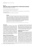
SARS-CoV genome organizationFigure 2
SARS-CoV genome organization. The structure of the SARS viral RNA is organized into 13–15 open reading frames (ORFs)
and contains an overall amount of approximately 30,000 nucleotides. The sequence can be separated into different elements
and genomic and subgenomic mRNAs.
SARS-CoV
mRNA2 – S protein
mRNA3
0 5 10 15 20 25 30
gene 1a
gene 1b
S
mRNAs3-9
mRNA1 –pp1a, pp1ba
mRNA4 –E protein
mRNA5 – M protein
mRNA6
mRNA7
mRNA8
mRNA9 – N protein
Respiratory Research 2005, 6:8 />Page 5 of 16
(page number not for citation purposes)
efficient and the gene products are not very abundant,
these ORFs typically encode nonstructural proteins.
Whereas the ORFs between the structural protein genes
are very heterogeneous among the different coronaviruses
and not essential for viral replication, recent studies sug-
gested that deletion of non-essential ORFs may result in a
reduced virulence [32]. In agreement with this, some of
these non-essential ORFs of the new SARS-CoV genome
may be responsible for the high SARS-CoV virulence.
So far, five to eight subgenomic mRNAs were found in
SARS-CoV-infected cells [17,27]. Thiel and colleagues per-
formed the first detailed study on mechanisms and
enzymes involved in SARS-CoV genome expression (Fig-
ure 2) [29]. They determined the sequence of the SARS-
CoV isolate Frankfurt 1 and characterized the major RNA
elements and protein functions involved in the genome
expression by characterizing regulatory mechanisms such
as the discontinuous synthesis of eight subgenomic
mRNAs, ribosomal frameshifting and post-translational
proteolytic processing. Also, the activities of SARS-CoV
enzymes such as the helicase or the two cysteine protein-
ases (PL2
pro
and M
pro
) were addressed as they are involved
in replication, transcription or post-translational polypro-
tein processing [29].
In conclusion, research in the area of coronavirus gene
expression is important to delineate components which
directly affect SARS-CoV virulence.
SARS virus structural proteins
The structural proteins of the new SARS-CoV are potential
targets for new treatment options. The new SARS-CoV
only contains the three envelope proteins, spike (S),
envelope (E), and membrane (M) but not the
hemagglutinin-esterase (HE) protein, which is present in
some coronaviruses of the second group.
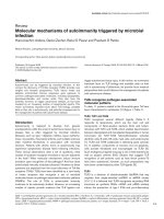
The spike glycoprotein is responsible for the characteristic
spikes of the SARS-CoV (Figure 3). Intra- and extracellular
proteases often cleave the S protein into S1 and S2
domains, with the cleavage process often increasing infec-
tivity of the virus. Molecular modelling has been per-
formed for the S1 and S2 units of the SARS-CoV spike
protein [33,34]. The spike proteins of coronaviruses are
reported to bind to receptors on their target cells and the
domains responsible for receptor-binding are commonly
situated in the N-terminal region of S1 [35-40]. The spikes
consist of oligomeric structures, that are formed by heptad
repeats of the S2 domain which also represent a fusion
peptide sequence. This peptide is responsible for the coro-
navirus fusion activity.
The SARS-CoV has also been reported to cause the forma-
tion of syncytia in vivo, but so far only under the condi-
tion of cultured Vero cells [6]. The SARS-CoV S protein
seems to have most of its characteristics in common with
the S proteins of other coronaviruses, but it will be impor-
tant for the understanding of the SARS-CoV pathogenic
properties to identify the exact conditions of membrane
fusion, i.e. pH dependency and protease sensitivity, which
can increase the infectivity. The envelope and membrane
proteins are integral membrane proteins and required for
virus assembly [41]. In the case of the murine coronavirus
MHV-A59 the coexpression of the E and M proteins but
SARS-CoV transmission electron microscopyFigure 3
SARS-CoV transmission electron microscopy. In the super-
natant of SARS-CoV infected cytopathic Vero E6 cells, char-
acteristic virus particles can be found. The diameter of the
viruses ranges between 60 nm and 120 nm and the virus
shapes are round or oval. There are many protrusions from
the envelope which are arranged in order with wide gaps
between them. There are also many virus particles in the
infected cells present. They often form a virus vesicle with an
encircling membrane. A: Higher magnification B: Lower mag-
nification. Scale bars represent 100 nm. Reproduced with
permission from Acta Biochimica et Biophysica Sinica 2003,
35(6):587–591 [126].
A
B
Respiratory Research 2005, 6:8 />Page 6 of 16
(page number not for citation purposes)
not the S or N proteins is needed for the release of virus-
like particles (VLP) [42]. The nucleocapsid and viral core
of the SARS-CoV are likely to be formed by the N protein.
An interesting feature of the SARS-CoV and other corona-
viruses is the resistance against the gastrointestinal fluids
despite the lipid composition of their envelope. It has
been reported that the SARS-CoV can survive in diarrheal
stool for four days and also, patients with SARS often suf-
fer from gastrointestinal symptoms with the virus to be
detected in the stool [4]. As the molecular basis for the
envelope's resistance against acidic environments and gas-
trointestinal enzymes is unclear, further research has to be
carried out in this area which is important for the control
of future SARS outbreaks.
Evolution of the SARS virus
It is unclear when and how novel pathogens such as the
SARS-CoV cross the barriers between their natural reser-
voirs and human populations, leading to the epidemic
spread of novel infectious diseases [43]. As with the SARS-
CoV, new pathogens are believed to emerge from animal
reservoirs and a variety of molecular mechanisms may
contribute to the evolution of the viruses or bacteria. Due
to the estimated error frequency of 1 × 10
-4
for RNA-
dependent RNA polymerases [44], RNA viruses such as
the SARS-CoV can undergo mutation at a high frequency.
The SARS-CoV seems to be relatively genetically stable as
the RNA sequences from different SARS patients were
quite homogeneous. Even the entire genomic sequences
of virus isolates from different continental areas did not
differ by more than ten amino acids and it seems that two
lineages of the virus can be traced [45]. This obvious con-
tradiction to the high potential error rate of the RNA-
dependent RNA polymerase suggests the presence of some
proofreading mechanism connected with this enzyme. In
fact, a detailed analysis of the SARS-CoV genome by bio-
informatics indicates the presence of an exonuclease activ-
ity [27].
Next to mutations, a further threat of the SARS-CoV is
based on the ability of coronaviruses to undergo RNA
recombination at a high frequency [15]. For a variety of
other coronaviruses, both recombination and mutation in
natural infections have been shown to contribute to the
diversification of the coronaviruses. Because of the dem-
onstrated ability of coronaviruses to recombinate, the
question whether the SARS-CoV will show a higher fre-
quency of mutations within possible future seasonal
changes or in respond to drug treatment is an issue of
major concern. It was reported that in the initial phases of
the SARS epidemic, the mutation rate was high in the gene
for the spike protein, but this stabilized during the middle
and final stages of the 2003 epidemy [28]. Thus, the virus
had experienced great pressure to adapt to the new host
after crossing the species barrier, but has then been opti-
mized [28].
Duration of infection
Although human coronaviruses are characteristically caus-
ing self-limiting short diseases, the question of potential
chronic SARS infections is of major importance for a
future disease control. If the SARS-CoV is able to cause a
chronic persistent infection, chronic carriers may serve as
sources for new SARS outbreaks. However, the detection
of SARS-CoV in stool of patients for longer periods than 6
weeks after hospital discharge has not been reported so
far. Therefore, the danger of chronic carriers may not be
relevant. In contrast to common human coronavirus
infections with short durations, most animal coronavi-
ruses cause persistent infections. As an example, the feline
coronavirus FIPV infects animals which then continue to
shed virus for periods reaching up to seven months after
infection without carrying disease symptoms [46]. Also,
TGEV and MHV tend to cause chronic infections as these
viruses may be found in the airways and small intestine
(TGEV) or the nervous system (MHV) several months
after infection [47,48]. Although the SARS-CoV has
jumped to humans it may still have this property of
inducing chronic infections. Thus, SARS-CoV RNA was
found in patients' stool specimen more than 30 days after
the infections.
Clinical picture of SARS
The mean incubation period of SARS was estimated to be
6.4 days (95% confidence interval, 5.2 to 7.7). The mean
reported time from the onset of clinical symptoms to the
hospital admission varied between three and five days
[49].
Main clinical features of the disease are in the initial
period common symptoms such as persistent fever, myal-
gia, chills, dry cough, dizziness, and headache. Further,
although less common symptoms are sore throat, sputum
production, coryza, vomiting or nausea, and diarrhea
[50,51]. Special attention has been paid to the symptom
of diarrhea: Watery diarrhea has also been reported in a
subgroup of patients one week after the initial symptoms
[52].
The clinical course of the disease seems to follow a bi- or
triphasic pattern. In the first phase viral replication and an
increasing viral load, fever, myalgia, and other systemic
symptoms can be found. These symptoms generally
improve after a few days. In the second phase representing
an immunopathologic imbalance, major clinical findings
are oxygen desaturation, a recurrence of fever, and clinical
and radiological progression of acute pneumonia. This
second phase is concomitant with a fall in the viral load.
The majority of patients is known to respond in the
Respiratory Research 2005, 6:8 />Page 7 of 16
(page number not for citation purposes)
second phase to treatment. However, about 20% of
patients may progress to the third and critical phase. This
phase is characterized by the development of an acute res-
piratory distress syndrome (ARDS) commonly necessitat-
ing mechanical ventilation.
SARS in adults and children
Rapid progress has been made in understanding the clin-
ical presentation of SARS in adults and children [53-56].
In comparison to adults, SARS seems to be less aggressive
in younger children, with no children in one case series
requiring supplementary oxygen [57] while in adults, sys-
temic infection as well as respiratory infection may be the
rule. SARS is much milder with non-specific cold-like
symptoms in children younger than 12 years than it is in
adolescents and adults [58]. The reason for the milder
clinical presentation of SARS in children is most likely due
to differences in developmental stage of the immune
system.
The course of the disease in teenagers more likely resem-
bles adults in concerning clinical presentation and disease
progression [58]. SARS may also develop severe illness
requiring intensive care and assisted ventilation in these
adolescent patients. The common presenting features are
fever, malaise, coryza, cough, chills or rigor, headache,
myalgia, leucopaenia, thrombocytopaenia, lymphopae-
nia, elevated lactate dehydrogenase levels and mildly pro-
longed activated partial thromboplastin times [59]. The
radiographic findings are non-specific: However, high-res-
olution computed chest tomography in clinically sus-
pected cases may prove to be an early diagnostic aid when
initial chest radiographs appeared normal. While rapid
diagnosis with the first-generation RT-PCR assay was not
satisfactory, improved RT-PCR assays may help to diag-
nose SARS in early stages. In this respect, a sensitivity
approaching 80% in the first 3 days of illness when per-
formed on nasopharyngeal aspirates may be achieved.
The best treatment strategy for SARS among children still
has to be determined while no case fatality has been
reported in children. In comparison to the prognosis in
adults, there is a relatively good short- to medium-term
outcome. However, it is crucial to emphasize that contin-
ued monitoring for long-term complications due to the
disease or its treatment is of major importance [60].
Molecular mechanisms of SARS virus pathogenesis
Cytocidal mechanisms
Coronaviruses are known to exert their effects by cytocidal
and immune-mediated mechanisms. In vitro studies
using cell culture assays have shown that coronavirus
infection commonly results in cytopathic effects such as
cellular lysis or apoptosis [61]. Also, the virus can cause
cellular fusion leading to the formation of syncytia. These
cytopathic effects are caused by steps of the viral replica-
tion such as the mobilisation of vesicles to form the viral
replication complex [18], leading to the disruption of
Golgi complexes [62]. Parallel to results on other corona-
viruses, SARS-CoV has been shown to cause cytopathic
effects in Vero cells and the formation of syncytia in lung
tissues. A further similarity with other coronaviruses
seems to be the potential of the SARS-CoV to cause tissue
fibrosis [63]. As molecular mechanism for this fibrosis
which has been reported for infections with the coronavi-
rus MHV, the N protein has been demonstrated to induce
promoter activity of the prothrombinase gene that corre-
lates with fibrin deposition [64].
Immune-mediated mechanisms
Next to cytocidal effects, also immune-mediated mecha-
nisms of both the innate and adaptive immune system
seem to contribute to the pathogenesis of SARS-CoV infec-
tions. In this respect, it has been shown that in MHV infec-
tion, T cells and cytokines play an important role in
development of the disease [65]. Also, humoral antibod-
ies have been reported to be crucial in infections caused
by coronaviruses such as FIPV. Herein, antibodies against
the spike protein were shown to be related to the induc-
tion of peritonitis [66].
For SARS-CoV infections, it has been reported that there
seems to be an inflammatory cell influx consisting in par-
ticular of macrophages in the airways, and a massive
release of cytokines during the peak of the infection
[67,68]. It is therefore crucial that these immune mecha-
nisms are further analysed on the molecular level as it
seems appropriate that not only antiviral but also anti-
inflammatory strategies are evaluated for a use in the clin-
ical management of future SARS cases.
The pharmacotherapy for SARS with anti-inflammatory
steroids is controversial and largely anecdotal [69]. It was
reported that the initial use of pulse methylprednisolone
therapy appears to be more efficacious and equally safe
when compared with regimens with lower dosage and
should therefore be considered as the preferred steroid
regimen in the treatment of SARS, pending data from
future randomized controlled trials [70]. A further prelim-
inary, uncontrolled study of patients with SARS, reported
that the use of interferon alfacon-1 plus steroids was asso-
ciated with reduced disease-associated impaired oxygen
saturation and more rapid resolution of radiographic lung
abnormalities [71].
Mechanisms of target cell specificity
The most obvious gene which is likely to be a key modifier
of SARS pathomechanisms is the spike (S) protein gene.
As known for other coronaviruses, it does not only affect
viral pathogenesis by determining the target cell specifi-
city but also by other mechanisms. In this respect, a single
Respiratory Research 2005, 6:8 />Page 8 of 16
(page number not for citation purposes)
mutation in the S gene of MHV has significant effects on
the viral virulence and tissue tropism [72]. Also, muta-
tions in the S gene led to the emergence of the weakly vir-
ulent PRCV from the virulent enteric TGEV [73]. Further
potentially important genes are the 'non-essential' ORFs
which show a significant divergence between SARS-CoV
and other coronaviruses. In this respect, it was reported
that the civet cat coronavirus has a 29-nucleotide deletion
leading to a fusion of two non-essential ORFs into one
new ORF in the SARS-CoV [18]. It was shown that dele-
tion mutants of 'non-essential' ORFs of the group 2
mouse hepatitis virus (MHV) leads to a lower virulence
without an impact on viral replication [74]. It has to be
established if this also applies to 'non-essential' ORFs of
SARS-CoV. Also, other viral gene products such as the M
or E proteins may have an impact on the pathogenesis of
the disease as they may induce interferon production or
apoptosis [75,76].
Molecular targets for antiviral treatment
The primary target cells of SARS-CoV infection are respira-
tory epithelial cells. As the virus can also be detected in
stool specimen and patients with SARS often also have
gastrointestinal symptoms, epithelial cells of the gastroin-
testinal tract also seem to be major target cells. Next to
these epithelial cells, the SARS-CoV has also been found
in macrophages and many other cells as it has been
detected in not only in the respiratory tract and stool spec-
imen but also in the blood, liver, kidney and urine [6]. In
this respect, pathological examination did not only show
changes in the respiratory tract, but also in splenic lym-
phoid tissues and lymph nodes. Furthermore, signs of a
systemic vasculitis were found which included edema,
localized fibrinoid necrosis, and infiltration of mono-
cytes, lymphocytes, and plasma cells into vessel walls in
the heart, lung, liver, kidney, adrenal gland, and the
stroma of striated muscles. There was also thrombosis
present in veins. Systemic toxic changes included necrosis
and degeneration of parenchymal cells of the lung, heart,
liver, kidney, and adrenal gland [77]. It may therefore be
concluded that SARS can induce a systemic disease and
thereby injuring many other organs apart from the respi-
ratory tract.
Target cell receptors
The SARS-CoV target cell specificity is determined by the
spike protein affinity to cellular receptors. In contrast to
the all group III coronaviruses and the SARS-CoV for
which the receptors have not been finally analyzed, it is
known that group I coronaviruses bind to aminopepti-
dase N (CD13) as receptors [78], while group II coronavi-
rus such as MHV use carcinoembryonic antigen (CEA) as
receptor [79].
Recently, it was shown that a metallopeptidase, angi-
otensin-converting enzyme 2 (ACE2), efficiently binds the
S1 domain of the SARS-CoV S protein. SARS-CoV
replicated efficiently on ACE2-transfected but not mock-
transfected 293T cells. Also, anti-ACE2 but not anti-ACE1
antibodies blocked viral replication on Vero E6 cells, indi-
cating that ACE2 is a functional receptor for SARS-CoV
[80] which was also identified by a further study [81].
Recently, the C-type lectin CD209L (also called L-SIGN)
was discovered to be a further human cellular glycopro-
tein that can serve as an alternative receptor for SARS-CoV
[82]. The interruption of virus-receptor interactions could
be a potential target for future therapeutic strategies (Fig-
ure 4). In this respect, the receptor-binding S1 domain of
the SARS-CoV S protein represents a possible target for
new SARS antiviral drugs. Also, antibodies against ACE2,
but not inhibitors binding to the active site of ACE2 may
be useful for the development of therapeutic strategies.
Virus entry
After binding to the receptor, the next molecular step of
potential use for the development of anti-SARS drugs is
the virus entry into the cells. While most coronaviruses
enter their target cells via plasma membrane fusion, a fur-
ther entry mechanism may be acidic pH-dependent
endocytosis [83]. Focusing on these mechanisms, it will
be crucial to gain further knowledge about SARS-CoV
fusion activity. As a drug development candidate, a puta-
tive fusion peptide has good potential (Figure 4).
Intracellular replication
After the binding to a host cell receptor and entry into the
cells, the molecular steps of transcription, translation and
protein processing display further potential targets for
new therapeutic strategies. In this respect, the RNA-
dependent RNA polymerases (SARS-CoV RdRp) may be a
potential target for a future anti-SARS therapy. A recent
study located its conserved motifs and built a three-
dimensional model of the catalytic domain [84]. The
authors suggested that potential anti-SARS-CoV RdRp
nucleotide-analog inhibitors should feature a hydrogen-
bonding capability for the 2' and 3' groups of the sugar
ring and C3' endo sugar puckering. Also, the absence of a
hydrophobic binding pocket for non-nucleoside analog
inhibitors similar to those observed in hepatitis C virus
RdRp and human immunodeficiency virus type 1 reverse
transcriptase seems to be crucial [84].
Also, protease activity is crucial for SARS-CoV RNA repli-
cation and protein processing [29,85], and the inhibition
of protease function leads to an immediate stop of viral
RNA synthesis. Most of the coronaviruses express one
major cysteine proteinase, called the main proteinase
(M
pro
) or the 3C-like proteinase (3CL
pro
), and two
Respiratory Research 2005, 6:8 />Page 9 of 16
(page number not for citation purposes)
Potential target sites for therapeutic strategiesFigure 4
Potential target sites for therapeutic strategies. In view of the viral life cycle, there are several potential targets for the develop-
ment of antiviral drugs. Starting from the binding of the virus to the target cell, the spike protein or receptors such as angioten-
sion-converting enzyme 2 (ACE2), cell entry or the different replication steps may be targeted. After replication, virus
assembly and exit mechanisms may also be used for antiviral strategies. VLP, virus-like particles.
SARS
CoV
extracellular
space
membrane
target
cell
receptor
E
N
T
R
Y
BINDING
ENTRY
REPLICATION
ASSEMBLY
SARS
CoV
SARS
CoV
SARS
CoV
SARS
CoV
E
X
I
T
EXIT
Potential target sites for therapeutic strategies
i.e. ACE2
S protein
fusion
peptide
SARS-CoV RdRp
SARS-CoV 3CLpro
E and M proteins
VLP
transcription translation protein processing
Respiratory Research 2005, 6:8 />Page 10 of 16
(page number not for citation purposes)
auxiliary, papain-like proteinases (PL1
pro
and PL2
pro
). The
latter two are responsible for the cleavage of the viral poly-
proteins, pp1a and pp1ab, at three sites near the amino-
terminus, while the M
pro
processes these proteins at as
many as 11 additional sites. Interestingly, SARS-CoV lacks
the PL1
pro
[16,17], but it can be assumed that its action is
taken over by the PL2
pro
[29]. This is conceivable since
operation of the PL2
pro
on PL1
pro
cleavage sites has been
shown in IBV and HCoV [86]. Roughly at the position of
the PL1
pro
gene in other coronavirus genomes, SARS-CoV
displays a domain within ORF1a that lacks any detectable
sequence homology and has therefore been named the
SARS-unique domain (SUD) [27]. It is not known
whether the SUD protein is ever expressed in the life cycle
of SARS-CoV but if it is, it may be connected to the high
pathogenicity of SARS-CoV compared to other human
coronaviruses and, therefore, it may constitute an attrac-
tive target for therapeutic intervention.
Crystal structures have been determined for the M
pro
s of
TGEV [87], HCoV 229E [85], and, more recently, SARS-
CoV [88]. They all show a similar overall architecture for
the 34 kD enzyme which forms a dimer in the crystals and
also at intermediate and high concentrations in solution.
The monomer consists of three domains of which the first
two are β-barrels with an overall similarity to the 3C pro-
teinases of picornaviruses and to the serine proteinase,
chymotrypsin. The third domain is α-helical and was
shown to be essential for dimerization [85,87,88]. The
active site of the enzyme is located in a cleft between
domains I and II and comprises a catalytic dyad of
Cys His, rather than the catalytic triad common for
cysteine and serine proteinases. Anand et al. [85] have
synthesized a substrate-analogous hexapeptidyl chlo-
romethylketone inhibitor and bound it to TGEV M
pro
in
the crystalline state. The X-ray structure of the complex
revealed binding of the P1 glutamine, P2 leucine, and P4
threonine side chains of this compound to the respective
subsites in the substrate-binding cleft, in agreement with
the pronounced specificity for cleavage by the M
pro
after
the substrate sequence (Thr, Val, Ser)-Xaa-Leu-Gln. The
structure also showed the expected covalent attachment of
the methyl ketone group at P1 of the inhibitor to the cat-
alytic cysteine of the enzyme.
In spite of 40% and 44% sequence identity, respectively,
to the M
pro
s of HCoV 229E and TGEV, the crystal structure
of the SARS-CoV M
pro
revealed some surprises [88].
Within the dimer, one molecule was in the active confor-
mation seen in the other structures, whereas the other one
adopted a catalytically incompetent conformation. This
enzyme had been crystallized at a pH value of <6, which
in one of the monomers apparently led to the protonation
of a histidine residue at the bottom of the S1 specificity
pocket. This resulted in major conformational rearrange-
ments leading to the collapse of this binding site for the
P1 glutamine residue of the substrate and to a catalytically
incompetent conformation of the oxyanion-binding
loop. However, when the crystals were equilibrated at
higher pH values, their X-ray structures revealed the active
conformation for both monomers in the dimer. This pH-
dependent activation mechanism allows interesting
conclusions to be made for the self-activation of the M
pro
from the viral polyprotein, which probably involves a pH-
dependent step.
The same hexapeptidyl chloromethylketone inhibitor
used by Anand et al. [85] in their crystallographic study of
the TGEV M
pro
was employed by Yang et al. [88] to char-
acterize the interaction of the SARS-CoV enzyme with sub-
strate. This was performed by soaking the inhibitor into
crystals grown at the low pH. In spite of the inactive con-
formation of one of the two monomers in the dimer being
preserved, the compound was found to bind to it, but
with its P1 glutamine side chain pointing towards bulk
solvent rather than into the S1 binding site, because of the
collapse of the latter. The binding mode of the inhibitor
to the active monomer was also somewhat unusual and is
not fully understood at present.
On the basis of their crystallographic work, Anand et al.
[85] found that the binding mode of their hexapeptidyl
chloromethylketone inhibitor to the TGEV M
pro
resem-
bled that of AG7088 in complex with its target, the 3C
proteinase of human rhinovirus [89], even though the
respective target enzymes displayed large structural differ-
ences except in the immediate neighbourhood of the
active site. AG7088 is in phase II/III clinical studies as an
inhalation treatment for the common cold as caused by
human rhinovirus. Anand et al. [85] therefore proposed
that AG7088 should be a good starting point for the
design of anti-SARS drugs, and indeed, the manufacturer
of AG7088 confirmed only a few days after their proposal
had appeared on-line that the compound was effective
against SARS coronavirus in cell culture. AG7088 is now
the subject of intensive optimization efforts [90].
Other studies used molecular dynamics simulations of the
M
pro
and screened 29 approved and experimental drugs
against a model of the SARS CoV proteinase as well as the
experimental structure of the transmissible gastroenteritis
virus (TGEV) proteinase [91]. It was suggested that exist-
ing HIV-1 protease inhibitors, L-700,417 for instance,
may have high binding affinities and may therefore pro-
vide another good starting point for the future design of
SARS-CoV proteinase inhibitors [92]. However, this has to
be proved experimentally.
Further potential targets are the E and M proteins (Figure
4) as they represent the minimum essential components
Respiratory Research 2005, 6:8 />Page 11 of 16
(page number not for citation purposes)
for the assembly of coronaviruses which form the virus-
like particles [41,42]. Ultrastructurally, the process of
SARS-CoV assembly is most likely localised to the ER-
Golgi intermediate compartment [93]. Together with
strategies that may focus on the inhibition of virus assem-
bly, the virus exit through secretory pathways is also of
interest for the development of new antiviral compounds.
With regard to the multitude of potential epithelial target
cells, specific endogenous drug delivery systems may also
be of relevance. In this respect, the family of peptide
transporters consisting of PEPT1 and PEPT2 which are dif-
ferentially expressed in potentially infected cells of the res-
piratory tract [94,95], small intestine [96], kidneys
[97,98], nervous system [99] and other organs [100], may
serve a target for the rational drug design of antiviral
drugs. So far, a variety of antiviral drugs or prodrugs such
as valacyclovir [101], valganciclovir [102] or the valyl
ester of zidovudine [103] have been shown to be trans-
ported via these systems and minimal structure require-
ments for substrate transport have been determined
[104]. A further tool which may be used to approach anti-
viral therapies is the technique of small interfering RNAs
(siRNAs). SiRNAs are double-stranded RNAs which lead
to a sequence-specific degradation of mRNAs [105].
Recent in vitro studies used six 21-mer siRNAs that were
targeted to different sites of the replicase 1A region of
SARS-CoV [106]. Monkey kidney cells (FRhk-4) were
infected with the SARS-CoV GZ50 strain and transfected
eight hours later with the siRNAs. Three of the six siRNAs
led to a marked inhibition of virus cytopathic effects and
a reduction of virus copies between 85 and 92 %, indicat-
ing that siRNAs may have a potency as antiviral treatment
options and that the 1A region displays a promising
region to suppress virus replication [106].
Vaccines against the SARS virus
As most patients develop an immunity against the SARS-
CoV and survive the infection, the possibility of creating
an effective and safe vaccine seems to exist [107]. There
are several options to develop vaccines against the SARS-
CoV [108].
Live-attenuated vaccines
Live-attenuated coronavirus vaccines can be generated by
deletions in "group-specific genes". The deletions of these
genes do not change replication properties but attenuate
the virus [109]. Examples for the use of live-attenuated
vaccines to prevent coronavirus infections are live attenu-
ated IBV vaccines which are used in broiler chickens
[110]. For the animal coronavirus infections, live attenu-
ated vaccines have been proven to be significantly more
effective than whole killed vaccines, indicating that cell-
mediated immunity is a crucial defence mechanism.
However, the great threat remains that a vaccine strain can
recombine with a circulating wild type strain [111] and
without evidence that recombination and reversion of a
live-attenuated SARS-CoV to virulence can not occur, it is
unlikely that a live attenuated SARS-CoV vaccines will be
developed and used.
Whole killed vaccines
Whole killed vaccines are generally safe and easy to gener-
ate. In fact, this technique has been applied in veterinary
medicine to generate vaccines for BoCV and IBV [112].
Also, an inactivated canine coronavirus vaccine has been
produced [113]. A SARS inactivated vaccine was recently
developed using the SARS coronavirus (SARS-CoV) strain
F69 treated with formaldehyde and mixed with
Al(OH)(3) [114]. However, killed vaccines may not pro-
tect against different strains of coronaviruses, and live
attenuated vaccines have been shown to be more effective
than whole killed vaccines in preventing coronavirus ani-
mal infections [115].
Recombinant subunit vaccines
Using molecular biology techniques to generate large
quantities of recombinant viral proteins, recombinant
subunit vaccines, e.g. against the spike protein, are
expected to be created relatively easy as shown by two
recent studies [116,117]. Eight recombinant human sin-
gle-chain variable region fragments (scFvs) against the S1
domain of spike (S) protein of the SARS-CoV from two
nonimmune human antibody libraries were screened and
one scFv 80R efficiently neutralized SARS-CoV and inhib-
ited syncytia formation between cells expressing the S pro-
tein and those expressing the SARS-CoV receptor
angiotensin-converting enzyme 2 (ACE2) [117]. A recent
study used the SARS-CoV spike protein receptor binding
domain (aa 318–510) for immunization, which resulted
in the induction of effective neutralizing antibodies [118].
However, recombinant subunit vaccines may have a lim-
ited ability to protect against SARS-CoV infections in view
of the variations which may arise in the viral genome in
future outbreaks. Therefore, the approach of recombinant
subunit vaccines may have to be supplemented by further
vaccine strategies which focus on cell-mediated
immunity.
Recombinant vectored vaccines
An approach using recombinant vectored vaccines with
DNA or a viral vector could be a promising target. The
DNA prime and adenovirus or MVA boost approach
which is currently analysed for a potential use in the
development of HIV vaccines, may also offer a strategy to
prevent SARS infections. In this respect, a multi-valent
approach which induces both humoral and T cell-medi-
ated host responses seems to be the most attractive
strategy.
Respiratory Research 2005, 6:8 />Page 12 of 16
(page number not for citation purposes)
From the field of veterinary medicine, data on this
approach are already available: A recombinant fowlpox
with the S1 gene of IBV was demonstrated to be relatively
protective against IBV [119]. Also, a DNA vaccine was
developed which contains the nucleocapsid protein gene
of porcine transmissible gastroenteritis virus (PTGV). This
vaccine was shown to initiate both humoral and cell-
mediated immune host responses [120]. Recently, three
murine studies demonstrated that DNA vaccines encod-
ing different SARS-CoV antigens are capable of generating
humoral and cellular immunity and may potentially be
useful for control of infection with SARS-CoV [121-123].
However, it was also shown that immunization with
modified vaccinia virus Ankara-based recombinant vac-
cine against SARS is associated with enhanced hepatitis in
ferrets [124].
Epitope-based vaccines
A further strategy is based on the use of epitopes which
can be delivered using viral or DNA vectors. Such an
epitope-based strategy for coronavirus vaccination has
already been reported [125] and the major advantages is
the prevention of a possible vaccine reversion to viru-
lence. A further benefit of this technique is the possibility
to eliminate any regions of the viral genomic sequence
which be associated with a potential autoimmune effects.
The limitation of this approach is mainly based on poten-
tial variations. In this respect, epitopes which frequently
undergo mutations will not protect against the SARS-CoV
infections if used in epitope-based vaccines. If the SARS-
CoV evolves as a highly variable virus, it will be crucial to
identify highly conserved epitopes of the virus.
In summary, the important development of SARS vaccines
can be approached using several techniques which should
ideally encompass the induction of both humoral and
cell-mediated mechanisms. As coronavirus vaccines in
animals have partly been reported to cause an enhance-
ment of viral infections [66], a cautious approach has to
be followed. A first study has investigated the ability of
adenoviral delivery of a codon-optimised SARS-CoV spike
protein S1 fragment, membrane protein, and nucleocap-
sid protein to induce immunity in rhesus macaques. The
immunization with a combination of these three Ad5-
SARS-CoV vectors and a booster vaccination on day 28
demonstrated antibody responses against the spike pro-
tein S1 fragment. Also T-cell responses against the nucleo-
capsid protein were found and all vaccinated animals
displayed strong neutralising antibody responses in vitro.
These results indicated that an adenoviral-based vaccine
can induce SARS-CoV-specific immune responses in
monkeys.
Conclusion
In summary, the onset of the SARS epidemic in different
continents has led to the formation of a successful labora-
tory network to identify the molecular mechanisms
underlying the SARS infection. Next to the development
of early diagnostic tests and effective treatment strategies,
it is most important to orchestrate research activities
which lead to the development of vaccines and antiviral
agents, as there is no established therapy to date. Even
now in a situation of only a handful of new cases, SARS
remains a major global health hazard which may
reappear.
Acknowledgements
Part of this work was supported by grants from the European Commission
and the DFG to RH (Hi 611/4-1) and to DAG (Gr 2014/2-1). Support from
the Fonds der Chemischen Industrie (RH) and the Deutsche Atemwegsliga
(DAG) is also gratefully acknowledged.
References
1. Groneberg DA, Zhang L, Welte T, Zabel P, Chung KF: Severe acute
respiratory syndrome: global initiatives for disease diagnosis.
QJM 2003, 96:845-852.
2. Groneberg DA, Fischer A, Chung KF, Daniel H: Molecular mecha-
nisms of pulmonary peptidomimetic drug and peptide
transport. Am J Respir Cell Mol Biol 2004, 30:251-260.
3. Groneberg DA, Witt C, Wagner U, Chung KF, Fischer A: Funda-
mentals of pulmonary drug delivery. Respir Med 2003,
97:382-387.
4. Peiris JS, Lai ST, Poon LL, Guan Y, Yam LY, Lim W, Nicholls J, Yee
WK, Yan WW, Cheung MT, Cheng VC, Chan KH, Tsang DN, Yung
RW, Ng TK, Yuen KY: Coronavirus as a possible cause of
severe acute respiratory syndrome. Lancet 2003,
361:1319-1325.
5. Drosten C, Gunther S, Preiser W, van der Werf S, Brodt HR, Becker
S, Rabenau H, Panning M, Kolesnikova L, Fouchier RA, Berger A, Bur-
guiere AM, Cinatl J, Eickmann M, Escriou N, Grywna K, Kramme S,
Manuguerra JC, Muller S, Rickerts V, Sturmer M, Vieth S, Klenk HD,
Osterhaus AD, Schmitz H, Doerr HW: Identification of a novel
coronavirus in patients with severe acute respiratory
syndrome. N Engl J Med 2003, 348:1967-1976.
6. Ksiazek TG, Erdman D, Goldsmith CS, Zaki SR, Peret T, Emery S,
Tong S, Urbani C, Comer JA, Lim W, Rollin PE, Dowell SF, Ling AE,
Humphrey CD, Shieh WJ, Guarner J, Paddock CD, Rota P, Fields B,
DeRisi J, Yang JY, Cox N, Hughes JM, LeDuc JW, Bellini WJ, Anderson
LJ: A novel coronavirus associated with severe acute respira-
tory syndrome. N Engl J Med 2003, 348:1953-1966.
7. Reilley B, Van Herp M, Sermand D, Dentico N: SARS and Carlo
Urbani. N Engl J Med 2003, 348:1951-1952.
8. A multicentre collaboration to investigate the cause of
severe acute respiratory syndrome. Lancet 2003,
361:1730-1733.
9. Oxford JS, Bossuyt S, Lambkin R: A new infectious disease chal-
lenge: Urbani severe acute respiratory syndrome (SARS)
associated coronavirus. Immunology 2003, 109:326-328.
10. Kuiken T, Fouchier RA, Schutten M, Rimmelzwaan GF, van
Amerongen G, van Riel D, Laman JD, de Jong T, van Doornum G, Lim
W, Ling AE, Chan PK, Tam JS, Zambon MC, Gopal R, Drosten C, van
der Werf S, Escriou N, Manuguerra JC, Stohr K, Peiris JS, Osterhaus
AD: Newly discovered coronavirus as the primary cause of
severe acute respiratory syndrome. Lancet 2003, 362:263-270.
11. Fouchier RA, Kuiken T, Schutten M, van Amerongen G, van Doornum
GJ, van den Hoogen BG, Peiris M, Lim W, Stohr K, Osterhaus AD:
Aetiology: Koch's postulates fulfilled for SARS virus. Nature
2003, 423:240.
12. Siddell S, Wege H, ter Meulen V: The structure and replication
of coronaviruses. Curr Top Microbiol Immunol 1982, 99:131-163.
13. Wege H, Siddell S, ter Meulen V: The biology and pathogenesis
of coronaviruses. Curr Top Microbiol Immunol 1982, 99:165-200.
Respiratory Research 2005, 6:8 />Page 13 of 16
(page number not for citation purposes)
14. Cavanagh D: Nidovirales: a new order comprising Coronaviri-
dae and Arteriviridae. Arch Virol 1997, 142:629-633.
15. Lai MM, Cavanagh D: The molecular biology of coronaviruses.
Adv Virus Res 1997, 48:1-100.
16. Marra MA, Jones SJ, Astell CR, Holt RA, Brooks-Wilson A, Butterfield
YS, Khattra J, Asano JK, Barber SA, Chan SY, Cloutier A, Coughlin SM,
Freeman D, Girn N, Griffith OL, Leach SR, Mayo M, McDonald H,
Montgomery SB, Pandoh PK, Petrescu AS, Robertson AG, Schein JE,
Siddiqui A, Smailus DE, Stott JM, Yang GS, Plummer F, Andonov A,
Artsob H, Bastien N, Bernard K, Booth TF, Bowness D, Czub M,
Drebot M, Fernando L, Flick R, Garbutt M, Gray M, Grolla A, Jones S,
Feldmann H, Meyers A, Kabani A, Li Y, Normand S, Stroher U, Tipples
GA, Tyler S, Vogrig R, Ward D, Watson B, Brunham RC, Krajden M,
Petric M, Skowronski DM, Upton C, Roper RL: The Genome
sequence of the SARS-associated coronavirus. Science 2003,
300:1399-1404.
17. Rota PA, Oberste MS, Monroe SS, Nix WA, Campagnoli R, Icenogle
JP, Penaranda S, Bankamp B, Maher K, Chen MH, Tong S, Tamin A,
Lowe L, Frace M, DeRisi JL, Chen Q, Wang D, Erdman DD, Peret TC,
Burns C, Ksiazek TG, Rollin PE, Sanchez A, Liffick S, Holloway B,
Limor J, McCaustland K, Olsen-Rasmussen M, Fouchier R, Gunther S,
Osterhaus AD, Drosten C, Pallansch MA, Anderson LJ, Bellini WJ:
Characterization of a novel coronavirus associated with
severe acute respiratory syndrome. Science 2003,
300:1394-1399.
18. Guan Y, Zheng BJ, He YQ, Liu XL, Zhuang ZX, Cheung CL, Luo SW,
Li PH, Zhang LJ, Guan YJ, Butt KM, Wong KL, Chan KW, Lim W,
Shortridge KF, Yuen KY, Peiris JS, Poon LL: Isolation and charac-
terization of viruses related to the SARS coronavirus from
animals in southern China. Science 2003, 302:276-278.
19. Stadler K, Masignani V, Eickmann M, Becker S, Abrignani S, Klenk HD,
Rappuoli R: SARS beginning to understand a new virus. Nat
Rev Microbiol 2003, 1:209-218.
20. Martina BE, Haagmans BL, Kuiken T, Fouchier RA, Rimmelzwaan GF,
Van Amerongen G, Peiris JS, Lim W, Osterhaus AD: Virology:
SARS virus infection of cats and ferrets. Nature 2003, 425:915.
21. Kamahora T, Soe LH, Lai MM: Sequence analysis of nucleocapsid
gene and leader RNA of human coronavirus OC43. Virus Res
1989, 12:1-9.
22. Chouljenko VN, Kousoulas KG, Lin X, Storz J: Nucleotide and pre-
dicted amino acid sequences of all genes encoded by the 3'
genomic portion (9.5 kb) of respiratory bovine coronavi-
ruses and comparisons among respiratory and enteric
coronaviruses. Virus Genes 1998, 17:33-42.
23. Guy JS, Breslin JJ, Breuhaus B, Vivrette S, Smith LG: Characteriza-
tion of a Coronavirus Isolated from a Diarrheic Foal. J Clin
Microbiol 2000, 38:4523-4526.
24. Horzinek MC: Molecular evolution of corona- and toroviruses.
Adv Exp Med Biol 1999, 473:61-72.
25. Gibbs AJ, Gibbs MJ, Armstrong JS: The phylogeny of SARS
coronavirus. Arch Virol 2004, 149:621-624.
26. Eickmann M, Becker S, Klenk HD, Doerr HW, Stadler K, Censini S,
Guidotti S, Masignani V, Scarselli M, Mora M, Donati C, Han JH, Song
HC, Abrignani S, Covacci A, Rappuoli R: Phylogeny of the SARS
coronavirus. Science 2003, 302:1504-1505.
27. Snijder EJ, Bredenbeek PJ, Dobbe JC, Thiel V, Ziebuhr J, Poon LL,
Guan Y, Rozanov M, Spaan WJ, Gorbalenya AE: Unique and con-
served features of genome and proteome of SARS-coronavi-
rus, an early split-off from the coronavirus group 2 lineage. J
Mol Biol 2003, 331:991-1004.
28. Consortium CSME: Molecular evolution of the SARS coronavi-
rus during the course of the SARS epidemic in China. Science
2004, 303:1666-1669.
29. Thiel V, Ivanov KA, Putics A, Hertzig T, Schelle B, Bayer S, Weissbrich
B, Snijder EJ, Rabenau H, Doerr HW, Gorbalenya AE, Ziebuhr J:
Mechanisms and enzymes involved in SARS coronavirus
genome expression. J Gen Virol 2003, 84:2305-2315.
30. Gorbalenya AE, Koonin EV, Donchenko AP, Blinov VM: Coronavi-
rus genome: prediction of putative functional domains in the
non-structural polyprotein by comparative amino acid
sequence analysis. Nucleic Acids Res 1989, 17:4847-4861.
31. Lai MM: SARS Virus: The Beginning of the Unraveling of a
New Coronavirus. J Biomed Sci 2003, 10:664-675.
32. Ortego J, Sola I, Almazan F, Ceriani JE, Riquelme C, Balasch M, Plana
J, Enjuanes L: Transmissible gastroenteritis coronavirus gene 7
is not essential but influences in vivo virus replication and
virulence. Virology 2003, 308:13-22.
33. Spiga O, Bernini A, Ciutti A, Chiellini S, Menciassi N, Finetti F, Causa-
rono V, Anselmi F, Prischi F, Niccolai N: Molecular modelling of
S1 and S2 subunits of SARS coronavirus spike glycoprotein.
Biochem Biophys Res Commun 2003, 310:78-83.
34. Wu XD, Shang B, Yang RF, Yu H, Ma ZH, Shen X, Ji YY, Lin Y, Wu
YD, Lin GM, Tian L, Gan XQ, Yang S, Jiang WH, Dai EH, Wang XY,
Jiang HL, Xie YH, Zhu XL, Pei G, Li L, Wu JR, Sun B: The spike pro-
tein of severe acute respiratory syndrome (SARS) is cleaved
in virus infected Vero-E6 cells. Cell Res 2004, 14:400-406.
35. Godet M, Grosclaude J, Delmas B, Laude H: Major receptor-bind-
ing and neutralization determinants are located within the
same domain of the transmissible gastroenteritis virus
(coronavirus) spike protein. J Virol 1994, 68:8008-8016.
36. Kubo H, Yamada YK, Taguchi F: Localization of neutralizing
epitopes and the receptor-binding site within the amino-ter-
minal 330 amino acids of the murine coronavirus spike
protein. J Virol 1994, 68:5403-5410.
37. Bosch BJ, van der Zee R, de Haan CA, Rottier PJ: The coronavirus
spike protein is a class I virus fusion protein: structural and
functional characterization of the fusion core complex. J Virol
2003, 77:8801-8811.
38. Liu S, Xiao G, Chen Y, He Y, Niu J, Escalante CR, Xiong H, Farmar J,
Debnath AK, Tien P, Jiang S: Interaction between heptad repeat
1 and 2 regions in spike protein of SARS-associated corona-
virus: implications for virus fusogenic mechanism and identi-
fication of fusion inhibitors. Lancet 2004, 363:938-947.
39. Tripet B, Howard MW, Jobling M, Holmes RK, Holmes KV, Hodges
RS: Structural characterization of the SARS-coronavirus
spike S fusion protein core. J Biol Chem 2004, 279:20836-20849.
40. Xu Y, Liu Y, Lou Z, Qin L, Li X, Bai Z, Pang H, Tien P, Gao GF, Rao
Z: Structural basis for coronavirus-mediated membrane
fusion. Crystal structure of mouse hepatitis virus spike pro-
tein fusion core. J Biol Chem 2004, 279:30514-30522.
41. Vennema H, Godeke GJ, Rossen JW, Voorhout WF, Horzinek MC,
Opstelten DJ, Rottier PJ: Nucleocapsid-independent assembly
of coronavirus-like particles by co-expression of viral enve-
lope protein genes. EMBO J 1996, 15:2020-2028.
42. Bos EC, Luytjes W, van der Meulen HV, Koerten HK, Spaan WJ: The
production of recombinant infectious DI-particles of a
murine coronavirus in the absence of helper virus. Virology
1996, 218:52-60.
43. Antia R, Regoes RR, Koella JC, Bergstrom CT: The role of evolu-
tion in the emergence of infectious diseases. Nature 2003,
426:658-661.
44. Huang J, Brieba LG, Sousa R: Misincorporation by wild-type and
mutant T7 RNA polymerases: identification of interactions
that reduce misincorporation rates by stabilizing the catalyt-
ically incompetent open conformation. Biochemistry 2000,
39:11571-11580.
45. Ruan YJ, Wei CL, Ee AL, Vega VB, Thoreau H, Su ST, Chia JM, Ng P,
Chiu KP, Lim L, Zhang T, Peng CK, Lin EO, Lee NM, Yee SL, Ng LF,
Chee RE, Stanton LW, Long PM, Liu ET: Comparative full-length
genome sequence analysis of 14 SARS coronavirus isolates
and common mutations associated with putative origins of
infection. Lancet 2003, 361:1779-1785.
46. Herrewegh AAPM, Mahler M, Hedrich HJ, Haagmans BL, Egberink HF,
Horzinek MC, Rottier PJM, de Groot RJ: Persistence and Evolu-
tion of Feline Coronavirus in a Closed Cat-Breeding
Colony*1. Virology 1997, 234:349-363.
47. Knobler RL, Haspel MV, Oldstone MB: Mouse hepatitis virus type
4 (JHM strains). induced fatal central nervous system dis-
ease. I. genetic control and murine neuron as the susceptible
site of disease. J Exp Med 1981, 153:832-843.
48. Underdahl NR, Mebus CA, Torres-Medina A: Recovery of trans-
missible gastroenteritis virus from chronically infected
experimental pigs. Am J Vet Res 1975, 36:1473-1476.
49. Donnelly CA, Ghani AC, Leung GM, Hedley AJ, Fraser C, Riley S,
Abu-Raddad LJ, Ho LM, Thach TQ, Chau P, Chan KP, Lam TH, Tse
LY, Tsang T, Liu SH, Kong JH, Lau EM, Ferguson NM, Anderson RM:
Epidemiological determinants of spread of causal agent of
severe acute respiratory syndrome in Hong Kong. Lancet
2003, 361:1761-1766.
50. Lee N, Hui D, Wu A, Chan P, Cameron P, Joynt GM, Ahuja A, Yung
MY, Leung CB, To KF, Lui SF, Szeto CC, Chung S, Sung JJ: A major
Respiratory Research 2005, 6:8 />Page 14 of 16
(page number not for citation purposes)
outbreak of severe acute respiratory syndrome in Hong
Kong. N Engl J Med 2003, 348:1986-1994.
51. Booth CM, Matukas LM, Tomlinson GA, Rachlis AR, Rose DB, Dwosh
HA, Walmsley SL, Mazzulli T, Avendano M, Derkach P, Ephtimios IE,
Kitai I, Mederski BD, Shadowitz SB, Gold WL, Hawryluck LA, Rea E,
Chenkin JS, Cescon DW, Poutanen SM, Detsky AS: Clinical fea-
tures and short-term outcomes of 144 patients with SARS in
the greater Toronto area. JAMA 2003, 289:2801-2809.
52. Peiris JS, Chu CM, Cheng VC, Chan KS, Hung IF, Poon LL, Law KI,
Tang BS, Hon TY, Chan CS, Chan KH, Ng JS, Zheng BJ, Ng WL, Lai
RW, Guan Y, Yuen KY: Clinical progression and viral load in a
community outbreak of coronavirus-associated SARS pneu-
monia: a prospective study. Lancet 2003, 361:1767-1772.
53. Bitnun A, Allen U, Heurter H, King SM, Opavsky MA, Ford-Jones EL,
Matlow A, Kitai I, Tellier R, Richardson S, Manson D, Babyn P, Read
S: Children hospitalized with severe acute respiratory syn-
drome-related illness in Toronto. Pediatrics 2003, 112:e261.
54. Chiu, Wk, Cheung, Pc, Ng, Kl, Ip, Pl, Sugunan, Vk, Luk, Dc, Ma, Lc,
Chan, Bh, Lo, Lai WM: Severe acute respiratory syndrome in
children: Experience in a regional hospital in Hong Kong.
Pediatr Crit Care Med 2003, 4:279-283.
55. Sit SC, Yau EK, Lam YY, Ng DK, Fong NC, Hui YW, Cheng WF, Leung
CW, Chiu MC: A young infant with severe acute respiratory
syndrome. Pediatrics 2003, 112:e257.
56. Tsou IY, Loh LE, Kaw GJ, Chan I, Chee TS: Severe acute respira-
tory syndrome (SARS) in a paediatric cluster in Singapore.
Pediatr Radiol 2003.
57. Hon KL, Leung CW, Cheng WT, Chan PK, Chu WC, Kwan YW, Li
AM, Fong NC, Ng PC, Chiu MC, Li CK, Tam JS, Fok TF: Clinical
presentations and outcome of severe acute respiratory syn-
drome in children. Lancet 2003, 361:1701-1703.
58. Wong GW, Li AM, Ng PC, Fok TF: Severe acute respiratory syn-
drome in children. Pediatr Pulmonol 2003, 36:261-266.
59. Ng PC, Leung CW, Chiu WK, Wong SF, Hon EK: SARS in new-
borns and children. Biol Neonate 2004, 85:293-298.
60. Li AM, Chan CH, Chan DF: Long-term sequelae of SARS in
children. Paediatr Respir Rev 2004, 5:296-299.
61. Liu C, Xu HY, Liu DX: Induction of Caspase-Dependent Apop-
tosis in Cultured Cells by the Avian Coronavirus Infectious
Bronchitis Virus. J Virol 2001, 75:6402-6409.
62. Lavi E, Wang Q, Weiss SR, Gonatas NK: Syncytia formation
induced by coronavirus infection is associated with fragmen-
tation and rearrangement of the Golgi apparatus. Virology
1996, 221:325-334.
63. Antonio GE, Wong KT, Hui DS, Wu A, Lee N, Yuen EH, Leung CB,
Rainer TH, Cameron P, Chung SS, Sung JJ, Ahuja AT: Thin-Section
CT in Patients with Severe Acute Respiratory Syndrome
Following Hospital Discharge: Preliminary Experience. Radi-
ology 2003, 228:810-815.
64. Ning Q, Liu M, Kongkham P, Lai MMC, Marsden PA, Tseng J, Pereira
B, Belyavskyi M, Leibowitz J, Phillips MJ, Levy G: The Nucleocapsid
Protein of Murine Hepatitis Virus Type 3 Induces Transcrip-
tion of the Novel fgl2 Prothrombinase Gene. J Biol Chem 1999,
274:9930-9936.
65. Marten NW, Stohlman SA, Bergmann CC: MHV infection of the
CNS: mechanisms of immune-mediated control. Viral Immunol
2001, 14:1-18.
66. Weiss RC, Scott FW: Antibody-mediated enhancement of dis-
ease in feline infectious peritonitis: comparisons with dengue
hemorrhagic fever. Comp Immunol Microbiol Infect Dis 1981,
4:175-189.
67. Nicholls JM, Poon LL, Lee KC, Ng WF, Lai ST, Leung CY, Chu CM,
Hui PK, Mak KL, Lim W, Yan KW, Chan KH, Tsang NC, Guan Y, Yuen
KY, Peiris JS: Lung pathology of fatal severe acute respiratory
syndrome. Lancet 2003, 361:1773-1778.
68. Franks TJ, Chong PY, Chui P, Galvin JR, Lourens RM, Reid AH, Selbs
E, McEvoy CP, Hayden CD, Fukuoka J, Taubenberger JK, Travis WD:
Lung pathology of severe acute respiratory syndrome
(SARS): A study of 8 autopsy cases from Singapore. Hum
Pathol 2003, 34:729.
69. Tsang K, Zhong NS: SARS: pharmacotherapy. Respirology 2003, 8
Suppl:S25-30.
70. Ho JC, Ooi GC, Mok TY, Chan JW, Hung I, Lam B, Wong PC, Li PC,
Ho PL, Lam WK, Ng CK, Ip MS, Lai KN, Chan-Yeung M, Tsang KW:
High-dose pulse versus nonpulse corticosteroid regimens in
severe acute respiratory syndrome. Am J Respir Crit Care Med
2003, 168:1449-1456.
71. Loutfy MR, Blatt LM, Siminovitch KA, Ward S, Wolff B, Lho H, Pham
DH, Deif H, LaMere EA, Chang M, Kain KC, Farcas GA, Ferguson P,
Latchford M, Levy G, Dennis JW, Lai EK, Fish EN: Interferon
alfacon-1 plus corticosteroids in severe acute respiratory
syndrome: a preliminary study. JAMA 2003, 290:3222-3228.
72. Leparc-Goffart I, Hingley ST, Chua MM, Phillips J, Lavi E, Weiss SR:
Targeted recombination within the spike gene of murine
coronavirus mouse hepatitis virus-A59: Q159 is a determi-
nant of hepatotropism. J Virol 1998, 72:9628-9636.
73. Ballesteros ML, Sanchez CM, Enjuanes L: Two Amino Acid
Changes at the N-Terminus of Transmissible Gastroenteri-
tis Coronavirus Spike Protein Result in the Loss of Enteric
Tropism. Virology 1997, 227:378-388.
74. de Haan CA, Masters PS, Shen X, Weiss S, Rottier PJ: The group-
specific murine coronavirus genes are not essential, but their
deletion, by reverse genetics, is attenuating in the natural
host. Virology 2002, 296:177-189.
75. An S, Chen CJ, Yu X, Leibowitz JL, Makino S: Induction of Apopto-
sis in Murine Coronavirus-Infected Cultured Cells and Dem-
onstration of E Protein as an Apoptosis Inducer. J Virol 1999,
73:7853-7859.
76. Charley B, Laude H: Induction of alpha interferon by transmis-
sible gastroenteritis coronavirus: role of transmembrane
glycoprotein E1. J Virol 1988, 62:8-11.
77. Ding Y, Wang H, Shen H, Li Z, Geng J, Han H, Cai J, Li X, Kang W,
Weng D, Lu Y, Wu D, He L, Yao K: The clinical pathology of
severe acute respiratory syndrome (SARS): a report from
China. J Pathol 2003, 200:282-289.
78. Delmas B, Gelfi J, L'Haridon R, Vogel LK, Sjostrom H, Noren O,
Laude H: Aminopeptidase N is a major receptor for the
entero-pathogenic coronavirus TGEV. Nature 1992,
357:417-420.
79. Williams RK, Jiang GS, Holmes KV: Receptor for mouse hepatitis
virus is a member of the carcinoembryonic antigen family of
glycoproteins. Proc Natl Acad Sci U S A 1991, 88:5533-5536.
80. Li W, Moore MJ, Vasilieva N, Sui J, Wong SK, Berne MA, Somasunda-
ran M, Sullivan JL, Luzuriaga K, Greenough TC, Choe H, Farzan M:
Angiotensin-converting enzyme 2 is a functional receptor for
the SARS coronavirus. Nature 2003, 426:450-454.
81. Wang P, Chen J, Zheng A, Nie Y, Shi X, Wang W, Wang G, Luo M,
Liu H, Tan L, Song X, Wang Z, Yin X, Qu X, Wang X, Qing T, Ding
M, Deng H: Expression cloning of functional receptor used by
SARS coronavirus. Biochem Biophys Res Commun 2004,
315:439-444.
82. Jeffers SA, Tusell SM, Gillim-Ross L, Hemmila EM, Achenbach JE, Bab-
cock GJ, Thomas WDJ, Thackray LB, Young MD, Mason RJ, Ambro-
sino DM, Wentworth DE, Demartini JC, Holmes KV: CD209L (L-
SIGN) is a receptor for severe acute respiratory syndrome
coronavirus. Proc Natl Acad Sci U S A 2004, 101:15748-15753.
83. Yang ZY, Huang Y, Ganesh L, Leung K, Kong WP, Schwartz O, Sub-
barao K, Nabel GJ: pH-dependent entry of severe acute respi-
ratory syndrome coronavirus is mediated by the spike
glycoprotein and enhanced by dendritic cell transfer through
DC-SIGN. J Virol 2004, 78:5642-5650.
84. Xu X, Liu Y, Weiss S, Arnold E, Sarafianos SG, Ding J: Molecular
model of SARS coronavirus polymerase: implications for
biochemical functions and drug design. Nucleic Acids Res 2003,
31:7117-7130.
85. Anand K, Ziebuhr J, Wadhwani P, Mesters JR, Hilgenfeld R: Corona-
virus main proteinase (3CLpro) structure: basis for design of
anti-SARS drugs. Science 2003, 300:1763-1767.
86. Ziebuhr J, Thiel V, Gorbalenya AE: The autocatalytic release of a
putative RNA virus transcription factor from its polyprotein
precursor involves two paralogous papain-like proteases
that cleave the same peptide bond. J Biol Chem 2001,
276:33220-33232.
87. Anand K, Palm GJ, Mesters JR, Siddell SG, Ziebuhr J, Hilgenfeld R:
Structure of coronavirus main proteinase reveals combina-
tion of a chymotrypsin fold with an extra {alpha}-helical
domain. EMBO J 2002, 21:3213-3224.
88. Yang H, Yang M, Ding Y, Liu Y, Lou Z, Zhou Z, Sun L, Mo L, Ye S, Pang
H, Gao GF, Anand K, Bartlam M, Hilgenfeld R, Rao Z: The crystal
structures of severe acute respiratory syndrome virus main
Respiratory Research 2005, 6:8 />Page 15 of 16
(page number not for citation purposes)
protease and its complex with an inhibitor. Proc Natl Acad Sci U
S A 2003, 100:13190-13195.
89. Matthews DA, Dragovich PS, Webber SE, Fuhrman SA, Patick AK,
Zalman LS, Hendrickson TF, Love RA, Prins TJ, Marakovits JT, Zhou
R, Tikhe J, Ford CE, Meador JW, Ferre RA, Brown EL, Binford SL,
Brothers MA, DeLisle DM, Worland ST: Structure-assisted design
of mechanism-based irreversible inhibitors of human rhino-
virus 3C protease with potent antiviral activity against mul-
tiple rhinovirus serotypes. Proc Natl Acad Sci U S A 1999,
96:11000-11007.
90. Chou KC, Wei DQ, Zhong WZ: Binding mechanism of corona-
virus main proteinase with ligands and its implication to drug
design against SARS. Biochem Biophys Res Commun 2003,
308:148-151.
91. Xiong B, Gui CS, Xu XY, Luo C, Chen J, Luo HB, Chen LL, Li GW,
Sun T, Yu CY, Yue LD, Duan WH, Shen JK, Qin L, Shi TL, Li YX, Chen
KX, Luo XM, Shen X, Shen JH, Jiang HL: A 3D model of
SARS_CoV 3CL proteinase and its inhibitors design by vir-
tual screening. Acta Pharmacol Sin 2003, 24:497-504.
92. Jenwitheesuk E, Samudrala R: Identifying inhibitors of the SARS
coronavirus proteinase. Bioorg Med Chem Lett 2003,
13:3989-3992.
93. Klumperman J, Locker JK, Meijer A, Horzinek MC, Geuze HJ, Rottier
PJ: Coronavirus M proteins accumulate in the Golgi complex
beyond the site of virion budding. J Virol 1994, 68:6523-6534.
94. Groneberg DA, Nickolaus M, Springer J, Doring F, Daniel H, Fischer
A: Localization of the peptide transporter PEPT2 in the lung:
implications for pulmonary oligopeptide uptake. Am J Pathol
2001, 158:707-714.
95. Groneberg DA, Eynott PR, Doring F, Thai Dinh Q, Oates T, Barnes
PJ, Chung KF, Daniel H, Fischer A: Distribution and function of
the peptide transporter PEPT2 in normal and cystic fibrosis
human lung. Thorax 2002, 57:55-60.
96. Groneberg DA, Doring F, Eynott PR, Fischer A, Daniel H: Intestinal
peptide transport: ex vivo uptake studies and localization of
peptide carrier PEPT1. Am J Physiol Gastrointest Liver Physiol 2001,
281:G697-704.
97. Groneberg DA, Doring F, Nickolaus M, Daniel H, Fischer A: Renal
assimilation of short chain peptides: visualization of tubular
peptide uptake. Pharm Res 2002, 19:1209-1214.
98. Rubio-Aliaga I, Frey I, Boll M, Groneberg DA, Eichinger HM, Balling R,
Daniel H: Targeted disruption of the peptide transporter
Pept2 gene in mice defines its physiological role in the
kidney. Mol Cell Biol 2003, 23:3247-3252.
99. Groneberg DA, Doring F, Nickolaus M, Daniel H, Fischer A: Expres-
sion of PEPT2 peptide transporter mRNA and protein in
glial cells of rat dorsal root ganglia. Neurosci Lett 2001,
304:181-184.
100. Groneberg DA, Doring F, Theis S, Nickolaus M, Fischer A, Daniel H:
Peptide transport in the mammary gland: expression and
distribution of PEPT2 mRNA and protein. Am J Physiol Endocri-
nol Metab 2002, 282:E1172-9.
101. Ganapathy ME, Huang W, Wang H, Ganapathy V, Leibach FH: Vala-
cyclovir: a substrate for the intestinal and renal peptide
transporters PEPT1 and PEPT2. Biochem Biophys Res Commun
1998, 246:470-475.
102. Sugawara M, Huang W, Fei YJ, Leibach FH, Ganapathy V, Ganapathy
ME: Transport of valganciclovir, a ganciclovir prodrug, via
peptide transporters PEPT1 and PEPT2. J Pharm Sci 2000,
89:781-789.
103. Han H, de Vrueh RL, Rhie JK, Covitz KM, Smith PL, Lee CP, Oh DM,
Sadee W, Amidon GL: 5'-Amino acid esters of antiviral nucleo-
sides, acyclovir, and AZT are absorbed by the intestinal
PEPT1 peptide transporter. Pharm Res 1998, 15:1154-1159.
104. Theis S, Hartrodt B, Kottra G, Neubert K, Daniel H: Defining min-
imal structural features in substrates of the H(+)/peptide
cotransporter PEPT2 using novel amino acid and dipeptide
derivatives. Mol Pharmacol 2002, 61:214-221.
105. Hannon GJ: RNA interference. Nature 2002, 418:244-251.
106. He ML, Zheng B, Peng Y, Peiris JS, Poon LL, Yuen KY, Lin MC, Kung
HF, Guan Y: Inhibition of SARS-associated coronavirus infec-
tion and replication by RNA interference. JAMA 2003,
290:2665-2666.
107. De Groot AS: How the SARS vaccine effort can learn from
HIV speeding towards the future, learning from the past.
Vaccine 2003, 21:4095-4104.
108. Groneberg DA, Poutanen SM, Low DE, Lode H, Welte T, Zabel P:
Treatment and vaccines for severe acute respiratory syn-
drome (SARS). Lancet Infect Dis 2005, In Press:.
109. Brim TA, VanCott JL, Lunney JK, Saif LJ: Cellular immune
responses of pigs after primary inoculation with porcine res-
piratory coronavirus or transmissible gastroenteritis virus
and challenge with transmissible gastroenteritis virus. Vet
Immunol Immunopathol 1995, 48:35-54.
110. Farsang A, Ros C, Renstrom LHM, Baule C, Soos T, Belak S: Molec-
ular epizootiology of infectious bronchitis virus in Sweden
indicating the involvement of a vaccine strain. Avian Pathol
2002, 31:229-236.
111. Takamura K, Matsumoto Y, Shimizu Y: Field study of bovine coro-
navirus vaccine enriched with hemagglutinating antigen for
winter dysentery in dairy cows. Can J Vet Res 2002, 66:278-281.
112. Pratelli A, Tinelli A, Decaro N, Cirone F, Elia G, Roperto S, Tempesta
M, Buonavoglia C: Efficacy of an inactivated canine coronavirus
vaccine in pups. New Microbiol 2003, 26:151-155.
113. Xiong S, Wang YF, Zhang MY, Liu XJ, Zhang CH, Liu SS, Qian CW, Li
JX, Lu JH, Wan ZY, Zheng HY, Yan XG, Meng MJ, Fan JL: Immuno-
genicity of SARS inactivated vaccine in BALB/c mice. Immunol
Lett 2004, 95:139-143.
114. Ladman BS, Pope CR, Ziegler AF, Swieczkowski T, Callahan JM, Dav-
ison S, Gelb Jr. J: Protection of chickens after live and inacti-
vated virus vaccination against challenge with
nephropathogenic infectious bronchitis virus PA/Wolge-
muth/98. Avian Dis 2002, 46:938-944.
115. Bisht H, Roberts A, Vogel L, Bukreyev A, Collins PL, Murphy BR, Sub-
barao K, Moss B: Severe acute respiratory syndrome corona-
virus spike protein expressed by attenuated vaccinia virus
protectively immunizes mice. Proc Natl Acad Sci U S A 2004,
101:6641-6646.
116. Sui J, Li W, Murakami A, Tamin A, Matthews LJ, Wong SK, Moore MJ,
Tallarico AS, Olurinde M, Choe H, Anderson LJ, Bellini WJ, Farzan M,
Marasco WA: Potent neutralization of severe acute respira-
tory syndrome (SARS) coronavirus by a human mAb to S1
protein that blocks receptor association. Proc Natl Acad Sci U S
A 2004, 101:2536-2541.
117. He Y, Zhou Y, Liu S, Kou Z, Li W, Farzan M, Jiang S: Receptor-bind-
ing domain of SARS-CoV spike protein induces highly potent
neutralizing antibodies: implication for developing subunit
vaccine. Biochem Biophys Res Commun 2004, 324:773-781.
118. Wang X, Schnitzlein WM, Tripathy DN, Girshick T, Khan MI: Con-
struction and immunogenicity studies of recombinant fowl
poxvirus containing the S1 gene of Massachusetts 41 strain
of infectious bronchitis virus. Avian Dis 46:831-838.
119. Liu C, Kokuho T, Kubota T, Watanabe S, Inumaru S, Yokomizo Y,
Onodera T: DNA mediated immunization with encoding the
nucleoprotein gene of porcine transmissible gastroenteritis
virus. Virus Res 2001, 80:75-82.
120. Zhu MS, Pan Y, Chen HQ, Shen Y, Wang XC, Sun YJ, Tao KH: Induc-
tion of SARS-nucleoprotein-specific immune response by
use of DNA vaccine. Immunol Lett 2004, 92:237-243.
121. Kim TW, Lee JH, Hung CF, Peng S, Roden R, Wang MC, Viscidi R, Tsai
YC, He L, Chen PJ, Boyd DA, Wu TC: Generation and character-
ization of DNA vaccines targeting the nucleocapsid protein
of severe acute respiratory syndrome coronavirus. J Virol
2004, 78:4638-4645.
122. Yang ZY, Kong WP, Huang Y, Roberts A, Murphy BR, Subbarao K,
Nabel GJ: A DNA vaccine induces SARS coronavirus neutral-
ization and protective immunity in mice. Nature 2004,
428:561-564.
123. Weingartl H, Czub M, Czub S, Neufeld J, Marszal P, Gren J, Smith G,
Jones S, Proulx R, Deschambault Y, Grudeski E, Andonov A, He R, Li
Y, Copps J, Grolla A, Dick D, Berry J, Ganske S, Manning L, Cao J:
Immunization with modified vaccinia virus Ankara-based
recombinant vaccine against severe acute respiratory syn-
drome is associated with enhanced hepatitis in ferrets. J Virol
2004, 78:12672-12676.
124. Koolen MJ, Borst MA, Horzinek MC, Spaan WJ: Immunogenic pep-
tide comprising a mouse hepatitis virus A59 B-cell epitope
and an influenza virus T-cell epitope protects against lethal
infection. J Virol 1990, 64:6270-6273.
125. Zhang QF, Cui JM, Huang XJ, Lin W, Tan DY, Xu JW, Yang YF, Zhang
JQ, Zhang X, Li H, Zheng HY, Chen QX, Yan XG, Zheng K, Wan ZY,
Huang JC: Morphology and morphogenesis of severe acute
Publish with Bio Med Central and every
scientist can read your work free of charge
"BioMed Central will be the most significant development for
disseminating the results of biomedical research in our lifetime."
Sir Paul Nurse, Cancer Research UK
Your research papers will be:
available free of charge to the entire biomedical community
peer reviewed and published immediately upon acceptance
cited in PubMed and archived on PubMed Central
yours — you keep the copyright
Submit your manuscript here:
/>BioMedcentral
Respiratory Research 2005, 6:8 />Page 16 of 16
(page number not for citation purposes)
respiratory syndrome (SARS)-associated virus. Sheng Wu Hua
Xue Yu Sheng Wu Wu Li Xue Bao (Shanghai) 2003, 35:587-591.