Báo cáo y học: "bench-to-bedside review: Endothelial cell dysfunction in severe sepsis: a role in organ dysfunction" pdf
Bạn đang xem bản rút gọn của tài liệu. Xem và tải ngay bản đầy đủ của tài liệu tại đây (97.27 KB, 9 trang )
130
APC = activated protein C; EC = endothelial cell; ecNOS = endothelial constitutive nitric oxide synthase; ICAM = intercellular adhesion molecule;
ICU = intensive care unit; LPS = lipopolysaccharide; NO = nitric oxide; PC = protein C; PGI
2
= prostacyclin; TF = tissue factor; TFPI = tissue
factor pathway inhibitor TM = thrombomodulin; vWF = von Willebrand factor.
Critical Care April 2003 Vol 7 No 2 Vallet
The vascular endothelium regulates the flow of nutrient sub-
stances, diverse biologically active molecules and the blood
cells themselves. This role of endothelium is achieved through
the presence of membrane-bound receptors for numerous mol-
ecules, including proteins, lipid transporting particles, metabo-
lites and hormones, as well as through specific junction
proteins and receptors that govern cell–cell and cell–matrix
interactions [1,2]. Endothelial dysfunction and/or injury with
subendothelium exposure facilitates leucocyte and platelet
aggregation, and aggravation of coagulopathy. Therefore,
endothelial dysfunction and/or injury should favour impaired
perfusion, tissue hypoxia and subsequent organ dysfunction.
The present review describes, within the context of sepsis,
why altered endothelial properties may be suspected to be
involved in organ failure (Table 1).
Endothelial injury
Endothelial injury describes a state in which microscopically
visible endothelial cell (EC) shape change or injury can be
identified, as well as defects in endothelial lining or elevated
soluble markers of endothelial injury [3]. Anatomical damage
to the endothelium during septic shock has been assessed in
several studies [4–6]. A single injection of bacterial
lipopolysaccharide (LPS) has long been demonstrated to be
a nonmechanical technique for removing endothelium [5]. In
endotoxic rabbits, observations tend to demonstrate that EC
surface modification occurs easily and rapidly [5,6], with ECs
being detached from the internal elastic lamina with an indica-
tion of subendothelial oedema. As early as 15 min after LPS
injection [7] cellular injuries are apparent, with nuclear vac-
uolization, cytoplasmic swelling and protrusion, cytoplasmic
fragmentation, and various degrees of detachment of the
endothelium from its underlying layer. This can also be
observed 10 hours after the onset of sepsis in a caecal liga-
tion and puncture rat model [8]. Proinflammatory cytokines
increase permeability of the ECs, and this is manifested
approximately 6 hours after inflammation is triggered and
becomes maximal over 12–24 hours as the combination of
cytokines exert potentiating effects [8,9]. Endothelial physical
disruption allows inflammatory fluid and cells to shift from the
blood into the interstitial space.
Review
Bench-to-bedside review: Endothelial cell dysfunction in severe
sepsis: a role in organ dysfunction?
Benoît Vallet
Professor, Department of Anesthesiology and Intensive Care and Department of Pharmacology, University Hospital, Lille, France
Correspondence: Benoit Vallet,
Published online: 6 January 2003 Critical Care 2003, 7:130-138 (DOI 10.1186/cc1864)
This article is online at />© 2003 BioMed Central Ltd (Print ISSN 1364-8535; Online ISSN 1466-609X)
Abstract
During the past decade a unifying hypothesis has been developed to explain the vascular changes that
occur in septic shock on the basis of the effect of inflammatory mediators on the vascular endothelium.
The vascular endothelium plays a central role in the control of microvascular flow, and it has been
proposed that widespread vascular endothelial activation, dysfunction and eventually injury occurs in
septic shock, ultimately resulting in multiorgan failure. This has been characterized in various models of
experimental septic shock. Now, direct and indirect evidence for endothelial cell alteration in humans
during septic shock is emerging. The present review details recently published literature on this rapidly
evolving topic.
Keywords coagulation, endothelial cell, monocyte, sepsis, shock, tissue factor, tissue oxygenation, tissue
perfusion, vascular reactivity
131
Available online />Plasma levels of thrombomodulin (TM), intercellular adhesion
molecule (ICAM)-1 and E-selectin may be measured in order
to assess EC injury [10,11]. von Willebrand factor (vWF) and
its propeptide can also be measured as circulating blood pro-
teins to assess endothelial injury. It has been demonstrated
that the half-life of mature vWF and that of its propeptide
differ fourfold to fivefold [12]. The molar ratio of the propep-
tide to mature vWF can serve as a tool with which to assess
the extent of EC injury and to distinguish between acute and
chronic disease [13]. In patients with diabetes mellitus
propeptide levels are only slightly elevated, whereas vWF
levels are elevated twofold to threefold. In acute sepsis, both
vWF and propeptide are elevated several fold. High levels of
TM, ICAM-1 and vWF have been reported in several inflam-
matory diseases, sepsis and acute lung injury in patients with
nonpulmonary sepsis, in which endothelial damage is thought
to be important [11,14,15].
In a recent report, Mutunga et al. [16] developed a method
for detecting circulating ECs that provides direct evidence of
EC shedding in human sepsis. Blood samples were subse-
quently taken from 11 healthy volunteers, nine ventilated
intensive care unit (ICU) control patients without sepsis, eight
patients with sepsis but without shock, and 15 patients with
septic shock. EC were identified by indirect immunofluores-
cence, using antibodies to vWF and the vascular endothelial
growth factor receptor EGFR. vWF-positive EC counts per
millilitre were significantly greater in patients with sepsis
(16.1 ± 2.7 [mean ± SEM]) and septic shock (30.1 ± 3.3) than
in healthy (1.9 ± 0.5) or ICU control individuals (2.6 ± 0.6).
EGFR-positive EC counts per ongoing EC lesions were also
significantly higher in patients with sepsis (4.2 ± 1.1) and
septic shock (10.4 ± 1.2) than in healthy (0.7 ± 0.3) or ICU
control individuals (0.5 ± 0.2). Cell counts measured using
anti-vWF antibody were consistently higher than those mea-
sured using anti-EGFR antibody, but correlation between the
two counts was high (r
2
= 0.93). The number of circulating
EGFR-positive ECs per millilitre was significantly higher in
patients who died of septic shock than in survivors
(12.0 ± 1.6 versus 7.1 ± 1.2; P = 0.026). An increase in circu-
lating ECs can therefore be identified during sepsis and
septic shock. That study was among the first to support the
hypothesis that endothelial damage occurs in human sepsis.
An important point is that EC injury is sustained over time. In
an endotoxic rabbit model, we demonstrated that endothelium
denudation is present at the level of the abdominal aorta as
early as after several hours following injury and persisted for at
least 5 days afterward [6,17]. After 21 days we observed that
the endothelial surface had recovered. The de-endothelialized
surface accounted for approximately 25% of the total surface.
Similarly, in 12 human volunteers receiving 4 ng/kg
Escherichia coli LPS by intravenous injection, Taylor and
coworkers [18] showed that the immediate symptomatic
inflammatory stage (0–8 hours after LPS injection) was fol-
lowed after 12 hours by an asymptomatic noninflammatory
stage (volunteers were back at work). The latter stage was
characterized by decreased tumour necrosis factor,
interleukin-10, thrombin–antithrombin and plasmin–antiplas-
min complexes, and levels of TM peaked at 24 hours, suggest-
ing ongoing EC lesions. Increased TM was associated with a
level of tissue factor (TF) that was still increasing at 48 hours,
suggesting risk for activated coagulation. Indeed, TF is the
principal activator of the extrinsic coagulation pathway, and as
such is responsible for an intravascular procoagulant state.
Taylor and coworkers concluded that sustained injury to the
vascular endothelium secondary to reperfusion of the
microvasculature occurred in those asymptomatic individuals.
In our endotoxic model, we also demonstrated that at 5 days
the rabbits had maximal monocyte TF expression, which coin-
cided with maximal endothelial injury [6,17]. This, together
with altered coagulation modulation properties, may ultimately
result in intravascular microthrombosis.
Endothelial injury associated abnormal
coagulation and fibrinolysis
The outer membrane of ECs normally expresses various
membrane-associated components with anticoagulant prop-
Table 1
Physiology and pathophysiology of endothelial cells
Properties of ECs In sepsis
Surface area: 1–7 m
2
ECs become injured, prothrombotic and antifibrinolytic
Weight: 1 kg/70 kg body weight They promote platelet adhesion
Number: 1–6 ×10
13
cells They promote leucocyte adhesion and inhibit vasodilation
They line vessels in every organ: ‘gate keeping role’
They favour vasodilatation
They promote antithrombosis and profibrinolysis
They inhibit platelet adhesion and leucocyte adhesion
Shown are key endothelial cell (EC) functions that are altered in inflammation or sepsis.
132
Critical Care April 2003 Vol 7 No 2 Vallet
erties, among which are cell surface heparin-like molecules.
These molecules accelerate inactivation of coagulation pro-
teases by antithrombin and represent a TF pathway inhibitor
(TFPI) reserve [19]. The EC surface thrombin-binding protein
TM is responsible for inhibition of thrombin activity. TM, when
bound to thrombin, forms a potent protein C (PC) activator
complex (Fig. 1). Whereas unperturbed ECs confer anticoag-
ulant properties (Fig. 2), exposure to inflammatory and/or
septic stimuli rapidly lead to procoagulant behaviour (Figs 1
and 3). Moreover, the profibrinolytic property of ECs is
blunted, because of decreased release of tissue plasminogen
activator. This occurs within the context of increased release
of plasminogen activator inhibitor-1. During sepsis the proco-
agulant activity of TF increases, with transcriptional upregula-
tion of its expression on monocytes and ECs among other
cell types, whereas levels of endothelium anticoagulant mem-
brane components decrease, with internalization of TM [20]
and release of inactive TM into the bloodstream (Fig. 3). Loss
of TM and associated PC activation represents a key event,
namely decreased endothelial coagulation modulation ability.
Cleavage of TM by neutrophil elastase and other proteases
certainly participate in the reduced expression of TM.
In severe meningococcal sepsis, Faust and coworkers [21]
recently demonstrated that PC activation is impaired – a
finding that is consistent with downregulation of the endothe-
lial TM–endothelial PC receptor pathway. In 21 children
(median age 41 months) with purpura fulminans (meningo-
coccal sepsis), purpuric lesion skin biopsies exhibited
decreased expression of endothelial TM and of the endothe-
lial PC receptor as compared with control specimens, both in
vessels with and in those without thrombosis. Plasma TM
levels in the children with meningococcal sepsis (median
6.4 ng/l) were higher than those in the controls (median
3.6 ng/l; P = 0.002). Plasma levels of PC antigen, protein S
antigen and antithrombin antigen were lower than those in the
controls. In two patients treated with unactivated PC concen-
trate, activated PC (APC) was undetectable at the time of
admission, and plasma levels remained low.
Activation of coagulation concomitant with impaired fibrinoly-
sis is associated with fibrin deposition, tissue ischaemia and
tissue necrosis [22], and in critically ill patients with increased
risk for death [23,24]. Conversely, inhibition of coagulation is
associated with prevention of organ dysfunction [25,26].
Three therapeutic strategies that employ coagulation modula-
tion – TFPI, antithrombin and APC – were recently proposed
to reduce organ dysfunction and mortality in septic shock. It
has clearly been shown in various animal models of septic
shock that these treatments reduce organ dysfunction and
mortality [27,28]. This was associated with a reduction in
cytokine production [25,26,29]. With APC, it was further
demonstrated that leucocyte–endothelial interactions were
reduced [30]. Of note is the demonstration that APC was
also able to improve fibrinolysis by inhibiting plasminogen
activator inhibitor-1 [31]. Clinical phase II trials suggested
that mortality might be reduced by using these coagulation
modulators in critically ill septic patients [32–35]. Three
phase III trials of antithrombin, TFPI and APC were subse-
quently performed and recently completed in large popula-
tions of patients with severe sepsis, the net effect being an
overall lack of efficacy with antithrombin [36] and TFPI
(unpublished results), and a 19.43% reduction in relative risk
for death with APC [37].
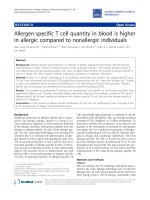
Figure 1
Thrombomodulin and protein C activation at the microcirculatory level.
The endothelial cell surface thrombin (Th)-binding protein
thrombomodulin (TM) is responsible for inhibition of thrombin activity.
TM, when bound to Th, forms a potent protein C activator complex.
Loss of TM and/or internalization results in Th–thrombin receptor (TR)
interaction. Loss of TM and associated protein C activation represents
the key event of decreased endothelial coagulation modulation ability
and increased inflammation pathways. Adapted from Iba and
coworkers [88]. ATIII, antithrombin III; NF-κ, nuclear factor-κB; PAI,
plasminogen activator inhibitor.
ENDOTHELIAL CELL
AT III
AT III
T
T
M
M
Th
Th
Th
Th
Protein C
Activated
pr
otein C
thrombomodulin
thrombomodulin
anti
coagulopathic
changes
Th
Th
Th
Th
Tissue factor ↑
PAI-1 ↑
Thrombomodulin ↓
Adhesion molecules ↑
Thrombin receptor ↑
Endothelin 1 ↑
Gap formation ↑
NF-
κB
T
T
R
R
Th
Th
thrombin
thrombin
receptor
receptor
pro
coagulopathic
changes
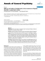
Figure 2
Coagulation and fibrinolysis pathways. Unperturbed endothelial cells
(ECs) provide anticoagulant (tissue factor pathway inhibitor [TFPI],
protein C [PC], protein S [PS], thrombomodulin [TM], heparan
sulphate [HS]) and fibrinolytic (tissue plasminogen activator [tPA])
properties. ATIII, antithrombin III; FXa, coagulation factor Xa; M,
activated monocyte; PAI, plasminogen activator inhibitor; SMC,
smooth muscle cell; TF, tissue factor.
EC
PAI
Antifibrinolysis Coagulation
Thrombin
M
Fibrin
Plasmin
PC
TM
Fibrinolysis Anticoagulation
ATIII
SMC
FXa
TF
tPA
TFPI
HS
PS
133
Although ECs probably have an important role in dissemi-
nated intravascular coagulation, there is also some evidence
favouring a major role for monocytes in the cellular mecha-
nisms of coagulation activation. We recently assessed the
relative impact of endothelial injury and monocyte activation
on coagulation disorders in our rabbit endotoxic shock model.
L-arginine and the angiotensin-converting enzyme inhibitor
perindopril were tested in that model for their demonstrated
ability to treat endothelial injury [38,39]. We found that both
L-arginine supplementation and perindopril could prevent
septic-shock-associated deterioration in endothelium-depen-
dent relaxation [40,41]. However, this preventive effect was
not associated with any reduction in TF expression, suggest-
ing that these two sepsis-associated abnormalities are not
strictly linked. In a subsequent study [42] we used an antigly-
coprotein IIb/IIIa, which attenuated endotoxin-induced mono-
cyte TF expression through decreased platelet activation.
This was associated with marked reduction in endothelial
injury, increased endothelium-derived relaxation and improved
survival rates in the treated animals. Those findings suggest
that monocyte activation and TF expression may be of impor-
tance in sepsis-associated injuries, and that coagulation acti-
vation may itself contribute to the EC injury observed during
sepsis.
Endothelial injury, in turn, exacerbates sepsis-induced coagu-
lation abnormalities. Indeed, release of endothelium-derived
factors such as nitric oxide (NO) and prostacyclin (PGI
2
) is
impaired. Because NO and PGI
2
not only control vascular
tone but also have antiadhesive and tissue plasminogen acti-
vator-like properties, loss of NO and PGI
2
release facilitates
leucocyte and platelet aggregation, and aggravation of coag-
ulopathy. Furthermore, when ECs generate adhesion mole-
cules during endotoxaemia that bind leucocytes and
monocytes, they favour enhancement in local procoagulant
reactions. The relationship between activation of innate immu-
nity and coagulation is phylogenetically ancient [43,44].
Localized activation of the coagulation system, as with the
innate immune response, serves to protect against a discrete
traumatic injury [43]. However, generalized intravascular
coagulation, as a generalized inflammatory response, is detri-
mental to the host, favouring widespread fibrin deposition and
altered tissue perfusion.
Endothelial activation
As a prelude to their migration into tissues, monocytes and
leucocytes must adhere to endothelium. Both adhesion to
and migration across endothelium are governed by the inter-
action of complementary adhesion molecules on the poly-
morphonuclear cells and endothelium [45]. The surface
expression, adhesion avidity and surface modulation of these
molecules are highly regulated by biological mediators such
as cytokines. Local synthesis of platelet-activating factor and
EC-derived cytokines such as interleukin-8, along with
tumour necrosis factor and interleukin-1, are important in
promoting neutrophil–EC interactions. ‘Endothelial activa-
tion’ refers to increased expression or release of endothelial
adhesion molecules.
The first step in migration consists of a ‘rolling’ of leucocytes
on endothelium, which involves the selectin family. Selectins
are molecules expressed on leucocytes (L-selectin), or even
on platelets (P-selectin) and on ECs (E-selectin); these act as
receptors that permit loose binding, which in turn facilitates
rolling. Selectins allow leucocytes to roll in the direction of
flow into the proximity of activating signals exhibited by ECs.
The second step involves receptors from the integrin family
(β
2
-integrin) and immunoglobulin-like receptors. These recep-
tors allow leucocyte arrest and adhesion strengthening. Three
heterodimers of β
2
-integrin are present on the outer cell mem-
brane of activated leucocytes and are collectively termed the
CD
11
/CD
18
complex. Stimulation of ECs induces expression
of cell surface adhesion molecules, which are members of the
immunoglobulin superfamily. Monoclonal antibodies to these
molecules have been shown to block leucocyte–EC interac-
tions and to improve sepsis-associated organ dysfunction
[46,47]. Endothelial adhesion molecules include ICAMs,
endothelial leucocyte adhesion molecules (E-selectins),
platelet EC adhesion molecules, and vascular cell adhesion
molecules.
In the third step, activated leucocytes migrate to the borders
of ECs to interact with ICAMs, endothelial leucocyte adhe-
sion molecules, platelet EC adhesion molecules or vascular
cell adhesion molecules (for review [48]). A large number of
experimental studies have documented the consequences of
inhibition of adhesion molecules. Inhibition of neutrophil
adherence to the ECs exerts significant protective effects in
these conditions [46,49].
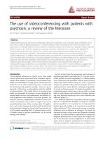
Available online />Figure 3
Sepsis and coagulation–fibrinolysis pathways. Exposure to
inflammatory and/or septic stimuli rapidly leads to procoagulant
behaviour. The profibrinolytic property of endothelial cells (ECs) is
blunted, due to decreased release of tissue plasminogen activator.
This occurs in a context of increased plasminogen activator inhibitor
(PAI)-1 release with antifibrinolysis. LPS, lipopolysaccharide; M,
activated monocyte; SMC, smooth muscle cell; TF, tissue factor; TM,
thrombomodulin.
EC
SMC
M
PAI
Antifibrinolysis Coagulation
LPS,
LPS,
cytokines
cytokines
inactive
inactive
TF
FXa
TM
TF
134
Interestingly, decreased reactive hyperaemia (suggesting
modified endothelial-derived relaxation) was also demon-
strated to coexist with increased leucocyte aggregation and
ICAM-1 levels [50]. It is also important to emphasize that
recent evidence suggested that adhesion can occur inde-
pendent of adhesion molecules in organs such as lung or
liver. This led to the hypothesis that stimulus-induced
increases in actin-containing stress fibres (such as LPS) at
the cell periphery lead to decreased deformability, prevent-
ing neutrophils from trafficking through the capillary bed and
therefore increasing their sequestration at sites of inflamma-
tion [51].
Sessler and coworkers [52] measured blood level of the
adhesion molecule ICAM-1 as a potential marker of EC acti-
vation in septic adults and healthy volunteers. Those investi-
gators established a relationship between increased ICAM-1
levels and consequences of sepsis (i.e. multiple organ failure
and death). Watanabe and coworkers [53] prevented endo-
toxin shock in rabbits by administering a specific monoclonal
antibody against CD
18
(integrin β
2
). In a mouse lethal septic
shock model, Xu and coworkers [54] observed that animals
deficient in ICAM-1 were markedly protected against death.
Whereas 80% of wild-type animals died within 48 hours
after receiving 40 mg/kg LPS, more than 90% of ICAM-1-
deficient animals survived for longer than 4 days. Interest-
ingly, EC dysfunction was found to involve a
CD
18
-dependent neutrophil adherent mechanism. Consis-
tently, Matsukawa and coworkers [55] recently provided evi-
dence on the contributions of E-selectin and P-selectin to
lethality in septic peritonitis. Mice that genetically lacked
endothelial selectins were shown to be resistant. The experi-
ments demonstrated that endothelial selectin mediated leu-
cocyte rolling impacts on mouse survival by influencing the
serum level of cytokines and by preventing renal dysfunction
– a potential cause of death in that context.
Endothelial dysfunction
The term ‘endothelial dysfunction’ refers to decreased
endothelial-dependent vascular relaxation or NO release, and
decreased expression or activity of endothelial constitutive
NO synthase (ecNOS). Endothelium-derived relaxation
and/or production of endothelium-derived NO from the amino
acid
L-arginine by ecNOS may be used as an indicator of EC
function. For example, the relaxing response of in vitro iso-
lated vascular rings to picomolar concentrations of acetyl-
choline is dependent on the presence and integrity of ECs
[56]. In vivo endothelial function can be determined by mea-
surement of forearm blood flow responses to intra-arterial
infusions of endothelium-dependent (i.e. acetylcholine) and
endothelium-independent vasodilators (i.e. sodium nitroprus-
side). Drugs are infused at a constant rate (1 ml/min) with an
infusion pump. Forearm blood flow is recorded for 10 s at
15-s intervals during the last 3 min of the drug and saline infu-
sion period using venous occlusion plethysmography com-
bined with a rapid cuff inflator [57–59].
Abnormal endothelial-dependent vascular relaxation has been
recognized in multiple sepsis conditions. Several investiga-
tions, including our own [17,60,61], have demonstrated
attenuated acetylcholine-induced relaxation in vascular rings
isolated from large arteries. Apart from anatomical injuries,
such abnormalities observed in these vessels may result from
several mechanisms: alteration in EC surface receptors; mod-
ified signal transduction pathways (receptor–ecNOS cou-
pling); altered function and/or density of the ecNOS;
changes in pathways that lead to release of NO; and/or
changes in mechanisms that participate in subsequent degra-
dation of NO.
In healthy volunteers, even brief exposure to endotoxin or
certain cytokines impairs endothelium-dependent relaxation
for many days [62,63]. This effect has been termed ‘endothe-
lial stunning’. After recovery from the acute insult, the
endothelium may remain dysfunctional (‘stunned’) for a long
period of time before full recovery. Hingorani and coworkers
[59] also demonstrated that a mild inflammatory response,
such as that generated by Salmonella typhi vaccine, is asso-
ciated with temporary but profound dysfunction of arterial
endothelium in both resistance and conduit vessels following
application of both physical and pharmacological dilator
stimuli. According to the concept of intrinsic metabolic regu-
lation, vasodilatation in tissues with relatively high metabolic
rates competes with sympathetic vasoconstrictor tone,
thereby adjusting the balance between local tissue oxygen
supply and demand. Although the nature of the oxygen-sensi-
tive structures that act at the local tissue level is not com-
pletely understood, ECs in direct contact with blood have a
number of properties that render them effective sensors. The
endothelium and smooth muscle of arteries and arterioles
appear to be coupled both structurally and functionally.
Sensing involves local depolarization/hyperpolarization of the
capillary EC, and communication is achieved by electronic
spread via endothelium/smooth muscle cell–cell gap junc-
tions [2,64]. During an hypoxic challenge, the ability of a
tissue to extract oxygen – and to minimize shunting through
areas with a high rate of perfusion relative to their oxygen
uptake – may therefore be considered an integrative test of
endothelial function and microcirculatory coordination [65].
We investigated the role of the endothelium in regulating the
balance between oxygen demand and supply within an indi-
vidual organ in an in vivo model of endothelial stripping in the
dog hind limb [66]. The hind limb vascular endothelium was
removed by injecting deoxycholate into the perfusing artery
before ischaemic challenge. Deoxycholate – a detergent
used to remove endothelium in in vitro studies – removes vas-
cular endothelium within arteries, arterioles, capillaries and
veins. It achieves this without causing apparent damage to
either the vascular smooth muscle layer or the skeletal muscle
parenchyma, as assessed by in vitro and in vivo studies of
pharmacological vascular reactivity, tissue histology and elec-
tron microscopy. Hind limb oedema or capillary plugging by
Critical Care April 2003 Vol 7 No 2 Vallet
135
endothelial fragments was not observed. During progressive
limitation of oxygen supply to the limb, a profound and signifi-
cant impairment in limb oxygen extraction ability (41.7%
versus 81% in controls) at critical oxygen delivery (at which
oxygen uptake begins to decrease) was observed. We con-
cluded that this severe limitation in the increase in oxygen
extraction capabilities during ischaemia suggested that vas-
cular endothelium plays an important role in matching oxygen
supply to demand.
In order to test the role of the endothelial-derived relaxing
factors NO and PGI
2
, we investigated, in a third group of
dogs, the influence of a combination of N
G
-nitro-L-arginine
methyl ester (an inhibitor of NO synthesis) and indomethacin
(an inhibitor of PGI
2
synthesis) [66]. In these dogs treated
with indomethacin plus N
G
-nitro-
L-arginine methyl ester, the
severity of the oxygen extraction defect was lower than that
observed in the deoxycholate-treated dogs, suggesting that
other mediators and/or mechanisms may be involved in
microcirculatory control during hypoxia. As suggested above,
one of these mediators or mechanisms could be related to
hyperpolarization. Membrane potential is an important deter-
minant of vascular smooth muscle tone through its influence
on calcium influx via voltage-gated calcium channels. Hyper-
polarization (as well as depolarization) has been shown to be
a means of cell–cell communication in upstream vasodilatation
and microcirculatory coordination [67]. It is important to
emphasize that intercell coupling exclusively involves ECs.
Interestingly, it was recently shown that sepsis, a situation that
is characterized by impaired tissue perfusion and abnormal
oxygen extraction, is associated with abnormal inter-EC cou-
pling and reduction in the arteriolar conducted response [68].
An intra-organ defect in blood flow related to abnormal vascu-
lar reactivity, cell adhesion and coagulopathy may account for
impaired organ oxygen regulation and function. If specific
classes of microvessels must or must not be perfused to
achieve efficient oxygen extraction during limitation in oxygen
delivery, then impaired vascular reactivity and vessel injury
might produce a pathological limitation in supply. In sepsis,
the inflammatory response profoundly alters circulatory
homeostasis, and this has been referred to as a ‘malignant
intravascular inflammation’ that alters vasomotor tone and the
distribution of blood flow among and within organs [69].
These mechanisms might coexist with other types of sepsis-
associated cell dysfunction. For example, data suggest that
endotoxin directly impairs oxygen uptake in ECs and indicate
the importance of endothelium respiration in maintaining vas-
cular homeostasis under conditions of sepsis [70].
Abnormal oxygen extraction is a key feature of severe sepsis
and septic shock. In an experimental study in dogs, an
ablated reactive hyperaemia was associated with endotox-
aemia-induced impaired oxygen extraction at the level of the
gastrointestinal tract [71]. Nevière and coworkers [72]
showed that reactive hyperaemia is attenuated in critically ill
patients with septic shock, despite normal or elevated whole-
body oxygen delivery. Proposed mechanisms to explain
blunted hyperaemia in septic patients might include impaired
vascular reactivity and/or microvascular obstruction that limits
the number of recruitable capillaries. In critically ill patients with
sepsis, it has been shown that decreased reactive hyperaemia
coexists with increased leucocyte adhesion and increased
release in surrogate markers of endothelium injury [50,73].
Thus, assessment of reactive hyperaemia might be used in
the near future to evaluate the effects of treatments aimed at
restoring endothelial function and tissue perfusion, such as
coagulation modulators or leucocyte adhesion antagonists.
Conclusion
How do all of these altered properties contribute to altered
perfusion and organ dysfunction? The combining effect of
altered vascular relaxation, altered blood flow distribution,
increased leucocyte adhesion and decreased coagulation
modulation should significantly contribute to microcirculatory
heterogeneity and lowered perfusion. Studies in the isolated
perfused rabbit heart [74], autoperfused rat cremaster [75]
and rat mesentery [76,77] suggested that these mechanisms
are operative in the microvasculature. On an intravital
microscopy extensor digitorum longus muscle model in rats
with peritonitis [78], it was shown that sepsis is associated
with a reduction in tissue perfused capillary density of up to
36%, increased perfusion heterogeneity and mean intercapil-
lary distance, contributing to functional shunting. In another
study [79], endotoxin administration resulted in a significant
enhancement in leucocyte–EC interaction, as indicated by
transiently increased number of leucocytes firmly attaching to
the microvascular endothelium of arterioles and venules [79].
Microvascular injury and/or the appearance of greater hetero-
geneity of microvascular distribution of oxygen supply with
respect to oxygen demand in endotoxin-treated animals is
consistent with the observation that endotoxin impairs oxygen
extraction [80–82]. The direct relationship between hetero-
geneity, decreased oxygen extraction and tissue acidosis was
recently confirmed in a pig model of endotoxic shock [83].
Consistent with the hypothesis that alteration in endothelium
plays a major in the pathophysiology of sepsis, it was
observed that chronic ecNOS overexpression in the endothe-
lium of mice resulted in resistance to LPS-induced hypoten-
sion, lung injury and death [84]. This observation was
confirmed by another group of investigators, who used trans-
genic mice overexpressing adrenomedullin [85] – a vasodilat-
ing peptide that acts at least in part via an NO-dependent
pathway. They demonstrated resistance of these animals to
LPS-induced shock, and lesser declines in blood pressure
and less severe organ damage than occurred in the control
animals. It might therefore be of importance to favour ecNOS
expression and function during sepsis. The recent negative
results obtained with therapeutic strategies aimed at blocking
inducible NOS with the nonselective NOS inhibitor N
G
-
Available online />136
monomethyl-
L-arginine in human septic shock [86] further
confirm the overall importance of favoring vessel dilatation. In
contrast, positive results obtained with corticosteroids [87]
and APC [37] suggest that improving haemodynamics while
decreasing vasopressor agents (corticosteroids) and limiting
coagulation activation are logical strategies that may greatly
favour tissue perfusion and improved oxygen delivery.
Competing interests
None declared.
References
1. Cines DB, Pollak ES, Buck CA, Loscalzo J, Zimmerman GA,
McEver RP, Pober JS, Wick TM, Konkle BA, Schwartz BS, Bar-
nathan ES, McCrae KR, Hug BA, Schmidt AM, Stern DM:
Endothelial cells in physiology and in the pathophysiology of
vascular disorders. Blood 1998, 91:3527-3561.
2. Sandow SL, Hill CE: Incidence of myoendothelial gap junc-
tions in the proximal and distal mesenteric arteries of the rat
is suggestive of a role in endothelium-derived hyperpolarizing
factor-mediated responses. Circ Res 2000, 86:341-346.
3. Stefanec T: Endothelial apoptosis: could it have a role in the
pathogenesis and treatment of disease. Chest 2000, 117:841-
854.
4. Reidy MA, Schwartz SM: Endothelial injury and regeneration.
IV. Endotoxin: a nondenuding injury to aortic endothelium. Lab
Invest 1983, 48:25-34.
5. Reidy MA, Bowyer DE: Scanning electron microscopy: mor-
phology of aortic endothelium following injury by endotoxin
and during subsequent repair. Atherosclerosis 1977, 26:319-
328.
6. Leclerc J, Pu Q, Corseaux D, Haddad E, Decoene C, Bordet R,
Six I, Jude B, Vallet B: A single endotoxin injection in the rabbit
causes prolonged blood vessel dysfunction and a procoagu-
lant state. Crit Care Med 2000, 28:3672-3678.
7. Lee M, Schuessler G, Chien S: Time dependent effetcs of
endotoxin on the ultrastructure of the aortic endothelium.
Artery 1998, 15:71-89.
8. Wang P, Wood TJ, Zhou M, Ba ZF, Chaudry IH: Inhibition of the
biological activity of tumor necrosis factor maintains vascular
endothelial cell function during hyperdynamic sepsis. J
Trauma 1996, 40:694-701.
9. Bauer PR: Microvascular responses to sepsis: clinical signifi-
cance. Pathophysiology 2002, 8:141-148.
10. Bajaj MS, Tricomi SM: Plasma levels of the three endothelial-
specific proteins von Willebrand factor, tissue factor pathway
inhibitor, and thrombomodulin do not predict the develop-
ment of acute respiratory distress syndrome. Intensive Care
Med 1999, 25:1259-1266.
11. Kayal S, Jais JP, Aguini N, Chaudiere J, Labrousse J: Elevated cir-
culating E-selectin, intercellular adhesion molecule 1, and von
Willebrand factor in patients with severe infection. Am J Respir
Crit Care Med 1998, 157:776-784.
12. Borchiellini A, Fijnvandraat K, ten Cate JW, Pajkrt D, van Deventer
SJ, Pasterkamp G, Meijer-Huizinga F, Zwart-Huinink L, Voorberg J,
van Mourik JA: Quantitative analysis of von Willebrand factor
propeptide release in vivo: effect of experimental endotox-
emia and administration of 1-deamino-8-D-arginine vaso-
pressin in humans. Blood 1996, 88:2951-2958.
13. Van Mourik JA, Boertjes R, Huisveld IA, Fijnvandraat K, Pajkrt D,
van Genderen PJ, Fijnheer R: von Willebrand factor propeptide
in vascular disorders: a tool to distinguish between acute and
chronic endothelial cell perturbation. Blood 1999, 94:179-185.
14. Blann AD, Babbs C, Neuberger JM: Endothelial cell damage in
primary biliary cirrhosis: influence of cholestasis and
immunological mechanisms. Clin Exp Immunol 1992, 90:88-
92.
15. Rubin DB, Wiener-Kronish JP, Murray JF, Green DR, Turner J,
Luce JM, Montgomery AB, Marks JD, Matthay MA: Elevated von
Willebrand factor antigen is an early plasma predictor of acute
lung injury in nonpulmonary sepsis syndrome. J Clin Invest
1990, 86:474-480.
16. Mutunga M, Fulton B, Bullock R, Batchelor A, Gascoigne A, Gille-
spie JI, Baudouin SV: Circulating endothelial cells in patients
with septic shock. Am J Respir Crit Care Med 2001, 163:195-
200.
17. Wiel E, Vallet B: Vascular endothelial cell dysfunction in septic
shock. Crit Care Med 2001, 29(suppl):S36-S41.
18. Taylor FB, Haddad PA, Hack E, Chang AC, Peer GT, Morrissey
JH, Li A, Allen R, Caa H, Kinasewitz GT: Two-stage response to
endotoxin infusion into normal human subjects: correlation of
blood phagocyte luminescence with clinical and laboratory
markers of the inflammatory, hemostatic response. Crit Care
Med 2001, 29:326-334.
19. Bombeli T, Mueller M, Haeberli A: Anticoagulant properties of
the vascular endothelium. Thromb Haemost 1997, 77:408-423.
20. Rapaport S, Rao L: Initiation and regulation of tissue factor-
dependent blood coagulation. Arterioscler Thromb 1992, 12:
1111-1121.
21. Faust SN, Levin M, Harrison OB, Goldin RD, Lockhart MS, Kon-
daveeti S, Laszik Z, Esmon CT, Heyderman RS: Dysfunction of
endothelial protein C activation in severe meningococcal
sepsis. N Engl J Med 2001, 345:408-416.
22. Regoeczi E, Brain MC: Organ distribution of fibrin in dissemi-
nated intravascular coagulation. Br J Haematol 1969, 17:73-
81.
23. Fourrier F, Chopin C, Goudemand J, Hendrycx S, Caron C, Rime
A, Marey A, Lestavel P: Septic shock, multiple organ failure,
and disseminated intravascular coagulation. Compared pat-
terns of antithrombin III, protein C, and protein S deficiencies.
Chest 1992, 101:816-823.
24. Gando S, Nakanishi Y, Tedo I: Cytokines and plasminogen acti-
vator inhibitor-1 in posttrauma disseminated intravascular
coagulation: relationship to multiple organ dysfunction syn-
drome. Crit Care Med 1995, 23:1835-1842.
25. Creasey AA, Chang AC, Feigen L, Wun TC, Taylor FB, Hinshaw
LB: Tissue factor pathway inhibitor reduces mortality from
Escherichia coli septic shock. J Clin Invest 1993, 91:2850-
2856.
26. Kessler CM, Tang Z, Jacobs HM, Szymanski LM: The supraphar-
macologic dosing of antithrombin concentrate for Staphylo-
coccus aureus-induced disseminated intravascular
coagulation in guinea pigs: substantial reduction in mortality
and morbidity. Blood 1997, 89:4393-4401.
27. Carr C, Bild GS, Chang AC: Recombinant E. coli-derived tissue
factor pathway inhibitor reduces coagulopathic and lethal
effects in the baboon gram-negative model of septic shock.
Circ Shock 1994, 44:126-137.
28. Camerota AJ, Creasey AA, Patla V, Larkin VA, Fink MP: Delayed
treatment with recombinant human tissue factor pathway
inhibitor improves survival in rabbits with gram-negative peri-
tonitis. J Infect Dis 1998, 177:668-676.
29. Grey ST, Tsuchida A, Hau H, Orthner CL, Salem HH, Hancock
WW: Selective inhibitory effects of the anticoagulant acti-
vated protein C on the responses of human mononuclear
phagocytes to LPS, IFN-gamma, or phorbol ester. J Immunol
1994, 153:3664-3672.
30. Grinnell BW, Hermann RB, Yan SB: Human protein C inhibits
selectin-mediated cell adhesion: role of unique fucosylated
oligosaccharide. Glycobiology 1994, 4:221-225.
31. Jackson CV, Bailey BD, Shetler TJ: Pharmacological profile of
recombinant, human activated protein C (LY203638) in a
canine model of coronary artery thrombosis. J Pharmacol Exp
Ther 2000, 295:967-971.
32. Smith OP, White B, Vaughan D, Rafferty M, Claffey L, Lyons B,
Casey W: Use of protein-C concentrate, heparin, and
haemodiafiltration in meningococcus-induced purpura fulmi-
nans. Lancet 1997, 350:1590-1593.
33. Fourrier F, Chopin C, Huart JJ, Runge I, Caron C, Goudemand J:
Double-blind, placebo-controlled trial of antithrombin III con-
centrates in septic shock with disseminated intravascular
coagulation. Chest 1993, 104:882-888.
34. Baudo F, Caimi TM, de Cataldo F, Ravizza A, Arlati S, Casella G,
Carugo D, Palareti G, Legnani C, Ridolfi L, Rossi R, D’Angelo A,
Crippa L, Giudici D, Gallioli G, Wolfler A, Calori G: Antithrombin
III (ATIII) replacement therapy in patients with sepsis and/or
postsurgical complications: a controlled double-blind, ran-
domized, multicenter study. Intensive Care Med 1998, 24:336-
342.
Critical Care April 2003 Vol 7 No 2 Vallet
137
Available online />35. Eisele B, Lamy M, Thijs LG, Keinecke HO, Schuster HP, Matthias
FR, Fourrier F, Heinrichs H, Delvos U: Antithrombin III in
patients with severe sepsis. A randomized, placebo-con-
trolled, double-blind multicenter trial plus a meta-analysis on
all randomized, placebo-controlled, double-blind trials with
antithrombin III in severe sepsis. Intensive Care Med 1998, 24:
663-672.
36. Warren BL, Eid A, Singer P, Pillay SS, Carl P, Novak I, Chalupa P,
Atherstone A, Penzes I, Kubler A, Knaub S, Keinecke HO, Hein-
richs H, Schindel F, Juers M, Bone RC, Opal SM: Caring for the
critically ill patient. High-dose antithrombin III in severe
sepsis: a randomized controlled trial. JAMA 2001, 286:1869-
1878
37. Bernard GR, Vincent JL, Laterre PF, LaRosa SP, Dhainaut JF,
Lopez-Rodriguez A, Steingrub JS, Garber GA, Helterbrand JD, Ely
EW, Fisher CJ: Efficacy and safety of recombinant human acti-
vated protein C for severe sepsis. N Engl J Med 2001, 344:
699-709.
38. Hamon M, Vallet B, Bauters C, Wernert N, McFadden EP,
Lablanche JM, Dupuis B, Bertrand ME: Long-term oral adminis-
tration of L-arginine reduces intimal thickening and enhances
neoendothelium-dependent acetylcholine-induced relaxation
after arterial injury. Circulation 1994, 90:1357-1362.
39. Van Belle E, Vallet B, Auffray JL, Bauters C, Hamon M, McFadden
EP, Lablanche JM, Dupuis B, Bertrand ME: NO synthesis is
involved in structural and functional effects of ACE inhibitors
in injured arteries. Am J Physiol 1996, 270:H298-H305.
40. Wiel E, Pu Q, Corseaux D, Robin E, Bordet R, Lund N, Jude B,
Vallet B: Effect of L-arginine on endothelial injury and hemo-
stasis in rabbit endotoxin shock. J Appl Physiol 2000, 89:
1811-1818.
41. Leclerc JF, Pu Q, Bordet R, Bauters C, Vallet B: Endothelial cell
dysfunction occurring during septic shock is prevented by
angiotensin converting enzyme inhibitor [abstract]. Am J
Respir Crit Care Med 2000, 157:A101.
42. Pu Q, Wiel E, Corseaux D, Bordet R, Azrin MA, Ezekowitz MD,
Lund N, Jude B, Vallet B: Beneficial effect of glycoprotein
IIb/IIIa inhibitor (AZ-1) on endothelium in Escherichia coli
endotoxin-induced shock. Crit Care Med 2001, 29:1181-
1188.
43. Opal SM: Phylogenetic and functional relationships between
coagulation and the innate immune response. Crit Care Med
2000, 28(suppl):S77-S80.
44. Opal SM, Palardy JE, Parejo NA, Creasey AA: The activity of
tissue factor pathway inhibitor in experimental models of
superantigen-induced shock and polymicrobial intra-abdomi-
nal sepsis. Crit Care Med 2001, 29:13-17.
45. Springer T: Traffic signals on endothelium for lymphocyte
recirculation and leukocyte emigration. Ann Rev Physiol 1995,
57:827-872.
46. Gardinali M, Borrelli E, Chiara O, Lundberg C, Padalino P, Conci-
ato L, Cafaro C, Lazzi S, Luzi P, Giomarelli PP, Agostoni A: Inhibi-
tion of CD11b/CD18 complex prevents acute lung injury and
reduces mortality after peritonitis in rabbits. Am J Respir Crit
Care Med 2000, 161:1022-1029.
47. Kuebler WM, Borges J, Sckell A, Kuhnle EH, Bergh K, Messmer
K, Goetz AE: Role of L-selectin in leukocyte sequestration in
lung capillaries in a rabbit model of endotoxemia. Am J Respir
Crit Care Med 2000, 161:36-43.
48. Varani J, Ward P: Mechanisms of endothelial cell injury in
acute inflammation. Shock 1994, 2:311-319.
49. Ma X, Tsao P, Lefer A: Antibody to CD-18 exerts endothelial
cell and cardiac protective effects in myocardial ischemia and
reperfusion. J Clin Invest 1995, 88:1237-1243.
50. Astiz ME, DeGent GE, Lin RY, Rackow EC: Microvascular func-
tion and rheologic changes in hyperdynamic sepsis. Crit Care
Med 1995, 23:265-271.
51. Doerschuk CM: Leukocyte trafficking in alveoli and airway pas-
sages. Respir Res 2000, 1:136-140.
52. Sessler C, Windsor A, Schwartz M: Circulating ICAM-1 is
increased in septic shock. Am J Respir Crit Care Med 1995,
151:1420-1427.
53. Watanabe S, Mukaida N, Ikeda N, Akiyama M, Harada A, Nakan-
ishi I, Nariuchi H, Watanabe Y, Matsushima K: Prevention of
endotoxin shock by an antibody against leukocyte integrin
beta 2 through inhibiting production and action of TNF. Int
Immunol 1995, 7:1037-1046.
54. Xu H, Gonzalo JA, St Pierre Y, Williams IR, Kupper TS, Cotran
RS, Springer TA, Gutierrez-Ramos JC: Leucocytosis and resis-
tance to septic shock in intercellular adhesion molecule 1-
deficient mice. J Exp Med 1994, 180:95-109.
55. Matsukawa A, Lukacs NW, Hogaboam CM, Knibbs RN, Bullard
DC, Kunkel SL, Stoolman LM: Mice genetically lacking endothe-
lial selectins are resistant to the lethality in septic peritonitis.
Exp Mol Pathol 2002, 72:68-76.
56. Furchgott R, Zawadzki J: The obligatory role of endothelial cells
in the relaxation of arterial smooth muscle by acetylcholine.
Nature 1980, 288:373-376.
57. Makimattila S, Virkamaki A, Groop PH, Cockcroft J, Utriainen T,
Fagerudd J, Yki-Jarvinen H: Chronic hyperglycemia impairs
endothelial function and insulin sensitivity via different mech-
anisms in insulin-dependent diabetes mellitus. Circulation
1996, 94:1276-1282.
58. Vakkilainen J, Makimattila S, Seppala-Lindroos A, Vehkavaara S,
Lahdenpera S, Groop PH, Taskinen MR, Yki-Jarvinen H: Endothe-
lial dysfunction in men with small LDL particles. Circulation
2000, 102:716-721.
59. Hingorani AD, Cross J, Kharbanda RK, Mullen MJ, Bhagat K,
Taylor M, Donald AE, Palacios M, Griffin GE, Deanfield JE, MacAl-
lister RJ, Vallance P: Acute systemic inflammation impairs
endothelium-dependent dilation in humans. Circulation 2000,
102:994-999.
60. Parker JL, Adams HR: Selective inhibition of endothelium-
dependent vasodilatator capacity by Escherichia coli endotox-
emia. Circ Res 1993, 72:539-551.
61. Umans JG, Wylam ME, Umans JG, Wylam ME, Samsel RW,
Edwards J, Schumacker PT: Effects of endotoxin in vivo on
endothelial and smooth-muscle function in rabbit and rat
aorta. Am Rev Respir Dis 1993, 148:1638-1645.
62. Bhagat K, Collier J, Vallance P: Local venous responses to
endotoxin in humans. Circulation 1996, 94:490-497.
63. Bhagat K, Moss R, Collier J, Vallance P: Endothelial ‘stunning’
following a brief exposure to endotoxin: a mechanism to link
infection and infarction? Cardiovasc Res 1996, 32:822-829.
64. Emerson GG, Segal SS: Endothelial cell pathway for conduc-
tion of hyperpolarization and vasodilation along hamster feed
artery. Circ Res 2000, 86:94-100.
65. Vallet B: Endothelial cell dysfunction and abnormal tissue per-
fusion. Crit Care Med 2002, 30(suppl):S229-S234.
66. Curtis SE, Vallet B, Winn MJ, Caufield JB, King CE, Chapler CK,
Cain SM: Role of the vascular endothelium in O
2
extraction
during progressive ischemia in canine skeletal muscle. J Appl
Physiol 1995, 79:1351-1360.
67. Segal SS: Microvascular recruitment in hamster striated
muscle: role for conducted vasodilation. Am J Physiol 1991,
261:H180-H189.
68. Tyml K, Wang X, Lidington D, Ouellette Y: Lipopolysaccharide
reduces intercellular coupling in vitro and arteriolar con-
ducted response in vivo. Am J Physiol 2001, 281:H1397-
H1406.
69. Pinsky MR: Regional blood flow distribution. In The Splanchnic
Circulation: No Longer a Silent Partner. Edited by Pinsky MR,
Dhainaut JF, Artigas A. Berlin: Springer-Verlag; 1995:1-13.
70. Motterlini R, Kerger H, Green CJ, Winslow RM, Intaglietta M:
Depression of endothelial and smooth muscle cell oxygen
consumption by endotoxin. Am J Physiol 1998, 275:H776-
H782.
71. Nelson DP, Samsel RW, Wood LDH, Schumacker PT: Pathologi-
cal supply dependence of systemic and intestinal O
2
uptake
during endotoxemia. J Appl Physiol 1988, 64:2410-2419.
72. Nevière R, Mathieu D, Chagnon JL, Lebleu N, Millien JP, Wattel F:
Skeletal muscle microvascular blood flow and oxygen trans-
port in patients with severe sepsis. Am J Respir Crit Care Med
1996, 153:191-195.
73. Kirschenbaum LA, Astiz ME, Rackow EC, Saha DC, Lin R:
Microvascular response in patients with cardiogenic shock.
Crit Care Med 2000, 28:1290-1294.
74. Smith R, Palmer R, Moncada S: Coronary vasodilatation
induced by endotoxin in the rabbit isolated perfused heart is
nitric oxide-dependent and inhibited by dexamethasone. Br J
Pharmacol 1991, 140:5-6.
75. Lubbe AS, Garrison RN, Cryer HM, Alsip NL, Harris PD: EDRF as
a possible mediator of sepsis-induced arteriolar dilatation in
skeletal muscle. Am J Physiol 1992, 262:H880-H887.
138
Critical Care April 2003 Vol 7 No 2 Vallet
76. Schneider F, Schott C, Stoclet JC, Julou-Schaeffer G: L-arginine
induces relaxation of small mesenteric arteries from endo-
toxin-treated rats. Eur J Pharmacol 1992, 211:269-272.
77. Wang P, Ba ZF, Chaudry IH: Endothelium-dependent relax-
ation is depressed at the macro- and microcirculatory levels
during sepsis. Am J Physiol 1995, 269:R988-R994.
78. Lam C, Tyml K, Martin C, Sibbald W: Microvascular perfusion is
impaired in a rat model of normotensive sepsis. J Clin Invest
1994, 94:2077-2083.
79. Hoffmann JN, Vollmar B, Inthorn D, Schilberg FW, Menger MD: A
chronic model for intravital microscopic study of microcircula-
tory disorders and leukocyte/endothelial cell interaction
during normotensive endotoxemia. Shock 1999, 12:355-364.
80. Bredle DL, Samsel RW, Schumacker PT, Cain SM: Critical O
2
delivery to skeletal muscle at high and low PO
2
in endotox-
emic dogs. J Appl Physiol 1989, 66:2553-2558.
81. Bredle DL, Cain SM: Systemic and muscle oxygen
uptake/delivery after dopexamine infusion in endotoxic dogs.
Crit Care Med 1991, 19:198-204.
82. Vallet B, Lund N, Curtis SE, Kelly D, Cain SM: Gut and muscle
tissue PO
2
in endotoxemic dogs during shock and resuscita-
tion. J Appl Physiol 1994, 76:793-800.
83. Tugtekin IF, Radermacher P, Theisen M, Matejovic M, Stehr A,
Ploner F, Matura K, Ince C, Georgieff M, Trager K: Increased
ileal-mucosal-arterial PCO
2
gap is associated with impaired
villus microcirculation in endotoxic pigs. Intensive Care Med
2001, 27:757-766.
84. Yamashita T, Kawashina S, Ohashi Y: Resistance to endotoxin
shock in transgenic mice overexpressing endothelial nitric
oxide synthase. Circulation 2000, 101:931-937.
85. Shindo T, Kurihara H, Maemura K, Kurihara Y, Kuwaki T, Izumida
T, Minamino N, Ju KH, Morita H, Oh-hashi Y, Kumada M, Kangawa
K, Nagai R, Yazaki Y: Hypotension and resistance to
lipopolysaccharide-induced shock in transgenic mice overex-
pressing adrenomedullin in their vasculature. Circulation
2000, 101:2309-2316.
86. Grover R, Lopez A, Lorente J, Steingrub J, Bakker J, Willatts S:
Multi-center, randomized, placebo-controlled, double bind
study of the nitric oxide synthase inhibitor 546C88: effect on
survival in patients in septic shock [abstract]. Crit Care Med
1999, 27:A33.
87. Annane D, Sebille V, Charpentier C, Bollaert PE, Francois B,
Korach JM, Capellier G, Cohen Y, Azoulay E, Troche G, Chaumet-
Riffaut P, Bellissant E: Effect of treatment with low doses of
hydrocortisone and fludrocortisone on mortality in patients
with septic shock. JAMA 2002 , 288:862-871.
88. Iba T, Kidokoro A, Yagi Y: The role of the endothelium in
changes in procoagulant activity in sepsis. J Am Coll Surg
1998, 187:321-329.