Báo cáo y học: " Molecular mechanisms of neuroinvasion by monocytes-macrophages in HIV-1 infection" pdf
Bạn đang xem bản rút gọn của tài liệu. Xem và tải ngay bản đầy đủ của tài liệu tại đây (1.28 MB, 11 trang )
Gras and Kaul Retrovirology 2010, 7:30
/>Open Access
REVIEW
BioMed Central
© 2010 Gras and Kaul; licensee BioMed Central Ltd. This is an Open Access article distributed under the terms of the Creative Commons
Attribution License ( which permits unrestricted use, distribution, and reproduction in
any medium, provided the original work is properly cited.
Review
Molecular mechanisms of neuroinvasion by
monocytes-macrophages in HIV-1 infection
Gabriel Gras*
1
and Marcus Kaul
2
Abstract
HIV associated neurocognitive disorders and their histopathological correlates largely depend on the continuous
seeding of the central nervous system with immune activated leukocytes, mainly monocytes/macrophages from the
periphery. The blood-brain-barrier plays a critical role in this never stopping neuroinvasion, although it appears
unaltered until the late stage of HIV encephalitis. HIV flux that moves toward the brain thus relies on hijacking and
exacerbating the physiological mechanisms that govern blood brain barrier crossing rather than barrier disruption. This
review will summarize the recent data describing neuroinvasion by HIV with a focus on the molecular mechanisms
involved.
Introduction
HIV-1 infection is often associated with neurocognitive
impairment and the various degrees of severity have
recently been categorized under the overarching term
HIV associated neurocognitive disorders (HAND) [1].
HAND defines three categories of clinical disorders
according to standardized measures of dysfunction: i)
asymptomatic neurocognitive impairment (ANI), ii) mild
neurocognitive disorder (MND) and iii) HIV-associated
dementia (HAD) [2].
HAD constitutes the most severe form of HAND [1]
which presented itself prominently at the beginning of
the AIDS epidemic but primarily in patients with low
CD4+ cell counts and advanced HIV disease [3]. Intro-
duction of combination anti-retroviral therapy (cART)/
highly active antiretroviral therapy (HAART) in the mid
1990 improved treatment of HIV infection and often pre-
vented or at least delayed the progression to AIDS and
HAD. In recent years, however, and since HIV patients
live longer, the incidence of dementia as an AIDS-defin-
ing illness has increased, and HAD now defines a signifi-
cant independent risk factor for death due to AIDS [4,5].
While in the HAART era MND appears to be more prev-
alent than frank dementia, it appears important to take
these long lasting disorders into account in patients' fol-
low up as they may profoundly affect quality of life, com-
plicate autonomy, modify treatment compliance and
induce a high level of vulnerability. Moreover, clinical
observations over more than 10 years also suggest that
HAART cannot completely protect from HAD [1,4-7]. In
addition, it is possible that life-long treatment with
HAART itself generates a toxicological problem which
may affect neurocognitive performance on its own [5,8].
The neuropathological correlates of HIV-1 infection
are generally referred to as HIV encephalitis (HIVE) and
comprise microglial nodules, activated resident micro-
glia, multinucleated giant cells, infiltration predomi-
nantly by monocytoid cells, including blood-derived
macrophages, widespread reactive astrocytosis, myelin
pallor, and decreased synaptic and dendritic density in
combination with distinct neuronal loss [9-11]. HIV-1
associated neuronal damage and loss have been reported
for numerous regions of the central nervous system
(CNS), including frontal cortex [12,13], substantia nigra
[14], cerebellum [15], and putamen [16].
The neuropathology of HIV infection and AIDS has
changed under the influence of HAART [6,7,17]. Neu-
roinflammation was commonly observed in HIV patients
at the beginning of the AIDS epidemic and before the
introduction of HAART, and usually increased through-
out the progression of infected individuals from the
latent, asymptomatic stage of the disease to AIDS and
HAD [18]. Surprisingly, neuroinflammation seems to
persist or even flourish since the advent of HAART
[17,19]. Autopsy studies in recent years found microglial
activation comparable to that in fully developed AIDS
* Correspondence:
1
Institute of Emerging Diseases and Innovative Therapies, Division of Immuno-
Virology, CEA, 18 Route du Panorama, F92265 Fontenay-aux Roses, France
Full list of author information is available at the end of the article
Gras and Kaul Retrovirology 2010, 7:30
/>Page 2 of 11
cases from the pre-HAART era, although the primary
sites of neuroinflammation are seemingly changed. Dur-
ing pre-HAART times a strong involvement of the basal
ganglia was observed whereas post-HAART specimen
displayed prominent signs of inflammation in the hip-
pocampus and adjacent parts of the entorhinal and tem-
poral cortex [17]. Interestingly, HAART appeared to limit
or even prevent lymphocyte infiltration into the CNS
with the exception of the occasionally occurring immune
reconstitution inflammatory syndrome (IRIS), that is
characterized by massive lymphocytosis, extensive demy-
elination and white matter damage [6,17].
HIV-1 enters the brain early in the course of infection,
presumably via infected macrophages and lymphocytes,
and then persists primarily in perivascular macrophages
and microglia [11,20,21]. The pathophysiological rele-
vance of CNS invading lymphocytes in HAND remains to
be established [22], but CD8+ T cells have been suggested
to control intrathecal HIV replication [23]. In contrast to
lymphocytes, an increased number of microglia and mac-
rophages correlates well with the severity of pre-mortem
HAND [11,18,24].
Infection of the CNS by HIV-1 can be detected and
monitored by measurement of viral RNA in cerebrospinal
fluid (CSF). Several groups have reported a positive cor-
relation between CSF viral load and the observed degree
of cognitive dysfunction in patients with HAND [25-27].
Moreover, CSF viral load appears to correlate with viral
load in brain measured by quantitative PCR [27,28], and
the highest concentrations of virus are observed in those
subcortical structures most frequently affected in
patients with severe HAND/HAD [28].
However, in addition to initial neuroinvasion and infec-
tion of perivascular macrophages and microglia, factors
associated with progressive HIV infection in the periph-
ery, thus outside the brain, may be required to eventually
trigger the development of HAND and dementia [29].
One such factor could be an elevated number of circulat-
ing monocytes expressing two markers of activated
monocytes, CD16 and CD69. Another important player
may be the blood brain barrier (BBB) which separates the
CNS from the periphery and supposedly controls the
traffic of low-molecular-weight nutrients, peptides, pro-
teins and cells in and out of the brain (see for BBB review
[30]). Thus the condition of the BBB may potentially
determine continuing or repeated neuroinvasion during
the course of HIV disease. However, the molecular mech-
anisms underlying HIV neuroinvasion are only slowly
emerging. This review will discuss recent progress in
studies of cellular and molecular factors affecting HIV
neuroinvasion and consequent neurocognitive sequelae.
Peripheral Factors Influencing HIV-1 Neuroinvasion
While interferons (IFNs) are important for an anti-viral
immune response, the lasting production of IFN-α and -γ
in HIV-1 infection has been linked to an erroneous and
exhaustive immune activation leading eventually to
immune suppression and progression to AIDS [31-33]. In
addition, the sustained presence of IFN-α in the HIV-
infected CNS correlates with neurocognitive impairment
[34,35]. Therefore, IFNs appear to have indeed a major
impact on the overall course of HIV disease and conse-
quently also on the development of HAND. However, it is
not well understood whether or not IFNs directly influ-
ence neuroinvasion of HIV-1. One possible effect may be
the IFN-induced expression in the human BBB of
APOBEC3G, which has been suggested to account for the
limited ability of human brain microvascular endothelial
cells (HBMEC) to support HIV-1 replication and thus
dissemination into the central nervous system [36].
Peripherally circulating, activated CD16+CD69+
monocytes are prone to adhere to normal endothelium of
the brain microvasculature; they transmigrate and might
subsequently trigger a number of deleterious processes
[29]. Moreover, CD16+ monocytes become an expanding
immune cell population during HIV infection [37], par-
ticularly with progression to AIDS [38]. These CD16+
monocytes are also more susceptible to HIV infection
than the CD16- subset and are the major HIV reservoir
among monocytes in vivo [39,40]. In fact, CD16+ mono-
cytes likely serve as a vector for HIV trafficking from the
periphery into the brain [29,41]. Indeed, although most
monocytes do not actively replicate the virus, the mac-
rophages that differentiate from these infected mono-
cytes likely produce large amounts of virus after they quit
the circulation, considering that differentiated mac-
rophages are more prone to replicating HIV than mono-
cytes [42-48]. Furthermore, CD16+ monocytes/
macrophages can support HIV replication in T-lympho-
cytes [49] and may be sequestered by tissues expressing
the δ-chemokine Fractalkine (Fkn/CX
3
CL1), which
include the brain besides lymph nodes and intestine [50-
52]. These activated monocytes that represent a latent
provirus reservoir in the blood [40] thus may continu-
ously re-seed the brain with infected macrophages and
microglia. In addition, macrophages and microglia do
replicate HIV in the brain [11,20,21,53] and are not sus-
ceptible to the virus' cytopathic effects [54,55] thus per-
mitting them to produce virions throughout their long
life span [56-58].
In both HIVE and simian immunodeficiency virus
encephalitis (SIVE), CD163
+
/CD16
+
macrophages are
detected in the parenchyma of the brain and seem to rep-
resent the primary productively infected cell population
[53]. The elevated number of CD163
+
/CD16
+
monocytes/
macrophages may reflect an alteration of peripheral
mononuclear cell homeostasis and is associated with
increased viral burden and reduction of CD4+ T cells. In
SIV infection increased viral burden is associated with
development of encephalitis, and suggests that the
Gras and Kaul Retrovirology 2010, 7:30
/>Page 3 of 11
CD163
+
/CD16
+
monocyte/macrophage subset may be
important in HIV/SIV-associated CNS disease [53]. The
critical role of macrophages in the HIV-infected brain is
further supported by the viral coreceptor usage. CCR5 is
the main coreceptor for HIV infection of macrophages
and microglia [59-61], and most virus isolates found in
the brain or the CSF use CCR5 [60,62-68]. Of note, the
very rare brain-derived R5X4 isolates exhibit tissue spe-
cific changes in the V3 region of gp120 that increase the
efficiency of CCR5 usage and enhance their tropism for
macrophages and microglia [69]. Moreover, macrophage
tropism rather than R5 tropism appears to predict neu-
rotropism [67], further emphasizing the role of these cells
in NeuroAIDS.
One recent study used fluorescein-positive monocytes
in acute simian immunodeficiency virus infection to
track neuroinvasion [70]. In this study employing rhesus
macaques, fluorescein dye-labeled autologous leukocytes
were introduced in the periphery from where the cells
subsequently entered into the choroid plexus stromata
and perivascular locations in the cerebra during acute
SIV infection. The infiltrated cells displayed both CD16
and CD68, both markers for macrophages and microglia.
The neuroinvasion of monocytes occurred simultane-
ously with detectable amounts of virus in CNS tissue and
CSF. Furthermore, neuroinvasion was accompanied by
the appearance of the proinflammatory chemokines
CXCL9/MIG and CCL2/MCP-1 in the brain. Interest-
ingly, before neuroinvasion became obvious, plasma viral
load peaked; counts of peripheral blood monocytes rap-
idly increased; and circulating monocytes displayed an
elevated capacity to generate CCL2/MCP-1. Acute infil-
tration of monocytes into the brain is thus central in early
neuroinvasion in the SIV animal model of AIDS. Besides
a prominent role of migratory monocytes for SIV/HIV
neuroinvasion, this study suggested that a disturbance
occurs at the barriers between blood and brain paren-
chyma as well as blood and CSF [70].
As an alternative to HIV entry via infected mac-
rophages, it has been suggested that the inflammatory
cytokine TNF-α promotes a para-cellular route for the
virus across the BBB [71]. However, in a study in the
feline immunodeficiency virus model, cell-free FIV
crossed the BBB only in very low quantities [72]. More-
over, the presence of TNF-α did not change viral transfer
or compromise BBB integrity. In contrast, FIV readily
crossed the BBB when cell-associated, yet without any
significant impairment of the BBB. In response to TNF-α,
the migratory activity of uninfected and infected lympho-
cytes increased in association with an up-regulation of
vascular endothelial adhesion molecule (VCAM)-1 and
some detectable disturbance of the BBB. Interestingly,
once infected cells and TNF-α were introduced on the
abluminal side of the BBB in the brain parenchyma, an
additional enhanced cell infiltration and more pro-
nounced disruption of the BBB ensued. Moreover, the
same study concluded that CNS invasion of lymphocyte-
tropic lentiviruses is essentially very similar to that of
macrophage-tropic strains [72].
HIV-1 infection compromises the structural integrity of
the intestinal tract and can cause leakage of bacteria into
the blood stream. Such microbial translocation results in
elevated plasma levels of bacterial lipopolysaccharide
(LPS), and in HIV-infected/AIDS patients, is associated
with increased monocyte activation and dementia [73-
75]. Another study suggests that HIV infection increases
the vulnerability of the BBB in response to LPS and facili-
tates the transmigration of peripheral monocytes/mac-
rophages [76]. These findings support an important role
for Toll-like receptors (TLRs) besides monocytes and
macrophages in HAD [75,76].
On the part of the host, a vicious cycle of immune dys-
regulation and BBB dysfunction might be required to
achieve sufficient entry of infected or activated immune
cells into the brain to cause neuronal injury [77,78]. On
the side of the virus, variations of the envelope protein
gp120 might also influence the timing and extent of
events allowing viral entry into the CNS and leading to
neuronal injury [79].
Blood-Brain-Barrier (BBB)
The BBB is widely believed to play an important role in
HIV infection of the CNS [29,80]. For example, an acute
relapsing brain edema with diffuse BBB alterations and
axonal damage was observed early during the AIDS epi-
demic [81]; and the extravasation of plasma protein
through an altered BBB has long been described in AIDS
and HIVE cases [82]. In vivo, increased permeability of
the BBB following HIV/SIV neuroinvasion is associated
with the disorganization of tight junctions [83]. In partic-
ular zonula occludens (ZO-1) expression is modified in
brains of patients with HIV encephalitis [71,84], and loss
of occludin and claudin-5 correlates with areas of mono-
cytes infiltration [85]. Such modifications of molecules
involved in BBB structure are also found in the brain of
SIV-infected macaques with SIVE [86,87]. Nevertheless,
these profound modifications of the BBB structure
appear to be late events associated with encephalitis
whereas neuroinvasion is an early and continuing pro-
cess.
Regarding the underlying molecular mechanisms
involved in BBB crossing by HIV, it appears appropriate
to consider in particular the following processes: HIV-
dependent cytotoxicity towards cellular BBB compo-
nents, chemotaxis, regulation of adhesion molecules and
tight junction proteins, and last not least the potential
influence of drugs of abuse.
Gras and Kaul Retrovirology 2010, 7:30
/>Page 4 of 11
Cytotoxicity Towards Cellular BBB Components
The HIV envelope protein gp120 apparently can trigger
cytotoxicity in human brain microvascular endothelial
cells (HBMEC) [88]. The process required the presence of
IFN-γ and activation of the p38 mitogen-activated pro-
tein kinase (MAPK). Interestingly, gp120-induced cyto-
toxicity occurred only in HBMEC from children but not
from adults. The treatment with IFN-γ resulted in an up-
regulation of the chemokine receptors CCR3 and CCR5
in HBMECs which in turn may have enhanced the toxic
interaction with the viral envelope protein [88].
Interestingly, alterations in the BBB occur even in the
absence of intact virus in transgenic mice expressing the
HIV envelope protein gp120 in a form that circulates in
plasma [89]. This finding suggests that circulating virus
or envelope proteins may provoke BBB dysfunction at
least during the viremic phase of primary infection.
Chemotaxis
Neurons, astrocytes and microglia all produce chemok-
ines - cell migration/chemotaxis inducing cytokines -
such as monocyte chemoattractant protein CCL2/MCP-1
and CX
3
CL1/Fkn, which appear to attract peripheral
blood mononuclear cells (PBMC) across the BBB into the
brain parenchyma [22,90].
In fact, an increased risk of HAD has recently been
connected to a mutant MCP-1 allele that causes
increased infiltration of mononuclear phagocytes into tis-
sues [91]. In HIV/SIV infection, macrophages/microglia
and astrocytes express increased quantities of MCP-1/
CCL2 [92-94], a chemokine that efficiently attracts
monocytes across the BBB. Numerous cell types, includ-
ing macrophages/microglia, astrocytes and endothelial
cells, produce MCP-1 in response to inflammatory stimu-
lation [95]. Of note, HIV infection of macrophages
increased their expression of the CCL2 receptor, CCR2,
and CCL2 mediated transmigration of HIV-infected
PBMC reduced tight junction proteins occludin, claudin-
1 and ZO-1 expression in a BBB model in vitro [94]. Stud-
ies by numerous groups suggested CCL2 in the CNS as a
key molecule for HIV encephalitis [96-100] during which
it accumulates in the CSF and brain parenchyma [97,101].
Macaques with SIVE behave similarly [100,102,103]. Of
importance in HIV infection [96] as well as in the SIV
model [100] is that the CCL2 concentration rises in the
CSF before neurological signs of the disease occur, con-
ferring to the concentration of CCL2 a potentially prog-
nostic value.
In a mouse model of HIVE based on animals with
severe combined immunodeficiency (HIVE-SCID
model), HIV-infected microglia and astrocytes seemed to
regulate monocyte migration across the BBB via the
release of β-chemokines [104]. On the other hand,
stromal cell-derived factor (SDF)-1/CXCL12, an α-
chemokine, has also been found to influence migration of
monocytes by regulating attachment of the cells to
HBMEC via the β2 integrin lymphocyte function-associ-
ated antigen (LFA)-1 in a Lyn kinase dependent fashion
[105]. CXCL12 is up-regulated in neuroinflammatory
diseases such as HAND/HAD or multiple sclerosis, and
the same study found that the α-chemokine concomi-
tantly reduced monocyte adherence to intercellular adhe-
sion molecule (ICAM)-1, which binds β2 integrins.
Interestingly, CXCL12 also counteracted the effect of
TNF-α, IL-1β and HIV gp120 regarding an increase of
monocyte attachment to HBMEC due to an up-regula-
tion of ICAM-1 [105]. In line with these observations and
important for the better understanding of HIV-CNS dis-
ease, we found that nerve growth factor (NGF) promotes
the attraction of monocytes by CXCL12 with a preferen-
tial effect on the CD16+ subset [106], while at the same
time decreasing HIV-1 replication in the attracted and
infected cells [107], suggesting a specific attraction of
uninfected monocytes.
Using an in vitro model of the BBB comprised of
endothelial cells and astrocytes, another study found that
both CXCL12 and CCL2 promoted transmigration of
uninfected monocytes and lymphocytes [108]. This
investigation also revealed that HIV-1 transactivator of
transcription (Tat) induced adhesion molecules and
chemokines in astrocytes and microglia which may fur-
ther increase the trafficking of PBMC into the brain. At
the cellular level of monocytes and macrophages, the pro-
migratory effect of CCL2 appears to involve K+ channels
[109].
A recent microarray study of HBMEC co-cultured with
HIV-infected macrophages found the induction of
numerous pro-inflammatory and IFN-inducible genes in
comparison to endothelial cells exposed to uninfected
immune cells [110]. In a separate investigation by the
same group, HIVgp120 was observed to trigger in
HBMEC the activation of signal transducer and activator
of transcription (STAT)-1 and the release of interleukin
(IL)-6 and IL-8 [111]. The eukaryotic interleukins and the
viral gp120 promoted, in an in vitro BBB model, the
attachment and transmigration of monocytes; and those
processes were prevented by inhibitors of MAPKs, phos-
phatidyl-inositol 3 kinase (PI3K) or STAT-1 [111]. Fur-
thermore, the pro-inflammatory and IFN-inducible gene
products released by HBMEC upon exposure to HIV-1
have been found to down-regulate the expression of tight
junction proteins claudin-5, ZO-1, and ZO-2 [112]. Inter-
estingly, an increase of active STAT1 and a reduction of
claudin-5 were also found in microvessels of brain speci-
mens from HAD patients [112]. Of note, the HIV-1 enve-
lope protein gp120 seems to be able to trigger many of the
effects leading to a compromised BBB and enhanced
monocyte transmigration [113].
Gras and Kaul Retrovirology 2010, 7:30
/>Page 5 of 11
In line with the altered gene expression of HBMEC
exposed to HIV-1 infected macrophages, a proteomic
study found that over 200 proteins were up-regulated
under the same conditions [114]. The affected cellular
components included metabolic pathways, ion channels,
cytoskeletal, heat-shock, calcium-binding and transport-
related proteins.
Translocation of bacterial LPS from the intestine in
HIV-1 infection may not only promote the capability of
peripheral monocytes to transmigrate into the brain, but
may also encounter a BBB weakened by the effects of a
systemic lentiviral infection. In a transgenic mouse
model, JR-CSF/EYFP mice, expressing both a long termi-
nal repeat-regulated full-length infectious HIV-1 provirus
(JR-CSF) and a ROSA-26-regulated enhanced yellow flu-
orescent protein (EYFP) as transgenes, peripheral mono-
cytes had an increased capability to enter the brain
through an intact or partially compromised BBB [76].
Partial impairment of the BBB was induced by systemic
LPS. Importantly, the BBB of JR-CSF/EYFP mice seemed
more susceptible to disturbance by LPS than the BBB of
HIV-1 free control animals. An earlier in vitro study by
others found that placing LPS-stimulated macrophages
on an artificial BBB led to the occurrence of gaps between
endothelial cells and caused a significant increase in
monocyte transmigration [115]. The activated mono-
cytes released TNF-α, IL-6 and IL-10, but viral infection
itself surprisingly did not increase transmigration under
these conditions, suggesting that the LPS exerted a domi-
nant effect. A more recent study found an alternate
mechanism where LPS enhanced the trans-cellular trans-
port of HIV-1 across the BBB via a p38 MAPK-dependent
pathway [116].
Tryptophan metabolism via the kynurenine pathway
occurs in the human BBB during HIV-1 infection and has
been linked to immune tolerance and neurotoxicity [117].
Endothelial cells and pericytes of the BBB, as well as
astrocytes [118], acquire upon immune stimulation the
capability to produce kynurenine, which when released
into the vicinity of macrophages and microglia could be
further metabolized to the neurotoxin quinolinic acid
[119]. Of note, IFNs and LPS are both able to activate
tryptophan catabolism in macrophages [120], a process
that may add to the effects of BBB activation during HIV
infection. Thus, peripheral HIV-1 infection and associ-
ated immune stimulation side by side with LPS transloca-
tion could potentially exert neurotoxicity across the BBB
even without the virus entering the brain.
Adhesion Molecules
Cell migration also engages adhesion molecules, and
increased expression of various adhesion molecules, such
as VCAM-1, has been implicated in mononuclear cell
migration into the brain during HIV and SIV infection
[80,115,121,122]. Astrocytes apparently control expres-
sion of ICAM-1 in endothelial cells of the BBB, and upon
exposure to TNF-α, produce themselves ICAM-1,
VCAM-1, IG9 and E-selectin, all of which may promote
monocyte attachment and transmigration [121].
HIV-infected macrophages, in particular when addi-
tionally stimulated with LPS, induce expression of E-
selectin and VCAM-1 in brain microvascular endothelial
cells (BMEC) [80]. In brain specimens from AIDS
patients with HIVE, detection of E-selectin and VCAM-1
correlated with HIV-1 and pro-inflammatory cytokines;
and an association of invading macrophages and
increased signal for endothelial adhesion molecules were
observed in HIVE samples.
Possibly counteracting the effects of pro-inflammatory
cytokines, the activation of peroxisome proliferator-acti-
vated receptor γ (PPARγ) in HBMECs can suppress the
activity of Rho GTPases (Rac1 and RhoA) and inhibit
adhesion and transendothelial migration of HIV-1
infected monocytes [123].
Tight Junction Proteins
Concomitant with the development of HIVE, the expres-
sion of tight junction proteins between BMECs of the
BBB decreases. The disruption of tight junctions between
BMECs is apparently mediated through the activation of
focal adhesion kinase (FAK) by phosphorylation at TYR-
397 [124]. Furthermore, HIV-1 gp120 seems capable of
inducing the disruption of tight junctions by triggering
proteasomal degradation of ZO-1 and ZO-2 in HBMEC
[125]. Interestingly, the scaffolding protein 14-3-3tau
appears to counteract the down-regulation by HIV gp120
of ZO-1 and ZO-2; and even more surprisingly, the viral
envelope protein specifically increases expression of 14-
3-3tau [125].
In addition to HIV gp120, Tat also affects tight junction
proteins [126]. As such Tat reduces the expression of
occludin, ZO-1, and ZO-2 in the caveolar compartment
of HBMECs. The effect of Tat is dependent on caveolin-1
and its modulation of Ras signaling.
Drugs of Abuse and Alcohol
Abuse of psycho-stimulatory and addictive drugs seems
to increase the risk of HIV-1 infection and of the develop-
ment of HAND [127-130].
HIV Tat and morphine apparently cooperate in dimin-
ishing the electrical resistance and increasing the trans-
migration across the BBB via the activation of pro-
inflammatory cytokines, the stimulation of intracellular
Ca
2+
release, and the activation of myosin light chain
kinase [131]. A similar effect is caused by both metham-
phetamine and HIV gp120 either alone or in combination
[132].
Cocaine also alters the expression of tight junction pro-
teins and induces stress fibers in BMECs, and it in addi-
tion up-regulates the pro-migratory CCL2/CCR2 ligand-
Gras and Kaul Retrovirology 2010, 7:30
/>Page 6 of 11
receptor system thus facilitating the passage of HIV-
infected monocytes through the BBB [133]. In an in vitro
BBB model comprising endothelial cells and astrocytes,
cocaine was also found to decrease barrier function,
increase expression of ICAM-1, VCAM-1 and platelet-
endothelial cell adhesion molecule (PECAM)-1, and to
enhance monocyte migration across the BBB [134].
In contrast to the before-mentioned drugs, cannabi-
noids have been reported to preserve in HBMECs, in the
presence of HIV gp120, the expression of tight junction
proteins. Cannabinoids decrease the permeability of the
BBB and inhibit the transmigration of HIV-infected
monocytes through the barrier [135].
Alcohol and HIV-1 gp120 both affect BBB permeability
and stress fiber formation in BMECs [136]. Interestingly,
all these effects can apparently be ameliorated by the
inhibition of reactive oxygen species [136].
General considerations and conclusion
HIV enters the CNS very early after infection, and then
maintains its presence in the brain throughout the indi-
vidual's life. Interestingly, major alterations of the BBB
occur only late in HIV-CNS disease and thus initial seed-
ing likely reflects the hijacking of physiological mecha-
nisms of BBB crossing, such as the Trojan horse strategy
initially proposed by Narayan and colleagues [137,138]. A
model of the multistep, multifactorial process of CNS
invasion by HIV-1, is illustrated in figure 1. It has for
years remained unclear whether the infected CNS consti-
tuted, after its initial seeding, a viral sanctuary indepen-
dent of the periphery or just reflected infection features
outside the brain. The introduction of HAART chal-
lenged our vision of the brain as an independent sanctu-
ary of HIV infection because the lower incidence of HAD
in treated patients, despite low brain penetration of the
molecules, strongly suggested that HIV induced CNS dis-
orders do require continuous immune activation in the
brain and neuroinvasion of activated and/or infected leu-
kocytes.
This interdependence is exemplified by the fact that, in
humans and in animal models, neurological complica-
tions of HIV infection correlate not only with innate
immunity [35] and macrophage/microglia activation
[11,18,24] within the brain tissue, but also with proviral
load in activated peripheral CD16+ monocytes/mac-
rophages [29,40,41]. In this context, BBB crossing by HIV
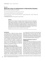
Figure 1 Mechanistic model of HIV-1 neuroinvasion. (1) The physiological expression of chemokines by brain cells, among which are soluble frac-
talkine (Fkn) and CXCL12, supports a slow but continuous entry of monocytes and macrophages into the central nervous system. Due to their expres-
sion of CX
3
CR1, CD16 positive, activated monocytes are the preferential targets for such attraction. These CD16 positive monocytes are the main
reservoir of monocyte/macrophage-harbored virus and are thus likely to be the predominant cell type carrying HIV into the brain. (2) Infiltrated HIV-
infected monocytes locally produce HIV and inflammatory mediators in perivascular areas. This activates neighbouring astrocytes as well as the blood
brain barrier (BBB) endothelium. (3) In response, endothelial cells up-regulate adhesion molecules, enhancing monocyte recruitment. However, mem-
brane-bound Fkn is also induced on endothelial cells and can arrest CD16 positive monocytes at the endothelium thus inhibiting their further infiltra-
tion. (4) CCL2 is overexpressed by infected, HIV-stimulated macrophages and activated astrocytes, attracting CD16 negative, CCR2 positive monocytes
toward the perivascular area. (5) Both CXCL12 and nerve growth factor (NGF) are overexpressed in the inflamed brain. NGF increases CXCR4 expres-
sion and promotes uninfected monocyte attraction by CXCL12. At the same time it limits entry of infected monocytes into the brain. (6) Activated
uninfected perivascular macrophages may be targets for de novo infection by locally produced HIV, amplifying the activation - attraction - infection
cycle. (7) Local inflammation as well as HIV products induce tight junction disorganization and lead to breaches in the BBB. Toxic serum proteins and
free virions may enter the brain, favouring more infection and further amplifying inflammation.
CD16+ Monocyte
Astrocyte
CD16- Monocyte
Breached BBB
Perivascular macrophage
Blood stream
Brain tissue
1
2
3
4
6
5
7
CD16
CX
3
CR1
CXCR4
CCR2
TrkA and/or p75
NTR
Tight junction
Adhesion molecules
Soluble FKN
Membrane-bound FKN
CXCL12
CCL2
HIV virion
NGF
4
5
Gras and Kaul Retrovirology 2010, 7:30
/>Page 7 of 11
infected and immune-activated macrophages appears to
be a critical target for future therapeutic developments.
The very complex and intricate mechanisms that govern
this crossing should thus be studied with particular atten-
tion.
HAND correlate with CSF viral load [25], which is
closely related to CSF pleocytosis [139]. In a recent study,
Sinclair et al. showed that HAART despite treatment fail-
ures with no effect on peripheral viral load, had neverthe-
less a significant beneficial impact on CSF viral load, CSF
pleocytosis, and immune activation [140]. This striking
and encouraging result further illustrates the critical
importance of an improved understanding of BBB func-
tion and neuroinvasion mechanisms. Furthermore, HIV
neuroinvasion and BBB likely will provide future thera-
peutic targets for coping with the anticipated increase in
HAND prevalence as more and more HIV patients come
of age.
Competing interests
The authors declare that they have no competing interests.
Authors' contributions
GG and MK wrote the article jointly. All authors read and approved the final
manuscript.
Acknowledgements
This review was inspired by discussions of the role of the cells of the mononu-
clear phagocyte lineage in HIV infection during meetings conducted by the
Association for Macrophage in Infection Research (AMIR). Article processing
charges of this review are paid for by the Concerted Action 31 - Dendritic cells,
Antigen Presentation and Innate Immunity of the "Agence Nationale de
Recherche sur le Sida et les Hépatites Virales" (ANRS). M. Kaul was supported by
NIH grant R01 NS050621. G. Gras was supported by grants from the "Agence
Nationale de Recherche sur le Sida et les Hépatites Virales" (ANRS), the « Fonda-
tion pour la Recherche Médicale » (FRM) and « Ensemble Contre le Sida »
(SIDACTION).
Author Details
1
Institute of Emerging Diseases and Innovative Therapies, Division of Immuno-
Virology, CEA, 18 Route du Panorama, F92265 Fontenay-aux Roses, France and
2
Infectious & Inflammatory Disease Center, Burnham Institute for Medical
Research, 10901 North Torrey Pines Road, La Jolla, CA 92037, USA
References
1. Antinori A, Arendt G, Becker JT, Brew BJ, Byrd DA, Cherner M, Clifford DB,
Cinque P, Epstein LG, Goodkin K, Gisslen M, Grant I, Heaton RK, Joseph J,
Marder K, Marra CM, McArthur JC, Nunn M, Price RW, Pulliam L, Robertson
KR, Sacktor N, Valcour V, Wojna VE: Updated research nosology for HIV-
associated neurocognitive disorders. Neurology 2007, 69:1789-1799.
2. Ghafouri M, Amini S, Khalili K, Sawaya BE: HIV-1 associated dementia:
symptoms and causes. Retrovirology 2006, 3:28.
3. McArthur JC, Hoover DR, Bacellar H, Miller EN, Cohen BA, Becker JT,
Graham NM, McArthur JH, Selnes OA, Jacobson LP, et al.: Dementia in
AIDS patients: incidence and risk factors. Multicenter AIDS Cohort
Study. Neurology 1993, 43:2245-2252.
4. Ellis RJ, Deutsch R, Heaton RK, Marcotte TD, McCutchan JA, Nelson JA,
Abramson I, Thal LJ, Atkinson JH, Wallace MR, Grant I: Neurocognitive
impairment is an independent risk factor for death in HIV infection.
San Diego HIV Neurobehavioral Research Center Group. Arch Neurol
1997, 54:416-424.
5. Liner KJ, Hall CD, Robertson KR: Effects of antiretroviral therapy on
cognitive impairment. Curr HIV/AIDS Rep 2008, 5:64-71.
6. Boisse L, Gill MJ, Power C: HIV infection of the central nervous system:
clinical features and neuropathogenesis. Neurol Clin 2008, 26:799-819. x
7. Brew BJ, Crowe SM, Landay A, Cysique LA, Guillemin G:
Neurodegeneration and ageing in the HAART era. J Neuroimmune
Pharmacol 2009, 4:163-174.
8. Letendre S, Marquie-Beck J, Capparelli E, Best B, Clifford D, Collier AC,
Gelman BB, McArthur JC, McCutchan JA, Morgello S, Simpson D, Grant I,
Ellis RJ, CHARTER Group: Validation of the CNS Penetration-Effectiveness
rank for quantifying antiretroviral penetration into the central nervous
system. Arch Neurol 2008, 65:65-70.
9. Adle-Biassette H, Bell JE, Creange A, Sazdovitch V, Authier FJ, Gray F, Hauw
JJ, Gherardi R: DNA breaks detected by in situ end-labelling in dorsal
root ganglia of patients with AIDS. Neuropathol Appl Neurobiol 1998,
24:373-380.
10. Masliah E, Heaton RK, Marcotte TD, Ellis RJ, Wiley CA, Mallory M, Achim CL,
McCutchan JA, Nelson JA, Atkinson JH, Grant I: Dendritic injury is a
pathological substrate for human immunodeficiency virus-related
cognitive disorders. HNRC Group. The HIV Neurobehavioral Research
Center. Ann Neurol 1997, 42:963-972.
11. Petito CK, Cho ES, Lemann W, Navia BA, Price RW: Neuropathology of
acquired immunodeficiency syndrome (AIDS): an autopsy review. J
Neuropathol Exp Neurol 1986, 45:635-646.
12. Everall IP, Luthert PJ, Lantos PL: Neuronal loss in the frontal cortex in HIV
infection. Lancet 1991, 337:1119-1121.
13. Ketzler S, Weis S, Haug H, Budka H: Loss of neurons in the frontal cortex
in AIDS brains. Acta Neuropathol 1990, 80:92-94.
14. Reyes MG, Faraldi F, Senseng CS, Flowers C, Fariello R: Nigral
degeneration in acquired immune deficiency syndrome (AIDS). Acta
Neuropathol 1991, 82:39-44.
15. Graus F, Ribalta T, Abos J, Alom J, Cruz-Sanchez F, Mallolas J, Miro JM,
Cardesa A, Tolosa E: Subacute cerebellar syndrome as the first
manifestation of AIDS dementia complex. Acta Neurol Scand 1990,
81:118-120.
16. Everall I, Luthert P, Lantos P: A review of neuronal damage in human
immunodeficiency virus infection: its assessment, possible mechanism
and relationship to dementia. J Neuropathol Exp Neurol 1993,
52:561-566.
17. Anthony IC, Bell JE: The Neuropathology of HIV/AIDS. Int Rev Psychiatry
2008, 20:15-24.
18. Glass JD, Fedor H, Wesselingh SL, McArthur JC: Immunocytochemical
quantitation of human immunodeficiency virus in the brain:
correlations with dementia. Ann Neurol 1995, 38:755-762.
19. Langford TD, Letendre SL, Larrea GJ, Masliah E: Changing patterns in the
neuropathogenesis of HIV during the HAART era. Brain Pathol 2003,
13:195-210.
20. Ho DD, Rota TR, Schooley RT, Kaplan JC, Allan JD, Groopman JE, Resnick L,
Felsenstein D, Andrews CA, Hirsch MS: Isolation of HTLV-III from
cerebrospinal fluid and neural tissues of patients with neurologic
syndromes related to the acquired immunodeficiency syndrome. N
Engl J Med 1985, 313:1493-1497.
21. Koenig S, Gendelman HE, Orenstein JM, Dal Canto MC, Pezeshkpour GH,
Yungbluth M, Janotta F, Aksamit A, Martin MA, Fauci AS: Detection of
AIDS virus in macrophages in brain tissue from AIDS patients with
encephalopathy. Science 1986, 233:1089-1093.
22. Asensio VC, Campbell IL: Chemokines in the CNS: plurifunctional
mediators in diverse states. Trends Neurosci 1999, 22:504-512.
23. Sadagopal S, Lorey SL, Barnett L, Basham R, Lebo L, Erdem H, Haman K,
Avison M, Waddell K, Haas DW, Kalams SA: Enhancement of human
immunodeficiency virus (HIV)-specific CD8+ T cells in cerebrospinal
fluid compared to those in blood among antiretroviral therapy-naive
HIV-positive subjects. J Virol 2008, 82:10418-10428.
24. Anthony IC, Ramage SN, Carnie FW, Simmonds P, Bell JE: Influence of
HAART on HIV-related CNS disease and neuroinflammation. J
Neuropathol Exp Neurol 2005, 64:529-536.
25. Brew BJ, Pemberton L, Cunningham P, Law MG: Levels of human
immunodeficiency virus type 1 RNA in cerebrospinal fluid correlate
with AIDS dementia stage. J Infect Dis 1997, 175:963-966.
26. Ellis RJ, Hsia K, Spector SA, Nelson JA, Heaton RK, Wallace MR, Abramson I,
Atkinson JH, Grant I, McCutchan JA: Cerebrospinal fluid human
immunodeficiency virus type 1 RNA levels are elevated in
Received: 1 October 2009 Accepted: 7 April 2010
Published: 7 April 2010
This article is available from: 2010 Gras and Kaul; licensee BioMed Central Ltd. This is an Open Access article distributed under the terms of the Creative Commons Attribution License ( ), which permits unrestricted use, distribution, and reproduction in any medium, provided the original work is properly cited.Retrovirolog y 2010, 7:30
Gras and Kaul Retrovirology 2010, 7:30
/>Page 8 of 11
neurocognitively impaired individuals with acquired
immunodeficiency syndrome. HIV Neurobehavioral Research Center
Group. Ann Neurol 1997, 42:679-688.
27. McArthur JC, McClernon DR, Cronin MF, Nance-Sproson TE, Saah AJ, St
Clair M, Lanier ER: Relationship between human immunodeficiency
virus-associated dementia and viral load in cerebrospinal fluid and
brain. Ann Neurol 1997, 42:689-698.
28. Wiley CA, Soontornniyomkij V, Radhakrishnan L, Masliah E, Mellors J,
Hermann SA, Dailey P, Achim CL: Distribution of brain HIV load in AIDS.
Brain Pathol 1998, 8:277-284.
29. Gartner S: HIV infection and dementia. Science 2000, 287:602-604.
30. Banks WA, Ercal N, Price TO: The blood-brain barrier in neuroAIDS. Curr
HIV Res 2006, 4:259-266.
31. Cho YY, Astgen A, Hendel H, Issing W, Perrot JY, Schachter F, Rappaport J,
Zagury JF: Homeostasis of chemokines, interferon production and
lymphocyte subsets: implications for AIDS pathogenesis. Biomed
Pharmacother 1997, 51:221-229.
32. Mandl JN, Barry AP, Vanderford TH, Kozyr N, Chavan R, Klucking S, Barrat
FJ, Coffman RL, Staprans SI, Feinberg MB: Divergent TLR7 and TLR9
signaling and type I interferon production distinguish pathogenic and
nonpathogenic AIDS virus infections. Nat Med 2008, 14:1077-1087.
33. Poli G, Biswas P, Fauci AS: Interferons in the pathogenesis and treatment
of human immunodeficiency virus infection. Antiviral Res 1994,
24:221-233.
34. Sas AR, Bimonte-Nelson H, Smothers CT, Woodward J, Tyor WR:
Interferon-alpha causes neuronal dysfunction in encephalitis. J
Neurosci 2009, 29:3948-3955.
35. Sas AR, Bimonte-Nelson HA, Tyor WR: Cognitive dysfunction in HIV
encephalitic SCID mice correlates with levels of Interferon-alpha in the
brain. Aids 2007, 21:2151-2159.
36. Argyris EG, Acheampong E, Wang F, Huang J, Chen K, Mukhtar M, Zhang
H: The interferon-induced expression of APOBEC3G in human blood-
brain barrier exerts a potent intrinsic immunity to block HIV-1 entry to
central nervous system. Virology 2007, 367:440-451.
37. Thieblemont N, Weiss L, Sadeghi HM, Estcourt C, Haeffner-Cavaillon N:
CD14lowCD16high: a cytokine-producing monocyte subset which
expands during human immunodeficiency virus infection. Eur J
Immunol 1995, 25:3418-3424.
38. Pulliam L, Gascon R, Stubblebine M, McGuire D, McGrath MS: Unique
monocyte subset in patients with AIDS dementia. Lancet 1997,
349:692-695.
39. Coleman CM, Wu L: HIV interactions with monocytes and dendritic
cells: viral latency and reservoirs. Retrovirology 2009, 6:51.
40. Ellery PJ, Tippett E, Chiu YL, Paukovics G, Cameron PU, Solomon A, Lewin
SR, Gorry PR, Jaworowski A, Greene WC, Sonza S, Crowe SM: The CD16+
monocyte subset is more permissive to infection and preferentially
harbors HIV-1 in vivo. J Immunol 2007, 178:6581-6589.
41. Shiramizu B, Gartner S, Williams A, Shikuma C, Ratto-Kim S, Watters M,
Aguon J, Valcour V: Circulating proviral HIV DNA and HIV-associated
dementia. Aids 2005, 19:45-52.
42. Kalter DC, Nakamura M, Turpin JA, Baca LM, Hoover DL, Dieffenbach C,
Ralph P, Gendelman HE, Meltzer MS: Enhanced HIV replication in
macrophage colony-stimulating factor-treated monocytes. J Immunol
1991, 146:298-306.
43. Naif HM, Li S, Alali M, Sloane A, Wu L, Kelly M, Lynch G, Lloyd A,
Cunningham AL: CCR5 expression correlates with susceptibility of
maturing monocytes to human immunodeficiency virus type 1
infection. J Virol 1998, 72:830-836.
44. Rich EA, Chen IS, Zack JA, Leonard ML, O'Brien WA: Increased
susceptibility of differentiated mononuclear phagocytes to productive
infection with human immunodeficiency virus-1 (HIV-1). J Clin Invest
1992, 89:176-183.
45. Schrier RD, Freeman WR, Wiley CA, McCutchan JA: CMV-specific immune
responses and HLA phenotypes of AIDS patients who develop CMV
retinitis. HNRC Group. HIV Neurobehavioral Research Center. Adv
Neuroimmunol 1994, 4:327-336.
46. Schrier RD, McCutchan JA, Wiley CA: Mechanisms of immune activation
of human immunodeficiency virus in monocytes/macrophages. J Virol
1993, 67:5713-5720.
47. Sonza S, Maerz A, Deacon N, Meanger J, Mills J, Crowe S: Human
immunodeficiency virus type 1 replication is blocked prior to reverse
transcription and integration in freshly isolated peripheral blood
monocytes. J Virol 1996, 70:3863-3869.
48. Wang X, Ye L, Hou W, Zhou Y, Wang YJ, Metzger DS, Ho WZ: Cellular
microRNA expression correlates with susceptibility of monocytes/
macrophages to HIV-1 infection. Blood 2009, 113:671-674.
49. Ancuta P, Kunstman KJ, Autissier P, Zaman T, Stone D, Wolinsky SM,
Gabuzda D: CD16+ monocytes exposed to HIV promote highly efficient
viral replication upon differentiation into macrophages and
interaction with T cells. Virology 2006, 344:267-276.
50. Ancuta P, Liu KY, Misra V, Wacleche VS, Gosselin A, Zhou X, Gabuzda D:
Transcriptional profiling reveals developmental relationship and
distinct biological functions of CD16+ and CD16- monocyte subsets.
BMC Genomics 2009, 10:403.
51. Ancuta P, Moses A, Gabuzda D: Transendothelial migration of CD16+
monocytes in response to fractalkine under constitutive and
inflammatory conditions. Immunobiology 2004, 209:11-20.
52. Ancuta P, Rao R, Moses A, Mehle A, Shaw SK, Luscinskas FW, Gabuzda D:
Fractalkine preferentially mediates arrest and migration of CD16+
monocytes. J Exp Med 2003, 197:1701-1707.
53. Fischer-Smith T, Bell C, Croul S, Lewis M, Rappaport J: Monocyte/
macrophage trafficking in acquired immunodeficiency syndrome
encephalitis: lessons from human and nonhuman primate studies. J
Neurovirol 2008, 14:318-326.
54. Gartner S, Markovits P, Markovitz DM, Betts RF, Popovic M: Virus isolation
from and identification of HTLV-III/LAV-producing cells in brain tissue
from a patient with AIDS. Jama 1986, 256:2365-2371.
55. Gartner S, Markovits P, Markovitz DM, Kaplan MH, Gallo RC, Popovic M:
The role of mononuclear phagocytes in HTLV-III/LAV infection. Science
1986, 233:215-219.
56. Embretson J, Zupancic M, Ribas JL, Burke A, Racz P, Tenner-Racz K, Haase
AT: Massive covert infection of helper T lymphocytes and macrophages
by HIV during the incubation period of AIDS. Nature 1993, 362:359-362.
57. Martin JC, Bandres JC: Cells of the monocyte-macrophage lineage and
pathogenesis of HIV-1 infection. J Acquir Immune Defic Syndr 1999,
22:413-429.
58. Orenstein JM, Fox C, Wahl SM: Macrophages as a source of HIV during
opportunistic infections. Science 1997, 276:1857-1861.
59. Alkhatib G, Combadiere C, Broder CC, Feng Y, Kennedy PE, Murphy PM,
Berger EA: CC CKR5: a RANTES, MIP-1alpha, MIP-1beta receptor as a
fusion cofactor for macrophage-tropic HIV-1. Science 1996,
272:1955-1958.
60. Choe H, Farzan M, Sun Y, Sullivan N, Rollins B, Ponath PD, Wu L, Mackay CR,
LaRosa G, Newman W, Gerard N, Gerard C, Sodroski J: The beta-
chemokine receptors CCR3 and CCR5 facilitate infection by primary
HIV-1 isolates. Cell 1996, 85:1135-1148.
61. Dragic T, Litwin V, Allaway GP, Martin SR, Huang Y, Nagashima KA,
Cayanan C, Maddon PJ, Koup RA, Moore JP, Paxton WA: HIV-1 entry into
CD4+ cells is mediated by the chemokine receptor CC-CKR-5. Nature
1996, 381:667-673.
62. Albright AV, Shieh JT, Itoh T, Lee B, Pleasure D, O'Connor MJ, Doms RW,
Gonzalez-Scarano F: Microglia express CCR5, CXCR4, and CCR3, but of
these, CCR5 is the principal coreceptor for human immunodeficiency
virus type 1 dementia isolates. J Virol 1999, 73:205-213.
63. He J, Chen Y, Farzan M, Choe H, Ohagen A, Gartner S, Busciglio J, Yang X,
Hofmann W, Newman W, Mackay CR, Sodroski J, Gabuzda D: CCR3 and
CCR5 are co-receptors for HIV-1 infection of microglia. Nature 1997,
385:645-649.
64. Li S, Juarez J, Alali M, Dwyer D, Collman R, Cunningham A, Naif HM:
Persistent CCR5 utilization and enhanced macrophage tropism by
primary blood human immunodeficiency virus type 1 isolates from
advanced stages of disease and comparison to tissue-derived isolates.
J Virol 1999, 73:9741-9755.
65. Shieh JT, Albright AV, Sharron M, Gartner S, Strizki J, Doms RW, Gonzalez-
Scarano F: Chemokine receptor utilization by human
immunodeficiency virus type 1 isolates that replicate in microglia. J
Virol 1998, 72:4243-4249.
66. Smit TK, Wang B, Ng T, Osborne R, Brew B, Saksena NK: Varied tropism of
HIV-1 isolates derived from different regions of adult brain cortex
discriminate between patients with and without AIDS dementia
complex (ADC): evidence for neurotropic HIV variants. Virology 2001,
279:509-526.
Gras and Kaul Retrovirology 2010, 7:30
/>Page 9 of 11
67. Gorry PR, Bristol G, Zack JA, Ritola K, Swanstrom R, Birch CJ, Bell JE, Bannert
N, Crawford K, Wang H, Schols D, De Clercq E, Kunstman K, Wolinsky SM,
Gabuzda D: Macrophage tropism of human immunodeficiency virus
type 1 isolates from brain and lymphoid tissues predicts neurotropism
independent of coreceptor specificity. J Virol 2001, 75:10073-10089.
68. Gorry PR, Taylor J, Holm GH, Mehle A, Morgan T, Cayabyab M, Farzan M,
Wang H, Bell JE, Kunstman K, Moore JP, Wolinsky SM, Gabuzda D:
Increased CCR5 affinity and reduced CCR5/CD4 dependence of a
neurovirulent primary human immunodeficiency virus type 1 isolate. J
Virol 2002, 76:6277-6292.
69. Gray L, Roche M, Churchill MJ, Sterjovski J, Ellett A, Poumbourios P, Sherieff
S, Wang B, Saksena N, Purcell DF, Wesselingh S, Cunningham AL, Brew BJ,
Gabuzda D, Gorry PR: Tissue-specific sequence alterations in the human
immunodeficiency virus type 1 envelope favoring CCR5 usage
contribute to persistence of dual-tropic virus in the brain. J Virol 2009,
83:5430-5441.
70. Clay CC, Rodrigues DS, Ho YS, Fallert BA, Janatpour K, Reinhart TA, Esser U:
Neuroinvasion of fluorescein-positive monocytes in acute simian
immunodeficiency virus infection. J Virol 2007, 81:12040-12048.
71. Fiala M, Looney DJ, Stins M, Way DD, Zhang L, Gan X, Chiappelli F,
Schweitzer ES, Shapshak P, Weinand M, Graves MC, Witte M, Kim KS: TNF-
alpha opens a paracellular route for HIV-1 invasion across the blood-
brain barrier. Mol Med 1997, 3:553-564.
72. Fletcher NF, Bexiga MG, Brayden DJ, Brankin B, Willett BJ, Hosie MJ, Jacque
JM, Callanan JJ: Lymphocyte migration through the blood brain barrier
(BBB) in feline immunodeficiency virus infection is significantly
influenced by the pre-existence of virus and TNF-alpha within the CNS:
studies using an in vitro feline BBB model. Neuropathol Appl Neurobiol
2009, 36:592-602.
73. Ancuta P, Kamat A, Kunstman KJ, Kim EY, Autissier P, Wurcel A, Zaman T,
Stone D, Mefford M, Morgello S, Singer EJ, Wolinsky SM, Gabuzda D:
Microbial translocation is associated with increased monocyte
activation and dementia in AIDS patients. PLoS One 2008, 3:e2516.
74. Brenchley JM, Price DA, Douek DC: HIV disease: fallout from a mucosal
catastrophe? Nat Immunol 2006, 7:235-239.
75. Brenchley JM, Price DA, Schacker TW, Asher TE, Silvestri G, Rao S, Kazzaz Z,
Bornstein E, Lambotte O, Altmann D, Blazar BR, Rodriguez B, Teixeira-
Johnson L, Landay A, Martin JN, Hecht FM, Picker LJ, Lederman MM, Deeks
SG, Douek DC: Microbial translocation is a cause of systemic immune
activation in chronic HIV infection. Nat Med 2006, 12:1365-1371.
76. Wang H, Sun J, Goldstein H: Human immunodeficiency virus type 1
infection increases the in vivo capacity of peripheral monocytes to
cross the blood-brain barrier into the brain and the in vivo sensitivity
of the blood-brain barrier to disruption by lipopolysaccharide. J Virol
2008, 82:7591-7600.
77. Bazan JF, Bacon KB, Hardiman G, Wang W, Soo K, Rossi D, Greaves DR,
Zlotnik A, Schall TJ: A new class of membrane-bound chemokine with a
CX3C motif. Nature 1997, 385:640-644.
78. Kaul M, Garden GA, Lipton SA: Pathways to neuronal injury and
apoptosis in HIV-associated dementia. Nature 2001, 410:988-994.
79. Power C, McArthur JC, Nath A, Wehrly K, Mayne M, Nishio J, Langelier T,
Johnson RT, Chesebro B: Neuronal death induced by brain-derived
human immunodeficiency virus type 1 envelope genes differs
between demented and nondemented AIDS patients. J Virol 1998,
72:9045-9053.
80. Nottet HS, Persidsky Y, Sasseville VG, Nukuna AN, Bock P, Zhai QH, Sharer
LR, McComb RD, Swindells S, Soderland C, Gendelman HE: Mechanisms
for the transendothelial migration of HIV-1-infected monocytes into
brain. J Immunol 1996, 156:1284-1295.
81. Gray F, Belec L, Chretien F, Dubreuil-Lemaire ML, Ricolfi F, Wingertsmann
L, Poron F, Gherardi R: Acute, relapsing brain oedema with diffuse
blood-brain barrier alteration and axonal damage in the acquired
immunodeficiency syndrome. Neuropathol Appl Neurobiol 1998,
24:209-216.
82. Petito CK, Cash KS: Blood-brain barrier abnormalities in the acquired
immunodeficiency syndrome: immunohistochemical localization of
serum proteins in postmortem brain. Ann Neurol 1992, 32:658-666.
83. Maclean AG, Belenchia GE, Bieniemy DN, Moroney-Rasmussen TA, Lackner
AA: Simian immunodeficiency virus disrupts extended lengths of the
blood brain barrier. J Med Primatol 2005, 34:237-242.
84. Dallasta LM, Pisarov LA, Esplen JE, Werley JV, Moses AV, Nelson JA, Achim
CL: Blood-brain barrier tight junction disruption in human
immunodeficiency virus-1 encephalitis. Am J Pathol 1999,
155:1915-1927.
85. Persidsky Y, Heilman D, Haorah J, Zelivyanskaya M, Persidsky R, Weber GA,
Shimokawa H, Kaibuchi K, Ikezu T: Rho-mediated regulation of tight
junctions during monocyte migration across the blood-brain barrier in
HIV-1 encephalitis (HIVE). Blood 2006, 107:4770-4780.
86. Luabeya MK, Dallasta LM, Achim CL, Pauza CD, Hamilton RL: Blood-brain
barrier disruption in simian immunodeficiency virus encephalitis.
Neuropathol Appl Neurobiol 2000, 26:454-462.
87. Mankowski JL, Queen SE, Kirstein LM, Spelman JP, Laterra J, Simpson IA,
Adams RJ, Clements JE, Zink MC: Alterations in blood-brain barrier
glucose transport in SIV-infected macaques. J Neurovirol 1999,
5:695-702.
88. Khan NA, Di Cello F, Stins M, Kim KS: Gp120-mediated cytotoxicity of
human brain microvascular endothelial cells is dependent on p38
mitogen-activated protein kinase activation. J Neurovirol 2007,
13:242-251.
89. Marshall DC, Wyss-Coray T, Abraham CR: Induction of matrix
metalloproteinase-2 in human immunodeficiency virus-1 glycoprotein
120 transgenic mouse brains. Neurosci Lett 1998, 254:97-100.
90. Boehme SA, Lio FM, Maciejewski-Lenoir D, Bacon KB, Conlon PJ: The
chemokine fractalkine inhibits Fas-mediated cell death of brain
microglia. J Immunol 2000, 165:397-403.
91. Gonzalez E, Rovin BH, Sen L, Cooke G, Dhanda R, Mummidi S, Kulkarni H,
Bamshad MJ, Telles V, Anderson SA, Walter EA, Stephan KT, Deucher M,
Mangano A, Bologna R, Ahuja SS, Dolan MJ, Ahuja SK: HIV-1 infection and
AIDS dementia are influenced by a mutant MCP-1 allele linked to
increased monocyte infiltration of tissues and MCP-1 levels. Proc Natl
Acad Sci USA 2002, 99:13795-13800.
92. El-Hage N, Wu G, Ambati J, Bruce-Keller AJ, Knapp PE, Hauser KF: CCR2
mediates increases in glial activation caused by exposure to HIV-1 Tat
and opiates. J Neuroimmunol 2006, 178:9-16.
93. El-Hage N, Wu G, Wang J, Ambati J, Knapp PE, Reed JL, Bruce-Keller AJ,
Hauser KF: HIV-1 Tat and opiate-induced changes in astrocytes
promote chemotaxis of microglia through the expression of MCP-1
and alternative chemokines. Glia 2006, 53:132-146.
94. Eugenin EA, Osiecki K, Lopez L, Goldstein H, Calderon TM, Berman JW:
CCL2/monocyte chemoattractant protein-1 mediates enhanced
transmigration of human immunodeficiency virus (HIV)-infected
leukocytes across the blood-brain barrier: a potential mechanism of
HIV-CNS invasion and NeuroAIDS. J Neurosci 2006, 26:1098-1106.
95. Gu L, Rutledge B, Fiorillo J, Ernst C, Grewal I, Flavell R, Gladue R, Rollins B: In
vivo properties of monocyte chemoattractant protein-1. J Leukoc Biol
1997, 62:577-580.
96. Cinque P, Vago L, Mengozzi M, Torri V, Ceresa D, Vicenzi E, Transidico P,
Vagani A, Sozzani S, Mantovani A, Lazzarin A, Poli G: Elevated
cerebrospinal fluid levels of monocyte chemotactic protein-1 correlate
with HIV-1 encephalitis and local viral replication. Aids 1998,
12:1327-1332.
97. Conant K, Garzino-Demo A, Nath A, McArthur JC, Halliday W, Power C,
Gallo RC, Major EO: Induction of monocyte chemoattractant protein-1
in HIV-1 Tat-stimulated astrocytes and elevation in AIDS dementia.
Proc Natl Acad Sci USA 1998, 95:3117-3121.
98. Kelder W, McArthur JC, Nance-Sproson T, McClernon D, Griffin DE: Beta-
chemokines MCP-1 and RANTES are selectively increased in
cerebrospinal fluid of patients with human immunodeficiency virus-
associated dementia. Ann Neurol 1998, 44:831-835.
99. Sozzani S, Introna M, Bernasconi S, Polentarutti N, Cinque P, Poli G, Sica A,
Mantovani A: MCP-1 and CCR2 in HIV infection: regulation of agonist
and receptor expression. J Leukoc Biol 1997, 62:30-33.
100. Zink MC, Coleman GD, Mankowski JL, Adams RJ, Tarwater PM, Fox K,
Clements JE: Increased macrophage chemoattractant protein-1 in
cerebrospinal fluid precedes and predicts simian immunodeficiency
virus encephalitis. J Infect Dis 2001, 184:1015-1021.
101. Sanders VJ, Pittman CA, White MG, Wang G, Wiley CA, Achim CL:
Chemokines and receptors in HIV encephalitis. Aids 1998,
12:1021-1026.
102. Buch S, Sui Y, Potula R, Pinson D, Adany I, Li Z, Huang M, Li S, Dhillon N,
Major E, Narayan O: Role of interleukin-4 and monocyte
chemoattractant protein-1 in the neuropathogenesis of X4 simian
human immunodeficiency virus infection in macaques. J Neurovirol
2004, 10(Suppl 1):118-124.
Gras and Kaul Retrovirology 2010, 7:30
/>Page 10 of 11
103. Hicks A, Potula R, Sui YJ, Villinger F, Pinson D, Adany I, Li Z, Long C, Cheney
P, Marcario J, Novembre F, Mueller N, Kumar A, Major E, Narayan O, Buch S:
Neuropathogenesis of lentiviral infection in macaques: roles of CXCR4
and CCR5 viruses and interleukin-4 in enhancing monocyte
chemoattractant protein-1 production in macrophages. Am J Pathol
2002, 161:813-822.
104. Persidsky Y, Ghorpade A, Rasmussen J, Limoges J, Liu XJ, Stins M, Fiala M,
Way D, Kim KS, Witte MH, Weinand M, Carhart L, Gendelman HE:
Microglial and astrocyte chemokines regulate monocyte migration
through the blood-brain barrier in human immunodeficiency virus-1
encephalitis. Am J Pathol 1999, 155:1599-1611.
105. Malik M, Chen YY, Kienzle MF, Tomkowicz BE, Collman RG, Ptasznik A:
Monocyte migration and LFA-1-mediated attachment to brain
microvascular endothelia is regulated by SDF-1 alpha through Lyn
kinase. J Immunol 2008, 181:4632-4637.
106. Samah B, Porcheray F, Gras G: Neurotrophins modulate monocyte
chemotaxis without affecting macrophage function. Clin Exp Immunol
2008, 151:476-486.
107. Samah B, Porcheray F, Dereuddre-Bosquet N, Gras G: Nerve growth factor
stimulation promotes CXCL-12 attraction of monocytes but decreases
human immunodeficiency virus replication in attracted population. J
Neurovirol 2009, 15:71-80.
108. Wu DT, Woodman SE, Weiss JM, McManus CM, D'Aversa TG, Hesselgesser
J, Major EO, Nath A, Berman JW: Mechanisms of leukocyte trafficking
into the CNS. J Neurovirol 2000, 6(Suppl 1):S82-85.
109. Gendelman HE, Ding S, Gong N, Liu J, Ramirez SH, Persidsky Y, Mosley RL,
Wang T, Volsky DJ, Xiong H: Monocyte chemotactic protein-1 regulates
voltage-gated K+ channels and macrophage transmigration. J
Neuroimmune Pharmacol 2009, 4:47-59.
110. Chaudhuri A, Duan F, Morsey B, Persidsky Y, Kanmogne GD: HIV-1
activates proinflammatory and interferon-inducible genes in human
brain microvascular endothelial cells: putative mechanisms of blood-
brain barrier dysfunction. J Cereb Blood Flow Metab 2008, 28:697-711.
111. Yang B, Akhter S, Chaudhuri A, Kanmogne GD: HIV-1 gp120 induces
cytokine expression, leukocyte adhesion, and transmigration across
the blood-brain barrier: modulatory effects of STAT1 signaling.
Microvasc Res 2009, 77:212-219.
112. Chaudhuri A, Yang B, Gendelman HE, Persidsky Y, Kanmogne GD: STAT1
signaling modulates HIV-1-induced inflammatory responses and
leukocyte transmigration across the blood-brain barrier. Blood 2008,
111:2062-2072.
113. Kanmogne GD, Schall K, Leibhart J, Knipe B, Gendelman HE, Persidsky Y:
HIV-1 gp120 compromises blood-brain barrier integrity and enhances
monocyte migration across blood-brain barrier: implication for viral
neuropathogenesis. J Cereb Blood Flow Metab 2007, 27:123-134.
114. Ricardo-Dukelow M, Kadiu I, Rozek W, Schlautman J, Persidsky Y,
Ciborowski P, Kanmogne GD, Gendelman HE: HIV-1 infected monocyte-
derived macrophages affect the human brain microvascular
endothelial cell proteome: new insights into blood-brain barrier
dysfunction for HIV-1-associated dementia. J Neuroimmunol 2007,
185:37-46.
115. Persidsky Y, Stins M, Way D, Witte MH, Weinand M, Kim KS, Bock P,
Gendelman HE, Fiala M: A model for monocyte migration through the
blood-brain barrier during HIV-1 encephalitis. J Immunol 1997,
158:3499-3510.
116. Dohgu S, Banks WA: Lipopolysaccharide-enhanced transcellular
transport of HIV-1 across the blood-brain barrier is mediated by the
p38 mitogen-activated protein kinase pathway. Exp Neurol 2008,
210:740-749.
117. Owe-Young R, Webster NL, Mukhtar M, Pomerantz RJ, Smythe G, Walker D,
Armati PJ, Crowe SM, Brew BJ: Kynurenine pathway metabolism in
human blood-brain-barrier cells: implications for immune tolerance
and neurotoxicity. J Neurochem 2008, 105:1346-1357.
118. Guillemin GJ, Kerr SJ, Smythe GA, Smith DG, Kapoor V, Armati PJ, Croitoru
J, Brew BJ: Kynurenine pathway metabolism in human astrocytes: a
paradox for neuronal protection. J Neurochem 2001, 78:842-853.
119. Guillemin GJ, Kerr SJ, Brew BJ: Involvement of quinolinic acid in AIDS
dementia complex. Neurotox Res 2005, 7:103-123.
120. Carlin JM, Borden EC, Sondel PM, Byrne GI: Interferon-induced
indoleamine 2,3-dioxygenase activity in human mononuclear
phagocytes. J Leukoc Biol 1989, 45:29-34.
121. Hurwitz AA, Berman JW, Lyman WD: The role of the blood-brain barrier
in HIV infection of the central nervous system. Adv Neuroimmunol 1994,
4:249-256.
122. Sasseville VG, Newman W, Brodie SJ, Hesterberg P, Pauley D, Ringler DJ:
Monocyte adhesion to endothelium in simian immunodeficiency
virus-induced AIDS encephalitis is mediated by vascular cell adhesion
molecule-1/alpha 4 beta 1 integrin interactions. Am J Pathol 1994,
144:27-40.
123. Ramirez SH, Heilman D, Morsey B, Potula R, Haorah J, Persidsky Y:
Activation of peroxisome proliferator-activated receptor gamma
(PPARgamma) suppresses Rho GTPases in human brain microvascular
endothelial cells and inhibits adhesion and transendothelial migration
of HIV-1 infected monocytes. J Immunol 2008, 180:1854-1865.
124. Ivey NS, Renner NA, Moroney-Rasmussen T, Mohan M, Redmann RK,
Didier PJ, Alvarez X, Lackner AA, Maclean AG: Association of FAK
activation with lentivirus-induced disruption of blood-brain barrier
tight junction-associated ZO-1 protein organization. J Neurovirol
2009:1-12.
125. Nakamuta S, Endo H, Higashi Y, Kousaka A, Yamada H, Yano M, Kido H:
Human immunodeficiency virus type 1 gp120-mediated disruption of
tight junction proteins by induction of proteasome-mediated
degradation of zonula occludens-1 and -2 in human brain
microvascular endothelial cells. J Neurovirol 2008, 14:186-195.
126. Zhong Y, Smart EJ, Weksler B, Couraud PO, Hennig B, Toborek M:
Caveolin-1 regulates human immunodeficiency virus-1 Tat-induced
alterations of tight junction protein expression via modulation of the
Ras signaling. J Neurosci 2008, 28:7788-7796.
127. Berman JW, Carson MJ, Chang L, Cox BM, Fox HS, Gonzalez RG, Hanson
GR, Hauser KF, Ho WZ, Hong JS, Major EO, Maragos WF, Masliah E,
McArthur JC, Miller DB, Nath A, O'Callaghan JP, Persidsky Y, Power C,
Rogers TJ, Royal W: NeuroAIDS, drug abuse, and inflammation: building
collaborative research activities. J Neuroimmune Pharmacol 2006,
1:351-399.
128. Bouwman FH, Skolasky RL, Hes D, Selnes OA, Glass JD, Nance-Sproson TE,
Royal W, Dal Pan GJ, McArthur JC: Variable progression of HIV-associated
dementia. Neurology 1998, 50:1814-1820.
129. Kapadia F, Vlahov D, Donahoe RM, Friedland G: The role of substance
abuse in HIV disease progression: reconciling differences from
laboratory and epidemiologic investigations. Clin Infect Dis 2005,
41:1027-1034.
130. Kopnisky KL, Bao J, Lin YW: Neurobiology of HIV, psychiatric and
substance abuse comorbidity research: workshop report. Brain Behav
Immun 2007, 21:428-441.
131. Mahajan SD, Aalinkeel R, Sykes DE, Reynolds JL, Bindukumar B, Fernandez
SF, Chawda R, Shanahan TC, Schwartz SA: Tight junction regulation by
morphine and HIV-1 tat modulates blood-brain barrier permeability.
J
Clin Immunol 2008, 28:528-541.
132. Mahajan SD, Aalinkeel R, Sykes DE, Reynolds JL, Bindukumar B, Adal A, Qi
M, Toh J, Xu G, Prasad PN, Schwartz SA: Methamphetamine alters blood
brain barrier permeability via the modulation of tight junction
expression: Implication for HIV-1 neuropathogenesis in the context of
drug abuse. Brain Res 2008, 1203:133-148.
133. Dhillon NK, Peng F, Bokhari S, Callen S, Shin SH, Zhu X, Kim KJ, Buch SJ:
Cocaine-mediated alteration in tight junction protein expression and
modulation of CCL2/CCR2 axis across the blood-brain barrier:
implications for HIV-dementia. J Neuroimmune Pharmacol 2008, 3:52-56.
134. Fiala M, Gan XH, Zhang L, House SD, Newton T, Graves MC, Shapshak P,
Stins M, Kim KS, Witte M, Chang SL: Cocaine enhances monocyte
migration across the blood-brain barrier. Cocaine's connection to AIDS
dementia and vasculitis? Adv Exp Med Biol 1998, 437:199-205.
135. Lu TS, Avraham HK, Seng S, Tachado SD, Koziel H, Makriyannis A, Avraham
S: Cannabinoids inhibit HIV-1 Gp120-mediated insults in brain
microvascular endothelial cells. J Immunol 2008, 181:6406-6416.
136. Shiu C, Barbier E, Di Cello F, Choi HJ, Stins M: HIV-1 gp120 as well as
alcohol affect blood-brain barrier permeability and stress fiber
formation: involvement of reactive oxygen species. Alcohol Clin Exp Res
2007, 31:130-137.
137. Narayan O, Wolinsky JS, Clements JE, Strandberg JD, Griffin DE, Cork LC:
Slow virus replication: the role of macrophages in the persistence and
expression of visna viruses of sheep and goats. J Gen Virol 1982,
59:345-356.
Gras and Kaul Retrovirology 2010, 7:30
/>Page 11 of 11
138. Peluso R, Haase A, Stowring L, Edwards M, Ventura P: A Trojan Horse
mechanism for the spread of visna virus in monocytes. Virology 1985,
147:231-236.
139. Spudich SS, Nilsson AC, Lollo ND, Liegler TJ, Petropoulos CJ, Deeks SG,
Paxinos EE, Price RW: Cerebrospinal fluid HIV infection and pleocytosis:
relation to systemic infection and antiretroviral treatment. BMC Infect
Dis 2005, 5:98.
140. Sinclair E, Ronquillo R, Lollo N, Deeks SG, Hunt P, Yiannoutsos CT, Spudich
S, Price RW: Antiretroviral treatment effect on immune activation
reduces cerebrospinal fluid HIV-1 infection. J Acquir Immune Defic Syndr
2008, 47:544-552.
doi: 10.1186/1742-4690-7-30
Cite this article as: Gras and Kaul, Molecular mechanisms of neuroinvasion
by monocytes-macrophages in HIV-1 infection Retrovirology 2010, 7:30