Sedation and Analgesia for Diagnostic and Therapeutic Procedures – Part 5 pot
Bạn đang xem bản rút gọn của tài liệu. Xem và tải ngay bản đầy đủ của tài liệu tại đây (226.4 KB, 33 trang )
Adult Sedation: Site and Procedure 121
7. Hiew, C.Y. Hart, G. K., Thomson, K. R., and Hennessy, O. F. (1995) Analge-
sia and sedation in interventional radiological procedures. Australas. Radiol.
39, 128–134.
8. Murphy, K. J. and Brunberg, J. A. (1997) Adult claustrophobia, anxiety and
sedation in MRI. Magn. Reson. Imaging 15(1), 51–54.
9. Hollenhorst, J., Münte, S., Friedrich, L., Heine, J., Leuwer, M., Becker, H., et
al. (2001) Using intranasal midazolam spray to prevent claustrophobia induced
by MR Imaging. American Journal of Radiology 176, 865–868.
10. Moss, M. L., Buongiorno, P. A., and Clancy, V. A. (1993) Intranasal midazo-
lam for claustrophobia in MRI. J. Comput. Assisted Tomogr. 17(6), 991–992.
11. Bluemke, D. A. and Breiter, S. N. (2000) Sedation procedures in MR Imag-
ing: Safety, effectiveness, and nursing effect on examinations. Radiology
216(3), 645–652.
12. Rao, C. C. and Krishna, G. (1994) Anaesthetic considerations for magnetic
resonance imaging. Annals Academy of Medicine Singapore 23, 531–535.
13. Prakash, U. B. S., Offord, K. P., and Stubbs, S. E. (1991) Bronchoscopy in
North America: The ACCP survey. Chest 100, 1668–1675.
14. Poi, P. J. H., Chuah, S. Y., Srinivas, P., and Liam, C. K. (1998) Common fears
of patients undergoing bronchoscopy. Eur. Respir. J. 11(5), 1147–1149.
15. Allen, M. B. (1995) Sedation in fibreoptic bronchoscopy. BMJ 310, 872–873.
16. Dubrawsky, C., Awe, R. J., and Jenkins, D. E. (1975) The effect of broncho-
fiberscopic examination on oxygen status. Chest 67, 137–140.
17. Milman, N., Faurschou, P., Grode, G., and Jorgensen, A. (1994) Pulse oxim-
etry during fiberoptic bronchoscopy in local anesthesia: Frequency of hypox-
emia and effect of oxygen supplementation. Respiration 61, 342–347.
18. Shelley, M. P., Wilson, P., and Norman, J. (1989) Sedation for fiberoptic bron-
choscopy. Thorax 44, 769–775.
19. Putinati, S., Ballerin, L., Corbetta, L., Trevisani, L., and Potena, A. (1999)
Patient satisfaction with conscious sedation for bronchoscopy. Chest 115(5),
1437–1440.
20. Matot, I. and Kramer, M. R. (2000) Sedation in outpatient bronchoscopy.
Respir. Med. 94, 1145–1153.
21. Landrum, L. (1997) Conscious sedation in the endoscopy setting. Critical Care
Nursing Clinics of North America 9(3), 355–360.
22. Bell, G. D. (2000) Premedication, preparation, and surveillance. Endoscopy
32(2), 92–100.
23. Rex, D. K., Imperiale, T. F., and Portish, V. (1999) Patients willing to try
colonoscopy without sedation: associated clinical factors and results of a ran-
domized controlled trial. Gastrointest. Endosc. 49(5), 554–559.
24. Mulcahy, H. E., Hennessy, E., Connor, P., Rhodes, B., Patchett, S. E., Far-
thing, M. J. G., et al. (2001) Changing patterns of sedation use for routine out-
patient diagnostic gastroscopy between 1989 and 1998. Aliment. Pharmacol.
Ther. 15, 217–220.
25. Zuccaro, G. (2000) Sedation and sedationless endoscopy. Gastrointest. Endosc.
10(1), 1–20.
122 Naughton
26. Early, D. S., Saifuddin, T., Johnson, J. C., King, P. D., and Marshall, J. B.
(1999) Patient attitudes toward undergoing colonoscopy without sedation. Am.
J. Gastroenterol. 94, 1892–1895.
27. Lazzaroni, M. and Bianchi-Porro, G. (1999) Premedication, preparation, and
surveillance. Endoscopy 31(1), 2–8.
28. Holm, C., Christensen, M., Rasmussen, V., Schulze, S., and Rosenberg, J.
(1998) Hypoxemia and myocardial ischaemia during colonoscopy. Scand. J.
Gastroenterol. 33, 769–772.
29. Wehrmann, T., Kokabpick, S., Lembcke, B., Caspary, W. F., and Seifert, H.
(1999) Efficacy and safety of intravenous propofol sedation during routine
ERCP: A prospective, controlled study. Gastrointest. Endosc. 49(6), 677–683.
30. Allgayer, H., Pohl, C., and Kruis, W. (1999) Arterial oxygen desaturation dur-
ing endoscopic ultrasonography: a safety evaluation in outpatients. Endoscopy
31, 447–451.
31. Assy, N., Rosser, B. G., Grahame, G. R., and Minuk, G. Y. (1999) Risk of
sedation for upper GI endoscopy exacerbating subclinical hepatic encephal-
opathy in patients with cirrhosis. Gastrointest. Endosc. 49(6), 690–694.
32. McCloy, R. (1992) Asleep on the job: Sedation and monitoring during endos-
copy. Scand. J. Gastroenterol. 27 (Suppl 192), 97–101.
33. Graber, R. G. (1999) Propofol in the endoscopy suite: an anesthesiologist’s
perspective. Editorial in Gastrointest. Endosc. 49(6), 803–806.
34. Bell G. D. and Charlton, J. E. (2000) Colonoscopy—Is sedation necessary and
is there any role for intravenous propofol? Endoscopy 32(3), 264–267.
35. Quine, M. A., Bell, G. D., McCloy, R. F., Charlton, J. E., Devlin, H. B., and
Hopkins, A. (1995) Prospective audit of upper gastrointestinal endoscopy in two
regions of England: safety, staffing, and sedation methods. Gut 36, 462–467.
36. Bell, G. D., Spickett, G. P., Reeve, P. A., Morden, A., and Logan, R. F. A.
(1987) Intravenous midazolam for upper gastrointestinal endoscopy: a study
of 800 consecutive cases relating dose to age and sex of patient. Brit. J. Clin.
Pharmacol. 23, 241–243.
37. Standards of Practice Committee of American Society for Gastrointestinal
Endoscopy. (2000) Modifications in endoscopic practice for the elderly.
Gastrointest. Endosc. 52(6), 849–851.
38. Waye, J. D. (2000) Intubation and sedation in patients who have emergency
upper GI endoscopy for GI bleeding. Gastrointest. Endosc. 51(6), 768–771.
39. Keeffe, E. B. and O’Connor, K. W. (1990) 1989 A/S/G/E survey of endoscopy
sedation and monitoring practices. Gastrointest. Endosc. 36(3), S13–S18.
40. Coulthard, P., Sano, K., Thomson, P. J., and Macfarlane, T. V. (2000) The
effects of midazolam and flumazenil on psychomotor function and alertness in
human volunteers. Br. Dent. J. 188(6), 325–328.
41. Manley MCG, Skelly, A. M., and Hamilton, A. G. (2000) Dental treatment for
people with challenging behaviour: general anaesthesia or sedation? Br. Dent.
J. 188(7), 358–360.
42. Jastak, J. T. and Peskin, R. M. (1991) Major morbidity or mortality from office
anesthetic procedures: a closed-claim analysis of 13 cases. Anesth. Prog. 38,
39–44.
Adult Sedation: Site and Procedure 123
43. Krippaehne, J. A. and Montgomery, M. T. (1992) Morbidity and mortality from
pharmacosedation and general anesthesia in the dental office. J. Oral
Maxillofac. Surg. 50, 691–698.
44. Bubien, R. S., Fisher, J. D., Gentzel, J. A., Murphy, E. K., Irwin, M. E., Shea,
J. B., et al. (1998) NASPE expert consensus document: Use of IV (Conscious)
sedation/analgesia by nonanesthesia personnel in patients undergoing arrhythmia
specific diagnostic, therapeutic, and surgical procedures. PACE 21, 375–385.
45. Tobin, M. G., Pinski, S. L., Tchou, P. J., Ching, E. A., and Trohman, R. G.
(1997) Cost effectiveness of administration of intravenous anesthetics for direct-
current cardioversion by nonanesthesiologists. Am. J. Cariol. 79, 686–688.
46. Tung, R. T. and Bajaj, A. K. (1995) Safety of implantation of a cardioverter-
defibrillator without general anesthesia in an electrophysiology laboratory. The
American Journal of Cardiology 75(14), 908–912.
47. Rodeman, B. J. (1997) Conscious sedation during electrophysiology testing
and radiofrequency catheter ablation. Critical Care Nursing Clinics of North
America 9:3, 313–324.
48. Craney, J. M. and Gorman, L. N. (1997) Conscious sedation and implantable
devices. Critical Care Nursing Clinics of North America 9:3, 325–334.
49. McGuire, B. M. (2001) Safety of endoscopy in patients with end-stage liver
disease. Gastrointest. Endosc. Clinics of North America 11(1), 111–130.
50. Eige, S., Pritts, E. A., Palter, S. F., and Olive, D. L. (1999) Anesthesia for
office endoscopy. Obstet. Gynecol. Clin. N. Am. 26(1), 99–108.
51. Iverson, R. E. (1999) Sedation and analgesia in ambulatory settings. Clinical
Guidelines in Plast. Reconstr. Surg. 1559–1564.
52. Christian, M., Yeung, L., Williams, R., Lapinski, P., and Moy, R. (2000) Con-
scious sedation in dermatologic surgery. Dermatology Surgery 26(10), 923–928.
53. Cohen, M. M., Doncan, P. G., and Tate, R. B. (1988) Does anesthesia contrib-
ute to operative mortality? JAMA 260, 2859.
Pharmacology of Sedative Agents 125
125
From: Contemporary Clinical Neuroscience: Sedation and Analgesia for Diagnostic and Therapeutic Procedures
Edited by: S. Malviya, N. N. Naughton, and K. K. Tremper © Humana Press Inc., Totowa, NJ
6
Pharmacology of Sedative Agents
Joseph D. Tobias, MD
1. INTRODUCTION
Over the years, various pharmacologic agents have been developed to pro-
vide sedation, anxiolysis, and amnesia. These agents have been used both as
therapeutic agents (barbiturates to control intracranial pressure, propofol to
treat refractory status epilepticus) and to provide sedation, anxiolysis, and
amnesia in various clinical scenarios. In the setting of diagnostic and thera-
peutic procedures, these agents usually are used to induce amnesia and to
provide a motionless patient, which may be required to facilitate a procedure
or achieve an accurate radiologic examination. When used during invasive
and/or diagnostic procedures, although these agents provide amnesia,
anxiolysis, and sedation, most—except for ketamine—possess limited intrin-
sic analgesic properties and therefore are often combined with an opioid if
analgesia is required (see Chapter 7). Although the majority of patients ex-
perience few and mild cardiorespiratory effects, these agents can be potent
respiratory depressants and may have adverse effects on cardiovascular func-
tion. Therefore, these agents should be administered only by those who are
well-acquainted with their use and pharmacologic properties and only in a
controlled, monitored setting (see Chapter 8). This chapter reviews the more
commonly used sedative agents, including propofol, ketamine, the barbitu-
rates, the benzodiazepines, nitrous oxide, and chloral hydrate.
2. SPECIFIC AGENTS
2.1. Propofol
Propofol is an intravenous (iv) anesthetic agent of the alkyl phenol group.
Because of its insolubility in water, it is commercially available in an egg
lecithin emulsion as a 1% (10 mg/mL) solution. Its chemical structure is
distinct from that of the barbiturates and other commonly used anesthetic
induction agents (1). Like the barbiturates, its mechanism of action involves
126 Tobias
an interaction with the gamma-aminobutyric acid (GABA) receptor system;
increasing the duration of time that the GABA molecule occupies the recep-
tor. This results in increased chloride conductance across the cell membrane.
Propofol is a sedative/amnestic agent and possesses no analgesic properties.
Therefore, it should be combined with an opioid when analgesia is required.
The anesthetic induction dose of propofol in healthy adults ranges from
1.5 to 3 mg/kg with recommended maintenance infusion rates of 50 to 200
mcg/kg/min, depending on the depth of sedation that is required. Following
iv administration, propofol is rapidly cleared from the central compartment
and undergoes hepatic metabolism to inactive water-soluble metabolites,
which are then renally cleared. Propofol’s clearance rate exceeds that of
hepatic blood flow, suggesting an extrahepatic route of elimination.
Propofol’s rapid clearance and metabolism account for its beneficial prop-
erty of rapid awakening when the infusion is discontinued. There is no evi-
dence to suggest altered clearance in patients with hepatic or renal dysfunction.
Following its introduction into anesthesia practice, propofol’s pharmaco-
dynamic profile—including a rapid onset, rapid recovery time, and lack of
active metabolites—eventually led to its evaluation as an agent for intensive
care unit (ICU) sedation (2,3), as well as for procedures outside of the oper-
ating room. When compared with midazolam for sedation in adult ICU
patients, propofol resulted in shorter recovery times, improved titration effi-
ciency, reduced post-hypnotic obtundation, and more rapid weaning from
mechanical ventilation (4). Lebovic et al. demonstrated the beneficial prop-
erties of propofol for sedation during cardiac catheterization in children (5).
Children received an initial dose of fentanyl (1 mcg/kg) followed by incre-
mental bolus doses of propofol (0.5 mg/kg) until the appropriate level of
sedation was achieved. Once an adequate level of sedation was achieved, a
propofol infusion was started with the hourly rate equivalent to 3 times the
induction dose. When compared with a group who received ketamine, the
authors noted significantly less time to full recovery with propofol (24 ± 19 min
vs 139 ± 87 min, p < 0.001).
In addition to its favorable properties with regard to sedation and recovery
times, propofol has beneficial effects on central nervous system (CNS)
dynamics including a decreased cerebral metabolic rate for oxygen (CMRO
2
),
cerebral vasoconstriction, and lowering of intracranial pressure (ICP) (6). The
latter effect is much the same as that seen with the barbiturates and etomidate.
These CNS effects suggest that propofol may be an effective and beneficial
agent for sedation in patients with altered intracranial compliance, provided
that ventilation is monitored and controlled when necessary to prevent
increases in P
a
CO
2
related to the respiratory depressant properties of propofol.
Pharmacology of Sedative Agents 127
The preliminary laboratory and clinical experience with propofol have
demonstrated its possible therapeutic role in regulating CNS dynamics and
controlling ICP. Nimkoff et al. evaluated the effects of propofol, metho-
hexital, and ketamine on cerebral perfusion pressure (CPP) and ICP in a
feline model of cytotoxic and vasogenic cerebral edema (7). Vasogenic
cerebral edema was induced by inflation of an intracranial balloon. Cyto-
toxic cerebral edema was induced by an acute reduction in blood osmolarity
using hemofiltration. Propofol lowered ICP and maintained CPP in vaso-
genic cerebral edema, but had no effect in cytotoxic cerebral edema. The
authors theorized that the loss of autoregulatory function with diffuse cyto-
toxic edema uncoupled CMRO
2
from cerebral blood flow (CBF) and thereby
eliminated propofol’s efficacy.
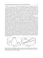
Watts et al. evaluated the effects of propofol and hyperventilation on ICP
and somatosensory evoked potentials (SEPs) in a rabbit model of intracra-
nial hypertension (8). Following inflation of an intracranial balloon to
increase the ICP to 26 ± 2 mmHg and produce a ≥ 50% reduction in SEPs,
the animals were randomized to: group 1 (propofol followed by hyperventi-
lation) or group 2 (hyperventilation followed by propofol). The ICP decrease
was significantly greater in group 1 (final ICP: 12 ± 2 mmHg vs 16 ± 5 mmHg,
p = 0.008). When comparing propofol with hyperventilation, propofol resulted
in a greater ICP decrease: 16 ± 2 mmHg with propofol vs 21 ± 5 mmHg with
hyperventilation, p = 0.007). When propofol was administered first, there
was a significant increase in the amplitude of the SEPs. The mean arterial
pressure (MAP) was maintained at baseline levels by the infusion of phe-
nylephrine. More phenylephrine (p < 0.02) was required to maintain the
MAP with propofol than with hyperventilation.
Despite these encouraging animal studies, the review of the literature con-
cerning propofol in humans provides somewhat contrasting results. Although
several studies demonstrate a decrease in ICP, propofol’s cardiovascular
effects with a lowering of the MAP can result in a decrease in the CPP.
Without the maintenance of MAP, a decrease occurs in CPP that may lead to
reflex cerebral vasodilation to maintain CBF, which may result in an in-
crease in ICP and negate the decrease in ICP induced by propofol.
Herregods et al. evaluated the effects of a propofol bolus (2 mg/kg
administered over 90 s) on ICP and MAP in six adults with an ICP greater
than 25 mmHg following traumatic brain injury (9). The mean ICP decreased
from 25 ± 3 to 11 ± 4 mmHg (p < 0.05). However, there was a decrease in
the MAP and consequently a decrease in the CPP from 92 ± 8 mmHg to a
low of 50 ± 7 mmHg. The CPP was less than 50 mmHg in four of six patients.
No vasoconstrictor agent was administered to maintain the MAP.
128 Tobias
Similar results were obtained by Pinaud et al. during their evaluation of
the effects of propofol on CBF, ICP, CPP, and cerebral arteriovenous oxy-
gen content difference in 10 adults with traumatic brain injury (10). Although
propofol decreased ICP (11.3 ± 2.6 to 9.2 ± 2.5 mmHg, p < 0.001), there
was also a decrease in MAP, which resulted in an overall decrease in CPP
from 82 ± 14 to 59 ± 7 mmHg, p < 0.01. Other investigators in patients with
traumatic brain injury (11) or during cerebral aneurysm surgery (12) have
noted similar effects of propofol on ICP and MAP with an overall lowering
of CPP caused by the greater decrease in MAP than ICP.
Farling et al. reported their experience with propofol for sedation in 10
adult patients with closed head injuries (13). Propofol was administered as a
continuous infusion of 2–4 mg/kg/h for 24 h. Additional therapy for increased
ICP included mannitol and hyperventilation. The mean rate of propofol infu-
sion was 2.88 mg/kg/h. There was a statistically significant decrease in the
mean ICP of 2.1 mmHg from baseline achieved at 2 h following the start of
the propofol infusion. No decrease in MAP was noted. The CPP increased
during the 24-h study period, and the difference was statistically significant
at the 24-h point (CPP increase of 9.8 mmHg, p = 0.028). The authors con-
cluded that propofol was a suitable agent for sedation in head-injury patients
who required mechanical ventilation.
Spitzfadden et al. reported their experience with the use of propofol to pro-
vide sedation and control ICP in two adolescents (14). Dopamine was used to
maintain MAP and CPP. Propofol resulted in adequate sedation and control of
ICP. When compared with barbiturates, the usual time-honored therapy for
pharmacologic control of ICP, the authors suggested that a significant advan-
tage of propofol was a much more rapid awakening. The latter effect may be
most evident following prolonged (>48 h) administration of barbiturates.
Further study will be required to fully evaluate the role of propofol in
controlling ICP. With control of MAP, the initial clinical and laboratory
evidence suggests that propofol can be used to decrease CMRO
2
, CBF, and
ICP. Additional benefits of propofol in patients with altered intracranial
compliance include maintenance of CBF autoregulation in response to
changes in MAP and P
a
CO
2
as well as preliminary evidence that suggests a
possible protective effect of propofol during periods of cerebral hypoperfu-
sion and ischemia (15,16). These latter effects are similar to those reported
with the use of barbiturates (17). It is postulated that the neuroprotective
effects may result from alterations in CMRO
2
or propofol’s antioxidant prop-
erties related to its phenol ring structure.
Following its increased use both in and outside of the operating room,
certain adverse effects have been reported with propofol (Table 1). Propo-
Pharmacology of Sedative Agents 129
fol’s cardiovascular effects are similar to those of the barbiturates, including
an overall lowering of the MAP related to both peripheral vasodilation and
negative inotropic properties (18). Propofol also alters the baroreflex responses,
thereby resulting in a smaller increase in heart rate for a given decrease in
blood pressure. These cardiovascular effects are especially pronounced fol-
lowing bolus administration. Although generally well-tolerated by patients
with adequate cardiovascular function, these effects may result in detrimen-
tal physiologic effects in patients with compromised cardiovascular func-
tion. Tritapepe et al. have demonstrated that the administration of calcium
chloride (10 mg/kg) prevented the deleterious cardiovascular effects of
propofol during anesthetic induction in patients undergoing coronary artery
bypass grafting (19).
In addition to the negative inotropic properties, central vagal tone may be
augmented, leading to bradycardia (20) or asystole when combined with
other medications known to alter cardiac chronotropic function (fentanyl,
succinylcholine) (21). Although the relative bradycardia is generally con-
sidered a beneficial effect in patients at risk for myocardial ischemia, it may
be detrimental in patients with fixed stroke volumes whose cardiac output is
heart-rate-dependent.
Unusual neurologic manifestations including opisthotonic posturing,
myoclonic movements (especially in children), and seizure-like activity have
Table 1
Adverse Effects Reported with Propofol
Hypotension
Negative inotropic effects
Vasodilation
Bradycardia, asystole
Neurologic sequelae
Opisthotonic posturing
Seizure-like activity
Myoclonus
Respiratory depression, apnea
Anaphylactoid reactions
Metabolic acidosis and cardiac failure (with prolonged
administration in the pediatric population)
Pain on injection
Bacterial contamination of solution
Hyperlipidemia
Hypercarbia
130 Tobias
been reported with propofol administration (22–25). Although some of the
initial reports suggested actual seizure activity, these concerns have most
likely been overemphasized, since no electroencephalographic evidence of
seizure activity has been documented during the abnormal movements seen
with propofol administration. Additionally, propofol is considered a valu-
able agent in the treatment of patients with refractory status epilepticus that
is unresponsive to conventional therapy (26).
Although many studies have examined the cardiovascular effects of
propofol, the respiratory-depressant effects of propofol should not be over-
looked. Although propofol has become a popular agent for deep sedation in
the spontaneously breathing patient, reports demonstrate a relatively high
incidence of respiratory effects including hypoventilation, upper airway
obstruction, and apnea (27). As with any sedative agent, some degree of
hypoventilation is likely to occur in all patients breathing spontaneously.
These effects may be detrimental related to the alterations in P
a
CO
2
and its
obvious deleterious effects on CBF, ICP, and CPP. Despite these potential
deleterious effects on respiratory function, recent laboratory and clinical
studies suggest that propofol may be advantageous when instrumenting the
airway of patients with reactive airway disease. In an animal model, Chih-
Chung et al. demonstrated that propofol attenuates carbachol-induced air-
way constriction (28). The mechanism involves a decrease in intracellular
inositol phosphate accumulation, thereby limiting intracellular calcium
availability. The latter results from a decrease in calcium release from intra-
cellular stores as well as a decrease in transmembrane movement.
In children, a significant issue with the prolonged use of propofol—such
as ongoing sedation in the pediatric ICU setting—are reports of unexplained
metabolic acidosis, brady-dysrhythmias, and fatal cardiac failure (29,30).
The initial report of Parke et al. published in 1992 included five children
with respiratory infections and respiratory failure who received prolonged
propofol infusions, although in higher than usual doses (up to 13.6 mg/kg/h).
Other anecdotal reports subsequently appeared, followed by a review by
Bray examining the reports from the medical literature of 18 children with
suspected propofol infusion syndrome (31). Risk factors for the syndrome
identified by Bray included propofol administration for more than 48 h or
doses greater than 4 mg/kg/h. However, several children received doses
greater than 4 mg/kg/h for longer than 48 h, suggesting that factors other
than dose and duration are necessary for development of the syndrome.
Other associated factors included age; 13 of the 18 patients were 4 yr of age
or younger, and only 1 of 18 was more than 10 yr of age. Since the review of
Bray et al, the syndrome has been reported in a 17-yr-old patient (32). As
suggested by the initial report of Parke et al., there may be an association of
Pharmacology of Sedative Agents 131
an respiratory tract infection in the etiology of the syndrome, as 82% of the
reported cases have been in children with such infections. In addition to the
cardiovascular manifestations, other features have included metabolic aci-
dosis, lipemic serum, hepatomegaly, and muscle involvement with rhabdo-
myolysis (32). Suggestions for treatment include discontinuation of the
propofol followed by symptomatic treatment of the cardiovascular dysfunc-
tion. In patients with rhabdomyolysis and renal failure, hemodialysis has
been used. Although hemodialysis has been effective in the management of
these patients, it is yet to be determined whether its only effect is in the
management of the renal dysfunction, or whether it may also have a thera-
peutic effect through the removal of a suspected toxic metabolite. Until fur-
ther data are available, caution is suggested with the administration of
propofol by continuous infusion in the pediatric ICU patient less than 10–12 yr
in doses exceeding 4 mg/kg/h or for longer than 48 h. However, because of
the previously described beneficial properties, propofol may have a role in
providing short-term sedation in younger patients and for more prolonged
use in older patients.
Additional problems with propofol relate to its delivery in a lipid emul-
sion. The latter is the same lipid preparation as that used in parenteral
hyperalimentation. There have been rare reports of anaphylactoid reactions
(33). These may be more likely in patients with a history of egg allergy. Pain
occurs with propofol administration through a peripheral infusion site. Vari-
able success in decreasing the incidence of pain has been reported with vari-
ous maneuvers, including the preadministration of lidocaine, pretreatment
with thiopental, mixing the lidocaine and propofol in a single solution, di-
luting the concentration of the propofol, or cooling it prior to bolus adminis-
tration (34,35). Another alternative is the administration of a small dose of
ketamine (0.5 mg/kg) prior to the administration of propofol (36). Since
propofol has limited analgesic properties, ketamine and propofol can be ad-
ministered together to take advantage of the analgesia provided by ketamine
and the rapid recovery with propofol. This combination can be used for brief
invasive procedures or for ICU sedation. For these purposes, ketamine can
be added to the propofol solution to produce a mixture containing 3–5 mg/
mL ketamine and 10 mg/mL propofol. For brief procedures, incremental
doses of 0.1 mL/kg can be administered, resulting in the delivery of 0.3–0.5
mg/kg of ketamine and 1 mg/kg of propofol.
Unlike many other medications, the initial formulation of propofol did
not contain preservatives. Laboratory investigation has demonstrated that
the lipid emulsion is a suitable culture medium for bacteria (37). Systemic
bacteremia and postoperative wound infections have been linked to extrinsically
contaminated propofol (38). A modification of the initial preparation by
132 Tobias
AstraZeneca Pharmaceuticals, manufacturer of propofol, included the addi-
tion of ethylenediaminetetraacetic acid (EDTA) as a preservative, which
may limit the risk of bacterial contamination and growth. Recently, another
preparation of propofol, manufactured by Baxter Pharmaceuticals, has been
released for clinical use. This latter preparation contains sodium metabisul-
fite as a preservative. There remains some controversy over the possible
association of sodium metabisulfite with allergic reactions, especially in
patients with asthma and other atopic conditions. Despite the recent changes,
meticulous aseptic technique is required when using propofol. Opened but
unused vials should be disposed of promptly and not saved for later use.
When used by continuous infusion for ICU sedation, the vial and tubing
should be changed every 12 h.
Additional problems related to the high lipid content of the solution have
included hypertriglyceridemia (39). A case report suggests the anecdotal
association of high-dose propofol infusion with an increasing PaCO
2
during
prolonged mechanical ventilation in the ICU setting (40). The latter report
describes a patient that required up to 200 mcg/kg/min of propofol to main-
tain an adequate level of sedation. This resulted in a total caloric intake of
4500 calories/d (53% from the lipid in the propofol diluent). The PaCO
2
increased from 67 mmHg to a maximum value of 78 mmHg, despite increas-
ing the minute ventilation from 11 to 13 L/min. The lipid content of propofol
should be taken into consideration when calculating the patient’s daily caloric
intake. A propofol infusion of 2 mg/kg/h provides roughly 0.5 gm/kg/d of fat.
Possible solutions to these problems include the potential production of a
2% solution to limit the total lipid administration.
2.2. Ketamine
Ketamine is a sedative/analgesic agent that is structurally related to phen-
cyclidine. It was introduced into clinical practice in the 1960s (41). A unique
feature of ketamine, which makes it particularly attractive for sedation dur-
ing procedures, is the provision of both amnesia and analgesia. Its molecu-
lar structure contains a chiral center at the C
2
carbon of the cyclohexanone
ring, resulting in both a (+) and (–) enantiomer. Ketamine’s anesthetic/anal-
gesic properties result from its interactions with the limbic/thalamic sys-
tems, resulting in what has been termed dissociative anesthesia. Additional
postulated sites/mechanisms of action include the NMDA receptor as well
as subgroups of opioid receptors.
Commercially available ketamine is a racemic mixture of these two opti-
cal (+,–) isomers. It is available in three different concentrations, including
1% (10 mg/mL), 5% (50 mg/mL), and 10% (100 mg/mL). Preliminary data
suggests that the (+) isomer may possess some clinical advantages, includ-
Pharmacology of Sedative Agents 133
ing a more potent anesthetic/analgesic effect with a more limited duration of
action allowing for a more rapid awakening and a more rapid return to nor-
mal cognitive function (42).
Metabolism occurs primarily by hepatic N-methylation to various metabo-
lites, including norketamine, which is further metabolized via hydroxylation
pathways with subsequent urinary excretion. Norketamine retains roughly
one-third of the analgesic and sedative properties of the parent compound.
Bioavailability is 100% following iv/intramuscular administration. How-
ever, the bioavailability is markedly decreased with oral or rectal adminis-
tration because of limited absorption and a high degree of first-pass
metabolism. Higher concentrations of norketamine are noted following oral/
rectal administration because of the greater degree of first-pass hepatic metabo-
lism and may account for a significant part of the anesthetic effect following
oral/rectal administration. As ketamine is primarily dependent on hepatic
metabolism, doses should be reduced in patients with hepatic dysfunction.
The beneficial properties of ketamine include preservation of cardiovas-
cular function and limited effects on respiratory mechanics. These proper-
ties make it an effective agent for the provision of amnesia and analgesia
during painful, invasive procedures while allowing the maintenance of spon-
taneous respiratory function (43).
In the majority of clinical scenarios, ketamine results in a dose-related
increase in heart rate and blood pressure, which are mediated through the
sympathetic nervous system response with the release of endogenous catechola-
mines (44,45). In most clinical circumstances, ketamine results in increased
heart rate and blood pressure, which can increase myocardial oxygen con-
sumption. These effects can alter the balance between myocardial oxygen
demand and delivery, inducing ischemia in patients with ischemic heart dis-
ease. The hypertension and tachycardia that occur with ketamine administra-
tion can be decreased by the administration of ketamine with a benzodiazepine,
a barbiturate, propofol, or synthetic opioids (fentanyl or sufentanil).
Ketamine’s indirect sympathomimetic effects generally overshadow its
direct negative inotropic properties. However, hypotension may occur in
patients with diminished myocardial contractility (46,47). In these patients,
it is postulated that ketamine’s direct negative inotropic properties predomi-
nate because the endogenous catecholamine stores have been depleted.
Although somewhat controversial, ketamine may adversely effect pul-
monary vascular resistance (PVR), and should be used with caution in adults
with diminished right ventricular function or altered PVR. This issue remains
controversial, as varying results have been reported in the literature, espe-
cially when considering both adult and pediatric patients. The initial studies
were performed during spontaneous ventilation, and the alterations in PVR
134 Tobias
may have been related to increases in PaCO
2
and not the direct effects of
ketamine on the pulmonary vasculature. Following ketamine administration
to infants with congenital heart disease during spontaneous ventilation,
Morray et al. noted statistically significant increases in pulmonary artery
pressure (from a mean of 20.6 mmHg to 22.8 mmHg) and increases in PVR
(48). In contrast, Hickey et al. found no change in PVR in intubated infants
with minimal ventilatory support (4 breaths/min and an F
i
O
2
of 0.4) (49).
The latter study included 14 patients—7 with normal and 7 with elevated
baseline PVR. Pending further investigations, ketamine should be used cau-
tiously in patients with pulmonary hypertension, especially during spontaneous
ventilation. However, the available literature in children with cyanotic and non-
cyanotic congenital heart disease continues to show beneficial effects of
ketamine on overall cardiovascular performance and oxygen saturation (50).
One significant advantage of ketamine over many other sedative/analgesic
agents is its lack of significant effects on respiratory function. Functional
residual capacity, minute ventilation, and tidal volume remain unchanged
following ketamine administration (51), while other investigators have dem-
onstrated improved pulmonary compliance, decreased resistance, and pre-
vention of bronchospasm (52). These effects on respiratory mechanics have
been partially attributed to effects from the release of endogenous catechola-
mines (53). Although minute ventilation is generally maintained, elevations
of P
a
CO
2
and a rightward shift of the CO
2
response curve have been reported
(54), and there remains controversy concerning ketamine’s effects on pro-
tective airway reflexes. Although clinical use and experimental studies sug-
gest that airway reflexes are maintained, aspiration and laryngospasm have
been reported following ketamine in spontaneously breathing patients with-
out a protected airway (55). In higher doses or in severely compromised
patients, ketamine can cause apnea, proving again that all sedative/analge-
sic agents, especially when administered to critically ill patients, should be
administered only in a controlled environment with appropriate monitoring.
An additional effect that may influence airway patency is increased oral
secretions. The concomitant administration of an anti-sialogogue such as
atropine or glycopyrrolate is recommended. Ketamine increases salivary and
bronchial gland secretion through stimulation of central cholinergic recep-
tors. Ketamine increases CBF/ICP, and should be avoided in patients with
altered intracranial compliance (56,57). The effects on ICP are the result of
direct cerebral vasodilatation, mediated through central cholinergic recep-
tors. They are not secondary to alterations in the CMRO
2
or changes in
PaCO
2
(58,59).
Perhaps the most well-known adverse effect related to ketamine is the
occurrence of emergence phenomena or hallucinations. Emergence phe-
Pharmacology of Sedative Agents 135
nomena are dose-related, occurring more commonly in adolescents and
adult patients. Their incidence can be decreased by the pre- or concomitant
administration of a barbiturate, propofol, or benzodiazepine (60). It is pos-
tulated that emergence phenomena result from the alteration of auditory
and visual relays in the inferior colliculus and the medical geniculate
nucleus, leading to the misinterpretation of visual and auditory stimuli (60).
The administration of a benzodiazepine (lorazepam or midazolam) 5 min
prior to the administration of ketamine is generally effective in preventing
emergence phenomena, and may allow for the use of ketamine even in older
patients. The combined use of propofol and ketamine has been previously
discussed.
Another option with ketamine is to use non-intravenous routes of deliv-
ery. Intramuscular (im) administration in doses of 3–4 mg/kg can be used in
uncooperative patients who lack venous access. Although the bioavailability
of im administration is 100%, the onset of action will be delayed, requiring
10–15 min to achieve a peak effect. Alternatively, in the pediatric popula-
tion, both intranasal and rectal administration of ketamine have been
reported for premedication for the operating room (61), and oral administra-
tion has been reported for sedation/analgesia during bone marrow aspiration
and for the suturing of lacerations in the emergency room setting (62,63).
When the non-parenteral routes are used, larger doses of 6–10 mg/kg are
required, since the bioavailability is only 10–20%.
Although it is most often administered in intermittent bolus doses, there
are limited reported clinical experiences with the use of ketamine for seda-
tion of the ICU patient. Tobias et al. reported their anecdotal experience
with the use of ketamine infusions for sedation in five pediatric ICU patients
(64). Four of the patients had experienced adverse cardiorespiratory effects
following the administration of benzodiazepines and/or opioids. Hartvig et
al. used a ketamine infusion to provide sedation and analgesia following
cardiac surgery in 10 infants and children ranging in age from 1 wk to 30 mo
(65). A continuous infusion of ketamine in a dose of 1 mg/kg/h was admin-
istered to five of the patients, and the other five received 2 mg/kg/h. Both
groups received intermittent, as-needed doses of midazolam. The mean
plasma clearance of ketamine was 0.94 ± 0.22 L/kg/h with an elimination
half-life of 3.1 ± 1.6 h. Norketamine demonstrated an elimination half-life
of 6.0 ± 1.8 h. Both ketamine infusion rates provided similar and acceptable
levels of sedation.
2.3. Etomidate
Etomidate is a carboxylated, imidazole-containing iv anesthetic agent that
was first synthesized in 1964 and introduced into clinical anesthesia prac-
136 Tobias
tice in 1972. Since the aqueous solution of etomidate is unstable at physi-
ologic pH, it is available in a 0.2% (20 mg/mL) solution with 35% propy-
lene glycol. The pH of 6.9 of this solution and the carrier vehicle—propylene
glycol—account for the high incidence of pain and the development of
thrombophlebitis with administration through peripheral iv sites. Although
the propylene glycol is not an issue with single, short-term administration,
toxicity from the carrier vehicle has been reported following long-term infu-
sions (66).
Like the barbiturates, propofol, and benzodiazepines, it is postulated that
etomidate provides its anesthetic effects by interactions with the GABA system
and alterations of chloride conductance across the cell membrane (67). Unlike
the barbiturates and propofol, etomidate has little effect on cardiovascular per-
formance, even in patients with altered myocardial contractility (68,69).
Anesthetic induction doses ranging from 0.2–0.4 mg/kg provide a rapid
onset of amnesia and sedation with a rapid emergence time following a
single bolus dose. Following iv administration, etomidate undergoes ester
hydrolysis by the liver with the formation of inactive water-soluble metabo-
lites. The elimination half-life is prolonged in the setting of hepatic dysfunc-
tion. As etomidate possesses limited analgesic properties, it may not
effectively blunt the hemodynamic response to endotracheal intubation in
patients with normal cardiovascular function. Co-administration of an opioid
such as fentanyl may provide a more stable hemodynamic profile.
Like the barbiturates and propofol, etomidate decreases the CMRO
2
, result-
ing in cerebral vasoconstriction and a decrease in CBF and ICP. With its
limited effects on cardiovascular function, CPP is maintained, making it a
suitable induction agent for patients with altered myocardial contractility and
increased ICP. Etomidate produces EEG changes similar to that seen with the
barbiturates; however, it can also produce epileptic-like EEG potentials in
patients with underlying seizure disorders. These potentials are produced with-
out accompanying motor activity, making it a useful intra-operative agent to
identify seizure foci during seizure surgery. Etomidate has also been used to
treat status epilepticus (70).
To date, the vast majority of experience with etomidate centers around its
use as a single dose for the induction of anesthesia in adults. Kay noted a
rapid onset of anesthesia with etomidate and limited effects on cardiovascu-
lar function in 198 children ranging in age from 1 d to 15 yr (71). However,
no data is given concerning the cardiovascular status of these patients.
Tobias reported anecdotal experience with the use of etomidate for anes-
thetic induction in three children including a 33-mo-old with a dilated cardi-
omyopathy, a 9-yr-old trauma victim with hypovolemia and increased ICP,
and a 10-yr-old with aortic stenosis and respiratory failure (72).
Pharmacology of Sedative Agents 137
Because of its limited effects on cardiovascular function, there is a con-
tinuing interest in the use of etomidate for sedation during procedures out-
side of the operating room. Ford et al. compared incremental doses of
thiopental at 50 mg or etomidate at 4 mg for sedation during cardioversion
in 16 ASA (American Society of Anesthesiologists) class II or III adult
males, age 55–66 yr (73). Both drugs provided adequate levels of sedation.
No significant difference was noted in heart rate and blood pressure. There
was a statistically significant increase in respiratory rate with etomidate,
and a decrease in respiratory rate with thiopental, and recovery times were
similar. Mild myoclonus was noted with the use of etomidate.
Canessa et al. evaluated four anesthetic agents (thiopental 3 mg/kg,
etomidate 0.15 mg/kg, midazolam 0.15 mg/kg, and propofol 1.5 mg/kg)
during cardioversion in 45 adults (74). All patients received 1.5 mcg/kg of
fentanyl 3 min prior to the procedure. Etomidate produced mild pain on
injection and myoclonus, but was the only one of the four agents that did not
lower MAP. Propofol resulted in hypotension and a higher incidence of apnea.
The duration of effect was similar with propofol, thiopental, and etomidate,
but was prolonged with midazolam.
The information concerning the use of etomidate for sedation in children
is more limited. McDowall et al. compared etomidate, propofol, and
ketamine for sedation during procedures in pediatric oncology patients (75).
Ketamine was associated with vomiting (14.6%), agitation (15%), and tachy-
cardia (19.5%). Etomidate was associated with vomiting (9.9%) and agita-
tion (1.2%). Propofol resulted in hypoxemia in 15.7% of patients, which
was usually managed by the administration of supplemental oxygen, but
occasionally required bag-mask ventilation. Propofol resulted in a low inci-
dence of vomiting (0.5%) and agitation (1.2%). Behrens et al. reported their
experience with the use of etomidate for sedation during placement of per-
cutaneous endoscopic gastrostomies in 139 patients (76).
Etomidate has also been administered by the non-parenteral route.
Streissand et al. evaluated the possible use of etomidate as a premedicant
administered as a transmucosal lozenge in 10 adult volunteers (77). The
volunteers ingested transmucosal etomidate in doses of 25, 50, 75, and 100 mg
on four study days. The peak plasma concentration was achieved at 20–30 min.
Two volunteers experienced brief episodes of involuntary tremor after the
100-mg dose. Drowsiness and light sleep occurred in a dose-related manner.
The authors concluded that this preparation might be effective when brief,
mild to moderate sedation was needed.
Various adverse effects have been reported with etomidate (Table 2).
Those related to the carrier vehicle include pain on injection, thrombophle-
bitis, and propylene glycol toxicity. The latter was reported only with pro-
138 Tobias
longed infusions. The most significant concern remains etomidate’s effect
on the endogenous production of corticosteroids. These effects limit its use
for prolonged sedation in the ICU setting (78). Etomidate inhibits the func-
tion of an enzyme system (11-beta hydroxylase), which is necessary for the
production of cortisol, aldosterone, and corticosterone. Although inhibition
is present after a single dose of etomidate (79), this effect is not believed to
be of clinical significance.
2.4. Barbiturates
The barbiturates are one of the oldest class of agents used in anesthesia
practice. They can be classified according to their duration of activity. Short-
acting agents include methohexital, thiopental, and thiamylal. Pentobarbital
is considered an intermediate-acting agent, and phenobarbital is considered
a long-acting agent. The short-acting agents have a duration of action of
5–10 min following a single bolus dose and are usually used by iv, bolus
administration for brief procedures such as the induction of anesthesia and
endotracheal intubation. When a more prolonged effect is needed, a con-
tinuous infusion may be used to maintain constant plasma levels. Thiopental
and thiamylal are thiobarbiturates, and methohexital is an oxybarbiturate.
Thiopental and thiamylal are commercially available as racemic mixtures of
the two optical isomers. The L-isomers of both drugs are twice as potent as
the D-isomers. Methohexital has two asymmetric centers, resulting in four
isomers. Since the beta isomers produce excessive motor activity, metho-
hexital is available as a mixture of the two alpha isomers. The three agents
are reconstituted with sterile saline to solutions of 1–2.5%. Induction doses
vary based on the potency of the agent. Methohexital is the most potent
(2.5–3 times that of thiopental) and thiopental is the least potent. Induction
doses are also higher in neonates and infants. Anesthetic induction doses for
thiopental vary from 3–5 mg/kg in healthy adults, 5–6 mg/kg in children, and
6–8 mg/kg in neonates and infants. The barbiturates undergo predominant
hepatic metabolism except for phenobarbital, which is also dependent on
Table 2
Adverse Effects Reported with Etomidate
Myoclonic movements
Nausea/vomiting
Pain on injection
Thrombophlebitis
Propylene glycol toxicity (with prolonged infusions)
Adrenal suppression (with prolonged infusions)
Pharmacology of Sedative Agents 139
renal elimination. The rapid dissipation of anesthetic effect is not related to
hepatic metabolism, but rather redistribution from the central compartment.
During prolonged infusions, the peripheral compartments are saturated and
a prolonged effect is seen.
Beneficial physiologic effects of the barbiturates include a decrease of
the CMRO
2
with a reduction in CBF, cerebral vasoconstriction, and a
decrease in ICP. They produce varying dose-dependent degrees of EEG sup-
pression, and in sufficient does produce electrical silence. The barbiturates
are potent anticonvulsants, and may be used to treat status epilepticus that is
unresponsive to other agents. Although still controversial, it has also been
suggested that the barbiturates may provide some degree of cerebral protec-
tion during periods of cerebral hypoxia or hypoperfusion. This effect has
not been shown to occur if these agents are administered after the event. The
CNS properties of the barbiturates are much the same as those described for
the benzodiazepines, etomidate, and propofol.
As with many of the agents described, the barbiturates’ effects on cardio-
respiratory function are dose-dependent. In healthy patients, sedative doses
have minimal effects on respiratory drive and airway protective reflexes,
yet larger doses—especially in patients with cardiorespiratory compro-
mise—can produce respiratory depression, apnea, or hypotension. The car-
diorespiratory effects are additive when the barbiturates are used with other
agents such as opioids. Hypotension results from both peripheral vasodilation
with a decrease in preload/afterload and a direct negative inotropic effect.
Although the barbiturates are used most often in the operating room for
the induction of anesthesia and in the ICU for their therapeutic effects (as
anticonvulsants or to decrease ICP), these agents may play a role in provid-
ing sedation outside of the operating room. Sanderson et al. reported the use
of iv pentobarbital to provide sedation during radiologic procedures in 149
children ranging in age from 3 mo to 7 yr (80). One hundred forty-one of the
patients received only pentobarbital, and eight also received midazolam and/
or fentanyl. The mean dose of pentobarbital was 4.6 mg/kg with a range of
2–10 mg/kg. The mean time from the start of sedation to the start of the scan
was 7 min (range: 2–50 min). Sedation was successful in all cases. Adverse
effects were noted in 22 of the 146 patients (14.7%), and included oxygen
desaturation, vomiting, airway secretions, airway obstruction, coughing, and
bronchospasm. No patient required endotracheal intubation or bag-mask
ventilation. Similar success with iv pentobarbital for radiologic procedures
has been reported by other investigators (81,82). In addition to iv adminis-
tration, rectal thiopental sodium has been successfully used in many centers
for sedation for radiologic procedures (83).
140 Tobias
The barbiturate, pentobarbital, has also been used for sedation during
mechanical ventilation in the pediatric ICU population. Tobias reported a
retrospective evaluation of pentobarbital use in 50 children for pediatric ICU
sedation (84). The 50 patients ranged in age from 1 mo to 14 yr, and ranged
in weight from 3.1 to 56 kg. Prior to switching to pentobarbital, the level of
sedation was inadequate despite midazolam doses of 0.4 mg/kg/h with either
fentanyl (10 mcg/kg/h) or morphine (100 mcg/kg/h). No significant adverse
affects related to pentobarbital were noted.
One problem that may limit the use of barbiturates in the ICU setting is
that the solution is alkaline, thereby making it incompatible with other medi-
cations and parenteral alimentation solutions. Therefore, the barbiturates
should be administered separately from other medications. Local erythema
and thrombophlebitis can occur with subcutaneous infiltration.
2.5. Nitrous Oxide
Nitrous oxide (N
2
O) was first synthesized in 1776 by Priestley, and its anes-
thetic properties were first described by Humphrey Davy in 1799. Despite
Davy’s suggestion of the potential effects of this agent, it was not until 1844 that
Gardner Colton used nitrous oxide as an anesthetic agent during a tooth extrac-
tion. Today, nitrous oxide remains one of the most widely used anesthetic agents.
Nitrous oxide possesses many of the characteristics of an ideal agent for
sedation. It has a rapid onset of action, is relatively easy and inexpensive to
use, its effects dissipate rapidly once discontinued, and it provides amnesia,
sedation, and analgesia. Because of its low blood-gas partition coefficient
(relative insolubility in blood), its alveolar concentration rises rapidly, result-
ing in a rapid onset of activity. Holst reported an astonishing experience of
3 million pediatric dental patients treated with 30–60% nitrous oxide with-
out a single serious complication (85). Griffin and colleagues describe its
use in children in an emergency room setting for treating burns, suturing
lacerations, and orthopedic reductions (86).
Nitrous oxide is not a complete anesthetic. Its minimum alveolar concen-
tration or MAC, (a measure of anesthetic potency, which describes the anes-
thetic concentration at which 50% of patients move in response to surgical
incision), is 105%. The latter is impossible to achieve at normal barometric
pressure. Even in concentrations of 70–80%, additional agents may be necessary.
Nitrous oxide can be administered by a face or nasal mask. Another op-
tion involves the use of a weighted mouthpiece that is held in place by the
patient during administration. If the patient becomes too sleepy, the device
falls from the patient, thereby stopping the administration of nitrous oxide.
Safety issues mandate that nitrous oxide be administered with several safety
Pharmacology of Sedative Agents 141
features including standard monitoring. Additional monitors include: a
monitor of the inspired oxygen concentration, a device that limits the ratio
of the flow rates of oxygen to nitrous oxide (a proportioning system so that
less than 30% oxygen cannot be administered), and a system that cuts off the
nitrous oxide flow if the oxygen supply fails. Without this latter device, the
nitrous oxide flow can continue without the addition of oxygen, leading to
the delivery of a hypoxic mixture or 100% nitrous oxide.
In the operating room, nitrous oxide and oxygen are generally adminis-
tered from the wall outlets connected to the hospital’s central supply. In
other areas when such a supply is not available, nitrous oxide can be admin-
istered from E cylinders and mixed with oxygen to provide the desired con-
centration. Alternatively, commercially available tanks are manufactured
that contain a 50/50 oxygen and nitrous oxide mixture, thereby limiting the
risk of a hypoxic mixture and the need for specialized equipment to mix
oxygen and nitrous oxide from separate tanks.
A scavenger device attached to the delivery system is also required to
remove waste gases and prevent environmental pollution. Repeated expo-
sure of the patient or healthcare workers to nitrous oxide can lead to ter-
atogenic effects, increased risk of spontaneous abortion, bone marrow
suppression or megaloblastic anemia, and peripheral neuropathy as a result
of its effects on B
12
metabolism and protein synthesis. Because of the
potential for abuse and/or illicit use, nitrous oxide tanks should be kept
under close surveillance.
Despite its widespread use and long safety record, significant physiologic
effects occur with nitrous oxide. Nitrous oxide exerts a dose-dependent
negative depressant effect on myocardial contractility and increases pulmo-
nary artery pressure. Like all sedative/analgesic agents, it also causes dose-
dependent respiratory depression, resulting in an elevation of the resting
P
a
CO
2
level and blunting of the central respiratory response to hypercarbia
and hypoxemia. Litman et al. evaluated the levels of sedation and respiratory
effects of oral midazolam (0.5 mg/kg) combined with increasing concentra-
tions of nitrous oxide in 20 children, age 1–3 yr (87). Four concentrations of
nitrous oxide were studied: 15%, 30%, 45%, and 60%. During nitrous oxide
inhalation, 12 of the 20 patients developed an increasing end-tidal CO
2
with
a decrease in the respiratory rate. At 30% nitrous oxide, one child met the
American Academy of Pediatrics (AAP) criteria for deep sedation. With
60% nitrous oxide, six children were not clinically sedated, six met the AAP
criteria for conscious sedation, six met the AAP criteria for deep sedation,
and one child developed an even deeper level of sedation with no response
to painful stimuli.
142 Tobias
Additional issues/concerns with nitrous oxide are listed in Table 3. Nitrous
oxide increases the incidence of postoperative nausea and vomiting. It dif-
fuses into air-filled spaces, increasing the volume and pressure of the space.
This can be an issue with any loculated collection of air, including bowel
obstruction, intrathoracic injuries with the risk of pneumothorax, the middle
ear, lung cysts, or in the presence of pneumocephalus. Nitrous oxide increases
CBF/ICP, and is relatively contraindicated in patients with closed head
injury and altered intracranial compliance.
Despite its relative insolubility in blood, during the administration of nitrous
oxide, a large amount is taken up into the blood. This latter effect, known as
the second gas effect of anesthesia, increases the alveolar PO
2
, resulting in
an added margin of safety during induction even if high concentrations of
nitrous oxide (80–90%) are administered. Once the administration of nitrous
oxide is discontinued, this effect occurs in the opposite direction, resulting
in a lowering of the alveolar PO
2
—which can result in hypoxemia unless
supplemental oxygen is administered until the nitrous oxide is eliminated
from the body.
2.6. Chloral Hydrate
In children, chloral hydrate remains one of the more commonly used
agents for sedation. It was originally synthesized in 1832 and introduced
into clinical practice in 1869 by Liebreich. For street and recreational use,
chloral hydrate is the ingredient combined with alcohol in mixtures known
as “knockout drops” and “Mickey Finns.” It is available as capsules (250 mg,
500 mg), syrup (250 mg/5 mL and 500 mg/5 mL), and suppositories (325 mg,
500 mg, and 650 mg). Chloral hydrate tends to be a GI irritant, especially
Table 3
Issues with Nitrous Oxide Administration
Potential for the administration of a hypoxic mixture
Contamination of tanks with nitrogen dioxide, nitric oxide
Healthcare worker exposure
Abuse potential
Megaloblastic anemia, bone marrow suppression
Teratogenesis
Myelopathy
Depressed myocardial contractility
Increased pulmonary artery pressure
Expansion of air-containing spaces
Increased cerebral blood flow, increased intracranial pressure
Pharmacology of Sedative Agents 143
when administered on an empty stomach, and results in a relatively high
incidence of nausea and vomiting. In younger children, these problems can
be avoided with the use of suppositories. It has no analgesic properties;
therefore, it should not be used to treat pain or during painful procedures
unless combined with an analgesic agent such as an opioid.
Chloral hydrate is rapidly absorbed from the gastrointestinal tract
with a bioavailability that approaches 100%. Its onset of action is within
20 min, with a peak effect at 30–60 min. Following absorption, it is metabo-
lized in the liver by the enzyme alcohol dehydrogenase to the active ingredi-
ent, trichloroethanol (TCE). TCE is then further metabolized by either
glucuronidation or oxidation to inactive metabolites. Less than 10% of
chloral hydrate undergoes renal excretion. The plasma half-life of TCE is
8–12 h in children, but may be prolonged up to 24–36 h in neonates and
infants (88). These prolonged half-lives account for the prolonged effects
that can occur in specific patient populations. Additive and prolonged
effects are commonly seen following repeated administration over a
period of days.
Chloral hydrate and its active metabolite TCE are CNS depressants. In
therapeutic doses, there are minimal effects on cardiorespiratory function
and airway control. Although apnea and hypotension can occur, these effects
are generally only seen in an overdose situation. In the setting of an over-
dose, central respiratory depression with apnea and cardiovascular compro-
mise is the leading cause of mortality. Cardiovascular effects relate to
decreased myocardial contractility, a shortened refractory period, and an
altered sensitivity of the myocardium to endogenous catecholamines (89).
The latter two effects account for the pro-arrythmogenic effects that are seen.
These effects primarily relate to TCE—which is a halogenated hydrocarbon—
and like halothane, shares the same myocardial effects. These properties
suggest against the use of chloral hydrate as a sedative in patients with toxic
ingestions that may predispose to arrhythmias such as tricyclic antidepres-
sants (90).
Several factors account for the continued use of chloral hydrate as a first-
line agent for sedation in the pediatric population. These include physicians’
familiarity with the agent, its wide therapeutic index, its cost, and a lack of
significant effects on cardiorespiratory function. Dose recommendations
vary widely, ranging from 25–30 mg/kg up to 80–100 mg/kg. The latter
doses generally provide a greater degree of success when attempting to pro-
vide a motionless patient during radiographic imaging. Because of the possi-
bility of a prolonged effect, extended post-procedure monitoring may be
required. Despite a long record of safety with minimal effects on respira-
144 Tobias
tory function in most patients, deaths from respiratory depression have
occurred. Like all other sedative agents, respiratory depression can occur
with chloral hydrate, and standard monitoring is mandatory. Additionally,
the patient should be monitored until fully awake. More than one patient
has been discharged before being fully awake, only to be found dead on
arrival at home.
2.7. Benzodiazepines
The benzodiazepines used most commonly in the United States for seda-
tion include diazepam, lorazepam, and midazolam. These agents bind to
specific receptor sites that are part of the GABA receptor system, increasing
the efficacy of the interaction between the GABA receptor and the chloride
channel. Benzodiazepines provide several therapeutic effects including seda-
tion, anxiolysis, amnesia, anticonvulsant properties, and spinally mediated
muscle relaxation. It has been suggested that increasing the rate of occu-
pancy of the benzodiazepine receptor results in an escalation of the benzodi-
azepine effect from anxiolysis to sedation to unconsciousness when 60% or
more of the receptor sites are occupied.
Diazepam and lorazepam are insoluble in water, and are commercially
available in a solution containing propylene glycol. The diluent, propylene
glycol, can result in tissue irritation, pain on injection, and local throm-
bophlebitis. These problems are not seen with midazolam, which is water-
soluble. Recently, diazepam has been made available in a lipid emulsion in
an attempt to limit the issues of local tissue irritation and pain on injection.
Metabolism of the benzodiazepines occurs via hepatic oxidation and
glucuronidation. Oxidative processes are primarily responsible for the
metabolism of diazepam and midazolam, and glucuronidation pathways are
responsible for lorazepam metabolism. Hepatic oxidative processes occur-
ring via the P
450
system are susceptible to hepatic dysfunction and the co-
administration of medications, including cimetidine and anticonvulsants.
Glucuronidation pathways are less influenced by hepatic dysfunction and
are not altered by the co-administration of other medications. As such,
lorazepam pharmacokinetics are not significantly altered, even in the setting
of hepatic dysfunction, yet significant variability in the pharmacokinetics of
midazolam and diazepam occur with hepatic disease processes. An addi-
tional issue of clinical importance concerning the metabolism of the ben-
zodiazepines is the production of active metabolites. Hepatic oxidative
metabolism of diazepam results in the active metabolites: desmethyl-
diazepam and 3-hydroxy-diazepam, both of which have significantly pro-
longed half-lives when compared with the parent compound. Oxidation
Pharmacology of Sedative Agents 145
of midazolam results in a hydroxy-metabolite, and following prolonged
administration, such as the ICU setting, this can result in prolonged
sedation.
Like the barbiturates, propofol, and etomidate, the benzodiazepines
decrease the CMRO
2
CBF and ICP. They are also effective anticonvulsants.
However, in contrast to these other agents, the benzodiazepines—even in
high doses—do not produce electrical silence on the electroencephalogram
(EEG). When used alone, especially in patients without underlying systemic
illness, they have limited effects on cardiorespiratory function. However, when
combined with opioids or other sedative agents or in patients with car-
diorespiratory compromise, they may produce respiratory depression and
apnea. Similar principles apply for the cardiovascular effects of these
agents. In high doses and in compromised patients, the benzodiazepines
decrease MAP by a direct negative inotropic effect as well as a decrease
in vascular resistance.
In addition to iv administration, multiple options for route of delivery of
midazolam have been investigated, especially in the pediatric population
(Table 4). These options may be considered when faced with the anxious
child in whom iv access has not yet been established. With oral administra-
tion, doses of 0.5 to 0.7 mg/kg will produce anxiolysis in 20–30 min. Oral
midazolam is currently the preferred agent for premedication in many of the
pediatric operating rooms across the country. The only significant disadvan-
tage to oral administration is that the iv preparation, when used for oral
administration, contains the preservative benzyl alcohol, which tastes bitter.
Therefore, the medication must be delivered in a solution that conceals the
bitter taste. One of the more popular alternatives currently used in many
operating rooms is to dilute the medication in double- or quadruple-strength
Kool-Aid or to mix it in Tylenol elixir, both of which are somewhat effec-
tive in hiding or masking the bitter taste. Additionally, Roche Pharmaceuti-
Table 4
Dosing Guidelines for Midazolam
Route of delivery Dose (mg/kg)
Intravenous 0.05–0.1
Oral 0.5–0.7
Rectal 0.7–1
Intranasal 0.2–0.4
Sublingual 0.2–0.4