Báo cáo y học: " MDM2 is a novel E3 ligase for HIV-1 Vif" pot
Bạn đang xem bản rút gọn của tài liệu. Xem và tải ngay bản đầy đủ của tài liệu tại đây (706.79 KB, 12 trang )
BioMed Central
Page 1 of 12
(page number not for citation purposes)
Retrovirology
Open Access
Research
MDM2 is a novel E3 ligase for HIV-1 Vif
Taisuke Izumi
1
, Akifumi Takaori-Kondo*
1
, Kotaro Shirakawa
1,2
,
Hiroaki Higashitsuji
3
, Katsuhiko Itoh
3
, Katsuhiro Io
1
, Masashi Matsui
1
,
Kazuhiro Iwai
4,5
, Hiroshi Kondoh
6
, Toshihiro Sato
7
, Mitsunori Tomonaga
7
,
Satoru Ikeda
7
, Hirofumi Akari
8
, Yoshio Koyanagi
9
, Jun Fujita
3
and
Takashi Uchiyama
1
Address:
1
Department of Hematology and Oncology, Graduate School of Medicine, Kyoto University, 54 Shogoin-Kawaracho, Sakyo-ku, Kyoto
606-8507, Japan,
2
Japanese Foundation for AIDS Prevention, 1-3-12 Misaki-cho, Chiyoda-ku, Tokyo 101-0061, Japan,
3
Department of Clinical
Molecular Biology, Graduate School of Medicine, Kyoto University, 54 Shogoin-Kawaracho, Sakyo-ku, Kyoto 606-8507, Japan,
4
Department of
Molecular Cell Biology, Graduate School of Medicine, Osaka City University, 1-4-3 Asahi-machi, Abeno-ku, Osaka 545-8585, Japan,
5
CREST,
Japan Science Technology Corporation, Kawaguchi, Saitama 332-0012, Japan,
6
Department of Geriatric Medicine, Graduate School of Medicine,
Kyoto University, 54 Shogoin-Kawaracho, Sakyo-ku, Kyoto 606-8507, Japan,
7
Central Pharmaceutical Research Institute, Japan Tobacco Inc., 1-1
Murasaki-cho, Takatsuki, Osaka 569-1125, Japan,
8
Laboratory of Disease Control, Tukuba Primate Research Center, National Institute of
Biomedical Innovation, Hachimandai-1, Tsukuba, Ibaraki 305-0843, Japan and
9
Laboratory of Viral Pathgenesis, Institute for Virus Research,
Kyoto University, 53 Shogoin-Kawaracho, Sakyo-ku, Kyoto 606-8507, Japan
Email: Taisuke Izumi - ; Akifumi Takaori-Kondo* - ;
Kotaro Shirakawa - ; Hiroaki Higashitsuji - ; Katsuhiko Itoh -
u.ac.jp; Katsuhiro Io - ; Masashi Matsui - ;
Kazuhiro Iwai - ; Hiroshi Kondoh - ;
Toshihiro Sato - ; Mitsunori Tomonaga - ;
Satoru Ikeda - ; Hirofumi Akari - ; Yoshio Koyanagi - ;
Jun Fujita - ; Takashi Uchiyama -
* Corresponding author
Abstract
The human immunodeficiency virus type 1 (HIV-1) Vif plays a crucial role in the viral life cycle by
antagonizing a host restriction factor APOBEC3G (A3G). Vif interacts with A3G and induces its
polyubiquitination and subsequent degradation via the formation of active ubiquitin ligase (E3)
complex with Cullin5-ElonginB/C. Although Vif itself is also ubiquitinated and degraded rapidly in
infected cells, precise roles and mechanisms of Vif ubiquitination are largely unknown. Here we
report that MDM2, known as an E3 ligase for p53, is a novel E3 ligase for Vif and induces
polyubiquitination and degradation of Vif. We also show the mechanisms by which MDM2 only
targets Vif, but not A3G that binds to Vif. MDM2 reduces cellular Vif levels and reversely increases
A3G levels, because the interaction between MDM2 and Vif precludes A3G from binding to Vif.
Furthermore, we demonstrate that MDM2 negatively regulates HIV-1 replication in non-permissive
target cells through Vif degradation. These data suggest that MDM2 is a regulator of HIV-1
replication and might be a novel therapeutic target for anti-HIV-1 drug.
Published: 7 January 2009
Retrovirology 2009, 6:1 doi:10.1186/1742-4690-6-1
Received: 16 September 2008
Accepted: 7 January 2009
This article is available from: />© 2009 Izumi et al; licensee BioMed Central Ltd.
This is an Open Access article distributed under the terms of the Creative Commons Attribution License ( />),
which permits unrestricted use, distribution, and reproduction in any medium, provided the original work is properly cited.
Retrovirology 2009, 6:1 />Page 2 of 12
(page number not for citation purposes)
Background
Host restriction factors protect hosts from viruses,
whereas viruses evade these proteins to replicate more
efficiently in host cells. The interplay between the host
restriction factors and viral proteins is therefore very
important for regulating viral replication [1,2]. A3G
(Apolipoprotein B mRNA editing enzyme, catalytic
polypeptide-like 3G) is a newly identified anti-HIV-1 host
factor [3], which belongs to the APOBEC superfamily of
cytidine deaminases, consisting of APOBEC1, APOBEC2,
AID (activation-induced cytidine deaminase),
APOBEC3(A-H), and APOBEC4 [4]. A3G is incorporated
into HIV-1 virions and inhibits HIV-1 replication by
inducing G-to-A hypermutation in viral cDNA during
reverse transcription [5-8]. HIV-1 Vif counteracts A3G by
targeting it for proteasomal degradation, thus supporting
HIV-1 replication in non-permissive target cells [9-11]. Vif
forms a ubiquitin ligase (E3) complex with Cullin5
(Cul5), Elongin B, and Elongin C and functions as a sub-
strate recognition subunit of this complex to induce ubiq-
uitination and subsequent degradation of A3G [12,13].
Vif also counteracts several APOBEC3 proteins including
APOBEC3F (A3F) [14,15]. These observations reconcile
the long-standing mystery of why Vif function is necessary
for HIV-1 to infect non-permissive cells. On the other
hand, it has been shown that intracellular levels of Vif are
maintained relatively low by ubiquitination in virus-pro-
ducing cells [16-18]. Although several groups have
reported E3 ligases important for Vif ubiquitination
[17,18], the precise roles and mechanisms of Vif ubiquiti-
nation remain unclear. Here we demonstrate that MDM2
is a novel E3 ligase for Vif and that it induces ubiquitina-
tion and degradation of Vif, thereby regulating HIV-1 rep-
lication.
Results
MDM2 downregulates cellular Vif levels by inducing its
degradation in a proteasome-dependent manner
To investigate the biological roles and molecular mecha-
nisms of Vif ubiquitination, we tried to identify a novel E3
ligase that may be involved in the ubiquitination of Vif.
During a search for Vif-interacting proteins in the HIV,
Human Protein Interaction Database of National Institute
for Allergy & Infectious Diseases http://
www.ncbi.nlm.nih.gov/RefSeq/HIVInteractions/, we were
struck by a protein called Gankyrin (proteasome 26S sub-
unit, non-ATPase, 10 (PSMD10)). We first examined the
biological effects of Gankyrin, but could not detect a
downregulation of Vif (data not shown). As we previously
reported that Gankyrin itself doesn't have an enzymatic
activity and that it rather enhances the E3 ligase activity of
MDM2 on p53 ubiquitination and degradation as a co-
factor [19], we tested the possibility that MDM2 plays an
important role in Vif ubiquitination as a novel E3 ligase.
We examined the effect of several E3 ligases including
MDM2 (a RING finger type E3 that mediates p53 ubiqui-
tination and degradation [20]), Cul5 (another RING fin-
ger type E3 that forms a complex with Vif and is reported
to induce Vif ubiquitination [17,21]), and Parkin
(another RING finger type E3) on cellular Vif levels (Fig.
1A). HEK293T cells were transfected with a subgenomic
expression vector pNL-A1 that expressed all HIV-1 pro-
teins except for gag and pol products [22], together with
the expression plasmids for these E3 ligases. We found
that the ectopic expression of MDM2 downregulated the
cellular levels of Vif as well as p53 in transfected cells in a
dose-dependent manner (Fig. 1A, lanes 8–10), whereas
Parkin and Cul5 did not affect their cellular levels (lanes
2–4 and 5–7, respectively), even though the latter proteins
were expressed more than MDM2. Our results are discrep-
ant with previous reports that demonstrated Cul5 induced
Vif ubiquitination and degradation [17,23]. We assume
that overexpression of Cul5 alone is insufficient to induce
Vif degradation, because other E3 components are not
overexpressed. Ectopic expression of MDM2 did not affect
cellular levels of another viral protein such as Nef, suggest-
ing that MDM2 specifically downregulated Vif levels; this
result also excluded the possibility that MDM2 affected
the transcriptional activity of the HIV-1 LTR.
Because it is well known that MDM2 regulates p53 levels
by modulating its protein stability, we next examined the
protein stability of Vif with the ectopic expression of
MDM2. HEK293T cells were transfected with pNL-A1
with or without a MDM2 expression vector and treated
with cycloheximide 21 hrs after transfection. After
cycloheximide treatment, cellular levels of Vif decreased
by 60% in MDM2-transfected cells and by 20% in control
cells, respectively (Fig. 1B &1C), indicating that Vif
decayed much faster when MDM2 was overexpressed. The
stability profile of Vif protein was similar to that of p53
(Fig. 1B). However, in our hands, the half-life of Vif pro-
tein was longer than those shown in previous studies from
several laboratories. We interpret that this difference is
attributable to divergent methods used in the studies
which employed radioisotopes or cycloheximide. Thus,
our findings suggest that MDM2 affects the stability of Vif
protein similar to its effect on p53. We also examined the
stability of Vif in MDM2-/- MEF cells. Vif decayed much
faster in p53-/- MEF cells than in p53-/-MDM2-/- double
knock-out (DKO) MEF cells (Additional file 1), suggesting
that endogenous MDM2 can also influence the stability of
Vif. We then tested a RING finger domain-deleted MDM2
mutant, ΔRF, which is inactive for the ubiquitination
activity of MDM2 [24]. Ectopic expression of MDM2 sup-
pressed cellular Vif levels, but the expression of ΔRF did
not (Fig. 1D). This result suggests that ubiquitination of
Vif by MDM2 is involved in the downregulation of cellu-
lar Vif levels. We further treated transfected cells with a
proteasome inhibitor MG132 to see whether the down-
Retrovirology 2009, 6:1 />Page 3 of 12
(page number not for citation purposes)
regulation of Vif by MDM2 was proteasome-dependent.
Treatment with MG132 clearly restored the cellular Vif
level that was downregulated by MDM2 (Fig. 1E, top
panel, lane 3 as compared with lane 1), supporting that
the MDM2-mediated downregulation of Vif was proteas-
ome-dependent. Taken together, we concluded that
MDM2 downregulates cellular Vif level by inducing its
degradation in a proteasome-dependent manner.
MDM2 specifically binds and downregulates Vif
To further investigate the molecular link between MDM2
and Vif, we next examined the physical interaction of
MDM2 with Vif. Immunoprecipitation assays showed
that Vif was co-precipitated with MDM2 (Fig. 2A). Glu-
tathione S-transferase (GST) pull-down assays showed
that MDM2 was found in GST-Vif-bound, but not GST-
bound, material (data not shown). Using a series of
MDM2 deletion mutants, we determined that the central
region of MDM2 (amino acids 168–320) was necessary
for Vif binding (Fig. 2B, left panel &2C). To more precisely
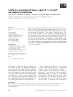
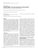
MDM2 downregulated cellular Vif levels in a proteasome dependent mannerFigure 1
MDM2 downregulated cellular Vif levels in a proteasome dependent manner. (A) MDM2 reduced cellular levels of
Vif as well as p53, but not that of Nef. HEK293T cells were cotransfected with expression vectors for the indicated E3 ligases
and a subgenomic HIV-1 expression vector pNL-A1. Cell lysates were subjected to immunoblotting with the indicated Abs.
We could not detect the expression of FLAG-MDM2 without MG132 treatment, because of a rapid degradation of MDM2.
MG132 treatment enabled us to detect expression of MDM2 only with anti-MDM2 Ab, but not with anti-FLAG mAb. (B)
Twenty-two hours after transfection, the cells were treated with cycloheximide (CHX)(80 μg/ml) for the indicated times, and
cell lysates were subjected to immunoblotting with the indicated Abs. (C) The amounts of Vif and Nef were quantified by den-
sitometry, and Vif protein levels were calculated using Nef protein levels as normalizing loading controls and presented as per-
centage values relative to that without CHX treatment set as 100%. Values are presented as averages of three independent
experiments. (D) MDM2 downregulated Vif, but a ΔRF mutant did not. HEK293T cells were cotransfected with expression
vectors for MDM2 and the mutant together with pNL-A1, and cell lysates were subjected to immunoblotting with the indi-
cated Abs. (E) p53
-/-
MDM2
-/-
DKO-MEF cells were cotransfected with expression vectors for MDM2 and Vif, and treated with
10 μM MG132 for 6 hrs, and cell lysates were subjected to immunoblotting with the indicated Abs.
Vif
Nef
p53
MDM2(+)
-actin
MDM2(-)
0 30 60 90 120
CHX treatment time (min)
0 30 60 90 120
CHX treatment time (min)
CHX treatment (min)
Protein levels (%)
0%
20%
40%
60%
80%
100%
120%
0 306090120
Vif+MDM2
Vif
P<0.05
P<0.01
Vif
-actin
p53
Nef
Mock
MDM2MDM2- RF
1 2 3 4 5 6 7
A
B
C
DE
Vif
MDM2
MG132
+
+
-
+
-
-
+
+
+
Vif
-actin
1 2 3 4
+
-
+
MDM2
MDM2Cul5
Vif
-actin
p53
Nef
Parkin
Mock
1 2 3 4 5 6 7 8 9 10
Anti-FLAG
Cul5
Parkin
Anti-FLAG
Cul5
Parkin
MDM2
Anti-MDM2
MG132
treatment
Retrovirology 2009, 6:1 />Page 4 of 12
(page number not for citation purposes)
determine a Vif-binding domain, we further tested
mutants deleted in a Zn Finger domain (ΔZn) or in an
acidic domain (ΔAD). Neither mutant could bind Vif,
whereas the mutant containing amino acids 168–411 was
able to bind Vif, suggesting that both domains are neces-
sary and that the central domain is sufficient for Vif bind-
ing (Fig. 2B, right panel &2C). Additionally, using a series
of Vif deletion mutants, we also found that the N-terminal
region of Vif (amino acids 4–22) is needed for MDM2
binding (Fig. 3A &3C). Furthermore, we examined the
MDM2-mediated downregulation of Vif mutants. MDM2
was able to efficiently downregulate cellular levels of the
MDM2-binding Vif mutants but not that of an MDM2-
non binding mutant, Δ4–45 (Fig. 3B). Collectively, these
results indicated that the Vif-MDM2 interaction is
required for MDM2-mediated downregulation of Vif (Fig.
3C).
MDM2 induces ubiqutination of Vif
Since we found that MDM2 bound Vif and promoted its
degradation via a proteasomal pathway, we next exam-
ined whether MDM2 is involved in the polyubiquitina-
tion of Vif. In vitro ubiquitination assays revealed that
bacterially expressed GST-MDM2 was able to induce the
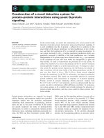
MDM2 bound Vif in its central domainFigure 2
MDM2 bound Vif in its central domain. (A) Immunoprecipitation assays revealed the interaction of MDM2 with Vif in vivo.
HEK293T cells were cotransfected with expression vectors for MDM2 and Vif and treated with MG132 for 6 hrs prior to har-
vest. Cell lysates were immunoprecipitated with anti-MDM2 mAb followed by immunoblotting with the indicated Abs (upper
two panels). Cell lysates were also subjected to immunoblotting with the indicated Abs (lower two panels). (B) The interaction
domain of MDM2 with Vif. HEK293T cells were cotransfected with expression vectors for HA-tagged MDM2 wild type (Wt)
and mutants together with pNL-A1, and cell lysates were immunoprecipitated with anti-HA mAb followed by immunoblotting
with the indicated Abs. Asterisk indicates immunoglobulin heavy chains from thenimmunoprecipitation. (C) Schematics of
MDM2 mutants binding to Vif are shown.
A
B
p
5
3
b
i
n
d
i
n
g
R
I
N
G
F
i
n
g
e
r
Zn
Fi
nge
r
C
N1
Binding to Vif
+
+
+
-
320
154
154 320
CN1
167
412
321
N2
+
-
168-411
Wt
A
c
i
d
i
c
168-411
168 411
+
AD
Zn
-
-
228
311
345292
C
D
MDM2
Vif
+
+
-
+
MDM2
Vif
IP :
anti-MDM2
Cell lysate
Vif
MDM2
1 2
C
C
N
1
N
1
N
2
1
6
8
-
4
1
1
W
t
M
o
c
k
Vif
Vif
IP : anti-HA
MDM2
Cell lysate
1 2 3 4 5 6 7
MDM2
1
6
8
-
4
1
1
W
t
M
o
c
k
Z
n
A
D
MDM2
1 2 3 4 5
MDM2
Retrovirology 2009, 6:1 />Page 5 of 12
(page number not for citation purposes)
polyubiquitination of purified GST-Vif protein in vitro
(Fig. 4A). The ubiquitination of Vif by MDM2 was spe-
cific, as the omission of ubiqutin, E1, E2, or MDM2 pre-
vented Vif-ubiquitination as shown in our previous
experiments [13]. We also performed in vitro ubiquitina-
tion assays using immunopurified MDM2 and Cul5.
Immunopurified MDM2 was able to induce ubiquitina-
tion of Vif in vitro to the same extent as Cul5 (Additional
file 2, part A), while it could not ubiquitinate the N-termi-
nal Vif deletion mutant Δ22 that was defective for binding
MDM2 (Additional file 2, part B). These findings suggest
that the interaction with MDM2 is important for Vif ubiq-
uitination. We performed in vivo ubiquitination assays to
further investigate the importance of MDM2 in Vif ubiq-
uitination. Lysates of cells co-expressing Vif, either with an
MDM2 wild type (Wt) or a ΔRF mutant, and His-tagged
Ubiquitin (His-Ub) were analyzed for the presence of
ubiquitinated Vif conjugates (Fig. 4B). Unfortunately, we
detected a Vif band that non-specifically bound to Ni-NTA
agarose (arrowhead) due to its nature as a sticky protein.
Overexpression of MDM2 induced a ladder detected by
anti-Vif Ab, even in the absence of His-Ub (lane 2), sug-
gesting that this ladder represented Vif protein polyubiq-
uitinated with endogenous Ub (arrows with asterisk).
Furthermore, in the presence of His-Ub, we detected a
doublet of ladder which presumably represented Vif pro-
tein polyubiquitinated with endogenous and His-tagged
Ub (arrows with asterisk and arrows, respectively). We
also obtained similar results using a UbiQapture™-Q Kit
(data not shown). We thus concluded that the overexpres-
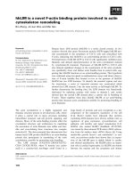
MDM2 specifically bound and downregulated VifFigure 3
MDM2 specifically bound and downregulated Vif. (A) The interaction domain of Vif with MDM2. HEK293T cells were
cotransfected with expression vectors for Vif and mutants together with pCMV/HA-MDM2, and cell lysates were immunopre-
cipitated with anti-Vif mAb followed by immunoblotting with the indicated Abs. Arrowhead indicates MDM2. (B) The down-
regulation of Vif protein by MDM2. HEK293T cells were cotransfected with expression vectors for Vif and mutants with or
without pCMV/HA-MDM2, and cell lysates were subjected to immunoblotting with the indicated Abs. The amounts of Vif were
quantified by densitometry and shown as the protein ratio relative to that without expression of MDM2. (C) Schematics of Vif
mutants bound by and downregulated by MDM2. NE: not examined.
W
t
23-
7
4
4-
45
74.9%62.0%70.7%61.5%99.9%
Vif
-actin
Nef
MDM2
+-
+-+-+-+-
Vif protein (%)
Vif
A
B
Wt
23-43
23-74
75-114
110-141
4-45
HCCH motif
BC box
22 44
22 75
74 115
109 142
463
+
+
+
+
+
-
SOCS box
Binding
to MDM2
+
NE
+
+
+
-
Downregulation
by MDM2
C
MDM2
MDM2
IP : anti-Vif
Vif
Vif
Cell lysate
W
t
23-
43
23-
74
75-
114
110-
141
4-
45
M
o
ck
1 2 3 4 5 6 7
Vif
Retrovirology 2009, 6:1 />Page 6 of 12
(page number not for citation purposes)
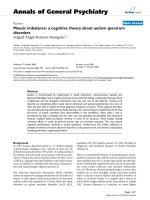
Figure 4 (see legend on next page)
A
GST-MDM2
GST-Vif
E1/ATP
E2
GST-Ub
-+++- +
-++-++
-+-+++
++++++
++++
GST-Vif
220
120
100
80
60
50
Ub
1
Ub
2
Ub
3
Ub
n
1 2 3 4 5 6
WB :
anti-Vif
Vif
MDM2
-actin
IP : Ni-agarose
His-Ub
Vif
MDM2
MDM2-
RF
Cell lysate
1 2 3 4 5 6
30
40
50
60
80
100
120
-
+
-
-
-
+
+
-
+
-
+
-
+
+
-
-
+
+
-
+
+
+
+
-
Ub
1
Ub
2
Ub
3
Ub
n
Vif
Vif
MDM2
-actin
+
+
+
-
-
+
+
+
WB :
anti-Vif
WB :
anti-HA
IP :
anti-Vif
HA-Ub
Vif
Control siRNA
siRNA
Cell
lysate
1 2
30
40
50
60
80
100
220
120
Ub
1
Ub
2
Ub
3
Ub
n
B
C
Ub
1
Retrovirology 2009, 6:1 />Page 7 of 12
(page number not for citation purposes)
sion of exogenous MDM2 efficiently induced polyubiqui-
tination of Vif in vivo. Furthermore, the knock-down of
endogenous MDM2 expression by introduction of
MDM2-specific short interfering RNA (siRNA) resulted in
a significant reduction in the amount of polyubiquiti-
nated Vif, commensurate with the extent of reduced
MDM2 expression (Fig. 4C). Collectively, these data indi-
cated that MDM2 mediates polyubiquitination of Vif both
in vitro and in vivo.
MDM2 negatively regulates HIV-1 replication in non-
permissive cells through ubiqutination and degradation of
Vif
Next, we examined the effect of MDM2 on HIV-1 replica-
tion. In a single round infection assay (Fig. 5A), in the
absence of A3G, viral replication was not affected by
expression of MDM2 and/or Vif (lanes 1–6). In contrast,
in the presence of A3G in a non-permissive cell setting,
without the expression of MDM2, the wild type virus
could replicate but the ΔVif virus could not, as previously
reported (lanes 7 & 8) [3,8]. Co-expression of MDM2
reduced the cellular level of Vif (Fig. 5B, upper panel,
lanes 5 & 11), resulting in the increased virion incorpora-
tion of A3G (Fig. 5B, 2nd lower panel, lane 11 as com-
pared with lanes 7) and the greater suppression of viral
replication (Fig. 5A, lane 11 as compared with lane 7).
We also tested the effect of MDM2 on HIV-1 replication in
the presence of A3F. MDM2 suppressed viral replication
in the presence of A3F, similar to results shown for A3G
(Additional file 3). These data indicated that the MDM2-
mediated Vif downregulation led to upregulated cellular
A3G and A3F levels in producer cells, resulting in less
infectious HIV-1 virions produced. Since MDM2 was pre-
viously reported to upregulate HIV-1 transcription by
ubiquitination of Tat, we further examined HIV-1 replica-
tion in macrophages knocked down for MDM2 (Fig. 5C).
We chose terminally differentiated macrophages as the
target, because the knockdown of MDM2 is lethal for pro-
liferating cells. HIV-1 replicated more efficiently in macro-
phages transfected with MDM2 siRNA than in control
siRNA-transfected macrophages. These data indicated that
MDM2 negatively regulated HIV-1 replication in non-per-
missive target cells through the ubiquitination and degra-
dation of Vif.
To obtain further insights into the mechanisms why our
MDM2 system did not induce the ubiquitination of A3G
which was bound to Vif, we tested the expression levels
and the binding affinity of A3G to Vif in transfected cells.
Co-expression of MDM2 reduced the cellular levels of Vif
and inversely increased the A3G levels in a dose depend-
ent manner (Fig. 5D). Immunoprecipitation assays
revealed that the co-expression of MDM2 blocked the
binding of A3G to Vif in a dose dependent manner (Fig.
5E). These data suggest that the interaction between
MDM2 and Vif precludes A3G from binding to Vif.
Discussion
In this study, we report that MDM2 is a novel E3 ligase for
HIV-1 Vif. MDM2 physically interacts with Vif and func-
tions as an E3 ligase for Vif to induce its polyubiquitina-
tion and proteasomal degradation. Several E3 ligases
including Cul5 [17], Nedd4, and AIP4 [18], have been
reported to induce Vif ubiquitination, and the roles of
Cul5 for Vif ubiquitination and degradation are especially
well documented. Dang et al. have recently reported that
Cul5 induces A3G degradation not by direct ubiquination
of A3G but indirectly through Vif ubiqutination and that
polyubiquitinated Vif might serve as a vehicle to transport
A3G into proteasomes for degradation [23]. In this man-
uscript, we show that MDM2 only targets Vif for degrada-
tion but not A3G, although MDM2 and Cul5 both induce
Vif ubiquitination (Additional file 2, part A). MDM2
reduced cellular Vif levels and inversely increased A3G
levels (Fig. 5B &5D), unlike Cul5. One possible explana-
tion is that the binding of MDM2 to Vif precluded A3G
from binding Vif (Fig. 5E), whereas a Cul5-Vif complex
MDM2 induced the polyubiquitination of Vif in vitro and in vivoFigure 4 (see previous page)
MDM2 induced the polyubiquitination of Vif in vitro and in vivo. (A) GST-MDM2 induced the polyubiquitination of Vif
in vitro. Bacterially expressed GST-Vif was subjected to in vitro ubiquitination assays. The reaction was performed in the pres-
ence or absence of E1, E2, GST-MDM2, and GST-Ubiquitin as indicated. Reactions were subjected to immunoblotting with
anti-Vif mAb. Arrows indicate GST-ubiquitin-conjugated Vif. (B) Overexpressed MDM2 induced the polyubiquitination of Vif in
vivo. HEK293T cells were cotransfected with expression vectors for MDM2 Wt and a ΔRF mutant together with expression
vectors for Vif and His-Ubiquitin (His-Ub) as indicated. Cells were treated with MG132 for 6 hrs, and cell lysates were precip-
itated with Ni-NTA agarose beads followed by immunoblotting with the indicated Abs. Since Vif naturally bound to Ni-NTA
agarose, we detected a Vif band itself (arrowhead), whereas no signal was detected in cells lacking Vif (lane 3). Arrows indicate
His-Ub-conjugated Vif. Arrows with asterisk indicate Vif conjugated with endogenous ubiquitin. (C) Transduction of siRNA
reduced cellular levels of endogenous MDM2 and polyubiquitination of Vif. HEK293T cells were cotransfected with expression
vectors for MDM2 siRNA and control siRNA together with expression vectors for Vif and HA-Ubiquitin (HA-Ub). Cell lysates
were immunoprecipitated with anti-Vif mAb followed by immunoblotting with the indicated Abs. Asterisk indicates immu-
noglobulin light chains from the immunoprecipitation.
Retrovirology 2009, 6:1 />Page 8 of 12
(page number not for citation purposes)
can bind A3G to form a ternary complex. MDM2 binds
the N-terminal region of Vif which does not overlap with,
but is close to the A3G/A3F binding domain [25]. This
binding might affect the interaction of Vif with A3G and/
or A3F. Furthermore, the evidence that an MDM2 ΔRF
mutant failed to protect A3G indicated that the ubiquiti-
nation and degradation of Vif is necessary to protect A3G
and A3F from Vif. These findings suggest that different E3
ligases might play different roles in Vif ubiquitination.
Further studies on the different roles of Vif ubiquitination
by different E3 ligases and their virological significance
should be investigated.
We demonstrate that MDM2 negatively regulated HIV-1
replication through Vif degradation. Through the degra-
dation of target proteins (p53, pRB, etc), MDM2 can exert
profound physiological effects on the regulation of cell
cycle, cell proliferation, DNA repairs and other processes.
To our knowledge, this is the first report to show that
MDM2 plays an important role in viral replication
MDM2 negatively regulated HIV-1 replication in non-permissive cells through the degradation of VifFigure 5
MDM2 negatively regulated HIV-1 replication in non-permissive cells through the degradation of Vif. (A) The
overexpression of MDM2 inhibited HIV-1 replication in the presence of A3G. NL-43 Wt and ΔVif viruses were produced from
HEK293T cells transfected with expression vectors for MDM2 Wt and a ΔRF mutant in the presence or absence of A3G. The
viral infectivity was examined using M8166 cells. Values are presented as averages of more than 3 independent experiments. (B)
MDM2 reduced cellular levels of Vif, resulting in more incorporation of A3G into HIV-1 virions. Immunoblotting for cell lysates
(upper 3 panels) and precipitated virions (lower 2 panels) was performed with the indicated Abs. Lane numbers correspond to
those in Fig. 4A. (C) HIV-1 replication in macrophages transfected with MDM2- and control-siRNA. MDM were transfected
with MDM2- and control-siRNA and challenged with R5 HIV-1
JR-FL
(left panel). Cell lysates were subjected to immunoblotting
with the indicated antibodies (right panels). (D) Coexpression of MDM2 reduced cellular levels of Vif and inversely increased
A3G levels in a dose dependent manner. HEK293T cells were cotransfected with expression vectors for A3G, Vif, GFP, and
MDM2 as indicated. Cell lysates were subjected to immunoblotting with the indicated Abs. (E) Immunoprecipitation assays
revealed that the coexpression of MDM2 blocked the binding of A3G to Vif in a dose dependent manner. HEK293T cells were
cotransfected with expression vectors for A3G, GFP-Vif, and MDM2 as indicated. Cell lysates were immunoprecipitated with
anti-GFP mAb followed by immunoblotting with the indicated Abs.
A
C
P<0.05
0%
20%
40%
60%
80%
100%
120%
140%
160%
180%
㪮㫋㩷㫍㫀㫉㫌㫊
㰱㪭㫀㪽㩷㫍㫀㫉㫌㫊
1 2 3 4 5 6 7 8 9 10 11 12
Mock RF MDM2 Mock RF MDM2
A3G (
-
) A3G (+)
Viral Infectivity (%)
B
0
200
400
600
800
1000
1200
1400
MDM2 siRNA 90pmol
control siRNA 90pmol
MDM2 siRNA 30pmol
control siRNA 30pmol
p24 (pg/ml)
Post infection (days)
4 7 11 14 18 21
MDM2
-actin
Control siRNA
MDM2 siRNA
90pmol
30pmol
M
D
M
2
R
F
M
o
c
k
A3G(+)A3G(-)
M
D
M
2
R
F
M
o
c
k
M
D
M
2
R
F
M
o
c
k
M
D
M
2
R
F
M
o
c
k
Cell lysate
Vif WT Vif WT
Vif
p55
APOBEC3G
APOBEC3G
Virion
p24
2 4 6 1 3 5 8 10 12 7 9 11
MDM2
APOBEC3G
Vif
APOBEC3G
Vif
GFP
-
+
-
-
+
+
+
+
+
++
+
+
+
+
-
1 2 3 4 5
+
-
+
+
+
+
+
+
+
+
+
-
HA-MDM2
HA-APOBEC3G
GFP-Vif
MDM2
APOBEC3G
Vif
Vif
APOBEC3G
MDM2
Cell Lysate IP : GFP
D
E
Retrovirology 2009, 6:1 />Page 9 of 12
(page number not for citation purposes)
through the degradation of viral proteins. Recently,
MDM2 was also reported to ubiquitinate HIV-1 Tat pro-
tein and activate its transcriptional activity in a non-prote-
olytic manner [26]. Our experiment using MDM2
knockdown macrophages showed that HIV-1 replication
in these macrophages was more efficient than in control
siRNA-transfected macrophages. These data are consistent
with MDM2 negatively regulateing HIV-1 replication
through Vif ubiquitination (Fig. 5C). However, the repli-
cation efficiency of HIV-1 in MDM2 knockdown macro-
phages was only 2-fold higher and was slower than in
control siRNA-transfected macrophages. This suggests the
possibilities that the ubiquitination of Tat might work as
a positive regulatory factor at an earlier phase of infection
and that MDM2 might be involved in both positive and
negative regulation of HIV-1 replication at different
stages. Further studies on the detailed effect of MDM2 on
HIV-1 replication are needed.
We also demonstrated that Vif can bind MDM2 directly.
We also mapped the interaction domain of MDM2 with
Vif to amino acids 168–320 which is located in its central
acidic and Zn finger domains. This central domain is dif-
ferent from the primary p53-binding site of MDM2 which
is located in its N-terminal region; however, this central
deomain was recently reported as a second p53-binding
site and was shown to be important for the regulation of
p53 stability [27-30] (Fig. 2B &2C). Interestingly, several
proteins including p300, p14
ARF
, and pRB bind to the cen-
tral domain of MDM2 and regulate the stability and func-
tion of p53 via MDM2 [28,31]. Thus, it is possible that Vif
might affect the stability and function of p53. Indeed, we
confirmed that Vif can stabilize p53 (Izumi et al., unpub-
lished data), which could explain why the effect of MDM2
on p53 degradation was weaker than that on Vif as shown
in Fig. 1A. A further study is under way to elucidate this
new function of Vif (Izumi et al., HIV-1 Vif induces G2 cell
cycle arrest via the p53 pathway, unpublished).
Finally, expanding evidence suggests that the ubiquitina-
tion system plays important roles in many aspects of HIV-
1 replication including the degradation of A3G by Vif [9-
11], the degradation of CD4 by Vpu [32], HIV-1 viral bud-
ding [33], Tat-mediated transactivation [26], and Vpr-
induced G2 cell cycle arrest [34,35]. The functional link-
age between Vif and MDM2 also suggests that ubiquitin
processes such as the A3G/Vif interplay is highly complex.
It is obvious that HIV-1 replication in target CD4+ T cells
is strongly affected by the interplay of these proteins.
From the viral point of view, this interplay might give an
advantage to HIV-1 replication. One possibility is that
MDM2 regulates cellular Vif levels appropriately, such as
not to affect viral replication [36] but just enough to
antagonize A3G. Recent studies suggest that the G-to-A
mutations induced by A3G may not be the mechanism by
which A3G restricts or controls viral replication [37] and
that a partially effective Vif inhibitor may actually acceler-
ate the evolution of drug resistance and immune escape
[38]. The inhibitory activity of MDM2 toward Vif could be
partially effective and therefore could lead to viral evolu-
tion of drug resistance and immune escape. More recently,
Nathans et al. have reported a small molecule that specif-
ically antagonizes Vif function and inhibits viral replica-
tion by targeting the A3G/Vif axis. This compound
enhances Vif degradation only in the presence of A3G, but
does not induce A3G degradation and rather stabilizes
A3G. They suggested the possibility of a new proteolytic
enzyme for Vif degradation and that their new compound
interferes with Vif interaction with a host protein in a Vif-
A3G-host protein complex, thereby making Vif less stable.
The precise biological significance of this Vif-A3G-host
protein complex requires future elucidation. Nevertheless,
modification or intervention of such Vif-A3G-host protein
interplay could lead to the development of new therapeu-
tic strategies for HIV-1 infection.
Conclusion
MDM2 is a novel E3 ligase for Vif which induces the poly-
ubiquitination and degradation of Vif to negatively regu-
late HIV-1 replication.
Methods
Plasmid constructs
Expression vectors for hemagglutinin (HA)- or FLAG-
tagged MDM2, pCMV4/HA-MDM2 or pCMV4/FLAG-
MDM2, and their mutants were constructed as previously
described [19]. An expression vector for HA-tagged
human APOBEC3G, pcDNA3/HA-hA3G [39], and HIV-1
reporter plasmids, pNL43/Δenv-Luc (WT) and pNL43/
ΔenvΔvif-Luc (ΔVif) [8], were constructed as previously
described. Expression vectors for FLAG-tagged Parkin and
Cul5 (pcDNA3/FLAG-Parkin and pcDNA3/FLAG-Cul5,
respectively) were constructed by the PCR method. Com-
plementary DNA for HIV-1 Vif was also cloned into
pDON-AI (TAKARA BIO INC.) and pDON/EGFP for
expression of Vif and EGFP-fused Vif (EGFP-Vif). The sub-
genomic expression vector pNL-A1, which expresses all
HIV-1 proteins except for gag and pol products, and its
mutants expressing Vif deletion mutants were kind gifts
from Dr. K. Strebel [22].
Co-immunoprecipitation assays
We performed an immunoprecipitation assay for protein-
protein interaction in vivo, as described previously [8].
HEK293T cells were cotransfected with pCMV4/HA-
MDM2 and pNL-A1 by the calcium phosphate method.
Two days after transfection, cells were lysed in lysis buffer
(25 mM HEPES pH7.4/150 mM NaCl/1 mM MgCl
2
/0.5%
TritonX-100/10% Glycerol) and complexes were immu-
noprecipitated with anti-MDM2 monoclonal antibody
Retrovirology 2009, 6:1 />Page 10 of 12
(page number not for citation purposes)
(mAb) (SMP-14, Santa Cruz Biotechnology, Inc., Santa
Cruz, CA and Ab-1, Calbiochem, EMD Biosciences, Inc,
Darmstadt, Germany) and Protein A-Sepharose beads
(Amersham Biosciences Corp.) at 4°C. The beads were
washed with RIPA buffer (50 mM Tris-HCl pH8.0/150
mM NaCl/1% Triton-X 100/0.1% SDS/0.1% DOC) and
analyzed by immunoblotting with anti-Vif mAb (#319)
(A kind gift from Dr. M. Malim through the AIDS
Research and Reference Reagent Program) [40] or anti-HA
mAb (12CA5). To map the regions of MDM2 necessary
for binding to Vif, HEK293T cells were cotransfected with
expression vectors for a series of MDM2 deletion mutants
together with pNL-A1. Complexes were immunoprecipi-
tated with anti-HA mAb and analyzed by immunoblot-
ting with anti-Vif mAb. To map the regions of Vif
necessary for binding to MDM2, HEK293T cells were
cotransfected with expression vectors for a series of Vif
deletion mutants together with pCMV4/HA-MDM2.
Complexes were immunoprecipitated with anti-Vif mAb
and analyzed by immunoblotting with anti-MDM2 mAb.
In all these experiments, transfected cells were treated
with MG132 for 6 hrs prior to harvesting in order to stabi-
lize both Vif and MDM2; otherwise we could not detect
the expression of MDM2 because of its rapid degradation,
as seen in Fig. 1A.
In vitro and in vivo ubiquitination assays
In vitro ubiquitination assays were carried out in ubiquitin
reaction buffer (50 mM Tris-HCl/2 mM ATP/5 mM
MgCl
2
/2 μM DTT) with E1(200 ng), E2(Ubc5c)(150 ng),
and GST-tagged ubiquitin (GST-Ub) (10 μg) as described
previously [13]. MDM2 and Vif were expressed as GST-
fusion proteins in Escherichia coli strain DH5α and BL21,
respectively. The reactions were incubated at 30°C for 90
min. The samples were subjected to immunoblotting with
anti-Vif mAb to detect GST-ubiquitin conjugated Vif.
For in vivo ubiquitination assays, HEK 293T cells were
cotransfected with plasmids expressing Vif, FLAG-MDM2
or its mutants, and His-tagged ubiquitin (His-Ub) as indi-
cated. Cells were treated with 10 μM MG132 for 6 hrs
prior to harvesting. Forty-eight hours post transfection,
cell lysates were affinity-purified with Ni-NTA-agarose
beads (Invitrogen corporation, Carlsbad, CA) and ana-
lyzed by immunoblotting with anti-Vif mAb.
For production of RNAi within the cells, we used the pSu-
per vector as described previously [19]. pSuper-MDM2-1
contained the 19 nt derived from the mdm2 cDNA (nt
404–422) as the target sequence. Double-stranded RNA
containing scrambled 19 nt was used as a control.
HEK293T cells were transfected with pSuper plasmids
together with plasmids expressing Vif and HA-Ub. Cell
lysates were immunoprecipitated with anti-Vif mAb fol-
lowed by immunoblottimg with anti-HA mAb.
Single round infection assays with HIV-1 luciferase
reporter virus
Luciferase reporter viruses with or without Vif were pre-
pared by cotransfection of pNL43/Δenv-Luc (Wt) or
pNL43/ΔenvΔvif-Luc (ΔVif) plus pVSV-G together with a
mock vector or an expression vector for MDM2 or a
mutant in the presence or absence of pcDNA3/hA3G by
calcium phosphate as previously described [8]. The
reporter viruses were adjusted according to p24 values and
used to infect M8166 target cells. Productive infection was
measured by luciferase activity and values were presented
as percent infectivity relative to the value of each virus
without the expression of hA3G.
Knockdown of MDM2 in macrophages and replication
assays
Monocyte-derived macrophages (MDM) were cultured for
7 days from CD14+ monocytes isolated from the periph-
eral blood of an HIV-1-negative healthy individual. Elec-
troporation with Stealth Select RNAi for MDM2 or
Control (Invitrogen Corporation) was performed using
the Nucleofector machine (Amaxa Inc., Gaithersburg,
MD) according to the manufacturer's instructions. Twenty
four hours after transfection, MDM were challenged with
R5 HIV-1
JR-FL
at multiplicity of infection of 0.1 at 37°C for
3 hrs. The cells were cultured from day 4 to 21 after infec-
tion, and the concentration of p24 antigen in the superna-
tant was measured with an HIV-1 p24 antigen enzyme-
linked immunosorbent assay [ELISA] kit (ZeptMetrix,
Buffalo, NY).
Competing interests
The authors declare that they have no competing interests.
Authors' contributions
TI. designed research, performed research, contributed
vital new reagents, analyzed data, and wrote the paper.
ATK designed research, analyzed data, wrote the paper,
and organized the research. KS, KIo, and MM prepared the
materials and performed a part of the research. KIwai, HK,
TS, MT, SI., and HA contributed vital new reagents. YK
contributed vital new reagents, performed a part of the
research, and analyzed the data. HH, KItoh, and JF
designed the research, contributed vital new reagents, and
analyzed the data. TU analyzed the data, drafted the
paper, and organized the research.
Retrovirology 2009, 6:1 />Page 11 of 12
(page number not for citation purposes)
Additional material
Acknowledgements
We thank Drs. K. Strebel for the pNL-A1 plasmid and its derivative
mutants, D. P. Lane for p53
-/-
MDM2
-/-
DKO-MEF, and M. Malim for the anti-
Vif mAb (#319) through the AIDS Research and Reference Reagent Pro-
gram, Division of AIDS, NIAID, NIH. This study was partly supported by
grants-in-aid from the Ministry of Education, Culture, Sports, Science, and
Technology, from the Ministry of Health, Labour and Welfare, Japan, from
the Naito Foundation, and from Mitsubishi Pharma Research Foundation.
References
1. Goff SP: Retrovirus restriction factors. Mol Cell 2004,
16:849-859.
2. Towers GJ: The control of viral infection by tripartite motif
proteins and cyclophilin A. Retrovirology 2007, 4:40.
3. Sheehy AM, Gaddis NC, Choi JD, Malim MH: Isolation of a human
gene that inhibits HIV-1 infection and is suppressed by the
viral Vif protein. Nature 2002, 418:646-650.
4. Goila-Gaur R, Strebel K: HIV-1 Vif, APOBEC, and intrinsic
immunity. Retrovirology 2008, 5:51.
5. Mangeat B, Turelli P, Caron G, Friedli M, Perrin L, Trono D: Broad
antiretroviral defence by human APOBEC3G through lethal
editing of nascent reverse transcripts. Nature 2003,
424:99-103.
6. Harris RS, Bishop KN, Sheehy AM, Craig HM, Petersen-Mahrt SK,
Watt IN, Neuberger MS, Malim MH: DNA deamination mediates
innate immunity to retroviral infection. Cell 2003,
113:803-809.
7. Zhang H, Yang B, Pomerantz RJ, Zhang C, Arunachalam SC, Gao L:
The cytidine deaminase CEM15 induces hypermutation in
newly synthesized HIV-1 DNA. Nature 2003, 424:94-98.
8. Shindo K, Takaori-Kondo A, Kobayashi M, Abudu A, Fukunaga K,
Uchiyama T: The enzymatic activity of CEM15/Apobec-3G is
essential for the regulation of the infectivity of HIV-1 virion
but not a sole determinant of its antiviral activity. J Biol Chem
2003, 278:44412-44416.
9. Marin M, Rose KM, Kozak SL, Kabat D: HIV-1 Vif protein binds
the editing enzyme APOBEC3G and induces its degradation.
Nat Med 2003, 9:1398-1403.
10. Sheehy AM, Gaddis NC, Malim MH: The antiretroviral enzyme
APOBEC3G is degraded by the proteasome in response to
HIV-1 Vif. Nat Med 2003, 9:1404-1407.
11. Stopak K, de Noronha C, Yonemoto W, Greene WC: HIV-1 Vif
blocks the antiviral activity of APOBEC3G by impairing both
its translation and intracellular stability.
Mol Cell 2003,
12:591-601.
12. Yu X, Yu Y, Liu B, Luo K, Kong W, Mao P, Yu XF: Induction of
APOBEC3G ubiquitination and degradation by an HIV-1 Vif-
Cul5-SCF complex. Science 2003, 302:1056-1060.
13. Kobayashi M, Takaori-Kondo A, Miyauchi Y, Iwai K, Uchiyama T:
Ubiquitination of APOBEC3G by an HIV-1 Vif-Cullin5-
Elongin B-Elongin C Complex Is Essential for Vif Function. J
Biol Chem 2005, 280:18573-18578.
14. Zheng Y-H, Irwin D, Kurosu T, Tokunaga K, Sata T, Peterlin BM:
Human APOBEC3F Is Another Host Factor That Blocks
Human Immunodeficiency Virus Type 1 Replication. J Virol
2004, 78:6073-6076.
15. Shirakawa K, Takaori-Kondo A, Kobayashi M, Tomonaga M, Izumi T,
Fukunaga K, Sasada A, Abudu A, Miyauchi Y, Akari H: Ubiquitina-
tion of APOBEC3 proteins by the Vif-Cullin5-ElonginB-
ElonginC complex. Virology 2006, 344:263-266.
16. Fujita M, Akari H, Sakurai A, Yoshida A, Chiba T, Tanaka K, Strebel K,
Adachi A: Expression of HIV-1 accessory protein Vif is con-
trolled uniquely to be low and optimal by proteasome deg-
radation. Microbes Infect 2004, 6:791-798.
17. Mehle A, Goncalves J, Santa-Marta M, McPike M, Gabuzda D: Phos-
phorylation of a novel SOCS-box regulates assembly of the
HIV-1 Vif-Cul5 complex that promotes APOBEC3G degra-
dation. Genes Dev 2004, 18:2861-2866.
18. Dussart S, Courcoul M, Bessou G, Douaisi M, Duverger Y, Vigne R,
Decroly E: The Vif protein of human immunodeficiency virus
type 1 is posttranslationally modified by ubiquitin. Biochem
Biophys Res Commun 2004, 315:66-72.
19. Higashitsuji H, Itoh K, Sakurai T, Nagao T, Sumitomo Y, Masuda T,
Dawson S, Shimada Y, Mayer RJ, Fujita J: The oncoprotein
gankyrin binds to MDM2/HDM2, enhancing ubiquitylation
and degradation of p53. Cancer Cell 2005, 8:75-87.
20. Honda R, Tanaka H, Yasuda H: Oncoprotein MDM2 is a ubiquitin
ligase E3 for tumor suppressor p53. FEBS Lett 1997, 420:25-27.
21. Yu Y, Xiao Z, Ehrlich ES, Yu X, Yu X-F: Selective assembly of HIV-
1 Vif-Cul5-ElonginB-ElonginC E3 ubiquitin ligase complex
through a novel SOCS box and upstream cysteines.
Genes
Dev 2004, 18:2867-2872.
22. Strebel K, Daugherty D, Clouse K, Cohen D, Folks T, Martin MA:
The HIV 'A' (sor) gene product is essential for virus infectiv-
ity. Nature 1987, 328:728-730.
23. Dang Y, Siew LM, Zheng YH: APOBEC3G is degraded by the
proteasomal pathway in a Vif-dependent manner without
being polyubiquitylated. J Biol Chem 2008, 283:13124-13131.
24. Honda R, Yasuda H: Activity of MDM2, a ubiquitin ligase,
toward p53 or itself is dependent on the RING finger domain
of the ligase. Oncogene 2000, 19:1473-1476.
25. He Z, Zhang W, Chen G, Xu R, Yu XF: Characterization of con-
served motifs in HIV-1 Vif required for APOBEC3G and
APOBEC3F interaction. J Mol Biol 2008, 381:1000-1011.
26. Brès V, Kiernan RE, Linares LK, Chable-Bessia C, Plechakova O,
Tréand C, Emiliani S, Peloponese JM, Jeang KT, Coux O, Scheffner M,
Benkirane M: A non-proteolytic role for ubiquitin in Tat-medi-
Additional file 1
Supplementary figure 1 – the stability of Vif protein in p53-/- MEF
and p53-/-MDM2-/- MEF cells. MEF cells were transfected with pDON/
Vif or pcDNA3/HA-A3G. Twenty-two hours after transfection, the cells
were treated with cycloheximide (CHX) for the indicated times, and cell
lysates were subjected to immunoblotting with the indicated Abs.
Click here for file
[ />4690-6-1-S1.pdf]
Additional file 2
Supplementary figure 2 – immunopurified MDM2 induced the polyu-
biquitination of Vif in vitro. (A) MDM2 as well as Cul5 induced the
polyubiquitination of Vif. HEK293T cells were transfected with expression
vectors for His-MDM2 and His-Cul5. His-tagged proteins were purified
using Ni-NTA agarose and subjected to in vitro ubiquitination assays as
described in a legend to Fig. 4A. Reactions were subjected to immunoblot-
ting with anti-Vif Ab. Arrows indicate GST-Ub-conjugated Vif. Asterisks
indicate non-specific bands associated with GST-Vif protein recognized by
anti-Vif Ab, as they are seen in lanes 1 and 3. (B) MDM2 induced the
polyubiquitination of Vif Wt but not that of
Δ
22 that was defective for
binding MDM2. Filled asterisks indicate non-specific bands associated
with GST-Vif protein, while white asterisks indicate those associated with
GST-Vif
Δ
22.
Click here for file
[ />4690-6-1-S2.pdf]
Additional file 3
Supplementary figure 3 – the overexpression of MDM2 inhibited HIV-
1 replication in the presence of A3F. Single round infection assays were
performed in the presence or absence of A3F as described in a legend to
Fig. 5A. Values are presented as averages of more than 3 independent
experiments.
Click here for file
[ />4690-6-1-S3.pdf]
Publish with BioMed Central and every
scientist can read your work free of charge
"BioMed Central will be the most significant development for
disseminating the results of biomedical research in our lifetime."
Sir Paul Nurse, Cancer Research UK
Your research papers will be:
available free of charge to the entire biomedical community
peer reviewed and published immediately upon acceptance
cited in PubMed and archived on PubMed Central
yours — you keep the copyright
Submit your manuscript here:
/>BioMedcentral
Retrovirology 2009, 6:1 />Page 12 of 12
(page number not for citation purposes)
ated transactivation of the HIV-1 promoter. Nat Cell Biol 2003,
5:754-761.
27. Argentini M, Barboule N, Wasylyk B: The contribution of the
acidic domain of MDM2 to p53 and MDM2 stability. Oncogene
2001, 20:1267-1275.
28. Iwakuma T, Lozano G: MDM2, an introduction. Mol Cancer Res
2003, 1:993-1000.
29. Kawai H, Wiederschain D, Yuan ZM: Critical contribution of the
MDM2 acidic domain to p53 ubiquitination. Mol Cell Biol 2003,
23:4939-4947.
30. Meulmeester E, Frenk R, Stad R, de Graaf P, Marine JC, Vousden KH,
Jochemsen AG: Critical role for a central part of Mdm2 in the
ubiquitylation of p53. Mol Cell Biol 2003, 23:4929-4938.
31. Ganguli G, Wasylyk B: p53-independent functions of MDM2.
Mol Cancer Res 2003, 1:1027-1035.
32. Margottin F, Bour SP, Durand H, Selig L, Benichou S, Richard V, Tho-
mas D, Strebel K, Benarous R: A novel human WD protein, h-
beta TrCp, that interacts with HIV-1 Vpu connects CD4 to
the ER degradation pathway through an F-box motif. Mol Cell
1998, 1:565-574.
33. Freed EO: Viral late domains. J Virol 2002, 76:4679-4687.
34. Wen X, Duus KM, Friedrich TD, de Noronha CM: The HIV1 pro-
tein Vpr acts to promote G2 cell cycle arrest by engaging a
DDB1 and Cullin4A-containing ubiquitin ligase complex
using VprBP/DCAF1 as an adaptor. J Biol Chem 2007,
282:27046-27057.
35. Schrofelbauer B, Hakata Y, Landau NR: HIV-1 Vpr function is
mediated by interaction with the damage-specific DNA-
binding protein DDB1. Proc Natl Acad Sci USA 2007,
104:4130-4135.
36. Akari H, Fujita M, Kao S, Khan MA, Shehu-Xhilaga M, Adachi A,
Strebel K: High level expression of human immunodeficiency
virus type-1 Vif inhibits viral infectivity by modulating prote-
olytic processing of the Gag precursor at the p2/nucleocap-
sid processing site. J Biol Chem 2004, 279:12355-12362.
37. Ulenga NK, Sarr AD, Hamel D, Sankale JL, Mboup S, Kanki PJ: The
level of APOBEC3G (hA3G)-related G-to-A mutations does
not correlate with viral load in HIV type 1-infected individu-
als. AIDS Res Hum Retroviruses 2008, 24:1285-1290.
38. Pillai SK, Wong JK, Barbour JD: Turning up the volume on muta-
tional pressure: is more of a good thing always better? (A
case study of HIV-1 Vif and APOBEC3). Retrovirology 2008,
5:26.
39. Kobayashi M, Takaori-Kondo A, Shindo K, Abudu A, Fukunaga K,
Uchiyama T: APOBEC3G Targets Specific Virus Species. J Virol
2004, 78:8238-8244.
40. Simon JH, Southerling TE, Peterson JC, Meyer BE, Malim MH: Com-
plementation of vif-defective human immunodeficiency
virus type 1 by primate, but not nonprimate, lentivirus vif
genes. J Virol 1995, 69:4166-4172.