Pocket Atlas of Human Anatomy 4th edition - part 7 doc
Bạn đang xem bản rút gọn của tài liệu. Xem và tải ngay bản đầy đủ của tài liệu tại đây (1.44 MB, 51 trang )
298
1
2
3
4
5
6
7
8
9
10
11
12
13
14
15
16
17
18
19
20
21
22
23
24
25
1 Third ventricle. Ventriculus tertius. Dien-
cephalic portion of the cerebral ventricular sys-
tem. It extends from the lamina terminalis to
the beginning of the cerebral aqueduct. A C
2 Hypothalamic sulcus. Sulcus hypothalamicus.
Furrow extending from the interventricular
foramen to the entrance into the cerebral aque-
duct. It separates the dorsal and ventral
thalami. A
3 Interventricular foramen. Foramen inter-
ventriculare. Opening between the lateral ven-
tricle and third ventricle behind the genu of the
fornix. A
4 Optic recess. Recessus opticus. Recess of third
ventricle above the optic chiasm. A
5 Recess of infundibulum. Recessus infundibuli
(infundibularis). Recess of third ventricle
within the infundibulum. A
6 Pineal recess. Recessus pinealis. Recess of third
ventricle extending partially into the epiphysis.
A
7 Supraspinal recess. Recessus supraspinalis.
Recess between the roof of the third ventricle
and the epiphysis. A
8 Tela choroidea. Forms the thin, narrow roof of
third ventricle and its choroid plexus. B C
9 Tenia of thalamus. Taenia thalami. Lateral at-
tachment line of the upper wall of the third
ventricle along the stria medullaris of the
thalamus. B C
10 Choroid plexus. Plexus choroideus. Paired,
highly vascularized villous infolding which
hangs down from the thin roof of the third ven-
tricle and is continuous anteriorly with the
choroid plexus of the 4
th
ventricle via the inter-
ventricular foramen. B C
11 Sections of thalamus and metathalamus. Sec-
tiones thalamici et metathalamici. See p. 409.
12 Reticular nucleus of thalamus. Nucleus reticu-
latus [thalami]. Thin layer lying mainly laterally
along the thalamus between the posterior limb
of the internal capsule and external medullary
lamina of the thalamus. It receives tributaries
from the entire cerebral cortex, globus pallidus
and reticular formation of the brainstem and
gives off efferent fibers to the reticular forma-
tion of the midbrain and thalamus. B
13 Anterior nuclei of thalamus. Nuclei anteriores
[thalami]. Cell group in the apex of the
thalamus. They receive fibers from the mamil-
lothalamic tract and have projections to the
cingulate gyrus.
14
Anterodorsal nucleus. Nucleus anterodorsalis
(anterosuperior). Narrow cell plate anterosupe-
riorly. B
15
Anteroventral nucleus. Nucleus anter-
oventralis (anteroinferior). Main nucleus of the
anterior nuclei. B
16
Anteromedial nucleus. Nucleus anterome-
dialis. Degenerating nuclear remains situated
medial and inferior to the anteroventral nu-
cleus. B
17 Median nuclei of thalamus. Nuclei mediani
[thalami]. Collective term for the nuclei located
medial and for the most part directly inferior to
the ependyma.
18
Anterior/posterior paraventricular nuclei.
Nuclei paraventriculares anteriores/posteri-
ores. Cell groups in the wall of the third ven-
tricle with neuronal function (among others,
vasopressin, angiotensin II, renin). C D E
19
Rhomboidal nucleus. Nucleus rhomboidalis. It
often forms the interthalamic adhesion. D
20
Nucleus reuniens. Nucleus extending from the
anterior end of the anterior tubercle to the
middle of the interthalamic adhesion. It may be
concerned with the formation of the latter
when present. It is absent in 28% of males and
14% of females. D
20 a
Paratenial nucleus of thalamus. Nucleus
parataenialis [thalami]. Located between the
stria medullaris, tenia, anterodorsal and para-
ventricular nuclei of the thalamus; it is prob-
ably involved in the processing of olfactor y
stimuli.
21 Medial nuclei of thalamus. Nuclei mediales
[thalami]. Mass of nuclei medial to the internal
medullary lamina with projections to other
thalamic nuclei and to the frontal lobe.
22
Dorsal medial nucleus. Nucleus medialis
dorsalis. Principal nucleus of this group. C D
23 Internal/external medullary lamina. Lamina
medullaris interna/externa. Layer of white
matter which, provided it is located internally,
has Y-shaped sections and divides the thalamus
into anterior, medial and lateral regions. B
24 Reticular (intralaminar) nuclei of thalamus.
Nuclei reticulares (intralaminares thalami). Nu-
clei located in the medullary lamina. They
correspond functionally to the reticular forma-
tion and thus are integrating nuclei.
25
Centromedian nucleus. Nucleus centromedi-
anus. It is the largest nucleus of this group and
has projections to the corpus striatum, hy-
pothalamus, and other structures. E
26
Paracentral nucleus. Nucleus paracentralis. It
lies in the internal medullary lamina lateral to
the centromedian nucleus. CD
27
Parafascicular nucleus. Nucleus parafascicu-
laris. It lies medial to the occipital region of the
centromedian nucleus. E
28
Lateral central nucleus. Nucleus centralis
lateralis. It lies dorsolateral and medial to the
centromedian nucleus. E
29
Medial central nucleus. Nucleus centralis me-
dialis. It lies at the lower medial end of the in-
ternal medullary lamina.
Brain
Feneis, Pocket Atlas of Human Anatomy © 2000 Thieme
All rights reserved. Usage subject to terms and conditions of license.
299
1
2
3
4
5
6
7
8
9
10
11
12
13
14
15
16
17
18
19
20
21
22
23
24
25
a
a
a
Brain
9
10
16
8
15
14
23
12
8
1
9
10
18
22
300.1
26
28
300.1
22
26
28
18
19
20
300.11
28
25
27
18
3
1
4
2
6
7
5
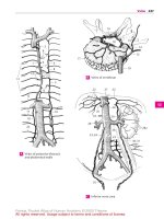
Diencephalon, sagittal sectionA
Diencephalon, cross section
B Diencephalon, cross section
C
Diencephalon, cross sectionD Diencephalon, cross sectionE
Feneis, Pocket Atlas of Human Anatomy © 2000 Thieme
All rights reserved. Usage subject to terms and conditions of license.
300
1
2
3
4
5
6
7
8
9
10
11
12
13
14
15
16
17
18
19
20
21
22
23
24
25
1 Nuclei ventrolaterales [thalami]. Ventrolateral
nuclei, the group of nuclei lateral to the internal
medullary lamina. B
2
Posterior lateral nucleus. Nucleus lateralis
posterior. Portion of the lateral nucleus situated
between the pulvinar and dorsal lateral nu-
cleus with connections to the parietal lobe. A
3
Dorsal lateral nucleus. Nucleus lateralis
dorsalis. Anterosuperior portion of the lateral
nucleus with projections to the region of the
posterior cingulum segment and the lower part
of the parietal lobe. A
4
Anterior ventral nucleus. Nucleus ventralis
anterior. Anterior portion of the ventral nucleus
with projections to the interlaminar nuclei,
globus pallidus and dentate nucleus and recip-
rocal connections to the precentral gyrus and
the area anterior to it. It plays a role in Parkin-
son’s disease. A
5
Intermediate ventral nucleus. Nucleus
ventralis intermedius. Portion of the ventral
nucleus situated behind the anterior ventral
nucleus; it is a synaptic station connecting the
cerebellum, red nucleus and motor cortex. A
6
Medial ventral nucleus. Nucleus ventralis me-
dialis. Poorly demarcated nuclear region sit-
uated anterior to the posterior ventral nuclei;
its function is unclear. A
7
Posterior ventral nuclei. Nuclei ventrales
posteriores. Collective term for the following
two nuclei.
8
Posterolateral ventral nucleus. Nucleus ventralis
posterolateralis. The lateral part of the poste-
rior ventral nucleus that receives the medial
lemniscus and spinothalamic tract and relays
their impulses to the postcentral gyrus via the
thalamocortical tract. A
9
Posteromedial ventral nucleus. Nucleus ventralis
posteromedialis. Part located between the cen-
tromedian and posterolateral nuclei. It receives
the trigeminal lemniscus. A
10 Posterior nuclei of thalamus. Nuclei posteri-
ores [thalami]. Collective term for the following
three parts of the thalamus.
11
Pulvinar nuclei. Nuclei pulvinares. Nuclei that
occupy the posterior portion of the thalamus;
they begin at the habenulae, receive tributar-
ies from the auditory and visual pathways as
well as from other thalamic nuclei and are
connected with the visual cortex, optic and
acoustic control centers, and other structures.
A
12
Lateral geniculate nucleus [dor sal par t].
Nucleus [corporis geniculati] lateralis [pars
dorsalis]. Part of the visual pathway. A
13
Medial geniculate nucleus [dorsal part]. Nu-
cleus [corporis geniculati] medialis [pars
dorsalis]. Part of medial geniculate body con-
taining small cells. A
14 Sections of ventral thalamus. Sectiones
thalami ventralis.
15 Lateral geniculate nucleus [ventral part]. Nu-
cleus corporis geniculati lateralis [pars
ventralis]. Small group of cells with fibers from
the retina: part of a light reflex tract. C
16 Medial geniculate nucleus [ventral part]. Nu-
cleus corporis geniculati medialis [pars
ventralis]. Possibly the true acoustic part of the
geniculate nucleus. C
17 Subthalamic nucleus. Nucleus subthalamicus
[corpus Luysii]. It lies between the lower end of
the internal capsule and the zona incerta. Of
clinical importance is its reciprocal connection
with the globus pallidus. B
18 Reticular nuclei of thalamus. Nuclei reticulares
[thalami]. Disaggregated cell layer on the
lateral surface of the thalamus between the ex-
ternal medullary lamina and internal capsule. B
19 Zona incerta. Basal continuation of the reticu-
lar nucleus of the thalamus and other struc-
tures. It lies in the path of the globus pallidus to
the tegmentum of the diencephalon. B
20
Nuclear regions H, H1 and H2. Nuclei re-
gionum H, H1 and H2. Dispersed neurons in the
corresponding Forel’s fields. Field H lies medial
to the zona incerta and in front of the red nu-
cleus, H1 between the thalamus and zona in-
certa, H2 between the zona incerta and sub-
thalamic nucleus. B
21 Thalamic tract and fasciculi. Tractus et
fasciculi thalamici.
22 Lateral lemniscus. Lemniscus lateralis. Audi-
tory pathway passing into the medial genicu-
late body. A
23 Medial lemniscus. Lemniscus medialis. Con-
tinuation of the tract from the posterior
funiculus radiating into the posterolateral ven-
tral nucleus. A
24 Spinal lemniscus. Lemniscus spinalis. Pain
pathway extending into the posterolateral ven-
tral nucleus. A
25 Trigeminal lemniscus. Lemniscus trigeminalis.
Fibers of the sensory trigeminal nucleus. They
pass into the posteromedial ventral nucleus. A
26 Brachium of inferior colliculus. Brachium col-
liculi inferioris. Outwardly visible connection
between the inferior colliculus and the medial
geniculate body. C
27 Acoustic radiation. Radiatio acustica. Portion of
auditory pathway extending from the medial
geniculate body to the transverse temporal gyn.
It passes through the occipital part of the poste-
rior limb of the internal capsule. A
28 Brachium of superior colliculus. Brachium col-
liculi superioris. Externally visible connection
between the superior colliculus and the lateral
geniculate body. Connection of the visual path-
way with the extrapyramidal system. C
29 Optic radiation. Radiatio optica [[Gratiolet]].
Portion of the visual pathway emanating from
the lateral geniculate body. Itpasses through the
occipital part of the posterior limb of the inter-
nal capsule and around the posterior horn of the
lateral ventricle to the area striata. A
Brain
Feneis, Pocket Atlas of Human Anatomy © 2000 Thieme
All rights reserved. Usage subject to terms and conditions of license.
301
1
2
3
4
5
6
7
8
9
10
11
12
13
14
15
16
17
18
19
20
21
22
23
24
25
a
a
a
26
151628
1
18
20
20
H
H
1
H2
19
17
11
12
25
22
298.21
9
23
8
6
5
4
3
2
298.13
13
29
24
27
Thalamic nuclei and pathwaysA
Subthalamic region
B Geniculate bodyC
Brain
Feneis, Pocket Atlas of Human Anatomy © 2000 Thieme
All rights reserved. Usage subject to terms and conditions of license.
302
1
2
3
4
5
6
7
8
9
10
11
12
13
14
15
16
17
18
19
20
21
22
23
24
25
1 Anterior thalamic radiations. Raditiones
thalamicae anteriores. Fibers of the anterior nu-
cleus passing to and from the cingulate gyrus
and likewise reciprocal connections between
the lateral nucleus and frontal lobe. The fibers
run in the anterior limb of theinternal capsule. A
2 Central thalamic radiations. Radiationes
thalamicae centrales. Reciprocal fibers passing
fan-like through the posterior limb of the inter-
nal capsule from the posterior lateral, anterior
ventral, lateral ventral and posterior ventral nu-
clei to the pre- and postcentral gyri in addition
to the connecting fields of the cortex. A
3 Posterior thalamic raditaions. Radiationes
thalamicae posteriores. They lie in the occipital
region of the posterior limb of the internal cap-
sule and contain fibers from the lateral genicu-
late body (optic radiation) and the pulvinar for
the occipital lobes and adjacent regions. A
4 Dentatothalamic tract. Tractus denta-
tothalamicus. It arises from the cerebellum and
radiates into the thalamic fasciculus and to the
lateral ventral nucleus. C
5 Thalamic fasciculus. Fasciculus thalamicus. It
lies below the thalamus, next to and above the
zona incerta in field H1 and is composed of the
ventricular fasciculus, ansa lenticularis and
fibers from the cerebellum. It is a conveyor of
impulses for the anterior ventral and lateral
ventral nuclei. C
6 Subthalamic fasciculus. Fasciculus subthalami-
cus. Fiber bundleextending from theglobus pal-
lidus to the subthalamic nucleus. C
7 Mamillot halamic fasciculus. Fasciculus mamil-
lothalamicus. Fiber tract extending from the
mamillary body to the anterior nucleus of the
thalamus. D
8 Inferior thalamic peduncle. Pedunculus
thalamicus inferior. Fibers between the hy-
pothalamus and thalamus. According to some
anatomists, it consists of fibers of the pulvinar
from and to the occipital lobes and its vicinity, as
well as fibers of the auditory tract.
9 Ansa lenticularis and fasciculus lenticularis.
Ansa et fasciculus lenticulares. Two fiber
bundles from the lentiform nucleus to the ven-
tral nuclei of the thalamus. One part passes
around the anterior margin of the internal cap-
sule (ansa lenticularis); the other part passes
through the internal capsule. Both tracts are
united in the thalamic fasciculus. C
10 Ansa peduncularis and fasciculus peduncu-
laris. Ansa et fasciculus pedunculares. Fiber
tract connecting the thalamus and claustrum,
thereby extending between the lentiform nu-
cleus and the amygdaloid body. B C
11 Intrathalamic fibers. Fibrae intrathalamicae.
Connections of individual thalamic nuclei.
12 Periventricular fibers. Fibrae periventriculares.
Fibers coursing beneath the ependyma of the
third ventricle between the medial nucleus and
the hypothalamic nucleus to enter the posterior
longitudinal fasciculus.
13 Sections of the hypothalamus. Sectiones hy-
pothalami.
14 Dorsal (posterior) hypothalamic region. Regio
(area) hypothalamica dorsalis. Area of the hy-
pothalamus next to the apex.
15
Nucleus of ansa lenticularis. Nucleus ansae
lenticularis. Groups of cells dispersed in the
ansa lenticularis.
16 Anterior (ventral) region of hypothalamus.
Regio hypothalamica anterior.
17
Medial/lateral preoptic nucleus. Nucleus
preopticus medialis/lateralis. Group of nuclei
located beneath the anterior commissure and
along the lamina terminalis with projections to
the stria terminalis, medial telencephalic
fasciculus and medial thalamic nuclei. D
18
Supraoptic nucleus. Nucleus supraopticus.
Nucleus lying above the optic chiasm with neu-
rosecretory fibers (oxytocin and vasopressin)
projecting to the posterior pituitary. D
19
Paraventricular nuclei. Nuclei paraventricu-
lares. Group of autonomic nuclei with neu-
rosecretory fibers (oxytocin and vasopressin)
projecting to the posterior lobe of the hypophy-
sis. They lie superiorly near the base of the hy-
pothalamic sulcus and behind the anterior hy-
pothalamic nucleus. D
20
Anterior hypothalamic nucleus. Nucleus hy-
pothalamicus anterior. Located behind the pre-
optic nucleus with projections to the hemi-
spheres, stria terminalis and thalamus, its effer-
ent fibers communicate with motor and auton-
omic nuclei. It influences heat regulation, glan-
dular activity and circulation. D
21 Intermediate hypothalamic region. Regio hy-
pothalamica intermedia. Area situated between
the anterior and posterior hypothalamic re-
gions.
22
Arcuate nucleus. [[Nucleus arcuatus]]. Mural
nucleus situated in the entrance to the infun-
dibulum. It belongs to the tuberal nuclei, i. e., it
regulates the release of hormones from the
anterior lobe by delivering an active substance
(neurohormone) to blood vessels of the hy-
pophysial stalk where its processes (axons) are
found. D
23
Tuberal nuclei. Nuclei tuberales. Groups of nu-
clei in the posterior wall of the infundibulum.
They function similar to the arcuate nucleus. D
24
Lateral hypothalamic region. Regio hy-
pothalamica lateralis. Area separated from the
medial hypothalamus by the fornix, mamil-
lothalamic fasciculus and medial telencephalic
fasciculus. It is occupied by the lateral preoptic
nucleus and thesupraoptic nucleus including its
lateral portion. D
Brain
Feneis, Pocket Atlas of Human Anatomy © 2000 Thieme
All rights reserved. Usage subject to terms and conditions of license.
303
1
2
3
4
5
6
7
8
9
10
11
12
13
14
15
16
17
18
19
20
21
22
23
24
25
a
a
a
1
2
3
10
304.17
19
17
20
18
22
23
24
24 7
5
6
9
910
4
Radiation of thalamusA
Ansa et fasciculus peduncularis
B
Subthalamic pathways
C
Nuclei of hypothalamus
D
Brain
Feneis, Pocket Atlas of Human Anatomy © 2000 Thieme
All rights reserved. Usage subject to terms and conditions of license.
304
1
2
3
4
5
6
7
8
9
10
11
12
13
14
15
16
17
18
19
20
21
22
23
24
25
1 Ventromedial hypothalamic nucleus. Nucleus
hypothalamicus ventromedialis. Lies in and
above the entrance into the infundibulum. This
nucleus belongs to the group of tuberal nuclei
and, like them, controls the release of regulating
hormones for the anterior lobe via the hypophy-
sial stalk. A
2 Dorsomedial hypothalamic nucleus. Nucleus
hypothalamicus dorsomedialis. It lies near the
apex of the ventromedial hypothalamic nucleus
and has similar functions. A
3 Dorsal hypothalamic nucleus. Nucleus hy-
pothalamicus dorsalis. Group of cells located
below the dorsal hypothalamic area (see
p. 302.14). A
4 Posterior periventricular nucleus. Nucleus
periventricularis posterior. Cell group located
below the ependyma in the posterior segment
of the 3
rd
ventricle. A
5 Infundibular (arcuate) nucleus. Nucleus infun-
dibularis (arcuatus). It lies near the apex of the
funnel of the infundibulum and has functions
similar to those of the tuberal nuclei. A
6 Posterior hypothalamic area. Regio hy-
pothalamica posterior. It contains the lateral
and medial nuclei of the mamillary body and
other structures.
7 Medial and lateral nuclei of mamillary body.
Nuclei corporis mamillaris mediales/laterales.
The medial nucleus forms the mamillary body
and is the origin of the mamillothalamic
fasciculus. The lateral nucleus lies ventrolateral
and receives the fornix. A B
8 Posterior hypothalamic nucleus. Nucleus hy-
pothalamicus posterior. It lies occipital to the
dorsomedial and ventromedial nuclei and
above the mamillary body up to the hy-
pothalamic sulcus and influences circulation,
peristalsis and the blood sugar level. A B
9 Neurohypophysis. In contrast to the two other
posterior lobes of the hypophysis, it is of neuro-
genic origin; so is the continuation of the infun-
dibulum. B
10 Hypothalamic tract and fasciculi. Tractus et
fasciculi hypothalamici. Tracts and fiber
bundles of the hypothalamus.
11 Periventricular fibers. Fibrae periventriculares.
Fiber tract directly under the ependyma of the
3
rd
ventricle. It is permeated by cells, connects
the thalamus with the hypothalamus and con-
tinues posteriorly into the posterior longitudi-
nal fasciculus. B
12 Dorsal supraoptic commissure. Commissura
supraoptica dorsalis [[Meynert]]. Decussation
lying directly above the chiasm. Passes to the
other side andmay connect the subthalamic nu-
cleus with the contralateral globus pallidus.
13 Ventral supraoptic commissure. Commissura
supraoptica ventralis [[Gudden]]. Crossing fibers
lying partially in the chiasm. Among other
things, it may connect the medial geniculate bo-
dies with one another.
14 Posterior (dorsal) longitudinal fasciculus.
Fasciculus longitudinalis dorsalis [[Schütz]].
Cranial continuation of a large portion of the
ventricular fibers. In the midbrain they lie close
to the cerebral aqueduct and connect the hy-
pothalamus with the rest of the brainstem. B
15 Mamillotegmental fasciculus. Fasciculus
mamillotegmentalis. Dissectible fiber bundle
between the mamillary body and the tegmental
nuclei of the midbrain. It arises as a common
trunk together with the mamillothalamic
fasciculus and branches off into the mesen-
cephalic tegmentum. B
16 Mamillothalamic fasciculus. Fasciculus mamil-
lothalamicus. It arises together with the mamil-
lotegmental fasciculus and passes to the ante-
rior thalamic nuclei. B
17 Fornix. It conveys fibers from the hippocampal
formation to the medial thalamic nuclei and hy-
pothalamus, and projects fibers to the lateral
nuclei of the mamillary body. B
18 Fibers of stria terminalis. Fibrae striae termi-
nalis. Fibers from the amygdaloid body which
communicate with the stria terminalis in the
hypothalamus. B
19 Medial prosencephalic fasciculus. Fasciculus
prosencephalicus medialis. Fib ers lying be-
tween the medial and lateral hypothalamus.
They connect individual hypothalamic nuclei
with one another and continue toward the oc-
ciput in the posterior longitudinal fasciculus. B
20 Hypothalamohypophysial tract. Tractus hy-
pothalamohypophysialis. Bundle of neu-
rosecretory fibers that arises after the union of
the fiber groups from the supraoptic and para-
ventricular nuclei. B
21
Supraoptic fibers. Fibrae supraopticae. Fibers
that arise in the supraoptic nucleus. B
22
Paraventricular f ibers. Fibrae paraventricu-
lares. Fibers that arise in the paraventricular nu-
cleus. B
23 Supraopticohypophysial tract. Tractus su-
praopticohypophysialis. Fibers that arise in the
supraoptic nucleus and form part of the hy-
pothalamohypophysial tract.
24 Paraventriculohypophysial tract. Tractus para-
ventriculohypophysialis. Fibers that arise in the
paraventricular nucleus and form part of the hy-
pothalamohypophysial tract.
Brain
Feneis, Pocket Atlas of Human Anatomy © 2000 Thieme
All rights reserved. Usage subject to terms and conditions of license.
305
1
2
3
4
5
6
7
8
9
10
11
12
13
14
15
16
17
18
19
20
21
22
23
24
25
a
a
a
17
14
19
15
7
11
16
8
18
20
21
22
9
318.1
304.17
3
4
8
2
1
7
5
Pathways of hypothalamusB
Nuclei of hypothalamus
A
Brain
Feneis, Pocket Atlas of Human Anatomy © 2000 Thieme
All rights reserved. Usage subject to terms and conditions of license.
306
1
2
3
4
5
6
7
8
9
10
11
12
13
14
15
16
17
18
19
20
21
22
23
24
25
1 ENDBRAIN. Telencephalon. The endbrain, which
is formed by invagination of the prosencephalon
(forebrain). It consists of the cerebral cortex to-
gether with the corpus callosum, corpus stri-
atum and olfactory brain.
2 CEREBRUM. In the present context, it comprises
the two cerebral hemispheres and their con-
tents.
3 Cerebral cortex. Cortex cerebralis (pallium).
Paired portion of the hemispheres covering
most of the brainstem.
4 Cerebral gyri. Gyri cerebrales. Convolutions of
the cerebral hemispheres, about 1 cm wide.
5 Cerebral sulci. Sulci cerebrales. Fissures be-
tween gyri.
6 Cerebral lobes. Lobi cerebrales. The four lobes
of the cerebrum: frontal, parietal, temporal and
occipital.
7 Longitudinal fissure of cerebrum. Fissura
longitudinalis cerebralis. Deep longitudinal
groove between the right and left cerebral
hemispheres. It lodges the falx cerebri. B
8 Transverse fissure of cerebrum. Fissura trans-
versa cerebralis [[fissura telodiencephalica]].
Fissure beneath the corpus callosum and fornix
as well as above the thalamus and roof of the 3
rd
ventricle. B
9 Lateral fossa of cerebrum. Fossa lateralis cere-
bralis. Space deep within the lateral sulcus. B
10 Superior (superomedial) margin. Margo su-
perior (superomedialis). Superior border of a
hemisphere between the superolateral and me-
dial surface. B
11 Inferior (inferolateral) margin. Margo inferior
(inferolateralis). Inferolateral border of a hemi-
sphere between the superolateral and inferior
surfaces. B
12 Medial (inferomedial) margin. Margo medialis
(inferomedialis). Inferomedial border of either
hemisphere between the inferior and medial
surfaces. B
13 [[Fissura limitans]]. Fissure between the insula
and opercula. The floor of this cleft, the sulcus
limitans, receives the insula.
14 Cerebral hemisphere. Hemispharium (cere-
bralis). Half of the telencephalon. B
15 Superolateral surface of hemisphere. Facies
superolateralis hemispherii. Upper and lateral
surface of the hemisphere. B
16 Central sulcus. Sulcus centralis. Furrow located
between the pre- and postcentral gyri and be-
tween the frontal and parietal lobes. A
17 Lateral sulcus. Sulcus lateralis. Deep cleft pass-
ing superiorly between the temporal and frontal
lobes and inferiorly between the temporal and
parietal lobes.
18
Anterior ramus. Ramus anterior. Short anteri-
orly directed branch of the lateral sulcus. A
19
Ascending ramus. Ramus ascendens. Short
branch of the lateral sulcus ascending into the
frontal lobe. A
20
Posterior ramus. Ramus posterior. Long poste-
rior branch of the lateral sulcus terminating
near the supramarginal gyrus. A
21 Interlobar sulci. Sulci interlobares. Furrows
which separate the cerebral lobes from one
another. They include the central and parieto-
occipital sulci and the lateral sulcus together
with its posterior ramus.
22 Frontal lobe. Lobus frontalis. Lobe extending
from the frontal pole to the central sulcus. A
23 Frontal pole. Polus frontalis. Anterior end of the
frontal lobe. A
24 Precentral sulcus. Sulcus precentralis. Furrow
in front of the precentral gyrus. A
25 Precentral gyrus. Gyrus precentralis. Convolu-
tion of the frontal lobe lying in front of the cen-
tral sulcus. Motor area of the cerebral cortex. A
26 Superior frontal gyrus.Gyrus frontalis superior
(primary motor area ???). A
27 Superior frontal sulcus. Sulcus frontalis super-
ior. Furrow below the superior frontal gyrus. A
28 Middle frontal gyrus. Gyrus frontalis medius. A
29 Inferior frontal sulcus. Sulcus frontalis inferior.
Furrow lying between the middle and inferior
frontal gyri. A
30 Inferior frontal gyrus. Gyrus frontalis inferior.
31
Opercular part (frontal operculum). Pars
opercularis [operculum frontale]. Part of infe-
rior frontal gyrus lying behind the ascending
ramus and covering the insula. A
32
Orbital part. Pars orbitalis. Part of the inferior
frontal gyrus located below the anterior ramus
of the lateral sulcus. A
33
Triangular part. Pars triangularis. Portion of
the inferior frontal gyrus located between the
anterior and descending rami of the lateral sul-
cus. Region of the motor speech center of Broca.
A
Brain
Feneis, Pocket Atlas of Human Anatomy © 2000 Thieme
All rights reserved. Usage subject to terms and conditions of license.
307
1
2
3
4
5
6
7
8
9
10
11
12
13
14
15
16
17
18
19
20
21
22
23
24
25
a
a
a
22
16
31
33
32
19
30
29
18
23
26
27
28
25
20
24
14
15
8
9
11
12
710
Brain, lateral viewA
Brain, frontal section
B
Brain
Feneis, Pocket Atlas of Human Anatomy © 2000 Thieme
All rights reserved. Usage subject to terms and conditions of license.
308
1
2
3
4
5
6
7
8
9
10
11
12
13
14
15
16
17
18
19
20
21
22
23
24
25
1 Parietal lobe. Lobus parietalis. It is bounded
anteriorly by the central sulcus, posteriorly by
the parieto-occipital sulcus. A
2 Postcentral sulcus. Sulcus postcentralis. Poste-
rior boundary of the postcentral gyrus. A
3 Postcentral gyrus. Gyrus postcentralis. Pre-
dominantly sensory area of the parietal lobe
that lies between the central and postcentral
sulci. A
4 Superior parietal lobule. Lobulus parietalis su-
perior. Upper half of parietal lobe situated be-
hind the postcentral gyrus and above the intra-
parietal sulcus. A
5 Intraparietal sulcus. Sulcus intraparietalis. In-
constant sagittal furrow between the superior
and inferior parietal lobulus. A
6 Inferior parietal lobue. Lobulus parietalis infe-
rior. Lower half of parietal lobe situated behind
the postcentral gyrus and below the intra-
parietal sulcus. A
7
Frontoparietal operculum. Operculum fron-
toparietale. Part of the cerebral segment located
above the posterior ramus of the lateral sulcus
and covering the insula. It extends toward the
occiput and approaches the site where the post-
erior ramus turns upward. A
8 Supramarginal gyrus. Gyrus supramarginalis.
Convolutioncurvingaround the posterior end of
the posterior ramus of the lateral sulcus. A
9 Angular gyrus. Gyrus angularis. Convolution
curving around the posterior endof the superior
temporal sulcus. A
10 Occipital lobe. Lobus occipitalis. It is in-
completely bounded by the parietal and
parieto-occipital sulci and the pre-occipital in-
cisure. A
11 Occipital pole.Polus occipitalis. Posteriorendof
occipital lobe. A
12 Transverse occipital sulcus. Sulcus occipitalis
transversus. Continuation of the intraparietalis
sulcus on the occipital lobe. A
13 Lunate sulcus. Sulcus lunatus. Sometimes con-
spicuous semilunar furrow that forms the ante-
rior boundary of the visual cortex. It lies on the
superolateral surface of the cerebrum near the
occipital pole of the hemisphere at the posterior
end of the calcarine fissure. A
14 Preoccipital incisure. Incisura preoccipitalis.
Notch near the inferolateral edge of the cerebral
hemisphere that marks the boundary between
the occipital and temporal lobes. On the bony
skull it is marked by the site where the petrous
ridge enters the lateral wall of the skull. A
15 Temporal lobe. Lobus temporalis. It is bounded
superiorly by the posterior ramus of the lateral
sulcus. A
16 Temporal pole. Polus temporalis. Anterior end
of temporal lobe. A
17 Transverse temporal sulci. Sulci temporales
transversi. Transverse furrows between the
transverse temporal gyri in the floor of the post-
erior ramus of the lateral sulcus. C
18 Transverse temporal gyri. Gyri temporales
transversi [Heschl’s transverse convolutions].
2−4 transverse convolutions in the floor of the
posterior ramus of the lateral sulcus. Acoustic
center. C
19 Superior temporal gyrus. Gyrus temporalis su-
perior. A C
20
Temporal operculum. Operculum temporale.
Part of superior temporal gyrus which covers
the insula. A
21 Superior temporal sulcus. Sulcus temporalis
superior. Cleft between the superior and middle
temporal gyri. A
22 Middle temporal gyrus. Gyrus temporalis me-
dius. A C
23 Inferior temporal sulcus. Sulcus temporalis in-
ferior. Cleft between the middle and inferior
temporal gyri. A
24 Inferior temporal gyrus. Gyrus temporalis in-
ferior. A
25 Insula (insular lobe). Lobus insularis (insula).
Originally exposed cerebral cortex overlapped
during ontogenesis. It lies on the floor of the
lateral cerebral fossa. B
26 Insular gyri. Gyri insulae. Gyri on the surface of
the insula.
27
Short gyri of insula Gyri breves insulae.
Short gyri located in the upper portion of the in-
sula. B
28
Long gyrus of insula. Gyrus longus insulae.
Long horizontal convolution located below the
short gyri. B
29 Limen insulae. Terminal portion of the insular
directed anteroinferiorly toward the anterior
perforated substance. It is covered by the middle
cerebral artery. B
30 Central sulcus of insula. Sulcus centralis in-
sulae. Cleft between the long and short gyri of
the insula. B
31 Circular sulcus of insula. Sulcus circularis in-
sulae. Limiting furrow of the insula. It is inter-
rupted by the limen insulae. B
Brain
Feneis, Pocket Atlas of Human Anatomy © 2000 Thieme
All rights reserved. Usage subject to terms and conditions of license.
309
1
2
3
4
5
6
7
8
9
10
11
12
13
14
15
16
17
18
19
20
21
22
23
24
25
a
a
a
25
1
15
10
29
31
30 28 31
27
13
14
2
4
5
6
3
7
8
9
12
11
23
24
16
20
19
21
22
18
19
17
22
Cerebrum, lateral viewA
InsulaB
Heschl
convolutions
C
Brain
Feneis, Pocket Atlas of Human Anatomy © 2000 Thieme
All rights reserved. Usage subject to terms and conditions of license.
310
1
2
3
4
5
6
7
8
9
10
11
12
13
14
15
16
17
18
19
20
21
22
23
24
25
1 Medial and inferior surfaces of a cerebral
hemisphere. Facies medialis et inferior
hemispherii cerebri.
2 Sulcus of corpus callosum. Sulcus corporis cal-
losi. Cleft between the corpus callosum and the
cingulate gyrus. A
3 Cingulate gyrus. Gyrus cinguli (cingulatum).
Convolution between the cingulate sulcus and
sulcus of the corpus callosum coursing parallel
to the corpus callosum. It is part of the limbic
cortex. A
4
Isthmus of cingulate gyrus. Isthmus gyri cin-
guli (cingulatus). Constricted area where the
cingulate gyrus connects with the parahippo-
campal gyrus posterior and inferior to the
splenium of the corpus callosum. A
5 Cingulate sulcus. Sulcus cinguli (cingulatus).
Furrow bounding the anterior portion of cingu-
late gyrus anteriorly and superiorly. A
6 Subparietal sulcus. Sulcus subparietalis. Fur-
row bounding the posterior portion of the cin-
gulate gyrus superiorly and posteriorly. A
7 Medial frontal gyrus. Gyrus frontalis medialis.
Convolution superior to the medial surface of
the frontal lobe, bounde d inferiorly by the cin-
gulate sulcus. A
8 Paracentral lobule. Lobulus paracentralis.
Hooklike connection between the pre- and
postcentral gyri on the medial surface. A
9 Precuneus. Precuneus.
3
Area in front of the
parieto-occipital sulcus. Anteriorly; it is partly
bounded by the subparietal sulcus. A
10 Parieto-occipital sulcus. Sulcus parieto-occipi-
talis. Deep cleft in front of the cuneus separating
the occipital and parietal lobes. A
11 Cuneus. Portion of the brain located between
the calcarine and parieto-occipital sulci. A
12 Calcarine sulcus. Sulcus calcarinus. Deep fur-
row below the cuneus near the primary visual
area. Anteriorly, it meets the parieto-occipital
sulcus at an acute angle. A
13 Dentate gyrus. Gyrus dentatus. Curved convo-
lution of gray matter with a serrated appearance
due to numerous indentations. It forms the infe-
rior continuation of the fasciolar gyrus, reaches
up to the medial surface of theuncusand lies be-
tween the hippocampus and parahippocampal
gyrus. A
14 Hippocampal sulcus. Sulcus hippocampi (hip-
pocampalis). Furrow situated between the para-
hippocampal and dentate gyri. It joins the uncus
anteriorly. A
15 Parahippocampal gyrus. Gyrus hippocampi
(parahippocampalis). Thick convolution located
below the hippocampal sulcus. A B
16
Uncus. Hooklike structure on the anterior end
of the parahippocampal gyrus. A B
17 Lingual gyrus. Gyrus lingualis. Occipitally
directed continuation of the parahippocampal
gyrus. A B
18 Collateral sulcus. Sulcus collateralis. Furrow
between the parahippocampal and medial oc-
cipitotemporal gyri extending into the occipital
lobe. A B
19 Rhinal sulcus. Sulcus rhinalis. Continuation of
the collateral sulcus occasionally present lateral
to the uncus. A B
20 Medial occipitotemporal gyrus. Gyrus occipi-
totemporalis medialis. Basal convolution be-
tween the collateral and occipitotemporal sulci.
AB
21 Occipitotemporal sulcus. Sulcus occipitotem-
poralis. Cleft between the medial and lateral oc-
cipitotemporal gyri located on the inferior sur-
face of the brain lateral to the collateral sulcus. A
B
22 Lateral occipitotemporal gyrus. Gyrus occipi-
totemporalis lateralis. Convolution adjoining
the occipitotemporal sulcus laterally. At the in-
ferior margin of the temporal lobe it becomes
continuous with the inferior temporal gyrus
without interruption. A B
23 Gyrus rectus. Elongated convolution located
above the orbit at its medial margin. B
24 Olfactory sulcus. Sulcus olfactorius. Groove for
the olfactory tract on the inferior surface of the
frontal lobe. B
25 Orbital gyri. Gyri orbitales. Frontal convolu-
tions located lateral to the gyrus rectus.
26 Orbital sulci. Sulci orbitales. Furrows between
the oribtal gyri. B
26 a Olfactory brain. Rhinencephalon.
27 Olfactory bulb. Bulbus olfactorius. Knob-like
enlargement containing dendrite-rich mitral
cells at the beginning of the olfactory tract. B
28
Olfactory tract. Tractus olfactorius. Connec-
tion between the olfactory bulb and trigone on
the inferior surface of the frontal lobe. B
29
Olfactory trigone. Trigonum olfactorium. Tri-
angular widening at the end of the olfactory
tract. B
30
Medial and lateral olfactory striae. Striae ol-
factoriae medialis et lateralis. Diverging fiber
bundles of theolfactory tract radiating fanlike at
the olfactory trigone. B
31 Medial and lateral olfactory gyri. Gyri olfac-
torii medialis et lateralis. Cellular continuations
of the corresponding olfactory striae.
Brain
Feneis, Pocket Atlas of Human Anatomy © 2000 Thieme
All rights reserved. Usage subject to terms and conditions of license.
311
1
2
3
4
5
6
7
8
9
10
11
12
13
14
15
16
17
18
19
20
21
22
23
24
25
a
a
a
11
9
7
5
3
16
15
22
20
18
17
21
8
4
14
13
26
24
23
29
25
16
20
22
17
15
2
19
2
6
12
10
27
28
30
19
18
21
Cerebrum, medial viewA
Base of brain
B
Brain
Feneis, Pocket Atlas of Human Anatomy © 2000 Thieme
All rights reserved. Usage subject to terms and conditions of license.
312
1
2
3
4
5
6
7
8
9
10
11
12
13
14
15
16
17
18
19
20
21
22
23
24
25
1 Olfactory brain. Its comprised of the substantia
perforata anterior, stria diagonalis, area subcal-
losa and gyrus paraterminalis.
2 Anterior perforated substance. Substantia
perforata anterior. Area posterior to the ol-
factory trigone with perforations for thepassage
of cerebral vessels. A
3 Diagonal stria (band) of Broca. Stria diagonalis
[Broca]. Bundle of myelinated fibers often
coursing obliquely over the anterior perforated
substance. It connects the precommissural sep-
tum with the uncus. A
4 Subcallosal area. Area subcallosa. Area on the
medial surface of thefrontallobe situated below
the genu and rostrum of the corpus callosum. A
5 Paraterminal gyrus. Gyrus paraterminalis.
Convolution on the medial surface below the
rostrum and in front of the laminal terminalis. A
6 Corpus callosum. Massive transverse fibers
connecting the right and left hemispheres atthe
base of the longitudinal fissure of the cerebrum.
ABC
7 Splenium. Thick, free posterior end of the cor-
pus callosum. B
8 Trunk. Truncus. Portion of corpus callosum be-
tween the splenium and genu. B
9 Genu. Bend in thecorpus callosum located ante-
riorly above the rostrum. B
10 Rostrum. Anterior end of corpus callosum that
tapers inferiorly to a point where it joins the
lamina terminalis. B
11 Radiation of corpus callosum. Radiatio cor-
poris callosi. Fib ers radiating from the corpus
callosum to the cerebral cortex. A D
12
Forceps minor. Forceps frontalis (minor). U-
shaped fibers passing through the genu of the
corpus callosum and connecting the frontal
lobes. D
13
Forceps major. Forceps occipitalis (major). U-
shaped fibers passing through the splenium of
the corpus callosum and connecting the poste-
rior parts of the occipital lobes. D
14
Tapetum. Continuous layer of fibers arching
laterallyandinferiorly from the corpus callosum
and forming the lateral wall of the inferior and
posterior horns of the lateral ventricle as well as
the roof of the posterior horn. C
15 Indusium griseum. Thin layer of gray matter on
the superior surface of the corpus callosum. B C
16
Medial longitudinal stria. Stria longitudinalis
medialis. A medial longitudinal band of white
fibers in the indusium griseum. It is part of the
olfactory brain. B C
17
Lateral longitudinal stria. Stria longitudinalis
lateralis. Paired longitudinal stripe embedded
in the indusium griseum and covered laterally
by the cingulate gyrus. It is part of the olfactory
brain. B C
18 Gyrus fasciolaris. Convolution that passes
around the spleniumof the corpus callosum and
forms a connection between the longitudinal
striae, including the indusium griseum and den-
tate gyrus. B
19 Lamina terminalis. Thin walled, anterior border
of the 3
rd
ventricle. A B
20 Anterior commissure. Commissura anterior.
Anterior, transverse connection between the
right and left halves of the cerebrum. It lies be-
hind the lamina terminalis and is visible in the
most anterior segment of the 3
rd
ventricle. A
21 Fornix. Curved bundle of fibers that pass in both
directions between the mamillary body and
hippocampus. B
22 Crus of fornix. Crus. The posterior limb of the
fornix that arises from the hippocampus as the
hippocampal fimbria, circles around the pulvi-
nar and unites with the contralateral limb to
form the body of the fornix. B
23 Body of fornix. Corpus. Unparied middle part of
fornix situated below the corpus callosum and
formed by the union of both crura. B
24 Tenia. Taenia. Thin, lateral margin of the fornix
that gives attachment to the choroid plexus of
the lateral ventricle. B
25 Column. Columna. Anterior partof the fornix lo-
cated partly in the lateral wall of the 3
rd
ven-
tricle. It extends as far as the mamillary body. B
26 Commissure. Commissura. Triangular connect-
ing plate situated between the crura of the for-
nix below the posterior part of the corpus callo-
sum. It contains fibers crossing from the hippo-
campal fimbriae of both sides. B
27 Septum pellucidum (lucidum). Bilayered, thin
plate extending between the corpus callosum
and fornix. It separates the anterior horns of the
lateral ventricles from one another. B
28 Cavity of septum pellucidum. Cavum septi pel-
lucidi. Enclosed cavity of variable size between
the two laminae of the septum pellucidum. B
29 Lamina of septum pellucidum. Lamina septi
pellucidi. Paired sheet forming the septum pel-
lucidum and the lateral wall of its cavity. B
30 Precommissural septum. Septum precommis-
surale. Area on the free medial surface of the
frontal lobe in front of the lamina terminalis.
Brain
Feneis, Pocket Atlas of Human Anatomy © 2000 Thieme
All rights reserved. Usage subject to terms and conditions of license.
313
1
2
3
4
5
6
7
8
9
10
11
12
13
14
15
16
17
18
19
20
21
22
23
24
25
a
a
a
Brain
6
16
11
11
6
11
6
4
4
4
2
19
5
20
17
8
23
7
26
18
22
24
25
28
29
27
19
10
9
21
6
14
11
13
12
3
16
17
Radiation of corpus
callosum and cingulum
A
Fornix with crura and
pellucid septum,
obliquely from behind
B
Tapetum
C Major and minor forcepsD
Feneis, Pocket Atlas of Human Anatomy © 2000 Thieme
All rights reserved. Usage subject to terms and conditions of license.
314
1
2
3
4
5
6
7
8
9
10
11
12
13
14
15
16
17
18
19
20
21
22
23
24
25
1 Lateral ventricle. Ventriculus lateralis. Paired
ventricle which communicates with the third
ventricle via the interventricular foramen. It
consists of the pars centralis and three horns
(anterior, posterior, inferior). A
2 Central part. Pars centralis. Middle portion of
lateral ventricle located above the thalamus and
below the corpus callosum. It contains a part of
the choroid plexus. A
3 Interventricular foramen. Foramen inter-
ventriculare. Passage behind and below the
genu of the fornix through which the lateral and
third ventricles communicate. D
4 Anterior horn. Cornu frontale (anterius). Part of
the lateral ventricle that extends forward from
the interventricular foramen. It is bounded me-
dially by the septum pellucidum, laterally by the
head of the caudate nucleus, superiorly by the
trunk of the corpus callosum, anteriorly and in-
feriorly by the genu and rostrum of the corpus
callosum. A
5 Posterior horn. Cornu occipitale (posterius).
Part of the lateral ventricle that extends into the
occipital lobe. A
6 Inferior horn. Cornu temporale (inferius). Part
of the lateral ventricle that accompanies the
hippocampus laterally and contains a part of the
choroid plexus. A
7 Stria terminalis. Longitudinal band of myeli-
nated fibers located in the angle between the
thalamus and caudate nucleus above the
thalamostriate vein. It arises from the amyg-
daloid body. B
8 Lamina affixa. Floor of lateral ventricle be-
tween the stria terminalis and tenia choroidea. B
9 Choroid fissure. Fissura choroidea. Cleft be-
tween the thalamus and fornix for passage of
the choroid plexus into the lateral ventricle. In
the inferior horn it lies between the fimbria of
the hippocampus and the stria terminalis. B
10 Tenia choroidea. Taenia choroidea. Line of at-
tachment of the lateral choroid plexus to the
thalamus. It becomes visibleafterremoval of the
choroid plexus. B
11 Choroid plexus of lateral ventricle. Plexus
choroideus ventriculi lateralis. Highly vascu-
larized, fringelike villous folds that protrude
into the lateral ventricle through thechoroid fis-
sure. They extend anteriorly to the interventric-
ular foramen and posteriorly into the inferior
horn. B
12 Bulb of posterior horn. Bulbus cornus occipi-
talis (posterioris). Enlargement on the medial
side of the posterior horn caused by fibers of the
splenium of the corpus callosum. C
13 Calcar avis. Enlargement on the medial side of
the posterior horn produced by the calcarine fis-
sure. C
14 Collateral eminence. Eminentia collateralis.
Elevation in the lateral floor of the inferior horn
near the hippocampus. It is caused by the col-
lateral sulcus. C
15 Collateral trigone. Trigonum collaterale.
Broadened triangular area near thebeginning of
the collateral eminence at the border between
the inferior and posterior horns. C
16 Hippocampus. Enlongated elevation in the infe-
rior horn formed by the hippocampal sulcus.Itis
a specifically structured part of the rhinen-
cephalon. C
17
Pes. Foot like elevation at the anterior end of the
hippocampus. C
18
Alveus. Thin layer of white matter on the hippo-
campus. C
19
Fimbria. Bundle ofwhitefibers emanating from
the alveus and passing medially and upward on
the hippocampus to continue into the fornix as
its crus. C
19 a Sections of the telencephalon. Sectiones telen-
cephalici.
20 Archicortex (archipallium). Archaecortex. Phy-
logenetically speaking, this is an older part of
the cerebral cortex. Unlike the neocortex, it has
three instead of six layers and is formed by the
hippocampus and dentate gyrus.
21 Paleocortex. Palaeocortex. Oldest part of cere-
bral cortex originally derived from the piriform
lobe.
22 Neocortex. The largest part of the cerebral cor-
tex comprised of six layers.
22 a Mesocortex. Incompletely differentiated zone
in the region of the insular cortex with visceral
functions.
Brain
Feneis, Pocket Atlas of Human Anatomy © 2000 Thieme
All rights reserved. Usage subject to terms and conditions of license.
315
1
2
3
4
5
6
7
8
9
10
11
12
13
14
15
16
17
18
19
20
21
22
23
24
25
a
a
a
1
2
4
6
5
9
7
10
11
3
12
13
15
14
16
17
19
16
18
8
Right and left lateral ventricles
with left caudate nucleus
A
Thalamus with fornix
B
Left hippocampus
C
Interventricular foramen
D
Brain
Feneis, Pocket Atlas of Human Anatomy © 2000 Thieme
All rights reserved. Usage subject to terms and conditions of license.
316
1
2
3
4
5
6
7
8
9
10
11
12
13
14
15
16
17
18
19
20
21
22
23
24
25
1 Sections through telencephalon. Sectiones tel-
encephali.
2 Cerebral cortex. Cortex cerebralis (pallium).
Gray matter, 1.5−4.5 mm thick, consisting
mainly of the following six layers. A
3 Molecular (plexiform) layer. 1
st
layer. Lamina
molecularis (plexiformis). It contains a few tan-
gential cells and a thick network of tangential
fibers from dendrites of pyramidal cells and
axons of other cells. None of its projections ex-
tend beyond the cerebral cortex. A
4 External granular layer. 2
nd
layer. Lamina
granularis externa. A layer of small cells in a fine
fiber network. A
5 External pyramidal layer (pyramidal cell
layer). 3
rd
layer. Lamina pyramidalis externa. It
contains medium-sized pyramidal cells which
do not form long tracts. A
6 Internal granular layer. 4
th
layer. Lamina granu-
laris interna. It consists predominantly of
closely packed stellate cells and receives im-
pulses primarily from thalamocortical fibers. Its
stria is comprised of densely stratified tangen-
tial fibers. A
7 Internal pyramidal (ganglionic) layer. 5
th
layer.
Lamina pyramidalis interna (ganglionaris). It
contains larger pyramidal cells and is the exit of
the corticonuclear and corticospinal tracts in
the corresponding regions of areas 4 and 6. A
8 Multiform (fusiform) layer. 6
th
layer. Lamina
multiformis. Poorly defined layer made up of
many, mostly small, fusiform cells extending
into the white matter. A
9 Tangential fibers. Neurofibrae tangentiales. Su-
perficial fiber complex comprised of the follow-
ing four layers.
10
Stria of molecular layer. Stria laminae
molecularis (plexiformis). Band of tangential
fibers in the 1
st
layer of the cerebral cortex. A
11
Stria of external granular layer. Stria laminae
granularis externa. Thinner band of tangential
fibers in the 2
nd
layer of the cerebral cortex. A
12
Stria of internal granular layer. Stria laminae
granularis interna. Band of tangential fibers in
the 4
th
layer of the cerebral cortex [[outer stripe
of Baillarger]]. A
13
Stria of internal pyramidal layer. Stria
laminae pyramidalis interna (ganglionaris).
Band of tangential fibers in the 5
th
layer of the
cerebral cortex [[inner stripe of Baillarger]]. A
14 Arcuate fibers of cerebrum. Fibrae arcuatae
cerebri. Arcuate fibers connecting the adjacent
cerebral gyri. F
15 Cingulum. Fiber bundle lying in the medulla of
the cingulate gyrus. It arises from the area sub-
callosa, arches around the corpus callosum,
passes the splenium and extends anteriorly up
to the uncus. C
16 Superior longitudinal fasciculus. Fasciculus
longitudinalis superior. Largest bundle of asso-
ciation fibers in the cerebrum, extending from
the frontal lobe to the temporal lobe via the
occipital lobe. E
17 Inferior longitudinal fasciculus. Fasciculus
longitudinalis inferior. Bundle of association
fibers connecting the temporal and occipital
lobes. E
18 Uncinate fasciculus. Fasciculus uncinatus. As-
sociation fib ers connecting the inferior surface
of the frontal lobe and the anterior part of the
temporal lobe. E
19 Radiation of corpus callosum. Radiatio cor-
poris callosi. Fibers connecting the right and left
cerebral cortices. See also p. 312.11−14. C
20 Basal nuclei. Nuclei basales. Basal ganglion.
21 Corpus striatum. The striate body, which is
comprised of basalganglia(caudatenucleus and
putamen) united by bundles of gray matter.
Central synaptic station of the extrapyramidal
system. D
22 Caudate nucleus. Nucleus caudatus. Elongated,
arched nucleus that arises from the ganglionic
mass of the telencephalon and curves around
the thalamus.
23
Head of caudate nucleus. Caput [[nuclei cau-
dati]]. Anteriorly situated structure that forms
the lateral wall of the anterior horn of the lateral
ventricle. B D
24
Body of caudate nucleus. Corpus [[nuclei cau-
dati]]. Middle part of caudate nucleus lying on
the thalamus. B D
25
Tail of caudate nucleus. Cauda [[nuclei cau-
dati]]. It accompanies the inferior horn and
forms the tapering posterior and inferior seg-
ments of the caudate nucleus. D
26 Lentiform nucleus. Nucleus lentiformis (lentic-
ularis). Part of the corpus striatum that arises
from the telencephalon and diencephalon. D
27
Putamen. Lateral, telencephalic portion of the
lentiform nucleus. B
28
Lateral medullary lamina. Lamina medullaris
lateralis. Medullary layer of the corpus striatum
situated between the globus pallidus and puta-
men. B
29
Lateral globus pallidus. Globus pallidus later-
alis. Part of the diencephalic globus pallidus lo-
cated between the lateral and medial medullary
laminae. B
30
Medial medullary lamina. Lamina medullaris
medialis. Medullary layer between the medial
and lateral parts of the globus pallidus. B
31
Medial globus pallidus.Globus pallidus medi-
alis. Part of the globus pallidus located medial to
the medial medullary lamina. B
32 Claustrum. Layer of gray matter between the
lentiform nucleus and the insular cortex. B
Brain
Feneis, Pocket Atlas of Human Anatomy © 2000 Thieme
All rights reserved. Usage subject to terms and conditions of license.
317
1
2
3
4
5
6
7
8
9
10
11
12
13
14
15
16
17
18
19
20
21
22
23
24
25
a
a
a
10
11
12
13
4
3
5
6
7
8
27
29
31
30
32
28
23
24
30
29
31
27
32
28
19
15
19
19
16
17
18
14
26
24
21
23
25
Cerebral cortex
Cells at left
Medullary sheaths at right
A
Horizontal and frontal
sections of the brain
B
Radiation of corpus
callosum and cingulum
C
Lateral ventricle
with left striate body
D
Association pathways
E
Arcuate fibers
F
Brain
Feneis, Pocket Atlas of Human Anatomy © 2000 Thieme
All rights reserved. Usage subject to terms and conditions of license.
318
1
2
3
4
5
6
7
8
9
10
11
12
13
14
15
16
17
18
19
20
21
22
23
24
25
1 Amygdaloid body (amygdala). Corpus amyg-
daloideum. Ovoid group of nuclei in front of the in-
ferior horn of the lateral ventricle that communi-
cates with the medial cerebral cortex. It forms part
of the rhinencephalon, has some autonomic func-
tions, and influences emotional behavior. D
2
Anterior amygdaloid area. Area amygdaloidea
anterior. Anterior group of cells directed toward
the anterior perforated substance. It receives
fibers from the lateral olfactory tract. The diago-
nal band of Broca arises from this area. D
3
Basolateral part. Pars basolateralis. In humans,
it contains the largest group of nuclei of the
amygdala complex. It receives no olfactory fibers
but has projections to the hypothalamus, hippo-
campus and other parts of the brain, as well as
with the stria terminalis. D
4
Corticomedial part. Pars corticomedialis [olfac-
toria]. Smaller superomedially directed group of
nuclei that receives fibers from the olfactory tract
and is involved in the formation of the stria ter-
minalis. D
5 Extreme capsule. Capsula extrema. White matter
between the cortex of the insula and the
claustrum. A B
6 External capsule. Capsula externa. White matter
between the claustrum and lentiform nucleus. A B
7 Internal capsule. Capsula interna. Very important
conduction band lying medial to the lentiformnu-
cleus and lateral to the thalamus and caudate nu-
cleus. A
8 Anterior limb of internal capsule. Crus anterius
capsulae internae. Structure lying between the
lentiform nucleus and the head of the caudate nu-
cleus. A
9
Anterior thalamic radiations. Radiationes
thalamicae anteriores. It contains fibers that con-
nect the frontal lobe and the medial nucleus of the
thalamus, as well as the anterior nucleus of the
thalamus and the anterior region of the cingulate
gyrus. B
10
Frontopontine tract. Tractus frontopontinus.
Fibers extendingfrom the frontal lobe to the nuclei
of the pons. B
11 Genu of internal capsule.Genu capsulae internae.
It lies between the anterior and posterior limbs of
the internal capsule and forms part of the lateral
wall of the ventricular system. A B
12
Corticonuclear tract. Tractus corticonuclearis.
Part of the pyramidal tract passing into the motor
nuclei of the cranial nerves. B
13 Posterior limb of internal capsule.Crus posterius
capsulae internae. The part of the internal capsule
that separates the lentiform nucleus from the
thalamus and body of caudate nucleus. A
14
Thalamolentiform part. Pars thalamolenti-
formis. Portion of the posterior limb of the internal
capsule that extends up to the posterior margin of
the lentiform nucleus. B
15
Corticospinal fibers. Fibrae corticospinales. Part of
the pyramidal tract that is organized somatotopi-
cally in such a way that the fibers for the most
caudal region of the body lie farthest toward the
occiput. B
16
Corticorubral fibers. Fibrae corticorubrales. Fib ers
extending from the frontal lobe to the red nucleus.
B
17
Corticoreticular fibers. Fibrae corticoreticulares.
Fibers passing from the region in front of and be-
hind the central sulcus to the reticular formation.
B
18
Corticothalamic fibers. Fibrae corticothalamicae.
Part of the thalamic radiation in the thalamus. B
19
Thalamoparietal fibers. Fibrae thalamoparietales.
Nerve fibers that project from the cerebral cortex
to the thalamus. B
20
Central thalamic radiations. Radiationes thalam-
icae centrales. Nerve fibers that radiate from the
superior portion of the cerebral cortex to the
thalamus. B See also p. 302.2.
21
Sublentiform par t. Pars sublentiformis. Portion
of internal capsule lying below the posterior part
of the lentiform nucleus. A B
22
Optic radiation [[Gratioleti]]. Radiatio optica. Fiber
tract that radiates from the lateral geniculate body
to the area striata in the occipital lobe. A B
23
Acoustic radiation.Radiatioacustica. Auditorytract
that radiates from the medial geniculate body to
the transverse temporal gyrus [[Heschl]]. A B
24
Corticotectal fibers. Fibrae corticotectales. Con-
necting fibers between thecerebral cortex and the
tectum. B
25
Temporopontine fibers. Fibrae temporopontinae.
Fibers of the cerebropontocerebellar tract that
arise from the temporal lobe. B
26
Retrolentiform par t. Pars retrolentiformis. Por-
tion of internal capsule situated occipital to the
lentiform nucleus. A B
27
Posterior thalamic radiations. Radiationes thalam-
icae posteriores. B
28
Parieto-occipitopontine fasciculus. Fasciculus
parieto-occipitopontinus. Portion of cerebropon-
tocerebellar tract arising from the parietal and
occipital lobes. B
29 Corona radiata. Fibers of the internal capsule
radiating fanlike in all directions toward the cere-
bral cortex. A
30 Anterior commissure. Commisura anterior. It lies
in front of the column of the fornix and is readily
visible in the anterior wall of the third ventricle. A
C
31
Anterior part. Pars anterior. It radiates into the
area subcallosa and is part of the phylogenetic
rhinencephalon. C
32
Posterior part. Pars posterior. Larger portion of
anterior commisure that connects the two tem-
poral lobes. C
33 Association neurofibers. Neurofibrae associa-
tiones. Association fibers connecting adjacent or
distant parts of the same side of the cerebrum.
They form some of the above-named tracts, e. g.,
cingulum, superior longitudinal fasciculus, etc.
34 Commissural neurofibers. Neurofibrae commis-
surales. They connect equivalent structures of
contralateral hemispheres.
35 Projecting neurofibers. Neurofibrae projec-
tiones. They form the longer tracts, e. g., py-
ramidal tract, optic and acoustic radiations,
thalamic radiation.
Brain
Feneis, Pocket Atlas of Human Anatomy © 2000 Thieme
All rights reserved. Usage subject to terms and conditions of license.
319
1
2
3
4
5
6
7
8
9
10
11
12
13
14
15
16
17
18
19
20
21
22
23
24
25
a
a
a
30
6
13
11
8
26
23
22
5
7
29
5
6
21
910
6
11
12
15
16; 17
14
18
19
20
21
23
24
25
28
26
27
22
5
30
32
31
1
4
2
3
Frontal and stepped horizontal
cut through cerebrum
A
Internal capsuleB Fornix with anterior commissure of cerebrum
C
Amygdaloid body
D
Brain
Feneis, Pocket Atlas of Human Anatomy © 2000 Thieme
All rights reserved. Usage subject to terms and conditions of license.
320
1
2
3
4
5
6
7
8
9
10
11
12
13
14
15
16
17
18
19
20
21
22
23
24
25
1 Peripheral nervous system. Pars peripherica
(systema nervosum periphericum). The periph-
eralpartofthenervoussystemwhichincludesall
peripheral conducting tracts (nerves). The
border between it and the central nervous sys-
temliesatthesurfaceofthebrain andspinalcord.
2 CRANIAL NERVES. Nervi craniales (en-
cephalici). The 12 pairs of nerves connected
with the brain. With the exception of the
trochlear (IV), all of them emerge from the base
of the brain and exit through the base of the
skull (in contrast to the spinal nerves). Area of
distribution: head, neck, as well as the thorax
and abdomen (via vagus nerve).
3 OLFACTORY NERVE (I). Nn. olfactorii (I). First
cranial nerve, which is formed by about 20
small bundles of nonmyelinated axons from the
olfactory cells. It passes through the cribriform
plate of the ethmoid into the olfactory bulb
(synaptic site). A
4 OPTIC NERVE (II). N. opticus [II]. Second cranial
nerve which leaves the eyeball medial to the
posterior optic pole and extends up to the optic
chiasm. B C
5 OCULOMOTOR NERVE (III). N. oculomotorius
[III]. Third cranial nerve, which exits from the
sulcus on the medial side of the cerebral
peduncle. This motor nerve (somatic and
visceral) passes into the orbit through the su-
perior orbital fissure. B C
6
Superior ramus (division). Ramus superior.
Superior branch for the superior rectus and le-
vator palpebrae superioris muscles. B
7
Inferior ramus (division). Ramus inferior. In-
ferior branch for the medial and inferior recti
and inferior oblique muscles. B
8 Ciliary ganglion. Ganglion ciliare. Located
about 2 cm behind the eyeball and lateral to the
optic nerve. This parasympathetic ganglion
serves as a relay station for fibers innervating
the ciliary and sphincter pupillae muscles. B
9
Parasympathetic (motor) root. Radix para-
sympathetica (oculomotoria). Branch of the
oculomotor nerve with preganglionic, para-
sympathetic fibers projecting to the ciliary gan-
glion. B
10 Short ciliary nerves. Nn. ciliares breves.
Several (up to 20) nerves penetrating the sclera
above and below the optic nerve and carrying
postganglionic, parasympathetic and sympa-
thetic fibers. B
11
Sympathetic root. Radix sympathetica. Fine,
postganglionic fiber tract from the internal
carotid plexus with no synapses in the ciliary
ganglion. B
12
Sensory root. Radix sensoria (nasociliaris).
Fine, long connection with afferent fibers to the
nasociliary nerve. B
13 TROCHLEAR NERVE (IV). N. trochlearis [IV].
Fourth cranial nerve. Thin nerve exiting dorsal
and caudal to the tectal lamina and supplying
the superior oblique muscle. B
14
Decussation of trochlear nerve. Decussatio
nervorum trochlearium. The crossing of
trochlear nerve fibers in the superior medul-
lary velum. B
15 TRIGEMINAL NERVE (V). N. trigeminus [V]. Fifth
cranial nerve (nerve of the 1
st
pharyngeal arch).
Nerve that exits laterally from the pons with
two groups of fibers, supplies the masticatory
muscles and provides sensory innervation to
the face. B C
16 Sensory root of trigeminal nerve. Radix sen-
soria [portio major]. Sensory part which exits
from the pons caudally and enters the trigemi-
nal ganglion. C
17 Trigeminal (semilunar, gasserian) ganglion.
Ganglion trigeminale [[semilunare; Gasseri]].
Semilunar ganglion that is equivalent to a spi-
nal ganglion. It is located in an outpocketing of
the subarachnoid space (cavum trigeminale)
above the foramen lacerum at the medial, ante-
rior border of the petrous part of the temporal
bone. C
18 Motor root. Radix motoria [portio minor].
Motor portion of trigeminal nerve for innerva-
tion of the masticatory muscles. It is situated
cranially at the exit of the trigeminal nerve and
below the trigeminal ganglion. C
19 Ophthalmic nerve. N. ophthalmicus. First divi-
sion (branch) of trigeminal nerve. It passes
through the superior orbital fissure. C
20
Tentorial (meningeal) branch. Ramus ten-
torii (meningeus). Recurrent nerve for the ten-
torium cerebelli and falx cerebri. C
21 Lacrimal nerve. N. lacrimalis. Passes laterally
through the superior orbital fissure and sup-
plies the lacrimal gland, conjunctiva and lateral
portion of upper eyelid. C
22
Communicating ramus with zygomatic
nerve.
Ramus communicans [cum. n. zygomat-
ico]. Connection to the zygomatic nerve with
autonomic fibers extending from the pterygo-
palatine ganglion to the lacrimal gland. C
23 Frontal nerve. N. frontalis. Nerve that enters
the orbit through the superior orbital fissure. It
lies on the levator palpebrae superioris and
continues toward the forehead. C; see also
p. 323 A
24
Supraorbital ner ve. N. supraorbitalis. Thickest
branch of the frontal nerve. It supplies the con-
junctiva, upper eyelid, frontal sinus and the
skin of the forehead. C
25 Lateral branch. Ramus lateralis. It passes
through the supra-orbital notch. C
26
Medial branch. Ramus medialis. It passes medi-
ally through the frontal notch. C
27
Supratrochlear nerve. N. supratrochlearis.
Thin, medial branch of frontal nerve. It divides
at the medial angle of the eye to form an as-
cending and descending branch. C
Cranial nerves
Feneis, Pocket Atlas of Human Anatomy © 2000 Thieme
All rights reserved. Usage subject to terms and conditions of license.
321
1
2
3
4
5
6
7
8
9
10
11
12
13
14
15
16
17
18
19
20
21
22
23
24
25
a
a
a
Cranial nerves
3
3
15
10
9
13 5
11 4
6
7
12
8
14
23 21
4
22
24
26
25
27
322.1
1715
18
16
20
5
19
Olfactory nerveA
Oculomotor and trochlear nerves
B
Ophthalmic nerve
C
Feneis, Pocket Atlas of Human Anatomy © 2000 Thieme
All rights reserved. Usage subject to terms and conditions of license.
322
1
2
3
4
5
6
7
8
9
10
11
12
13
14
15
16
17
18
19
20
21
22
23
24
25
1 Nasociliary nerve. N. nasociliaris. Most medial
branch of the ophthalmic nerve. It courses below
the superior rectus and between the superior ob-
lique and medial rectus. A
2
Communicating branch of nasociliary ner ve
with ciliary ganglion.
Ramus communicans
[cum ganglio ciliari]. Any branch carrying
sensory fibers from the eye through the ciliary
ganglion to the nasociliary nerve. A
3
Long ciliary nerves. Nn. ciliares longi. Two long,
fine twigs with sympathetic fibers supplying the
dilatator pupillae muscle and afferent fibers from
the iris, ciliary body and cornea. A
4
Posterior ethmoidal nerve. N. ethmoidalis
posterior. Thin nerve at the posterior end of the
orbit supplying the sphenoidal sinus and poste-
rior ethmoidal cells. A
5
Anterior ethmoidal nerve. N. ethmoidalis ante-
rior. Nerve that enters the cranial cavity (ex-
tradural part) through the anterior ethmoidal
foramen. It then courses through the cribriform
plate of the ethmoid into the nasal cavity. A B C
6
Nasal branches of anterior ethmoidal nerve. Rami
nasales [n. ethmoidalis anterior]. Collective term
for the following four branches of the anterior
ethmoidal nerve.
7
Internal nasal branches. Rami nasales interni.
Rami that innervate the nasal mucosa in front of
the conchae and for the anterior nasal septum. B
8
Lateral nasal branches. Rami nasales laterales. In-
nervate the anterior part of lateral nasal wall. B
9
Medial nasal branches. Rami nasales mediales. In-
nervate the anterior part of the nasal septum. C
10
External nasal branch. Ramus nasalis externus. In-
nervates the skin on the tip of the nose and the
nasal ala; it passes through the ethmoidal sulcus
of the nasal bone. B
11
Infratrochlear nerve. N. infratrochlearis. It
passes below the trochlea of the superior oblique
at the inner angle of the eye and supplies the lacri-
mal sac, lacrimal caruncle and surrounding skin. A
12
Palpebral branches. Rami palpebrales. Rami for
part of the upper and lower eyelids. A
13 Maxillary nerve. N. maxillaris. Second division
(branch) of trigeminal nerve. It passes through the
foramen rotundum to the pterygopalatine fossa
and subsequently through the inferior orbital fis-
sure into the orbit. A C
14 Meningeal nerve. R amus meningeus [medius].
Branch given offin front of the foramen rotundum.
It supplies the dura in the region of the frontal
branch of the middle meningeal artery. A
15 Ganglionic branches. Rami ganglionici (ganglion-
ares). Usually two rami from the pterygopalatine
ganglion. They contain autonomic fibers for the
lacrimal gland and sensory fibers from the perios-
teum of the orbit. A
16 Pterygopalatine ganglion. Ganglion pterygo-
palatinum. Parasympathetic ganglion located in
the corresponding fossa close to the spheno-
palatine foramen. Their postganglionic fibers in-
nervate the lacrimal and nasal glands. A B C
16 a
Parasympathetic root. Radix parasympathetica.
Communicates with the greater petrosal nerve.
16 b Sympathetic root. Radix sympathetica. Com-
municates with the deep petrosal nerve.
16 c
Sensory root. Radix sensoria. Communicates
with the maxillary nerve.
17
Orbital branches. Rami orbitales. Two to three
fine rami which pass into the orbit through the in-
ferior orbital fissure, then through the bone to the
posterior ethmoidal cells and to the sphenoidal
sinus. B C
18
Lateral posterior superior nasal branches.
Rami nasales posteriores superiores laterales. Up
to 10 fine rami which pass through the spheno-
palatine foramen to the superior and middle nasal
conchae and to the posterior ethmoidal cells. B
19
Medial posterior superior nasal branches.
Rami nasales posteriores superiores mediales.
Two to three branches which pass through the
sphenopalatine foramen to the upper part of the
nasal septum. C
20 Nasopalatine nerve. N. nasopalatinus [[incisi-
vus]]. It passes between the periosteum and mu-
cosa of the nasal septum, then through the incisive
canal to the anterior part of the palatine mucosa
and the gingiva of the upper incisor teeth. C
20 a
Long nasopalatine ner ve. Nervus nasopalatinus
longus.
20 b
Branches to nasal septum. Rami septales nasales.
20 c
Short nasopalatine ner ves. Nervi nasopalatini
breves.
20 d
Lateral nasal branches. Rami nasales laterales.
20 e
Branches to maxillary sinus. Rami sinus maxillaris.
21 Pharyngeal nerve. Nervus pharyngeus. Fine
nerve for the pharyngeal mucosa. B
22 Greater palatine nerve. N. palatinus major. After
passing through the greater palatine canal, it
courses through the corresponding foramen and
supplies the mucosa of the hard palate and its
glands. B
23
Posterior inferior nasal branches. Rami nasales
posteriores inferiores. Rami for the middle and in-
ferior nasal meatuses as well as the inferior nasal
concha. B
24 Lesser palatine nerves. Nn. palatini minores.
They travel in their respective, slender canals, exit
through the lesser palatine foramina and supply
the soft palate. B
24 a
Tonsillar branches. Rami tonsillares. Branches to
the palatine tonsil.
25 Zygomatic nerve. N. zygomaticus. It branches
into the pterygopalatine fossa, passes through the
inferior orbital fissure to the lateral wall of the
orbit and provides an anastomotic branch to the
lacrimal gland. A
26
Zygomaticotemporal branch. Ramus zygomati-
cotemporalis. It passes through the respective
foramen to the lateral wall of the orbit. A
27
Zygomaticofacial branch. Ramus zygomtico-
facialis. It passes through the corresponding fora-
men to the skin on the zygomatic bone. A
Cranial nerves
Feneis, Pocket Atlas of Human Anatomy © 2000 Thieme
All rights reserved. Usage subject to terms and conditions of license.