Báo cáo y học: " Lung cancer induced in mice by the envelope protein of jaagsiekte sheep retrovirus (JSRV) closely resembles lung cancer in sheep infected with JSRV" pot
Bạn đang xem bản rút gọn của tài liệu. Xem và tải ngay bản đầy đủ của tài liệu tại đây (6.94 MB, 15 trang )
Retrovirology
BioMed Central
Open Access
Research
Lung cancer induced in mice by the envelope protein of jaagsiekte
sheep retrovirus (JSRV) closely resembles lung cancer in sheep
infected with JSRV
Sarah K Wootton1, Michael J Metzger1, Kelly L Hudkins2, Charles E Alpers2,
Denis York3, James C DeMartini4 and A Dusty Miller*1,2
Address: 1Division of Human Biology, Fred Hutchinson Cancer Research Center, Seattle, Washington 98109, USA, 2Department of Pathology,
University of Washington, Seattle, Washington 98195, USA, 3Molecular Diagnostic Services, Westville 3630, South Africa and 4Department of
Microbiology, Immunology, and Pathology, Colorado State University, Fort Collins, Colorado 80523, USA
Email: Sarah K Wootton - ; Michael J Metzger - ;
Kelly L Hudkins - ; Charles E Alpers - ; Denis York - ;
James C DeMartini - ; A Dusty Miller* -
* Corresponding author
Published: 19 December 2006
Retrovirology 2006, 3:94
doi:10.1186/1742-4690-3-94
Received: 20 October 2006
Accepted: 19 December 2006
This article is available from: />© 2006 Wootton et al; licensee BioMed Central Ltd.
This is an Open Access article distributed under the terms of the Creative Commons Attribution License ( />which permits unrestricted use, distribution, and reproduction in any medium, provided the original work is properly cited.
Abstract
Background: Jaagsiekte sheep retrovirus (JSRV) causes a lethal lung cancer in sheep and goats. Expression
of the JSRV envelope (Env) protein in mouse lung, by using a replication-defective adeno-associated virus
type 6 (AAV6) vector, induces tumors resembling those seen in sheep. However, the mouse and sheep
tumors have not been carefully compared to determine if Env expression alone in mice can account for
the disease features observed in sheep, or whether additional aspects of virus replication in sheep are
important, such as oncogene activation following retrovirus integration into the host cell genome.
Results: We have generated mouse monoclonal antibodies (Mab) against JSRV Env and have used these
to study mouse and sheep lung tumor histology. These Mab detect Env expression in tumors in sheep
infected with JSRV from around the world with high sensitivity and specificity. Mouse and sheep tumors
consisted mainly of well-differentiated adenomatous foci with little histological evidence of anaplasia, but
at long times after vector exposure some mouse tumors did have a more malignant appearance typical of
adenocarcinoma. In addition to epithelial cell tumors, lungs of three of 29 sheep examined contained
fibroblastic cell masses that expressed Env and appeared to be separate neoplasms. The Mab also stained
nasal adenocarcinoma tissue from one United States sheep, which we show was due to expression of Env
from ovine enzootic nasal tumor virus (ENTV), a virus closely related to JSRV. Systemic administration of
the AAV6 vector encoding JSRV Env to mice produced numerous hepatocellular tumors, and some
hemangiomas and hemangiosarcomas, showing that the Env protein can induce tumors in multiple cell
types.
Conclusion: Lung cancers induced by JSRV infection in sheep and by JSRV Env expression in mice have
similar histologic features and are primarily characterized by adenomatous proliferation of peripheral lung
epithelial cells. Thus it is unnecessary to invoke a role for insertional mutagenesis, gene activation, viral
replication, or expression of other viral gene products in sheep lung tumorigenesis, although these
processes may play a role in other clinically less important sequelae of JSRV infection such as metastasis
observed with variable frequency in sheep.
Page 1 of 15
(page number not for citation purposes)
Retrovirology 2006, 3:94
Background
JSRV is the cause of a contagious lung cancer in sheep and
goats that occurs in many countries worldwide [1]. Disease progression leading to death may take years in adult
sheep but lung tumors can appear in as little as 10 days in
experimentally-infected animals [2]. Disease and death is
primarily the result of tumor growth and the production
of excess lung fluid that lead to breathing difficulty [3].
The disease was originally called jaagsiekte, an Afrikaans
term derived from "jaag" (to chase or hunt) and "siekte"
(sickness), as diseased sheep appear to have been chased
even when at rest and particularly when driven. JSRV-associated lung cancer has been called sheep pulmonary adenomatosis, ovine pulmonary carcinoma, or ovine
pulmonary adenocarcinoma, the latter being the currently
accepted name [3].
Several mechanisms have been proposed for JSRV oncogenesis, including the expression of an oncogene carried
by the virus, by insertional activation of host cell oncogenes, or by inactivation of host cell tumor suppressor
proteins. The Env protein of JSRV can transform a variety
of cultured cell types [4-9] and can induce lung tumors in
mice [10] and in sheep [11], indicating that Env is the primary determinant of oncogenesis. Expression of JSRV Env
in mouse lung was achieved by nasal administration of a
replication-defective AAV6 vector that encodes only the
JSRV Env protein. Env-induced tumor number showed a
linear correlation with vector dose [12], indicating singlehit kinetics of tumor formation and arguing against a
requirement for host oncogene activation by vector insertion into the host cell genome in these mice. Others have
attempted to find common integration sites for JSRV in
tumor tissue from sheep to identify oncogenes that might
be activated by JSRV, but only one common integration
site (2 proviruses 2.5 kb apart out of 37 studied) has been
identified, no activated oncogene has been found, and
tumors appear multiclonal [13,14]. Localization of the
gene encoding the receptor for JSRV cell entry, Hyal2, to a
tumor suppressor locus in human chromosome 3
(3p21.3) led to speculation that inactivation of Hyal2 by
Env might play a role in oncogenesis [4]. However, mouse
Hyal2 is not functional as a receptor for JSRV nor does it
bind JSRV Env [4,15-17], yet JSRV Env is able to induce
tumors in mice [10], indicating that Env interaction with
Hyal2 is not required for tumorigenesis. Together these
results indicate that JSRV oncogenesis is mediated entirely
by Env through pathways independent of Env interaction
with the virus receptor Hyal2.
Here we have addressed the question of how closely
tumors induced by JSRV Env in mice resemble those
induced by JSRV in sheep, in part to determine if the oncogenic activity of Env can entirely account for the disease
observed in sheep. To facilitate these studies we have gen-
/>
erated high-specificity high-sensitivity mouse Mab against
JSRV Env that detect tumor cells expressing Env in sheep
with JSRV disease from North and South America, Africa,
and Europe. JSRV is not known to be associated with
tumors originating in tissues other than the lung in JSRVinfected sheep, but we wanted to see if JSRV Env could
induce tumors in other tissues in mice. Tail vein injection
of the AAV6 vector encoding JSRV Env resulted in the production of various tumor types, showing that JSRV Env
can induce tumors in tissues other than the lung in mice.
Overall we conclude that the oncogenic activity of JSRV
Env displayed in mice can entirely account for the adenomatous proliferative histological phenotype of the vast
majority of lung tumors induced in sheep by JSRV.
Results
Generation of JSRV Env Mab
We previously showed that administration of an AAV6
vector encoding JSRV Env to the lungs of immunocompetent C57BL/6 mice results in the production of high-titer
neutralizing antibodies that can be used to detect Env in
histologic sections of tumors induced by JSRV Env in
immunodeficient mice [10]. However, due to the polyclonal nature of the antibodies, it is possible that the antibodies recognize tumor antigens in addition to JSRV Env,
and there was low-level background binding of the antibodies to lung tissue from mice not expressing Env.
To make Env-specific antibodies, we generated Mab
against the surface (SU) domain of JSRV Env as follows.
C57BL/6 mice were exposed to a replication-defective
AAV6 vector encoding JSRV Env (ARJenv) [10] by nasal
aspiration. Antibody titers in blood were measured every
week until they plateaued, at which time one mouse
received an injection of a hybrid JSRV Env SU-human IgG
constant fragment protein, produced and purified as
described [16], followed by a second injection three weeks
later. Three days after the last injection, the mouse was
killed and spleen cells were used to make monoclonal cell
lines by fusion with mouse myeloma cells. Cell clones
were screened for production of antibodies against JSRV
Env or human IgG by ELISA assay. Clones producing antibodies against human IgG were discarded and 8 clones
isolated from different master plates that produced antibodies against JSRV Env were chosen for further analysis.
These Mab brightly stained cultured rat cells that
expressed JSRV Env (data not shown).
Mab staining of lung tumors from mice
All eight of the selected Mab brightly stained tumors in
histologic sections of lungs from immunodeficient mice
exposed to an AAV6 vector that expresses JSRV Env,
ARJenv [10], with little to no staining of histologicallynormal lung tissue (Fig. 1, left panels; data not shown).
Notably, Env expression appears to be required for tumor-
Page 2 of 15
(page number not for citation purposes)
Retrovirology 2006, 3:94
/>
igenesis in this system, because we never observed masses
of epithelial cells (tumors) that did not stain with the Env
Mab in sections of lungs from different animals that in
total contained over 500 Env+ tumors. Mab clones B3 and
C9 were chosen for subsequent studies. These two Mab
appear to recognize different epitopes since optimal antigen recognition in histological sections requires an antigen retrieval step for the C9 Mab but not for the B3 Mab.
However, both Mab recognize the same cells in serial sections of JSRV Env-induced lung tumors in mice (not
shown). Neither Mab recognized histologically-similar
lung tumors induced in mice by urethane [18] (samples
kindly provided by Alvin M. Malkinson; data not shown).
In addition to their histological similarity, both Env- and
urethane-induced tumors are primarily composed of cells
that express the alveolar type II cell marker surfactant protein C and do not expresses the non-ciliated bronchiolar
Clara cell marker CC-10 [10,18]. These data indicate that
Mouse
the Mab are specific for JSRV Env and do not recognize
mouse tumor antigens expressed by this type of tumor.
Ovine enzootic nasal tumor virus (ENTV) is closely
related to JSRV, and like JSRV, the Env protein of ENTV
can induce lung tumors in mice following AAV6 vectormediated Env gene transfer [12]. The SU domain of JSRV
Env, against which the Mab were made, is 96% identical
to that of ENTV, and we tested whether the Mab would
recognize ENTV Env also. Indeed, the Mab recognized
ENTV Env in mouse tumors induced by administration of
an AAV6 vector that expresses only the ENTV Env protein
[12].
Mab staining of tumors from sheep
We next tested the Mab for staining of lung tumors in
sheep with confirmed JSRV disease following experimental infection with the JS7 strain of JSRV. Lung tumors in
Sheep
400 µm
400 µm
100 µm
100 µm
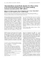
Figure 1
Mab staining of mouse and sheep lung tumors
Mab staining of mouse and sheep lung tumors. Left panels are from a mouse exposed 2 months previously to an AAV6
vector encoding JSRV Env (ARJenv) [10], and right panels are from sheep 85RS14 (Table 1) that was experimentally infected
with JSRV. Sections were stained with the Env Mab C9 and were counterstained with methyl green. Arrow in lower right panel
indicates inflammatory cells that do not stain for Env expression.
Page 3 of 15
(page number not for citation purposes)
Retrovirology 2006, 3:94
/>
these sheep were brightly stained by the Mab B3, C9, or a
mixture of the two, with no staining of histologically-normal lung tissue (Table 1; Fig. 1, right panels). The appearance of many of the Mab-stained sheep lung tumors was
remarkably similar to that of mouse lung tumors induced
by exposure to ARJenv, the AAV6 vector that only encodes
JSRV Env (Fig. 1, left panels). The majority of lung tumors
in sheep and mice appeared as adenomas consisting of
well-differentiated epithelial cells. There was more lung
inflammation in the immunocompetent sheep in comparison to the immunodeficient mice (Fig 1), as might be
expected. However, the Mab clearly differentiated tumor
cells from Env-negative immune cells, connective tissue,
and myxomatous tissue [19,20] that were often found
within and around the sheep tumors.
Table 1: JSRV Env-antibody staining of histologic sections of lung
tissue from sheep
Country of origin
Sheep number
antibody
B3 C9
USA
(experimentallyinfected)
+
84RS18
85RS1
85RS14
85RS22
USA
(naturally- infected)
84RS17
+
+
84RS28
+
+
85RS65
98RS1
98RS3
99RS27
99RS33
Peru
polyclonal
+
+
B3+C9
81R15
81R16
81R22
81R71
81R78
+
+
+
+
+
+
+
+
+
+
+
+
+
+
+
+
+
+
+
+
+
+
+
+
Sequencing of the env regions of different JSRV isolates
from sheep has revealed several strains that fall into two
groups, those from Africa and those from the United Kingdom and United States [21-24]. Our Mab were generated
using the JS7 strain of Env [24], an isolate from Scotland,
and we wanted to know if the Mab would recognize Env
from wild-type strains of JSRV from countries spanning
North and South America, Europe and Africa. The Mab
recognized tumors in all sheep with JSRV-induced disease
from the United States, Peru, Spain, Kenya and South
Africa (Table 1, Fig. 2). Because all tumors were recognized by Mab B3, C9, or both, we conclude that the mixture of Mab B3 and C9 is capable of recognizing JSRV Env
in tumors caused by wild-type JSRV in multiple geographic regions, in particular, from regions where infection by either of the two major types of JSRV predominate.
This may in part result from the fact that the Mab were
raised against the SU domain of Env, which is relatively
well conserved among JSRV strains that have been
sequenced to date.
+
+
+
+
Spain
B-96/00
+
+
Kenya
92K3
+
+
South Africa
93141
95195
95205
95211
95226
95227
95229
95234
95251
96238
96269
+
+
+
+
+
+
+
+
+
+
+
The majority of sheep tumors examined by Mab staining
had the histologic appearance of adenomas with little evidence of anaplasia (Figs. 1 and 2). In contrast, adenocarcinomas were occasionally found in mice at long times (4
to 6 months) after vector administration (Fig. 3). All of
these tumors were Env+ as determined by Mab staining
(not shown). In some sheep, large adenomatous tumors
were present in airways (Fig. 2 panel F), and some mice
exhibited similar tumors at long times (4 to 6 months)
after exposure to the ARJenv vector encoding JSRV Env
(not shown).
In three sheep (85RS65 and 99RS27 from the United
States and 96238 from South Africa) we found proliferative lesions consisting of fibroblasts or other connective
tissue cells that expressed Env and that appeared to be separate neoplasms. Low-power views of these lesions
revealed relatively round Env+ masses of cells (Fig. 4A, B,
E) that were sometimes flanked by typical well-differentiated Env+ epithelial cell tumors (Fig. 4A). High-power
views of cells in the fibroblastic areas (Fig. 4C, D) revealed
a histological similarity to connective tissue found at the
edges of some sheep lungs (Fig. 4F). Such connective tissue lined the lungs and septae projected into the interior
of the lungs of some sheep, but none of these tissues
stained with Env Mab, including the area shown in Fig. 4F
(data not shown). Thus there was a clear differentiation
between the streams of Env-negative connective tissue in
the lung and the Env+ masses consisting of disorganized
immature connective tissue cells. We did not observe such
Env+ fibroblastic masses of connective tissue cells in mice
transduced with the ARJenv AAV6 vector that encodes
JSRV Env, but did observe streams of Env-negative connective tissue by histologic analysis in some mice.
Page 4 of 15
(page number not for citation purposes)
Retrovirology 2006, 3:94
/>
A
B
C
D
E
F
G
H
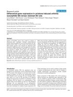
Figure 2
Mab staining of JSRV-infected sheep lung tumors from around the world
Mab staining of JSRV-infected sheep lung tumors from around the world. Sheep numbers and countries of origin are:
A, 96238 from South Africa; B, 95234 from South Africa; C, 92K3 from Kenya; D, 81R16 from Peru; E and F, 85RS1 from the
USA (experimentally-infected); G, 84RS28 from the USA; and H, B-96/00 from Spain. Sections were stained with Mab B3, C9,
or both. Scale bars indicate a distance of 100 µm.
Page 5 of 15
(page number not for citation purposes)
Retrovirology 2006, 3:94
/>
100 µm
100 µm
Figure 3
Adenocarcinoma in a mouse 6 months after exposure to the ARJenv vector
Adenocarcinoma in a mouse 6 months after exposure to the ARJenv vector. Tissues were stained with hematoxylin
and eosin. Left panel, adenoma (left) and adenocarcinoma (right). Right panel, close-up of an adenocarcinoma showing atypical
nuclei.
The JSRV Env Mab did not recognize any cross-reacting
antigens in lung samples from sheep and goats diagnosed
with a variety of diseases that were not the result of JSRV
infection. These included lung samples from a sheep and
a goat with mucinous goblet cell adenocarcinoma, a
sheep infected with ovine lentivirus, and two sheep with
inflammatory diseases, one classified as follicular bronchiolitis and the other as lymphoid follicular hyperplasia
due to verminous pneumonia.
Interestingly, the Mab did stain tumor cells in nasal adenocarcinoma from a sheep (no. 99RS39 from the United
States) (Fig. 5, top and middle panels), presumably
caused by infection with ENTV as it is in Europe. Env
staining in the nasal tumor appears to be almost exclusively localized to the apical cell membrane, as opposed
to JSRV Env staining which also appears at high levels in
the cytoplasm (compare Fig. 5 top and middle panels to
Fig. 1, right panels, and Fig. 2). Furthermore, tumors
induced in the lungs of mice by an AAV6 vector encoding
ENTV Env [12] showed the same apical localization of
ENTV Env (Fig. 5, bottom panel) compared to the apical
and cytoplasmic localization of JSRV Env (Fig. 1, left panels). PCR amplification followed by direct sequencing of
nasal tumor DNA using ENTV-specific primers [25] that
amplify a portion of the cytoplasmic tail of Env that is relatively divergent between ovine ENTV (ENTV-1), caprine
ENTV (ENTV-2), JSRV, and sheep endogenous retrovirus
sequences, revealed that this sheep was indeed infected by
a virus with a unique sequence [GenBank: EF184579]
most closely related to ENTV-1, with up to 97% identity
to existing ENTV-1 sequences. The sequence also contains
a 2 bp frameshift in the C-terminus of the Env coding
region, a characteristic of the ENTV-1 lineage. These
results confirm the suspected presence of ENTV in the
United States [26-28].
Tumors induced in mice following intravenous injection of
an AAV6 vector encoding JSRV Env
JSRV DNA and RNA can be detected in lymph nodes,
spleen, thymus, bone marrow, and blood cells of sheep
infected with JSRV [29,30], and in natural settings systemic infection can be present over long periods without
induction of lung tumors [31]. Although oncogenesis
originating in tissues other than the lung has not been
reported in JSRV-infected sheep, we wanted to determine
whether JSRV Env could induce tumors in other tissues.
We determined that tail vein administration of an AAV6
vector (ARAP4), that expresses human placental alkaline
phosphatase (AP) from the same strong Rous sarcoma
virus promoter present in the ARJenv vector [10], led to
transduction of multiple tissues in mice, including liver,
spleen, heart, kidney, and lung (data not shown). We next
administered the ARJenv AAV6 vector to two 1.5-monthold mice. Both mice showed a lack of weight gain starting
at 4.5 months of age, showed visible signs of disease starting at 6.5 months of age, and were killed for analysis at 7.5
months of age, 6 months after vector exposure. Only a few
of the tissues that can be transduced by an AAV6 vector
showed evidence of hyperplasia and/or overt tumor formation. The vector did induce multiple tumors in the liver
(Fig. 6A, B). Immunohistochemical staining for Env
revealed that Env expression corresponded to the areas of
Page 6 of 15
(page number not for citation purposes)
Retrovirology 2006, 3:94
/>
Env Antibody
A
400 µm
C
100 µm
E
400 µm
H&E
B
400 µm
D
100 µm
F
100 µm
Figure 4
Fibroblastic cell masses found in some JSRV-infected sheep
Fibroblastic cell masses found in some JSRV-infected sheep. Left panels show Mab C9 staining and right panels show
hematoxylin and eosin staining of sheep lung sections. Panels A and B show a round proliferative fibroblastic cell mass (myxomatous tissue) flanked at upper right and left by typical epithelial tumors from South African sheep 96238. Panels C and D
show magnified views of the fibroblastic cell mass corresponding to the boxed areas in Panels A and B. Panel E shows Env staining of a large fibroblastic cell mass from naturally-infected United States sheep 85RS65. Panel F shows connective tissue containing fibroblasts at the edge of the lung from South African sheep 93141. These cells were all Env-negative (not shown).
Page 7 of 15
(page number not for citation purposes)
Retrovirology 2006, 3:94
/>
200 µm
100 µm
100 µm
Figure 5
Mab staining of ENTV Env
Mab staining of ENTV Env. Top and middle panels show Env Mab staining of nasal adenocarcinoma from sheep 99RS39
infected with ENTV. Bottom panel shows Env Mab staining of lung tumor from mouse 5-3 exposed 4 months earlier to an
AAV6 vector encoding ovine ENTV Env [12].
Page 8 of 15
(page number not for citation purposes)
Retrovirology 2006, 3:94
/>
A
B
C
D
E
F
Figure 6
Hepatocellular tumors induced by intravenous injection of an AAV6 vector expressing JSRV Env
Hepatocellular tumors induced by intravenous injection of an AAV6 vector expressing JSRV Env. Panels A and B
show low-magnification views of the same area of a liver stained with a mixture of the B3 and C9 Env Mab (light methyl green
counterstain) (Panel A) or hematoxylin and eosin (Panel B). Panels C and D show a mixed tumor with adenomatous features in
the upper right portion and adenocarcinomatous features in the lower left portion. Panel C shows staining with the Mab
(hematoxylin counterstain) and Panel D shows staining with hematoxylin and eosin. Note the compression of liver tissue near
the lower left side of the tumor. Panel E shows a high-magnification view of the same tumor in the panel above, with the division between adenoma and adenocarcinoma running from the top left to the bottom right of the panel. Note the linear staining
between cells that likely represents Env in bile canaliculi. Panel F shows a tumor with a foamy appearance stained with the Env
Mab (hematoxylin counterstain). Scale bars = 100 µm.
Page 9 of 15
(page number not for citation purposes)
Retrovirology 2006, 3:94
/>
1 mm
1 mm
RBC
RBC
100 µm
100 µm
50 µm
50 µm
Figure 7
Hemangiomas and hemangiosarcomas induced by intravenous injection of the AAV6 vector expressing JSRV Env
Hemangiomas and hemangiosarcomas induced by intravenous injection of the AAV6 vector expressing JSRV
Env. Left panels show tumors stained with a mixture of the B3 and C9 Mab (light methyl green counterstain) and right panels
show hematoxylin and eosin staining of the same areas shown in the left panels. Top panels show a large cavernous hemangiosarcoma arising in subcutaneous fat. Boxes indicate the areas shown in the middle panels (black boxes) and lower panels
(white boxes). Middle panels show an area typical of hemangioma composed of cavernous blood vessels containing residual red
blood cells (RBC) that are lined with a single layer of well differentiated, flattened endothelial cells. Note the intense Env Mab
staining of endothelial cells but that the collagen and other cells between the vascular spaces do not stain with the Env Mab.
Bottom panels show a high-magnification view of an area of hemangiosarcoma comprised of Env+ pleomorphic endothelial cells
forming a solid cellular mass. Note examples of pleomorphic nuclei with prominent nucleoli (yellow arrows, lower right panel).
Page 10 of 15
(page number not for citation purposes)
Retrovirology 2006, 3:94
hyperplastic growth, indicating that Env was responsible
for lesion formation (Fig. 6A, B). Liver lesions included
foci of hepatocellular hyperplasia, adenoma, and rare adenocarcinoma (Fig. 6). In particular, one lesion had a
mixed phenotype consisting of adenoma (top right) and
adenocarcinoma (bottom left) (Fig. 6C, D, E). A highmagnification view of this tumor showed striking linear
staining that likely represents accumulation of Env protein in bile canaliculi (Fig. 6E). Hemangiomas and
hemangiosarcomas were observed in multiple fat tissues,
most notably in subcutaneous (Fig. 7) and peritesticular
(not shown) fat. These lesions ranged from hemangioma
(Fig. 7, middle row panels) to hemangiosarcoma (Fig. 7,
bottom panels), and all stained positive for JSRV Env
expression (Fig. 7 and data not shown). These data show
that Env can induce tumors in various cell types besides
lung epithelial cells, and some of these tumors have a relatively aggressive histologic appearance.
Discussion
We have developed Mab against the Env protein of JSRV
that give intense staining of lung tumors in sheep infected
with JSRV and in mice exposed to an AAV6 vector encoding JSRV Env. These Mab recognized tumors in all JSRVinfected sheep examined (n = 29) from multiple countries. The antibodies did not recognize similar urethaneinduced lung tumors in mice. Both urethane- and JSRV
Env-induced lung tumors have the same histologic
appearance, express the type II alveolar cell marker surfactant protein C and most do not express the Clara cell
marker CC-10 [10,18,32]. The Mab did not recognize
alveolar type II cell hyperplasia or other cell types in a variety of diseases in sheep that were not the result of JSRV
infection. We also found that at least two of the Mab recognized the Env from ovine ENTV in tumors induced in
mice by exposure to an AAV6 vector encoding ENTV Env
[12], and in a sheep with nasal adenocarcinoma associated with ENTV infection. Together these results indicate
that the Mab are highly specific for ovine betaretrovirus
Env expression, and would provide a useful diagnostic test
for JSRV, and possibly for ENTV as well.
The current accepted nomenclature for lung cancer resulting from JSRV infection is ovine pulmonary adenocarcinoma. The primary reason for its characterization as a
malignant disease is because of the observation of metastases consisting of lung tumor epithelial cells, which
occurs to a variable extent in sheep [3]. However, the main
tumor type we see in JSRV-infected sheep and in JSRV Envexpressing mice is adenoma, consistent with the previous
description of the disease as an adenomatosis. Given our
results in sheep and in mice, and the fact that what kills
these animals is breathing difficulty, it seems the primary
effect of JSRV infection, mediated through the Env protein, is to cause proliferation of lung epithelial cells. In
/>
JSRV-infected sheep, such proliferation typically increases
lung fluid production and thereby facilitates aerosol transmission of the virus produced by epithelial cells. Metastasis may occur as the result of additional genetic changes
resulting from virus replication and integration, or resulting directly from Env expression and stimulation of cell
proliferation, but these appear not to be the primary
effects of virus infection or Env expression.
It is remarkable how little Env Mab staining we observe
outside of tumors in mice and sheep. Others have
reported similar results in sheep by using polyclonal antibodies to detect JSRV Env or capsid proteins [31-33], but
our use here of highly specific Mab that give intense staining of Env-expressing cells helps to rule out the presence
of low levels of Env expression outside of tumors. In mice
we know that an AAV6 vector encoding AP (ARAP4) can
transduce all epithelial cell populations in the airway with
relatively high efficiency [34], yet we see no Env staining
in large or small airways or in histologically-normal alveoli in mice exposed to the AAV6 vector ARJenv, which like
ARAP4 contains a strong Rous sarcoma virus promoter to
drive gene expression. It is known that oncoproteins can
have both growth-promoting and toxic effects in cultured
cells [35], and perhaps only lung stem cells that are the
progenitors of tumors can tolerate expression of the
potent Env oncoprotein, while Env expression is toxic to
the more differentiated cells.
Lack of Env expression outside of tumors in lungs of sheep
infected with JSRV is particularly surprising given the presence of replicating virus in the sheep. Perhaps spread of
JSRV is inhibited in sheep by an immune response,
despite the finding that sheep mount a poor response
against JSRV because of immune tolerance induced by
proteins made by related endogenous retroviruses [36].
Alternatively, like other simple retroviruses, JSRV may
only infect dividing cells, and most potential target cells in
the lung are not actively dividing. Most intriguingly, it
may be that Env is toxic to most differentiated lung cell
types in sheep, as proposed above for mice.
Our results provide further support for the conclusion
that the JSRV cell-entry receptor Hyal2 plays no role in
sheep tumorigenesis beyond its role as a receptor for virus
entry. Mouse Hyal2 does not serve as a cell-entry receptor
for retrovirus vectors bearing the JSRV Env protein [4,1517] nor does it bind the SU domain of JSRV Env [16], yet
we have shown here that lung tumors induced in mice by
Env expression alone are quite similar to lung tumors
induced by JSRV in sheep having a functional Hyal2 virus
receptor.
Our results also argue against a role for insertional oncogene activation or insertional mutagenesis in sheep tum-
Page 11 of 15
(page number not for citation purposes)
Retrovirology 2006, 3:94
origenesis. An AAV6 vector was used to transfer and
express JSRV Env in the mice analyzed here, and tumor
induction followed single-hit kinetics [12], a result that is
inconsistent with a requirement for insertional events in
addition to Env expression for tumorigenesis. In addition,
inclusion of an excess of a non-oncogenic AAV6 vector
during transduction by the JSRV Env-expressing AAV6 vector reduced the number of tumors [12], again indicating
that additional genetic changes that might be caused by
the AAV6 vector are not important for tumorigenesis.
Together with results shown here that tumors induced by
JSRV Env in mice are quite similar to tumors induced by
JSRV in sheep, these results indicate that JSRV tumorigenesis is primarily dependent on the oncogenic activity of
the JSRV Env protein and does not require genetic changes
resulting from JSRV integration.
The main tumor type induced by systemic administration
of the AAV6 vector encoding JSRV Env was hepatocellular
adenoma. Generation of this non-malignant proliferative
tumor is consistent with the activity of JSRV Env in the
lung to generate adenomas arising from lung epithelial
cells. Given the low frequency of hepatocellular adenocarcinomas following JSRV Env vector administration, it is
likely that additional events are required for adenocarcinoma formation, as they appear to be following expression of other oncoproteins such as Myc [37].
Systemic administration of the AAV6 vector encoding
JSRV Env to mice induced multiple hemangiomas and
some hemangiosarcomas, tumors that arise from uncontrolled and disorganized proliferation of endothelial cells.
Endothelial cells in these tumors were uniformly and
uniquely stained by the Env Mab, indicating a direct effect
of Env on endothelial cells in these tumors.
Oncogenes from other viruses can also induce hemangiomas and have helped elucidate a common pathway for
hemangiogenesis that involves phosphatidylinositol 3kinase (PI3K) activation, downstream activation of Akt,
and increased vascular endothelial growth factor production; the latter being a key stimulus for hemangiogenesis.
For example, avian sarcoma virus 16 induces hemangiomas and was found to express a viral oncogene derived
from the gene encoding the catalytic subunit of PI3K [38].
Viral vectors expressing the viral or cellular forms of the
PI3K catalytic subunit could induce hemangiosarcomas in
chickens and could transform chicken embryo fibroblasts
in culture [38]. Transformation in culture was accompanied by Akt activation and VEGF production, and overexpression of a myristylated form of Akt or VEGF itself could
induce hemangiosarcoma formation in chicken embryos
[39]. Interestingly, JSRV Env has been shown to transform
cultured fibroblasts from mice, rats, and chickens [4-6],
and transformation is accompanied by activation of PI3K
/>
and Akt in these cells [8,40,41], suggesting that JSRV Env
may induce hemangioma formation by activation of the
PI3K-Akt-VEGF pathway in mouse endothelial cells.
Another retrovirus that induces hemangiomas is avian
hemangioma virus, and like JSRV, this appears to be due
to expression of the viral Env protein [42,43]. However,
the avian hemangioma virus Env protein shows no similarity to that of JSRV, so it is difficult to predict if the mechanisms of hemangiogenesis are similar. It will be
interesting to see if AHV also activates members of the
PI3K-Akt-VEGF pathway.
In 10% of JSRV-infected sheep studied we observed
masses of Env+ fibroblastic cells that appear to be separate
neoplasms. The ability of JSRV Env to transform fibroblasts from several species in tissue culture [4-6], and the
uniform Env+ staining of the fibroblastic cell masses in
sheep, make it tempting to speculate that these masses
represent a novel tumor type. However, the frequent
observation of non-neoplastic fibroblast or mesenchymal
cell proliferation in response to a number of tissue insults
complicates this interpretation. Others have observed
similar proliferation of connective tissue in association
with epithelial tumors in JSRV-infected sheep [3], but
immunohistochemical analysis for Env expression was
not performed. We did not see such Env+ fibroblastic
masses in mice, but this could simply be due to a lower
frequency of the parental cell type in mouse lung. Regardless, these fibroblastic masses were an infrequent occurrence in sheep and thus do not account for the typical
disease observed in JSRV-infected sheep.
Nasal administration of the AAV6-Jenv vector to normal
C57BL/6 mice results in strong immune responses against
Env that limit tumor formation, therefore we have used
immunodeficient C57BL/6 Rag-2 mice to model tumor
formation by JSRV Env. The question arises whether an
immunodeficient mouse is an appropriate model for a
disease that occurs in immunocompetent sheep. In fact,
expression of multiple endogenous retroviruses related to
JSRV in sheep results in immunotolerance toward JSRV
infection [36], thus immunodeficient mice appear to be a
good model in which to study this intriguing viral disease.
Conclusion
We have generated Mab against the SU domain of JSRV
Env and have shown that these Mab allow robust detection of Env protein synthesis from wild-type strains of
JSRV from around the world. The histologic appearance of
the majority of lung tumors in sheep infected with JSRV
and in mice expressing only the JSRV Env protein is
remarkably similar, indicating that Env expression alone
can explain much of the disease phenotype in sheep.
Indeed, some tumors in mice exhibit a more aggressive
Page 12 of 15
(page number not for citation purposes)
Retrovirology 2006, 3:94
adenocarcinomatous histology than do tumors observed
in sheep. While JSRV infection in sheep is not known to
induce tumors originating in organs other than the lung,
systemic expression of JSRV Env in mice induced hepatocellular tumors, hemangiomas, and hemangiosarcomas,
showing that Env can induce tumors in cells other than
lung epithelial cells. Our results indicate that Env interaction with the virus-entry receptor Hyal2, insertional activation of cellular oncogenes, and insertional mutagenesis
do not play major roles in sheep tumorigenesis by JSRV.
Overall, the primary effect of JSRV infection is to drive
localized proliferation of lung epithelial cells.
Methods
Animal studies and safety precautions
Experiments involving mice were performed using procedures approved by the Institutional Animal Care and Use
Committee of the Fred Hutchinson Cancer Research
Center. Special safety precautions employed during production and use of the AAV6 vectors encoding oncogenic
Env proteins were as previously described [10]. All sheep
tissue samples were obtained from archival materials collected as part of previously approved studies.
Mouse immunization protocol for production of Mab
5 × 10¹⁰ vector genomes of a replication-defective AAV6
vector expressing JSRV Env (ARJenv) [10] was administered intranasally to lightly anesthetized eight-week-old
C57BL/6 mice. Blood samples were collected weekly and
sera were screened for the presence of antibodies to JSRV
Env protein by ELISA. At 6 weeks post-infection, mice
were boosted intraperitoneally with 50 µg JSU-IgG protein in incomplete Freund's adjuvant. JSU-IgG is a hybrid
protein consisting of the JSRV Env surface domain (SU)
fused to a human IgG Fc [16]. At 9 weeks post-infection,
mice were subjected to a second and final boost consisting
of 50 µg JSU-IgG (without adjuvant) delivered both intraperitoneally and intravenously.
Hybridoma generation and characterization of Mab by
ELISA
Three days after the last injection, mice were killed and
their spleens were removed. Splenocytes were harvested
and fused with FOX-NY myeloma cells [44], and hybridomas were selected in medium containing adenine, aminopterin and thymidine as described [44]. Hybridoma
supernatants were screened for antibodies against JSRV
Env by antigen-dependent ELISA. Briefly, purified JSU-IgG
or human IgG was passively adsorbed onto 96 well U-bottom non-tissue culture treated plates (Falcon) at a concentration of 1 µg/ml in Dulbecco's PBS overnight at 4°C.
Plates were rinsed with PBS containing 0.05% Tween-20
(PBST) and blocked with PBS containing 5% nonfat milk
extract and 2% goat serum for 1 h at 37°C. Antibodies
(tissue culture supernatants) were reacted with antigens
/>
for 1 h at 37°C, rinsed with PBST, and incubated for an
additional hour with a 1:10,000 dilution of HRP conjugated goat anti-mouse IgG (γ chain) (Southern Biotech).
Plates were washed and developed using ABST peroxidase
substrate (KPL). At 10 and 30 min, the absorbance at 492
nm was determined using a microplate reader. Selected
hybridomas were cloned by limiting dilution, and the isotypes of their antibody products were determined by an
indirect-capture ELISA. Of the 564 clones that were generated using this vaccination protocol, 52 clones demonstrated specificity of varying degree for JSU-IgG as
determined by ELISA. Eight hybridomas that produced
antibodies against JSRV Env were selected for further characterization. Of those, 6 were IgG1 (including clones B3
and C9), one was IgG2a and one was IgG2b isotype.
Immunohistochemistry
Sheep tissues were fixed in 10% formalin, and mouse lung
tissue was fixed in 2% paraformaldehyde in phosphatebuffered saline. After fixation tissues were embedded in
paraffin wax using an automatic tissue processor and tissue sections (5 µm) were cut and placed on positively
charged slides. Samples were deparaffinized and antigen
retrieval was performed in a pressure cooker (heat to
120°C, hold for 3 min, allow to cool to 90°C, hold for 3
min) using Antigen Unmasking Solution (Vector Laboratories, Burlingame CA USA). After cooling, endogenous
peroxidase was quenched with 3% hydrogen peroxide for
5 min. Mouse IgG was blocked with unconjugated antimouse IgG (Vector Laboratories AI-2000) at 1:50 dilution
for 15 min. Slides were washed two times for 10 min each
with PBS, and medium exposed to hybridoma cells that
produce Mab (1:50 dilution in PBS) was incubated with
the tissue for 1 h at room temperature. Slides were washed
and biotinylated horse-anti-mouse IgG (Vector Laboratories) at a 1:300 dilution was added for 30 min at room
temperature. Slides were washed again and incubated
with avidin:biotinylated enzyme complex (Vectastain
Elite ABC kit; Vector Laboratories). 3,3'-diaminobenzidine tetrahydrochloride (DAB) with nickel chloride
enhancement was used as a peroxidase substrate and the
sections were counterstained with methyl green.
Systemic administration of AAV6 vectors
5 × 10¹⁰ vector genomes of ARJenv or ARAP4 [45] was
administered intravenously to C57BL6/RAG2 mice by tail
vein injection. Vectors were suspended in Dulbecco's PBS
and administered in a total volume of 0.4 ml. A heating
pad was placed in the mouse cage 10 minutes prior to
injection in order to dilate tail veins and facilitate delivery
of virus. At one-week post infection, mice were given a second injection of 5 × 10¹⁰ vector genomes of ARJenv intraperitoneally. Mice were killed 6 months post infection
and a full body necropsy was performed. All tissues, with
the exception of the lung, were fixed in 2% paraformalde-
Page 13 of 15
(page number not for citation purposes)
Retrovirology 2006, 3:94
hyde for 48 h, dehydrated, embedded in paraffin, sectioned and stained with hematoxylin and eosin by
standard methods. Mouse lungs were perfused with 2%
paraformaldehyde and fixed for 4 h. Immunohistochemical staining for JSRV Env was performed as described
above. Alkaline phosphatase staining of tissues was performed as described previously [10].
Competing interests
The author(s) declare that they have no competing interests.
Authors' contributions
SW generated the Mab, the AAV6 vectors encoding JSRV
and ENTV Env, and the AAV6 vector-transduced mice; JD
and DY provided sheep samples and helped with data
interpretation; MM identified the ENTV virus in the nasal
adenocarcinoma sample; KH and CA performed the histologic and antibody staining of mouse and sheep tissues;
and AM coordinated the experiments, analyzed the data,
and wrote the manuscript. All authors read and approved
the final manuscript.
Acknowledgements
We thank Helle Bielefeldt-Ohmann and Susan Knoblaugh for advice regarding histological interpretation of tumor phenotype, Alvin Malkinson for
providing samples of urethane-induced lung tumors from mice, and Luis
Luján for providing a lung tumor sample from a Spanish sheep. SW was
funded by postdoctoral fellowships from the National Sciences and Engineering Research Council of Canada and the Canadian Institutes of Health
Research, and MM was funded by NIH training grant CA09229. Overall
funding was primarily provided by a pilot and feasibility grant from the Fred
Hutchinson Cancer Research Center.
References
1.
2.
3.
4.
5.
6.
7.
8.
Sharp JM, DeMartini JC: Natural history of JSRV in sheep. Curr
Top Microbiol Immunol 2003, 275:55-79.
Sharp JM, Angus KW, Gray EW, Scott FM: Rapid transmission of
sheep pulmonary adenomatosis (jaagsiekte) in young lambs.
Brief report. Arch Virol 1983, 78(1-2):89-95.
De las Heras M, Gonzalez L, Sharp JM: Pathology of ovine pulmonary adenocarcinoma.
Curr Top Microbiol Immunol 2003,
275:25-54.
Rai SK, Duh FM, Vigdorovich V, Danilkovitch-Miagkova A, Lerman MI,
Miller AD: Candidate tumor suppressor HYAL2 is a glycosylphosphatidylinositol (GPI)-anchored cell-surface receptor
for jaagsiekte sheep retrovirus, the envelope protein of
which mediates oncogenic transformation. Proc Natl Acad Sci
USA 2001, 98(8):4443-4448.
Maeda N, Palmarini M, Murgia C, Fan H: Direct transformation of
rodent fibroblasts by jaagsiekte sheep retrovirus DNA. Proc
Natl Acad Sci USA 2001, 98(8):4449-4454.
Allen TE, Sherrill KJ, Crispell SM, Perrott MR, Carlson JO, DeMartini
JC: The jaagsiekte sheep retrovirus envelope gene induces
transformation of the avian fibroblast cell line DF-1 but does
not require a conserved SH2 binding domain. J Gen Virol 2002,
83(11):2733-2742.
Danilkovitch-Miagkova A, Duh FM, Kuzmin I, Angeloni D, Liu SL,
Miller AD, Lerman MI: Hyaluronidase 2 negatively regulates
RON receptor tyrosine kinase and mediates transformation
of epithelial cells by jaagsiekte sheep retrovirus. Proc Natl Acad
Sci USA 2003, 100(8):4580-4585.
Liu SL, Lerman MI, Miller AD: Putative phosphatidylinositol 3kinase (PI3K) binding motifs in ovine betaretrovirus Env pro-
/>
9.
10.
11.
12.
13.
14.
15.
16.
17.
18.
19.
20.
21.
22.
23.
24.
25.
26.
27.
28.
29.
teins are not essential for rodent fibroblast transformation
and PI3K/Akt activation. J Virol 2003, 77(14):7924-7935.
Liu SL, Miller AD: Transformation of Madin-Darby canine kidney epithelial cells by sheep retrovirus envelope proteins. J
Virol 2005, 79(2):927-933.
Wootton SK, Halbert CL, Miller AD: Sheep retrovirus structural
protein
induces
lung
tumours.
Nature
2005,
434(7035):904-907.
Caporale M, Cousens C, Centorame P, Pinoni C, De las Heras M, Palmarini M: Expression of the jaagsiekte sheep retrovirus envelope glycoprotein is sufficient to induce lung tumors in
sheep. J Virol 2006, 80(16):8030-8037.
Wootton SK, Halbert CL, Miller AD: Envelope proteins of
jaagsiekte sheep retrovirus and enzootic nasal tumor virus
induce similar bronchioalveolar tumors in lungs of mice. J
Virol 2006, 80(18):9322-9325.
Cousens C, Bishop JV, Philbey AW, Gill CA, Palmarini M, Carlson JO,
DeMartini JC, Sharp JM: Analysis of integration sites of
Jaagsiekte sheep retrovirus in ovine pulmonary adenocarcinoma. J Virol 2004, 78(16):8506-8512.
Philbey AW, Cousens C, Bishop JV, Gill CA, Demartini JC, Sharp JM:
Multiclonal pattern of Jaagsiekte sheep retrovirus integration sites in ovine pulmonary adenocarcinoma. Virus Res 2006,
117(2):254-263.
Rai SK, DeMartini JC, Miller AD: Retrovirus vectors bearing
jaagsiekte sheep retrovirus Env transduce human cells by
using a new receptor localized to chromosome 3p21.3. J Virol
2000, 74(10):4698-4704.
Liu SL, Duh FM, Lerman MI, Miller AD: Role of virus receptor
Hyal2 in oncogenic transformation of rodent fibroblasts by
sheep betaretrovirus env proteins.
J Virol 2003,
77(5):2850-2858.
Duh FM, Dirks C, Lerman MI, Miller AD: Amino acid residues that
are important for Hyal2 function as a receptor for jaagsiekte
sheep retrovirus. Retrovirology 2005, 2:59.
Mason RJ, Kalina M, Nielsen LD, Malkinson AM, Shannon JM: Surfactant protein C expression in urethane-induced murine
pulmonary tumors. Am J Pathol 2000, 156(1):175-182.
Cutlip RC, Young S: Sheep pulmonary adenomatosis
(jaagsiekte) in the United States. Am J Vet Res 1982,
43(12):2108-2113.
Garcia-Goti M, Gonzalez L, Cousens C, Cortabarria N, Extramiana
AB, Minguijon E, Ortin A, De las Heras M, Sharp JM: Sheep pulmonary adenomatosis: characterization of two pathological
forms associated with jaagsiekte retrovirus. J Comp Pathol
2000, 122(1):55-65.
York DF, Vigne R, Verwoerd DW, Querat G: Nucleotide
sequence of the jaagsiekte retrovirus, an exogenous and
endogenous type D and B retrovirus of sheep and goats. J
Virol 1992, 66(8):4930-4939.
Bai J, Bishop JV, Carlson JO, DeMartini JC: Sequence comparison
of JSRV with endogenous proviruses: envelope genotypes
and a novel ORF with similarity to a G-protein-coupled
receptor. Virology 1999, 258(2):333-343.
Palmarini M, Sharp JM, De las Heras M, Fan H: Jaagsiekte sheep retrovirus is necessary and sufficient to induce a contagious
lung cancer in sheep. J Virol 1999, 73(8):6964-6972.
DeMartini JC, Bishop JV, Allen TE, Jassim FA, Sharp JM, De las Heras
M, Voelker DR, Carlson JO: Jaagsiekte sheep retrovirus proviral
clone JSRV(JS7), derived from the JS7 lung tumor cell line,
induces ovine pulmonary carcinoma and is integrated into
the surfactant protein A gene. J Virol 2001, 75(9):4239-4246.
Kane Y, Rosati S, Diop OM, Profiti M, Niang I, Kadja M, Kaboret YY,
Alogninouwa T, Lena P: Enzootic nasal tumour virus demonstrated in sheep in Senegal by direct detection of provirus
from tumour DNA. Vet Rec 2004, 155(17):526-528.
Young S, Lovelace SA, Hawkins WW Jr., Catlin JE: Neoplasms of
the olfactory mucous membrane of sheep. Cornell Vet 1961,
51(1):96-112.
Duncan JR, Tyler DE, Van Der Maaten MJ, Andersen JR: Enzootic
nasal adenocarcinoma in sheep. J Am Vet Med Assoc 1967,
151(6):732-734.
Rings DM, Rojko J: Naturally occurring nasal obstructions in 11
sheep. Cornell Vet 1985, 75(2):269-276.
Palmarini M, Holland MJ, Cousens C, Dalziel RG, Sharp JM:
Jaagsiekte retrovirus establishes a disseminated infection of
Page 14 of 15
(page number not for citation purposes)
Retrovirology 2006, 3:94
30.
31.
32.
33.
34.
35.
36.
37.
38.
39.
40.
41.
42.
43.
44.
45.
the lymphoid tissues of sheep affected by pulmonary adenomatosis. J Gen Virol 1996, 77(12):2991-2998.
Holland MJ, Palmarini M, Garcia-Goti M, Gonzalez L, McKendrick I,
De las Heras M, Sharp JM: Jaagsiekte retrovirus is widely distributed both in T and B lymphocytes and in mononuclear
phagocytes of sheep with naturally and experimentally
acquired pulmonary adenomatosis.
J Virol 1999,
73(5):4004-4008.
Caporale M, Centorame P, Giovannini A, Sacchini F, Di Ventura M,
De las Heras M, Palmarini M: Infection of lung epithelial cells and
induction of pulmonary adenocarcinoma is not the most
common outcome of naturally occurring JSRV infection during the commercial lifespan of sheep.
Virology 2005,
338(1):144-153.
Platt JA, Kraipowich N, Villafane F, DeMartini JC: Alveolar type II
cells expressing jaagsiekte sheep retrovirus capsid protein
and surfactant proteins are the predominant neoplastic cell
type in ovine pulmonary adenocarcinoma. Vet Pathol 2002,
39(3):341-352.
Palmarini M, Dewar P, De las Heras M, Inglis NF, Dalziel RG, Sharp
JM: Epithelial tumour cells in the lungs of sheep with pulmonary adenomatosis are major sites of replication for
Jaagsiekte retrovirus. J Gen Virol 1995, 76(11):2731-2737.
Halbert CL, Allen JM, Miller AD: Adeno-associated virus type 6
(AAV6) vectors mediate efficient transduction of airway epithelial cells in mouse lungs compared to that of AAV2 vectors. J Virol 2001, 75(14):6615-6624.
Deng Q, Liao R, Wu BL, Sun P: High intensity ras signaling
induces premature senescence by activating p38 pathway in
primary human fibroblasts. J Biol Chem 2004, 279(2):1050-1059.
DeMartini JC, Carlson JO, Leroux C, Spencer T, Palmarini M: Endogenous retroviruses related to jaagsiekte sheep retrovirus.
Curr Top Microbiol Immunol 2003, 275:117-137.
Wu Y, Renard CA, Apiou F, Huerre M, Tiollais P, Dutrillaux B, Buendia MA: Recurrent allelic deletions at mouse chromosomes 4
and 14 in Myc-induced liver tumors.
Oncogene 2002,
21(10):1518-1526.
Chang HW, Aoki M, Fruman D, Auger KR, Bellacosa A, Tsichlis PN,
Cantley LC, Roberts TM, Vogt PK: Transformation of chicken
cells by the gene encoding the catalytic subunit of PI 3kinase. Science 1997, 276(5320):1848-1850.
Jiang BH, Zheng JZ, Aoki M, Vogt PK: Phosphatidylinositol 3kinase signaling mediates angiogenesis and expression of
vascular endothelial growth factor in endothelial cells. Proc
Natl Acad Sci USA 2000, 97(4):1749-1753.
Palmarini M, Maeda N, Murgia C, De-Fraja C, Hofacre A, Fan H: A
phosphatidylinositol 3-kinase docking site in the cytoplasmic
tail of the Jaagsiekte sheep retrovirus transmembrane protein is essential for envelope-induced transformation of NIH
3T3 cells. J Virol 2001, 75(22):11002-11009.
Zavala G, Pretto C, Chow YH, Jones L, Alberti A, Grego E, De las
Heras M, Palmarini M: Relevance of Akt phosphorylation in cell
transformation induced by Jaagsiekte sheep retrovirus. Virology 2003, 312(1):95-105.
Soffer D, Resnick-Roguel N, Eldor A, Kotler M: Multifocal vascular
tumors in fowl induced by a newly isolated retrovirus. Cancer
Res 1990, 50(15):4787-4793.
Alian A, Sela-Donenfeld D, Panet A, Eldor A: Avian hemangioma
retrovirus induces cell proliferation via the envelope (env)
gene. Virology 2000, 276(1):161-168.
Taggart RT, Samloff IM: Stable antibody-producing murine
hybridomas. Science 1983, 219(4589):1228-1230.
Allen JM, Halbert CL, Miller AD: Improved adeno-associated
virus vector production with transfection of a single helper
adenovirus gene, E4orf6. Mol Ther 2000, 1(1):88-95.
/>
Publish with Bio Med Central and every
scientist can read your work free of charge
"BioMed Central will be the most significant development for
disseminating the results of biomedical researc h in our lifetime."
Sir Paul Nurse, Cancer Research UK
Your research papers will be:
available free of charge to the entire biomedical community
peer reviewed and published immediately upon acceptance
cited in PubMed and archived on PubMed Central
yours — you keep the copyright
BioMedcentral
Submit your manuscript here:
/>
Page 15 of 15
(page number not for citation purposes)