Báo cáo y học: "Downregulation of protein disulfide isomerase in sepsis and its role in tumor necrosis factor-alpha release" pps
Bạn đang xem bản rút gọn của tài liệu. Xem và tải ngay bản đầy đủ của tài liệu tại đây (328.01 KB, 8 trang )
Open Access
Available online />Page 1 of 8
(page number not for citation purposes)
Vol 12 No 4
Research
Downregulation of protein disulfide isomerase in sepsis and its
role in tumor necrosis factor-alpha release
Mian Zhou
1,2
, Asha Jacob
1,2
, Natalie Ho
2
, Michael Miksa
1,2
, Rongqian Wu
1,2
, Subir R Maitra
2
and
Ping Wang
1,2
1
The Feinstein Institute for Medical Research, North Shore University Hospital and Long Island Jewish Medical Center, 350 Community Drive,
Manhasset, NY 11030, USA
2
Department of Surgery, North Shore University Hospital and Long Island Jewish Medical Center, 300 Community Drive, Manhasset, NY 11030, USA
Corresponding author: Ping Wang,
Received: 27 Feb 2008 Revisions requested: 26 Mar 2008 Revisions received: 8 Jul 2008 Accepted: 4 Aug 2008 Published: 4 Aug 2008
Critical Care 2008, 12:R100 (doi:10.1186/cc6977)
This article is online at: />© 2008 Zhou et al.; licensee BioMed Central Ltd.
This is an open access article distributed under the terms of the Creative Commons Attribution License ( />),
which permits unrestricted use, distribution, and reproduction in any medium, provided the original work is properly cited.
Abstract
Introduction Protein disulfide isomerase (PDI) is an important
factor for the protein modification step in the post-translational
event. PDI plays an essential role in cell survival under various
stress conditions. It has been reported that PDI can serve as a
negative regulator of nuclear factor-kappa-B (NF-
κ
B) and that it
can inhibit lipopolysaccharide (LPS)-induced proinflammatory
cytokine production in macrophages. Thus, PDI may be an
intracellular anti-inflammatory molecule. Although we have
previously shown that Kupffer cell-derived proinflammatory
cytokines cause liver injury in sepsis, the effect of sepsis on PDI
expression as well as the effect of PDI inhibition on cytokine
production have not been investigated. We therefore
hypothesized that sepsis downregulates PDI expression and
that the inhibition of PDI promotes proinflammatory cytokine
production.
Method Adult male rats were subjected to sepsis by cecal
ligation and puncture (CLP) or endotoxemia (continuous
infusion of 1 μg/kg body weight LPS by an osmotic pump) for
20 hours. Hepatic tissues were collected and PDI gene
expression was determined. In additional experiments, cells from
a macrophage-like cell line, RAW 264.7, were treated with 100
ng/mL LPS for 4 hours and protein expressions were measured.
RAW 264.7 cells were also treated with bacitracin, a specific
PDI inhibitor, for 24 hours, and tumor necrosis factor-alpha
(TNF-
α
) gene and protein expression as well as its release in the
cell supernatant were determined. To further confirm the
beneficial effect of PDI in sepsis, RAW 264.7 cells were
transfected with PDI short interfering RNA (siRNA) and PDI
gene expression and TNF-
α
release were measured by
quantitative polymerase chain reaction and enzyme-linked
immunosorbent assay, respectively.
Results PDI gene expression was significantly decreased by
28% and 69% at 20 hours after CLP or LPS infusion,
respectively. LPS also decreased PDI protein expression by
33% in RAW 264.7 cells. Incubation of RAW 264.7 cells with
bacitracin significantly increased TNF-
α
gene expression and
TNF-
α
release as well as its cellular levels in a dose-dependent
manner. Transfection of RAW 264.7 cells with PDI siRNA
produced an average 36.8% inhibition of the PDI gene
expression. This downregulation was correlated with a 3.19-fold
increase in TNF-
α
release into the cell supernatant.
Conclusion Taken together, these results suggest that
downregulation of PDI by sepsis significantly increases
proinflammatory cytokine production. Thus, prevention of PDI
downregulation in sepsis may be a novel approach to attenuate
hyperinflammation and to reduce tissue injury under such
conditions.
Introduction
Infection and sepsis continue to be the most common causes
of death in noncardiac intensive care units [1-4]. Evidence
indicates that, in the US alone, more than 750,000 patients
develop sepsis and septic shock each year with an overall
mortality of 28.6% [5]. Severe sepsis is a common, expensive,
and frequently fatal condition with as many deaths annually as
those from acute myocardial infarction. The sepsis model of
BW = body weight; CLP = cecal ligation and puncture; ELISA = enzyme-linked immunosorbent assay; G3PDH = glyceraldehyde 3-phosphate dehy-
drogenase; IL = interleukin; LPS = lipopolysaccharide; NF-κB = nuclear factor-kappa-B; PCR = polymerase chain reaction; PDI = protein disulfide
isomerase; RAW 264.7 = murine macrophage-like cell line; RT-PCR = reverse transcription-polymerase chain reaction; siRNA = short interfering
RNA; TNF-α = tumor necrosis factor-alpha.
Critical Care Vol 12 No 4 Zhou et al.
Page 2 of 8
(page number not for citation purposes)
cecal ligation and puncture (CLP) mimics many features of
clinical sepsis-peritonitis [6-14]. By using the CLP model of
sepsis in the rat, we have shown that organ dysfunction
occurred early after sepsis [14-18] and that the liver residen-
tial macrophages, Kupffer cells, play an important role in pro-
ducing proinflammatory cytokines (for example, tumor
necrosis factor-alpha [TNF-
α
]) in sepsis [19,20]. It is encour-
aging, however, that the complex pathophysiology of sepsis is
becoming better understood as more studies are being
reported. These studies are shedding light on the fundamental
mechanisms of the pathogenesis of sepsis and are providing
novel therapeutic approaches to modulate various pathologi-
cal processes under such conditions.
Protein disulfide isomerase (PDI) catalyses the formation,
breakage, and rearrangement of disulfide bonds within a mol-
ecule. This catalysis is an important post-translational event in
the biosynthesis of many extracellular proteins that are usually
coupled to the process of protein folding [21]. Disulfide forma-
tion involves the endogenous oxidized and reduced forms of
glutathione and is catalysed by PDI in the endoplasmic reticu-
lum[22]. The highly oxidative environment of the endoplasmic
reticulum directs the catalytic action of the PDI-related pro-
teins mainly toward the formation of disulfide bonds of proteins
[23,24]. Among various tissues, the liver contains the largest
amount of PDI protein, followed by the kidneys and fat tissues,
and it has been shown that fasting and refeeding affect the
PDI protein and its enzyme activities [25]. PDI is one of the
endoplasmic reticulum stress proteins and it plays an essential
role in cell survival under stress conditions [26]. These pro-
teins also have other properties, such as proteolytic activities
and the capacity of binding calcium, ATP, or other small lig-
ands [26].
Previous studies have demonstrated that proinflammatory
cytokines play a critical role in the initiation and progression of
sepsis syndrome and that TNF-
α
, interleukin (IL)-1
β
, and IL-6
are important mediators of hemodynamic, metabolic, and
immunologic alterations in the host during sepsis [27-31]. In
this regard, it has been reported that PDI is a negative regula-
tor of nuclear factor-kappa-B (NF-
κ
B) and can inhibit cytokine
production in macrophages after lipopolysaccharide (LPS)
stimulation, suggesting that PDI may serve as an intracellular
anti-inflammatory molecule [32]. Although PDI has been impli-
cated in tumor- or apoptosis-associated conditions [33,34], its
role in sepsis has not been investigated. In the present study,
we determined PDI gene expression in the liver during sepsis
and endotoxemia. Because previous studies have shown that
Kupffer cell-derived proinflammatory cytokines play a major
role in sepsis-induced liver injury [19,20], we also investigated
the expression of PDI in cells of the macrophage-like cell line,
RAW 264.7, after incubation with LPS. In addition, the spe-
cific PDI inhibitor, bacitracin, was used to determine the effect
of PDI inhibition on TNF-
α
gene expression and production in
the RAW 264.7 cells.
Materials and methods
Experimental model of sepsis
Polymicrobial sepsis was induced in adult male rats by CLP as
we have previously described [35-37]. Briefly, male Sprague-
Dawley rats (275 to 325 g; Charles River Laboratories, Wilm-
ington, MA, USA) were housed in a temperature-controlled
room on a 12-hour light/dark cycle and fed on a standard
Purina rat chow diet (Nestlé Purina PetCare Company, St.
Louis, MO, USA). Prior to the experiment, rats were fasted
overnight but were allowed water ad libitum. The animals were
anesthetized by isoflurane inhalation and a 2-cm ventral mid-
line abdominal incision was made. The cecum was then
exposed, ligated just distal to the ileocecal valve to avoid intes-
tinal obstruction, punctured twice with an 18-gauge needle,
and returned to the abdominal cavity. The incision was closed
in layers and the animals were resuscitated by 3 mL/100 g
body weight (BW) normal saline subcutaneously immediately
after CLP to provide fluid resuscitation. Sham-operated ani-
mals underwent the same surgical procedure with the excep-
tion that the cecum was neither ligated nor punctured. Hepatic
tissues were then harvested at 5 hours (early sepsis) and 20
hours (late sepsis) after CLP or sham operation for further
analysis. This project was approved by the Animal Care and
Use Committee of the Feinstein Institute for Medical Research
(Manhasset, NY, USA).
Administration of lipopolysaccharides
Male rats were fasted overnight but were allowed water ad
libitum. The animals were anesthetized with isoflurane inhala-
tion and a 1-cm ventral midline abdominal incision was made.
A 200-μL mini-osmotic pump (Model 2ML1; Durect Corpora-
tion, Cupertino, CA, USA) was prefilled with LPS (Escherichia
coli O55:B5; Sigma-Aldrich, St. Louis, MO, USA) solution (2
μg/mL in saline) and connected to a silastic catheter. The pre-
filled pump was primed in sterile normal saline for 2 hours at
37°C. The primed osmotic pump was then implanted subcuta-
neously in the rat and the silastic catheter was inserted into the
abdominal cavity for the continuous infusion of LPS at a rate of
8 μL/hour for 20 hours (total dose: 1 μg/kg BW). Following
the closure of the incision, the animals received 3 mL/100 g
BW normal saline subcutaneously. Control animals underwent
the same surgical procedure except that normal saline was
infused. Hepatic tissues were collected at 20 hours after the
infusion for further analysis.
Cell culture and tumor necrosis factor-alpha
measurement
Cells of the murine macrophage-like cell line, RAW 264.7,
were obtained from the American Type Culture Collection
(Manassas, VA, USA) and cultured in Dulbecco's modified
Eagle's medium containing 10% heat-inactivated fetal bovine
serum, supplemented with 15 mM HEPES (pH 7.4), 2 mM
L-
glutamine, 100 U/mL penicillin, and 100 μg/mL streptomycin,
and placed in an incubator at 37°C in 5% CO
2
/95% air. Cells
were incubated for 4 hours with LPS (100 ng/mL) and PDI
Available online />Page 3 of 8
(page number not for citation purposes)
gene expression was determined by reverse transcription-
polymerase chain reaction (RT-PCR), as described below. In
addition, RAW 264.7 cells were incubated for 24 hours with
bacitracin, a specific PDI inhibitor (Sigma-Aldrich, 0.25, 1.25,
and 3.75 mM) and the supernatant and cell lysate were col-
lected for the measurement of TNF-
α
. The levels of TNF-
α
were determined by using commercially obtained enzyme-
linked immunosorbent assay (ELISA) kits specific for rat TNF-
α
(BioSource International, Camarillo, CA, USA). The assay
was carried out according to the instructions provided by the
manufacturer.
Assessment of protein disulfide isomerase and tumor
necrosis factor-alpha gene expression
Hepatic tissues harvested from animal experiments or cells
from the in vitro studies were fixed in RNAlate solution
(Ambion, Inc., Austin, TX, USA). Total RNA was extracted
using TRIzol reagent (Invitrogen, Carlsbad, CA, USA) and 4 μg
RNA from hepatic tissues was reverse-transcribed to cDNA.
The resulting cDNAs were amplified by PCR using specific
primers for rat PDI (forward CTA CGA TGG CAA ATT GAG
CA and reverse CTT CCA CCT CAT TGG CTG TT) and rat
glyceraldehyde 3-phosphate dehydrogenase (G3PDH) (for-
ward TTG TAA CCA ACT GGG ACG ATA TGG and reverse
GAT CTT GAT CTT CAT GGT GCT AGG). For TNF-
α
gene
expression, 1.8 μg RNA from RAW 264.7 cells was reverse-
transcribed to cDNA and amplified by PCR using specific
primers for mouse TNF-
α
(forward TTC TGT CCC TTT CAC
TCA CTG G and reverse TTG GTG GTT TGC TAC GAC
GTG G) and mouse β-actin (forward GTG GGC CGC TCT
AGG CAC CAA and reverse CTC TTT GAT GTC ACG CAC
GAT TTC). For both PDI and the TNF-
α
gene expression, the
PCR was conducted at 30 cycles, each cycle consisting of 30
seconds at 94°C, 30 seconds at 60°C, and 1 minute at 72°C.
Following the RT-PCR procedure, the reaction products were
electrophoresed on 1.6% TBE (Tris borate-ethylenediamine-
tetraacetic acid)-agarose gel containing 0.22 μg/mL ethidium
bromide. The gel was then photographed and the band den-
sity was analyzed by a digital image system.
Transfection of RAW 264.7 cells with protein disulfide
isomerase short interfering RNA
Silencer select predesigned PDI specific short interfering
RNA (siRNA) (catalog number 4390771) previously annealed
was obtained from Ambion, Inc., Austin, TX, USA. RAW 264.7
cells were plated at 5 × 10
5
cells in 12-well dishes and incu-
bated overnight at 37°C and 5% CO
2
. Cells were then trans-
fected with 100 nM PDI siRNA or negative control siRNA
using Dharmafect Reagent 4 (Dharmacon RNAi Technologies,
Chicago, IL, USA) in 1 mL media containing 10% serum
according to the manufacturer's instructions. The transfected
cells were incubated at 37°C for 48 hours. Afterward, cells
were harvested for RNA isolation and the supernatant was col-
lected for cytokine measurement. Total RNA isolated was
reverse-transcribed to cDNA and used in real-time PCR with
relative quantification analysis using primers specific for
mouse PDI: forward 5'-TACCTGCTGGTGGAGTTCTATGC-
3' and reverse 5'-TCGGGAGCCAGAGCTTTG-3'. The
mouse β-actin primers were used as a control to quantitate the
fold change in PDI gene expression. The supernatant col-
lected from the transfected cells was used to measure TNF-
α
levels using ELISA kits specific for mouse TNF-
α
. The GPDH
siRNA (100 nM) was used as a positive control for the trans-
fection studies.
Statistical analysis
All data were expressed as mean ± standard error and com-
pared by one-way analysis of variance and Tukey's test or Stu-
dent t test. Differences in value were considered significant if
the P value was less than 0.05.
Results
Protein disulfide isomerase gene expression in the liver
after cecal ligation and puncture and in RAW 264.7 after
lipopolysaccharide incubation
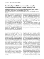
As shown in Figure 1, despite the fact that the PDI gene
expression in hepatic tissues decreased by 19% at 5 hours
after CLP, such a decrease was not statistically significant. In
contrast, hepatic PDI gene expression decreased by 28% at
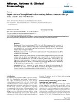
20 hours after CLP (P < 0.05, Figure 1). At 20 hours after the
continuous infusion of LPS (1 μg/kg BW) in normal rats, the
hepatic PDI gene expression markedly decreased by 69% (P
< 0.05, Figure 2). This suggests that LPS may be responsible
for the downregulation of the PDI gene expression observed
20 hours after the onset of sepsis. In cells of the cultured mac-
rophage-like cell line, RAW 264.7, the PDI protein expression
Figure 1
Alterations in the protein disulfide isomerase (PDI) gene expression in hepatic tissues at 5 and 20 hours after cecal ligation and puncture (CLP)Alterations in the protein disulfide isomerase (PDI) gene expression in
hepatic tissues at 5 and 20 hours after cecal ligation and puncture
(CLP). The ratio of PDI and the housekeeping gene glyceraldehyde 3-
phosphate dehydrogenase (G3PDH) is calculated. Values (n = 4 to 5/
group) are presented as mean ± standard error and are compared by
one-way analysis of variance and Tukey's test: *P < 0.05 versus
respective sham-operated animals.
Critical Care Vol 12 No 4 Zhou et al.
Page 4 of 8
(page number not for citation purposes)
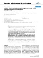
was also significantly reduced (by 33%) after incubation with
LPS (100 ng/mL) for 4 hours (Figure 3).
Effects of protein disulfide isomerase inhibition on
tumor necrosis factor-alpha gene expression and
production in RAW 264.7 cells
To investigate the role of PDI in the regulation of proinflamma-
tory cytokine TNF-
α
, we incubated RAW 264.7 cells with a
specific PDI inhibitor, bacitracin (24-hour culture). Figure 4
shows the effect of bacitracin on the TNF-
α
gene expression
in RAW 264.7 cells. Bacitracin significantly increased TNF-
α
gene expression in a dose-dependent manner. The TNF-
α
gene expression was increased by 33%, 84%, and 93% at
0.25, 1.25, and 3.75 mM bacitracin, respectively (Figure 4).
Alterations in the supernatant and cellular TNF-
α
levels in cells
cultured with bacitracin are shown in Figures 5 and 6. As
shown in Figure 5, the supernatant levels of TNF-
α
signifi-
cantly increased (by 55%) at 0.25 mM bacitracin and further
increased by 317% and 327% at the higher concentrations,
1.25 and 3.75 mM, respectively (Figure 5). Similarly, cellular
concentrations of TNF-
α
were markedly elevated by bacitracin
in the range of 12- to 54-fold in a dose-response fashion (Fig-
ure 6).
Effect of protein disulfide isomerase inhibition by short
interfering RNA on tumor necrosis factor-alpha gene
expression and release in RAW 264.7 cells
To further confirm the role of PDI in the regulation of proinflam-
matory cytokine TNF-
α
, RAW 264.7 cells were transfected
with PDI siRNA for 48 hours and TNF-
α
release into the cell
supernatant was assessed. Transfection with 100 nM PDI
siRNA produced an average 36.8% inhibition of the PDI gene
expression (Figure 7a, P < 0.001). Interestingly, the PDI
downregulation by siRNA caused a 3.19-fold increase in TNF-
α release (Figure 7b, P < 0.001).
Figure 2
Alterations in the protein disulfide isomerase (PDI) gene expression in hepatic tissues after continuous infusion of lipopolysaccharide (LPS) or normal saline (control)Alterations in the protein disulfide isomerase (PDI) gene expression in
hepatic tissues after continuous infusion of lipopolysaccharide (LPS) or
normal saline (control). The ratio of PDI and the housekeeping gene
glyceraldehyde 3-phosphate dehydrogenase (G3PDH) is calculated.
Values (n = 4 to 6/group) are presented as mean ± standard error and
are compared by Student t test: *P < 0.05 versus control.
Figure 3
Alterations in the protein disulfide isomerase (PDI) protein expression in RAW 264.7 cells after stimulation of lipopolysaccharide (LPS) (100 ng/mL) for 4 hoursAlterations in the protein disulfide isomerase (PDI) protein expression
in RAW 264.7 cells after stimulation of lipopolysaccharide (LPS) (100
ng/mL) for 4 hours. The ratio of PDI and the housekeeping gene β-actin
is calculated. Values (n = 4/group) are presented as mean ± standard
error and are compared by Student t test: *P < 0.05 versus control.
Figure 4
Alterations in tumor necrosis factor-alpha (TNF-
α
) gene expression in RAW 264.7 cells after culture with bacitracin (0.25, 1.25, and 3.75 mM) for 24 hoursAlterations in tumor necrosis factor-alpha (TNF-
α
) gene expression in
RAW 264.7 cells after culture with bacitracin (0.25, 1.25, and 3.75
mM) for 24 hours. The ratio of TNF-
α
and the housekeeping gene β-
actin is calculated. Values (n = 4 to 5/group) are presented as mean ±
standard error and are compared by one-way analysis of variance and
Tukey's test: *P < 0.05 versus control;
#
P < 0.05 versus 0.25 mM
bacitracin.
Available online />Page 5 of 8
(page number not for citation purposes)
Discussion
The notion that reduced/denatured proteins would spontane-
ously reoxidize and refold to form their native conformation led
to the search for a physiological catalyst of this process. An
enzyme was found that catalyzed the formation of native pro-
teins from the reduced/denatured state and has been termed
as PDI [38]. PDI is widely distributed and has been detected
in most vertebrate tissues, although detailed studies have
been confined to the enzyme from the liver. In the mammalian
liver homogenates, PDI is found in crude microsomal
membrane fractions [39]. In the rat liver, the enzyme co-sedi-
ments with markers of the endoplasmic reticulum [38]. PDI is
a membrane-associated enzyme of the endoplasmic reticulum
and its function, in part, is translational modification of proteins
[40]. PDI may also catalyze the covalent crosslinking of native
proteins or the covalent immobilization of biologically active
molecules to the extracellular matrix.
In the present study, by using animal models of sepsis or endo-
toxemia, we have shown that the PDI gene expression is
decreased at 20 hours after CLP or LPS infusion. Similarly,
PDI gene expression is downregulated in a macrophage-like
cell line after stimulation by LPS for 4 hours. These results indi-
cate that PDI gene expression is downregulated under inflam-
matory conditions and that LPS plays an important role in the
downregulation of PDI. In addition, to evaluate the role of PDI
Figure 5
Alterations in supernatant tumor necrosis factor-alpha (TNF-
α
) levels in RAW 264.7 cells after culture with bacitracin (0.25, 1.25, and 3.75 mM) for 24 hoursAlterations in supernatant tumor necrosis factor-alpha (TNF-
α
) levels in
RAW 264.7 cells after culture with bacitracin (0.25, 1.25, and 3.75
mM) for 24 hours. TNF-
α
levels were determined by enzyme-linked
immunosorbent assay. Values (n = 7 to 8/group) are presented as
mean ± standard error and are compared by one-way analysis of vari-
ance and Tukey's test: *P < 0.05 versus control;
#
P < 0.05 versus 0.25
mM bacitracin.
Figure 6
Alterations in cellular tumor necrosis factor-alpha (TNF-
α
) levels in RAW 264.7 cells cultured with bacitracin (0.25, 1.25, and 3.75 mM) for 24 hoursAlterations in cellular tumor necrosis factor-alpha (TNF-
α
) levels in
RAW 264.7 cells cultured with bacitracin (0.25, 1.25, and 3.75 mM)
for 24 hours. TNF-
α
levels were determined by enzyme-linked immuno-
sorbent assay. Values (n = 7 to 8/group) are presented as mean ±
standard error and are compared by one-way analysis of variance and
Tukey's test: *P < 0.05 versus control;
#
P < 0.05 versus 0.25 mM
bacitracin.
Figure 7
Alterations in the protein disulfide isomerase (PDI) gene expression and supernatant tumor necrosis factor-alpha (TNF-
α
) levels in RAW 264.7 cells transfected with PDI short interfering RNA (siRNA) for 48 hoursAlterations in the protein disulfide isomerase (PDI) gene expression
and supernatant tumor necrosis factor-alpha (TNF-
α
) levels in RAW
264.7 cells transfected with PDI short interfering RNA (siRNA) for 48
hours. (a) PDI gene expression was determined by real-time polymer-
ase chain reaction using specific PDI primers. (b) The TNF-
α
release
into the cell supernatant was measured by enzyme-linked immunosorb-
ent assay. Values (n = 3 to 6/group) are presented as mean ± standard
error and are compared by paired Student t test. *P < 0.05 versus
control.
Critical Care Vol 12 No 4 Zhou et al.
Page 6 of 8
(page number not for citation purposes)
on TNF-
α
gene expression, we have used bacitracin, a
specific inhibitor of PDI, on the TNF-
α
release and the expres-
sion in 24-hour-cultured RAW 264.7 cells. TNF-
α
levels in the
supernatant and cellular TNF-
α
in RAW 264.7 cells cultured
with bacitracin were significantly increased. In addition, we fur-
ther confirmed that downregulation of PDI using PDI siRNA
significantly increased TNF-
α
release from cells. These results
suggest that PDI plays an important role in the production of
proinflammatory cytokine TNF-
α
.
PDI has been found to be secreted from a variety of cell types,
including hepatocytes [41], pancreatic exocrine cells [42],
endothelial cells [43], and activated platelets [44]. While the
biological importance of these secreted proteins remains in
most cases obscure, the function of PDI secreted by
thyrocytes into the lumen of the thyroid follicles has been iden-
tified [45]. It has been shown that the enzyme is involved in the
control of thyroglobulin folding and multimerization, probably
by reducing the intermolecular disulfide bridges and thus lim-
iting the extent of multimer formation. While the full biological
importance of the protein disulfide activity must still be under-
stood, some interesting examples of PDI in pathological
events such as Sindbis virus [46] and HIV [47] have been
demonstrated. It has been suggested that PDI is specifically
upregulated in response to hypoxia/ischemia in astrocytes
[48]. In addition, the overexpression of this gene into neurons
protects against apoptopic cell death induced by hypoxia/
brain ischemia. Further studies by the same group indicate
that ubiquilin, an endoplasmic reticulum-associated protein,
together with PDI, has critical functions as a regulatory protein
for cell death and therefore that upregulation of these proteins
may result in the acquisition of tolerance against ischemic
stress in glial cells [48]. A recent report also indicates that the
transcriptional activity of NF-
κ
B is negatively regulated by PDI
[32]. Overexpression of PDI in RAW 264.7 cells strongly sup-
pressed the LPS-induced production of inflammatory
cytokines as well as NF-
κ
B-dependent luciferase activity. This
negative regulation of NF-
κ
B was reversed by bacitracin, a
PDI inhibitor. Finally, PDI expression was induced by the anti-
inflammatory cytokine IL-10, and IL-10-mediated inhibition of
LPS-induced IL-6 expression was reduced by bacitracin.
These findings clearly demonstrate that PDI is a negative reg-
ulator of NF-
κ
B and may act downstream of IL-10 in this signal
pathway [32].
Our present study with septic rats, in which immunomodula-
tion is known, also indicates that PDI is a regulator of inflam-
matory cytokines. Previous studies have demonstrated that
proinflammatory cytokines play a critical role in the initiation
and progression of sepsis syndrome and that TNF-
α
, IL-1
β
,
and IL-6 are important mediators of hemodynamic, metabolic,
and immunologic alterations in the host during sepsis [27-31].
Studies have also shown that circulating concentrations of
TNF-
α
, IL-1
β
, and IL-6 increase significantly in the early,
hyperdynamic stage of sepsis and remain elevated in the late,
hypodynamic stage of sepsis [27,49]. In the present study, we
have provided a clue that TNF-
α
release increased signifi-
cantly in RAW 264.7 cells treated with bacitracin, which is an
inhibitor of PDI. This result indicates the important role of PDI
in TNF-
α
release in sepsis.
Conclusion
In summary, our results indicate that PDI gene expression is
downregulated in sepsis or endotoxemia. In addition, PDI
gene expression is attenuated in a macrophage-like cell line
after stimulation with LPS. Since the PDI inhibitor bacitracin
significantly increases TNF-
α
release in a macrophage cell
line, it appears that prevention of PDI downregulation may be
a novel approach to reduce proinflammatory cytokine release
in sepsis. Further studies are necessary in this direction.
Competing interests
The authors declare that they have no competing interests.
Authors' contributions
MZ designed the study, collected data, interpreted the data,
performed statistical analysis, and drafted the manuscript. NH
is a summer student who helped MZ to collect the data. MM
and RW participated in the design of the study. SRM and AJ
participated in the critical revision of the manuscript. PW con-
ceived of the study, participated in its design and interpreta-
tion, and helped to draft the manuscript. All authors read and
approved the final manuscript.
Acknowledgements
This study was supported by National Institutes of Health grants R01
GM053008 and R01 GM057468 (PW).
Key messages
• Protein disulfide isomerase (PDI), an important factor
for the protein modification step in the post-translational
event, plays an essential role in cell survival under stress
conditions.
• In an experimental model, PDI gene and protein expres-
sions were significantly downregulated in late sepsis.
• Similar downregulation was also observed in lipopoly-
saccharide-treated RAW 264.7 cells, a macrophage-
like cell line.
• Bacitracin, a specific PDI inhibitor, significantly
increased tumor necrosis factor-alpha (TNF-
α
) gene
expression and TNF-
α
release as well as its cellular lev-
els in a dose-dependent manner.
• Collectively, the data suggest that prevention of down-
regulation of PDI in sepsis attenuates hyperinflamma-
tion and reduces tissue injury.
Available online />Page 7 of 8
(page number not for citation purposes)
References
1. Bernard GR, Vincent JL, Laterre PF, LaRosa SP, Dhainaut JF,
Lopez-Rodriguez A, Steingrub JS, Garber GE, Helterbrand JD, Ely
EW, Fisher CJ Jr: Efficacy and safety of recombinant human
activated protein C for severe sepsis. N Engl J Med 2001,
344:699-709.
2. Martin GS, Mannino DM, Eaton S, Moss M: The epidemiology of
sepsis in the United States from 1979 through 2000. N Engl J
Med 2003, 348:1546-1554.
3. Hotchkiss RS, Karl IE: The pathophysiology and treatment of
sepsis. N Engl J Med 2003, 348:138-150.
4. Martin GS, Mannino DM, Moss M: The effect of age on the devel-
opment and outcome of adult sepsis. Crit Care Med 2006,
34:15-21.
5. Angus DC, Linde-Zwirble WT, Lidicker J, Clermont G, Carcillo J,
Pinsky MR: Epidemiology of severe sepsis in the United
States: analysis of incidence, outcome, and associated costs
of care. Crit Care Med 2001, 29:1303-1310.
6. Deitch EA: Animal models of sepsis and shock: a review and
lessons learned. Shock 1998, 9:1-11.
7. Remick DG, Bolgos GR, Siddiqui J, Shin J, Nemzek JA: Six at six:
interleukin-6 measured 6 h after the initiation of sepsis pre-
dicts mortality over 3 days. Shock 2002, 17:463-467.
8. Zingarelli B, Sheehan M, Hake PW, O'Connor M, Denenberg A,
Cook JA: Peroxisome proliferator activator receptor-gamma
ligands, 15-deoxy-delta(12,14)-prostaglandin J2 and ciglita-
zone, reduce systemic inflammation in polymicrobial sepsis
by modulation of signal transduction pathways. J Immunol
2003, 171:6827-6837.
9. Chung CS, Song GY, Lomas J, Simms HH, Chaudry IH, Ayala A:
Inhibition of Fas/Fas ligand signaling improves septic survival:
differential effects on macrophage apoptotic and functional
capacity. J Leukoc Biol 2003, 74:344-351.
10. Chaudry IH: Sepsis: lessons learned in the last century and
future directions. Arch Surg 1999, 134:922-929.
11. Wang P, Chaudry IH: Mechanism of hepatocellular dysfunction
during hyperdynamic sepsis. Am J Physiol
1996,
270:R927-R938.
12. Wang H, Liao H, Ochani M, Justiniani M, Lin X, Yang L, Al Abed Y,
Wang H, Metz C, Miller EJ, Tracey KJ, Ulloa L: Cholinergic ago-
nists inhibit HMGB1 release and improve survival in experi-
mental sepsis. Nat Med 2004, 10:1216-1221.
13. Wichterman KA, Baue AE, Chaudry IH: Sepsis and septic shock:
a review of laboratory models and a proposal. J Surg Res
1980, 29:189-201.
14. Yang S, Cioffi WG, Bland KI, Chaudry IH, Wang P: Differential
alterations in systemic and regional oxygen delivery and con-
sumption during the early and late stages of sepsis. J Trauma
1999, 47:706-712.
15. Wang P, Ba ZF, Chaudry IH: Hepatocellular dysfunction occurs
earlier than the onset of hyperdynamic circulation during
sepsis. Shock 1995, 3:21-26.
16. Wang P, Ba ZF, Chaudry IH: Hepatic extraction of indocyanine
green is depressed early in sepsis despite increased hepatic
blood flow and cardiac output. Arch Surg 1991, 126:219-224.
17. Wang P, Zhou M, Rana MW, Ba ZF, Chaudry IH: Differential
alterations in microvascular perfusion in various organs dur-
ing early and late sepsis. Am J Physiol 1992, 263:G38-G43.
18. Wang P, Ba ZF, Chaudry IH: Mechanism of hepatocellular dys-
function during early sepsis: key role of increased gene
expression and release of proinflammatory cytokines tumor
necrosis factor and interleukin-6. Arch Surg 1997,
132:364-370.
19. Koo DJ, Chaudry IH, Wang P: Kupffer cells are responsible for
producing inflammatory cytokines and hepatocellular dys-
function during early sepsis. J Surg Res 1999, 83:151-157.
20. Yang S, Zhou M, Fowler DE, Wang P: Mechanisms of the bene-
ficial effect of adrenomedullin and adrenomedullin-binding
protein-1 in sepsis: down-regulation of proinflammatory
cytokines. Crit Care Med 2002, 30:2729-2735.
21. Darby NJ, Creighton TE: Functional properties of the individual
thioredoxin-like domains of protein disulfide isomerase. Bio-
chem 1995, 34:11725-11735.
22. Freedman RB, Hirst TR, Tuite MF: Protein disulphide isomerase:
building bridges in protein folding.
Trends Biochem Sci 1994,
19:331-336.
23. Frand AR, Kaiser CA: Ero1p oxidizes protein disulfide isomer-
ase in a pathway for disulfide bond formation in the endoplas-
mic reticulum. Mol Cell 1999, 4:469-477.
24. Cabibbo A, Pagani M, Fabbri M, Rocchi M, Farmery MR, Bulleid
NJ, Sitia R: ERO1-L, a human protein that favors disulfide bond
formation in the endoplasmic reticulum. J Biol Chem 2000,
275:4827-4833.
25. Mikami T, Genma R, Nishiyama K, Ando S, Kitahara A, Natsume H,
Yoshimi T, Horiuchi R, Nakamura H: Alterations in the enzyme
activity and protein contents of protein disulfide isomerase in
rat tissues during fasting and refeeding. Metabolism 1998,
47:1083-1088.
26. Turano C, Coppari S, Altieri F, Ferraro A: Proteins of the PDI fam-
ily: unpredicted non-ER locations and functions. J Cell Physiol
2002, 193:154-163.
27. Ertel W, Morrison MH, Wang P, Ba ZF, Ayala A, Chaudry IH: The
complex pattern of cytokines in sepsis. Association between
prostaglandins, cachectin, and interleukins. Ann Surg 1991,
214:141-148.
28. Waage A, Halstensen A, Espevik T: Association between tumour
necrosis factor in serum and fatal outcome in patients with
meningococcal disease. Lancet 1987, 1:355-357.
29. Debets JM, Kampmeijer R, Linden MP van der, Buurman WA,
Linden CJ Van Der: Plasma tumor necrosis factor and mortality
in critically ill septic patients. Crit Care Med 1989, 17:489-494.
30. Marano MA, Fong Y, Moldawer LL, Wei H, Calvano SE, Tracey KJ,
Barie PS, Manogue K, Cerami A, Shires GT: Serum cachectin/
tumor necrosis factor in critically ill patients with burns corre-
lates with infection and mortality. Surg Gynecol Obstet 1990,
170:32-38.
31. Busund R, Lindsetmo RO, Rasmussen LT, Rokke O, Rekvig OP,
Revhaug A: Tumor necrosis factor and interleukin 1 appear-
ance in experimental gram-negative septic shock. The effects
of plasma exchange with albumin and plasma infusion. Arch
Surg 1991, 126:591-597.
32. Higuchi T, Watanabe Y, Waga I: Protein disulfide isomerase
suppresses the transcriptional activity of NF-kappaB. Bio-
chem Biophys Res Commun 2004, 318:
46-52.
33. Kaiser BK, Yim D, Chow IT, Gonzalez S, Dai Z, Mann HH, Strong
RK, Groh V, Spies T: Disulphide-isomerase-enabled shedding
of tumour-associated NKG2D ligands. Nature 2007,
447:482-486.
34. Short DM, Heron ID, Birse-Archbold JL, Kerr LE, Sharkey J, McCul-
loch J: Apoptosis induced by staurosporine alters chaperone
and endoplasmic reticulum proteins: identification by quanti-
tative proteomics. Proteomics 2007, 7:3085-3096.
35. Wang P, Chaudry IH: A single hit model of polymicrobial sep-
sis: cecal ligation and puncture. Sepsis 1998, 2:227-233.
36. Zhou M, Simms HH, Wang P: Adrenomedullin and adrenom-
edullin binding protein-1 attenuate vascular endothelial cell
apoptosis in sepsis. Ann Surg 2004, 240:321-330.
37. Zhou M, Wu R, Dong W, Jacob A, Wang P: Endotoxin downreg-
ulates peroxisome proliferator-activated receptor-{gamma}
via the increase in TNF-{alpha} release. Am J Physiol Regul
Integr Comp Physiol 2008, 294:R84-R92.
38. Ibbetson AL, Freedman RB: Thiol-protein disulphide oxidore-
ductases. Assay of microsomal membrane-bound glutath-
ione-insulin transhydrogenase and comparison with protein
disulphide-isomerase. Biochem J 1976, 159:377-384.
39. Goldberger RF, Epstein CJ, Anfinsen CB: Acceleration of reacti-
vation of reduced bovine pancreatic ribonuclease by a micro-
somal system from rat liver. J Biol Chem 1963, 238:628-635.
40. Freedman RB: Protein disulfide isomerase: multiple roles in
the modification of nascent secretory proteins. Cell 1989,
57:1069-1072.
41. Terada K, Manchikalapudi P, Noiva R, Jauregui HO, Stockert RJ,
Schilsky ML: Secretion, surface localization, turnover, and
steady state expression of protein disulfide isomerase in rat
hepatocytes. J Biol Chem 1995, 270:20410-20416.
42. Yoshimori T, Semba T, Takemoto H, Akagi S, Yamamoto A, Tashiro
Y: Protein disulfide-isomerase in rat exocrine pancreatic cells
is exported from the endoplasmic reticulum despite possess-
ing the retention signal. J Biol Chem 1990, 265:15984-15990.
43. Hotchkiss KA, Matthias LJ, Hogg PJ: Exposure of the cryptic Arg-
Gly-Asp sequence in thrombospondin-1 by protein disulfide
isomerase. Biochim Biophys Acta 1998, 1388:478-488.
Critical Care Vol 12 No 4 Zhou et al.
Page 8 of 8
(page number not for citation purposes)
44. Chen K, Lin Y, Detwiler TC: Protein disulfide isomerase activity
is released by activated platelets. Blood 1992, 79:2226-2228.
45. Delom F, Mallet B, Carayon P, Lejeune PJ: Role of extracellular
molecular chaperones in the folding of oxidized proteins.
Refolding of colloidal thyroglobulin by protein disulfide iso-
merase and immunoglobulin heavy chain-binding protein. J
Biol Chem 2001, 276:21337-21342.
46. Abell BA, Brown DT: Sindbis virus membrane fusion is medi-
ated by reduction of glycoprotein disulfide bridges at the cell
surface. J Virol 1993, 67:5496-5501.
47. Ryser HJ, Levy EM, Mandel R, DiSciullo GJ: Inhibition of human
immunodeficiency virus infection by agents that interfere with
thiol-disulfide interchange upon virus-receptor interaction.
Proc Natl Acad Sci USA 1994, 91:4559-4563.
48. Ko HS, Uehara T, Nomura Y: Role of ubiquilin associated with
protein-disulfide isomerase in the endoplasmic reticulum in
stress-induced apoptotic cell death. J Biol Chem 2002,
277:35386-35392.
49. Blackwell TS, Christman JW: Sepsis and cytokines: current
status. Br J Anaesth 1996, 77:110-117.