Báo cáo y học: "Insights gained from the reverse engineering of gene networks in keloid fibroblasts" pdf
Bạn đang xem bản rút gọn của tài liệu. Xem và tải ngay bản đầy đủ của tài liệu tại đây (464.63 KB, 17 trang )
RESEARCH Open Access
Insights gained from the reverse engineering of
gene networks in keloid fibroblasts
Brandon NS Ooi
1*
and Toan Thang Phan
2
* Correspondence:
1
Graduate Programme in
Bioengineering, National University
of Singapore, Singapore
Full list of author information is
available at the end of the article
Abstract
Background: Keloids are protrusive claw-like scars that have a propensity to recur
even after surgery, and its molecular etiology remains elusive. The goal of reverse
engineering is to infer gene networks from observational data, thus providing insight
into the inner workings of a cell. However, most attempts at modeling biological
networks have been done using simulated data. This study aims to highlight some
of the issues involved in working with experimental data, and at the same time gain
some insights into the transcriptional regulatory mechanism present in keloid
fibroblasts.
Methods: Microarray data from our previous study was combined with microarray
data obtained from the literature as well as new microarray data generated by our
group. For the physical approach, we used the fREDUCE algorithm for correlating
expression values to binding motifs. For the influence approach , we compared the
Bayesian algorithm BANJO with the information theoretic method ARACNE in terms
of performance in recovering known influence networks obtained from the KEGG
database. In addition, we also compared the performance of different normalization
methods as well as different types of gene networks.
Results: Using the physical approach, we found cons ensus sequences that were
active in the keloid condition, as well as some sequences that were responsive to
steroids, a commonly used treatment for keloids. From the influence approach, we
found that BANJO was better at recovering the gene networks compared to ARACNE
and that transcriptional networks were better suited for network recovery compared
to cytokine-receptor interaction networks and intracellular signaling networks. We
also found that the NFKB transcriptional network that was inferred from normal
fibroblast data was more accurate compared to that inferred from keloid data,
suggesting a more robust network in the keloid condition.
Conclusions: Consensus sequences that were found from this study are possible
transcription factor binding sites and could be explored for developing future keloid
treatments or for improving the efficacy of current steroid treatments. We also found
that the combination of the Bayesian algorithm, RMA normalization and
transcriptional networks gave the best reconstructio n results and this could serve as
a guide for future influence approaches dealing with experimental data.
Ooi and Phan Theoretical Biology and Medical Modelling 2011, 8:13
/>© 2011 Ooi and Phan; licensee BioMed Central Ltd. This is an Open Access article distributed under the terms of the Creative
Commons Attribution License ( which permits unrest ricted use, distribution, and
reproduction in any medium, provided the original work is properly cited.
Background
Keloids are large protruding claw-like scars that extend well beyond the confines of the
original wound and do not subside with time [1]. They uniquely affect only humans,
and may develop even after the most minor of skin wounds, such as insect bites or
acne [2]. Keloids are frequently associated with itchiness, pain and, when involving the
skin overlying a joint, restricted range of motion [3]. It is not well documented how
commonly keloids occur in the general population but the reported incidence range
from a high of 16% among adults in Zaire to a low of less than 1% among adults in
England [4]. In a study assessing the quality of life of patients with keloid and hyper-
trophic scarring, it was demonstrated for the first time that the quality of life of these
patients was reduced due t o physical and/or psychological effects [5]. The problem is
further exacerbated by the fact that there is no particularly effective treatment to date
[6,7]. Keloids also have a propensity to recur after surgery and have been considered as
benign tumours [4].
The goal of reverse engineering methods is to infer gene networks from observa-
tional data, thus providing insight into the inner workings of a cell [8,9]. There are two
general strategies for reve rse engineering gene networks - a physical approach whe re
physical interactions between transcription factors (TFs) and their promoters are mod-
eled, and an influence approach where the mechanistic process is abstracted out as a
black box [10]. The advantage of the physical approach is that it enables the use of
genome sequence data, in combination with RNA expression data, to enhance the sen-
sitivity and specificity of predicted interactions, but its limitation is that it cannot
describe regulatory control by mechanisms other than transcription factors. On the
other hand, an advantage of the influence strategy is that the model can implicitly cap-
ture regulatory mechanisms at the protein and metabolite level that are not physically
measur ed, but the limitation is that it can be difficult to interpret in terms of the phy-
sical structure of the cell. Moreover, the implicit description of hidden regulatory fac-
tors may lead to prediction errors [10].
In addition to these two modeling approaches, reverse en gineering methods also dif-
fer in terms of the mathematical formalisms used and can be static or dynamic, contin-
uous or discrete, linear or nonlinear and deterministic or stochastic [ 11]. For the
purposes of this study, we have chosen to use both the physical as well as the influence
approach for reconstructing the networks. For the physical approach, we will use the
regression method fREDUCE (fast-Regulatory Element Detection Using Correlation
with Expression) [12] with the objective of identifying important cis-binding motifs
and their targets in keloid fibroblasts. For the influence approach, we will compare the
performance of the information theoretic method ARACNE (Algorithm for the Recon-
struction of Accurate Cellular Networks) [13] and the Bayesian package BANJO (Baye-
sian Network Inference with Java Objects) [14] in uncovering regulatory interactions in
keloid and normal fibroblasts. The effect of different normalization/ summarization
methods and lowly expressed probes on gene network inference will also be examined
in this system.
Microar ray data from previous studies will be used to learn the networks. However,
learning the structure of a gene network using the influence approach is difficult as
the number of possibilities scale exponentially with the number of variables. Therefore,
modeling and testing such large structures would require large amounts o f data for
Ooi and Phan Theoretical Biology and Medical Modelling 2011, 8:13
/>Page 2 of 17
accuracy. Due to our limite d data, we have decided to focus on small networks of
genes that have been found to be differentially expressed from ou r previous wo rk.
Furthermore, to increase the number of samples, we will also use data from Smith et
al [15], which is the only keloid fibroblast data publicly available at the Gene Expres-
sion Omnibus (GEO) database. For the physical approach, since the binding motif
repeats are regressed against the expression levels of each gene, it is the number of
genes that constitute the sample size. Therefore, the full range of genes is used for this
approach instead of the smaller transcriptional networks that have found to be differ-
entially expressed.
In total, we have four different treatment conditions (serum-treated, serum-free,
hydrocortisone-treated and HDGF-treated) and two different cell derivations (keloid
and normal) from multiple patients. Although some of our datasets consist of time-ser-
ies data, the gap between each time point is very large (in the order of days) and may
lead to inaccurate results if used to infer time-series regulatory networks. Therefore,
we have limited our study to steady state conditions with the assumption that each
time point is statis tically independ ent from others. This is a possibly valid assumption
as the sampling time is very long. Furthermore, the genes were not directly perturbed
by knockdown or overexpression in our experiments and it is very likely that the dif-
ferent conditions used will result in multiple unknown perturbations. As such, infer-
ence algorithms such as dynamic Bayesian networks (which require numerous closely
spaced time points) and differential equation approaches (which require either time
series data or knowledge of perturbations) cannot be applied in our case.
To date, most attempts at modeling biological networks have been done using simu-
lated data. We hope that this work would highlight some of the issues involved in
working with experimental data. Furthermore, we also hope that insights gained from
this endeavor would provide some clues about the different transcriptional r egulatory
mechanisms present in keloid and normal fibroblasts.
Methods
Keloid and normal fibroblast database
Keloid and normal fibroblasts were selected from a specimen bank of fibroblast strains
derived from excised keloid specimens. All patients had received no previous treatment
for the keloids before surgical excision. A full history was taken and an examination
performed, complete with color slide photographic documentation before taking
informed consent prior to excision. Approval by the NUS Institutional Review Board,
NUS-I RB was sought before excision of human tissue and collection of ce lls. Remnant
dermis from keloid or normal skin was minced and incubated in a solution of collage-
nase type I (0.5 mg/ml) and tryp sin (0.2 mg/ml) for 6 h at 37°C. The cells were pel-
leted and grown in tissue culture flasks. The cell strains were maintained and stored in
liquid nitrogen until use.
Cell culture
Five different keloid fibroblast samples and five different normal fibroblast samples that
were previously maintained and stored at -150°C were thawed and used for the experi-
ments. Fibroblasts were seeded in 15 cm dishes at a density of 1 × 10
4
cells/ml in 10%
FCS until confluenc y and subsequently starved in a serum-free medium for 48 hrs.
Ooi and Phan Theoretical Biology and Medical Modelling 2011, 8:13
/>Page 3 of 17
After 48 hrs, the serum free medium was replaced and fibroblasts were harvested after
another 24 hrs (day 1), 72 hrs (day 3) and 120 hrs (day 5). Cells were grown and pro-
cessed in five batches. Each batch consisted of one keloid and one normal sample har-
vested at the three different time points. KF1, NF1, KF2, NF2, KF4, NF4 and KF5, NF5
were samples from different patients while KF3 and NF3 were s amples from the same
patient. In another experiment, one keloid fibroblast sample was grown, treated with
hepatoma derived growth factor (HDGF) and harvested for RNA after 6 hours, day 1
and day 2.
RNA extraction, cRNA preparation and labeling
RNA was extracted using the RNeasy-kit (Qiagen, Hilden, Germany) according to the
manufacturer’s protocol. Purified RNA was quantified by UV absorbance at 260 and
280 nm on a ND1000 spectrophotometer (Nanodrop™, ThermoScientific). Labeled
complementary RNA (cRNA) was produced from total RNA using the GeneChip One-
Cycle or Two-Cycle Eukaryotic Target Labeling and Control Reagents (Affymetrix,
Santa Clara, USA) according to the manufacturer’s protocol.
Affymetrix chip hybridization and scanning
Fragmented cRNA was then hybridized to preequilibrated Affymetrix GeneChip
U133A or the newer Genechip U133 2.0 Plus arrays at 45 °C for 15 hours. The cock-
tails were removed after hybridization and the chips were washed an d stained using
Affymetrix wash buffers and stain cocktails in an automated fluidic station. The chips
were then scanned in a Hewlett-Packard ChipScanner (Affymetrix, Santa Clara, USA)
to detect hybridization signals.
Data preprocessing
In addition to microarray data generated by our lab, raw microarray data in the form
of .CELS files from Smith et al’s experiments were also downloaded from the GEO
database [15]. Following data collection, RMA and MAS 5.0 normalization and sum-
marization were done using the R Bioconductor package. The four different datasets
(serum starvation dataset using U133A arrays, serum starvation dataset using U133
Plus 2.0 arrays, HDGF dataset using U133 Plus 2.0 arrays and Smith’s dataset using
U133 Plus 2.0 arrays) were normalized and summarized independently. Two different
custom Chip Definition Files (CDF) were used [16]. T he first CDF was based on the
Ensembl Gene database for analysis with fREDUCE as it is easy to obtain the upstream
sequence which is requir ed by fREDUCE from the Ensembl database. The second was
based on the Entrez Gene database for influence based reverse engineering methods
such as BANJO and ARACNE as these probe mappings allow one to ignore any differ-
ential signal due to multiple probesets and gives a single value for a given gene. In
addition, two lists were produced. In the first list, no filtering was done while in the
second list, 25% of the lowly expressed genes were filtered.
Application of the fREDUCE algorithm
Human genomic sequences 1000 base pairs upstream from the transcriptional start site
if known, or from the initiation codon, were extracted from the Ensembl database [17].
As fREDUCE requires only a single expression dataset and makes use of the entire
Ooi and Phan Theoretical Biology and Medical Modelling 2011, 8:13
/>Page 4 of 17
genomic dataset (both signal and background), the datasets were compared as follows :
A: Keloid versus normal fibroblasts under serum starvation conditions (only KF1, KF2,
NF1 and NF2 were used to keep the number of samples close to the other conditions),
B: Keloid versus normal fibroblasts under serum conditions (from Smith et al’sdata-
set), C: Keloid treated with steroid versus serum induced keloid fibroblasts (from
Smith et al ’s dataset), D: Normal tr eated with steroid versus serum induced normal
fibroblasts (from Smith et al’s dataset), E: Keloid versus normal fibroblasts both treated
with steroid (from Smith et al’ s dataset) and F: Keloid treated with HDGF versus
untreated keloid fibroblasts (from HDGF dataset). The expression value for each gene
is represented as the following t-statistic:
t
g
=
µ
e
g
− µ
c
g
Var
e
g
n
e
+
Var
c
g
n
c
where g is the index over genes, μ
e
g
is the mean value of gene g under our condition
of interest, μ
c
g
os the mean value of gene g under control conditions, Var
e
g
is the var-
iance of gene g under our condition of interest, Var
c
g
is the variance of gene g u nder
control conditions, and n
e
and n
c
are the number of sa mples under our condition of
interest and under control conditions respectively. This statisitic is similar to the z-sta-
tistic used by the fREDUCE creators [14]. We then ran fREDUCE on the t-statistic for
RMA normalized and MAS 5.0 normalize d as well as unfiltered and filtered gene l ists
on the basis that a higher t-statistic translat es to higher expression. Four different sets
of parameters were run on each replicate: length 6 with 0 IUPAC substitutions, length
6 with 1 IUPAC substitution, length 7 with 0 IUPAC substitutions and length 7 with 1
IUPAC substitution. Top and consistent binding sequences obtained from fREDUCE
above were then searched through the TRANSFAC database [18] for possible gene tar-
gets and their corresponding transcription factors. Only gene targets identified from
Homo sapiens were collected, and binding sites for all these targets were reconfi rmed
to be located within the 1000 base pair upstream sequences collected from the Ensem-
ble database previously.
Pathways selected for influence approach
KEGG pathways that were found to be enriched when comparing keloid to normal
fibroblasts from a previous study were used for the influence approach (unpublished
data). These were the a ntigen presentation and processing pathway, cytokine-cytokine
receptor interacti on and toll-li ke receptor signaling pathway. Genes that were used as
nodes for modeling were chosen on the basis that there is only one gene representing
that particular node, all other genes will be assumed to be hidden nodes. The following
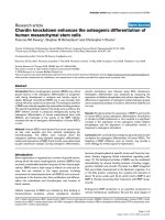
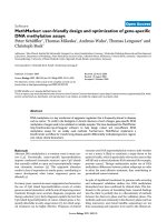
5 pathways were eventually selected for the influence approach (Figure 1). Pathways
were also chosen such that 1A and 1B represent cytokine recep tor interactions, 1C, 1E
and 1G represent transcriptional networks and 1D and 1F represent intracellular
signaling.
Application of the ARACNE and BANJO algorithms
Expression values of selected genes from all the different data sets available were used
for the influence approach. To enable comparison between the different data sets, gene
Ooi and Phan Theoretical Biology and Medical Modelling 2011, 8:13
/>Page 5 of 17
STAT1
MIG
I-TAC
IP-10
IRF3
G
CXCL1
CXCL2
CXCL7
CXCL6
IL8
CXCL3
CXCL5
IL8RA
IL8RB
CXCL9
CXCL10
CXCL11
CXCR3
CREB
B2M
CIITA
Ii
A
B
C
TLR1
RAC1
NFKB
TNFA
IL1B
IKBA
IL8
RANTES
IL6
TLR2
NFKB
E
TLR3
TRIF
TRAF3
TBK1
IRF3
F
D
Figure 1 KEGG pathways used for the influence approach. (A and B) Pathways taken from the
cytokine-cytokine receptor interaction map. (C) Transcriptional pathway taken from antigen processing and
presentation map. (D, E, F and G) Pathways taken from the toll-like receptor signaling map.
Ooi and Phan Theoretical Biology and Medical Modelling 2011, 8:13
/>Page 6 of 17
expression for all the relevant nodes were normalized using the average of GAPDH
and B-actin expression. GAPDH and B-actin were fir st plotted to determi ne their cor-
relation and outliers were removed from the dataset. Three keloid experiments from
the serum starvation U133A dataset di d not meet this criteria and was removed giving
a total of 28 keloid experiments and 24 normal experiments. We ran ARACNE and
BANJO on the keloid and normal inputs separately, and also on the MAS 5 and RMA
normalized expression values separately. All parameters were left at their default
values. For ARACNE, kernel width and number of bins were automatically detected by
the software while DPI tolerance to remove false positives was set at 0.15. For BANJO,
the Proposer/Searcher strategies were chosen as random local move and simulated
annealing, respectively, and the amount of time BANJO uses to explore the Bayesian
Network space was se t to one minute. All the other parameters such as reannealing-
Temperature, coolingFactor, and so on, were left with their default values. Parameter
values were selected as bes t values (in terms of network inference accuracy) as shown
by Bansal et al [19]. In order to estimate the joint probability distribution of all vari-
ables in the network, BANJO requires discrete data. The data was therefore discretized
into 7 discrete states using the quantile discretization procedure in the software.
Furthermore, as the simulated annealing algorithm in BANJO does not guarantee a
global maximum, the runs were repeated three times and the result with the hi ghest
maximum score was taken.
Estimation of the performance of the algorithms
In order to assess the inference performances we computed the Positive Predicted
Value (PPV) and the Sensitivity scores as described by Bansal et al [19]. The following
definitions were used:
TP = Number of True Positives = number of edges in the real network that are cor-
rectly inferred; FP = Number of False Positives = number of inferred edges that are
not in the real network; FN = Number of False Negatives = number of edges in the
real network that are not inferred.
The following were then computed:
PPV =
TP
TP + FP
Sensitivity =
TP
TP + F
N
In order to compute the random PPV we considered the expected value of a hyper-
geometrically distributed random variable whose distribution function and expected
value are, respectively:
P
x
=
M
C
X
N−M
C
n−x
N
C
x
E[x]=M
N−1
C
n−1
N
C
n
= M
n
N
where N = number of possible edges in the network, M = number of true edges and
n = number of predicted edges. Then,
Ooi and Phan Theoretical Biology and Medical Modelling 2011, 8:13
/>Page 7 of 17
PPV
rand
=
TP
rand
TP + FP
=
E[x]
n
=
M
N
All statistical tests are done using the one tailed paired t-test.
Results
Binding motifs found from fREDUCE for keloid versus normal fibroblasts under serum
starvation condition
Binding motifs found using the gene expression values from set A (keloid versus nor-
mal fib roblasts under serum starvation conditions)areshowninTable1.Highlighted
motifs indicate top motifs or motifs found in at least two variations of the conditions /
parameters. Both MAS5 and RMA normalization as well as filtered and unfiltered gene
lists provided hits for the binding motifs. Of particular note are the binding motifs
CGCCGA (found in 5 of the conditions), GCCGAC (found in 3 of the conditions), and
CACATAT (found in 3 of the conditions). A search through the TRANSFAC database
did not produce any results for the binding motif CACATAT, but found possible gene
targets for CGCCGA (MYB) and GCCGAC (ATF2) (Table 2).
Binding motifs found from fREDUCE for keloid versus normal fibroblasts under serum
induced condition
No binding motifs were found for unfiltered RMA normalized set B (keloid versus nor-
mal fibrob lasts under serum conditions), but binding mo tifs were found for the other
conditions (Table 3). Of particular note is the binding motif GGGGCTC which was
found to be consistent in 4 of the conditions, although all these 4 conditions were
Table 1 Binding motifs found from fREDUCE for keloid versus normal fibroblasts under
serum starvation condition (P > 1.3)
Normalization Parameters Binding Motif P-value Correlation
MAS 5
(unfiltered)
Length 7
(0 IUPAC)
CCGGCC 5.31 0.0558
GCCGAC 1.99 0.0432
Length 7
(1 IUPAC)
CGCBGA 5.30 0.0605
MCGGAA 1.42 0.0469
RMA
(unfiltered)
Length 7
(0 IUPAC)
GCCGAC 3.35 0.0487
CACATAT 2.56 -0.0480
Length 7
(1 IUPAC)
GBCGAC 3.56 0.0549
CACATAT 2.02 -0.0470
MAS 5
(filtered)
Length 7
(0 IUPAC)
CGCCGA 2.86 0.0616
Length 7
(1 IUPAC)
CGCCBA 3.65 0.0726
RMA
(filtered)
Length 7
(0 IUPAC)
CGCCGA 2.58 0.0561
TATACAC 1.95 -0.0560
Length 7
(1 IUPAC)
CACAKAT 2.33 -0.0649
CGCCGA 2.03 0.0548
Note: P-values are shown as -log
10
values.
IUPAC characters M = C/A; Y = C/T; K = T/G; B = C/T/G, V = A/C/G
Ooi and Phan Theoretical Biology and Medical Modelling 2011, 8:13
/>Page 8 of 17
using the MAS 5 normalization. A search through the TRANSFAC database found
ADA as a possible gene with this binding motif (Table 4).
Binding motifs found from fREDUCE for sets C and D suggest consistent effects from
steroid induction for both keloid and normal fibroblasts
Binding motifs were found for set C (keloid treated with steroid versus serum induced
keloid fibroblasts) and D (normal treated with steroid versus serum induced normal fibro-
blasts) when fREDUCE was run using parameters length 6 with 0 IUPAC substitutions.
Other parameters did not produce any results. Furthermore, results were only obtained
when MAS 5 normalization was used. The effect of hydrocortisone appears to be realized
through the binding motifs GGAGGG and GCCCCC and this was consistent for both
keloid (Table 5) and normal (Table 6) fibroblasts. A search through the TRANSFAC data-
base using these binding motifs found a large list of genes containing these binding motifs,
including COL1A2, FN, TGFB1, PDGF1 and IGF2 (Table 7). Of particular note is the fact
that most of the genes found in this list have SP1 as its transcription factor (Table 7).
Not many binding motifs found from fREDUCE for sets E and F
fREDUCE f ound few binding motifs for set E (keloid versus normal fibroblasts both
treated with steroid) and no binding motifs for set F (keloid treated with HDGF versus
Table 2 Possible gene targets and TFs found from the TRANSFAC database for top
binding motifs from Table 1
Binding Motif Possible gene targets Possible TFs
CCGGCC MC2R (melanocortin 2 receptor) SF-1
MT1G (metallothionein 1G) -
EPO (erythropoietin) Tf-LF1 and Tf-LF2
SURF1 and SURF2 (surfeit 1 and 2) YY1
GCCGAC ATF2 (activating transcription factor 2) SP1
CGCCGA c-myb MZF-1
CACATAT
Table 3 Binding motifs found from fREDUCE for keloid versus normal fibroblasts under
serum induced condition (P > 1.3)
Normalization Parameters Binding Motif P-value Correlation
MAS 5
(unfiltered)
Length 7
(0 IUPAC)
CCACACA 2.44 -0.0376
GGGGCTC 2.19 -0.0368
Length 7
(1 IUPAC)
CCACACA 2.14 -0.0376
GGVCTC 1.91 -0.0386
MAS 5
(filtered)
Length 7
(0 IUPAC)
GGGGCTC 2.28 -0.0500
Length 7
(1 IUPAC)
GGGGHTC 2.56 -0.0573
RMA
(filtered)
Length 7
(0 IUPAC)
GCGCCA 2.52 -0.0432
GTCCCG 1.46 -0.0388
Length 7
(1 IUPAC)
GTCVCG 4.29 -0.0545
Note: P-values are show n as -log
10
values.
IUPAC characters R = A/G; W = T/A; H = A/T/C, V = A/C/G
Ooi and Phan Theoretical Biology and Medical Modelling 2011, 8:13
/>Page 9 of 17
untreated keloid fibroblasts). Binding motifs for set E were found only when the MAS
5 unfiltered condition and the RMA filtered condition were used (Tab le 8). Further-
more, binding motifs found in these conditions were not very consistent. A search
through the TRANSFAC database using the top binding motifs from Table 8 found
EGFR, ADM and CGA as possible gene targets (Table 9).
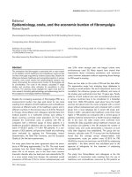
Mean sensitivity performance of BANJO in recovering influence networks was
significantly better than that of ARACNE
On average, BANJO was significantly more sensitive compared to ARACNE in recover-
ing influence networks (Figure 2C). However, there was no significant difference i n
average accuracy (PPV) between BANJO and ARACNE (Figure 2A). Furthermore,
there was n o significant difference between RMA and MAS 5 normalization both in
terms of mean accuracy (PPV) (Figure 2B) as well as mean sensitivity (Figure 2D)
although p-values were fairly close to 0.05, wi th RMA being the better choice for both
measures.
Transcriptional networks were better suited for network inference compared to cytokine
receptor interactions and intracellular signaling networks
Transcriptional networks (networks from Figure 1C, E and 1G) were better suited for
network inf erence compared to cytokine receptor interactions (networks from Figure
Table 4 Possible gene targets and TFs found from the TRANSFAC database for top
binding motifs from Table 3
Binding Motif Possible gene targets Possible TFs
CCACACA
GGGGCTC ADA (adenosine deaminase) SP1
GTCCCG EGFR (EGF receptor) -
ATF2 (activating transcription factor 2) SP1
CCNE1 (cyclin E1) E2F-1
MET (hepatocyte growth factor receptor) PAX-3
Table 5 Binding motifs found from fREDUCE for steroid treated versus control keloid
fibroblasts (P > 1.3)
Normalization Parameters Binding Motif P-value Correlation
MAS 5
(filtered)
Length 6
(0 IUPAC)
GGAGGG 24.62 -0.108
GCCCCC 11 -0.0765
CCTGGG 7.33 -0.0654
TGTGTG 3.93 -0.0531
GGCTGG 3.45 -0.0511
CTGTGC 1.73 -0.0434
Length 6
(1 IUPAC)
GGWGGG 30.68 -0.122
CCDGGG 12.92 -0.0856
CTCCCH 6.23 -0.0666
TGTGDG 4.52 -0.0609
HACGAA 3.63 0.0577
ACCGCD 2.03 0.0514
Note: P-values are show n as -log
10
values.
IUPAC characters W = T/A; H = A/T/C; V = A/C/G; D = A/T/G
Ooi and Phan Theoretical Biology and Medical Modelling 2011, 8:13
/>Page 10 of 17
1A and 1B) and intracellular signaling networks (network s from Figure 1D and 1F).
The full list of results for all data sets is given in Table 10 with bold typeface indicating
performance better than random. In particular, RMA no rmalization for transcrip tional
networks (C, E and G) had consistently better accuracy (PPV) compared to random,
and also had sensitivity values higher than 0.5, regardless of the algorithm used.
Table 6 Binding motifs found from fREDUCE for steroid treated versus control normal
fibroblasts (P > 1.3)
Normalization Parameters Binding Motif P-value Correlation
MAS 5
(filtered)
Length 6
(0 IUPAC)
GCCCCC 30.08 -0.119
GGAGGG 19.87 -0.0984
CTGGGG 10.31 -0.0745
TGGGCC 5.00 -0.0573
CCCAGA 2.67 -0.0478
AGAACG 2.44 0.0468
TGGGTG 2.25 -0.0459
GCGAAA 1.53 0.0425
Note: P-values are show n as -log
10
values.
Table 7 Possible gene targets and TFs found from the TRANSFAC database for top
binding motifs from Tables 5 and 6
Binding Motif Possible gene targets Possible TFs
GGAGGG EPO (erythropoietin) Tf-LF1 and Tf-LF2
ATF2 (activating transcription factor 2) SP1
RARG (retinoic acid receptor, gamma) SP1
ACTC1 (actin, alpha, cardiac muscle 1) SP1
FN (fibronectin) -
c-myc Pur factor
CEACAM5 (carcinoembryonic antigen-related cell adhesion molecule 5) SP1
CYP17 (cytochrome P450, subfamily XVII) PBX1B
SFTPB (surfactant protein B) NKX2-1
PDGFA (platelet derived growth factor A chain) SP1, WT1
ADA (adenosine deaminase) SP1
SA-ACT (skeletal alpha actin) COUP-TF2
MIP (major intrinsic protein of lens fiber) SP1
c-myb MZF-1
COL1A2 (collagen I alpha 2) SP1
ALDC (aldolase C) -
GCCCCC TGFB1 (transforming growth factor beta 1) SP1 and AP1
apoE (apolipoprotein E) -
c-jun SP1
ACTC1 (actin, alpha, cardiac muscle 1) SP1
ATF2 (activating transcription factor 2) SP1
apoB (apolipoprotein B) -
GFAP (glial fibrillary acidic protein) NF1, SP1
Cyclin D1 c-Ets-2
Insulin -
ALDC (aldolase C) -
HRAS (transforming protein p21) SP1
PFKM (muscle phosphofructokinase) SP1
DRD1 (dopamine receptor D1) -
IGF2 (insulin-like growth factor 2) WT1
Ooi and Phan Theoretical Biology and Medical Modelling 2011, 8:13
/>Page 11 of 17
BANJO appears to perform particularly well fo r intracellular signaling network F, but
did not do so well for network D. For the NFKB transcriptional network (network E),
performance using keloid sets were consistently lower than performance using normal
sets, but there was very litt le difference in performance between the keloid and normal
datasets for the other networks (Table 10).
Discussion
Reverse engineering gene networks from expression data is a considerably difficult pro-
blem, with ch allenges arising from the nature of the data which is typically noisy, high
dimensional, and significantly undersampled. Most evaluations of reverse engineering
techniques are done on simulated data [11,20]althoughsomehaveextendedthisto
small sets of experimental data [19,21]. While simulated data can model the high
dimensionality as well as the indeterminacy of the problem accurately, the nature of
noise as well as the underlying function governing the regulatory interactions has to be
assumed apriori. A major problem of working with experimental data, however, is
that not enough is known about the real networks and this could lead to diffic ulties in
validating the inferred networks.
Our results from the physical approach show that MAS 5 normalization was better
suited for the recovery of significant binding motifs as more binding motifs were
obtained when the fREDUCE method was used. However, the results from influence
methods show that RMA is better for the inference of gene networks especially for th e
case of transcriptional networks. The per formance of different normalization
approaches have been assessed in a previous study by Lim et al [22]. In the study, the
Spearman rank correlation was used to compare between gene expression profile pairs
from replicate samples as well as from samples with randomly permuted probe values.
The authors found that t he GCRMA procedure produced significant correlation arte-
facts (false p ositives), and that the MAS 5 procedure was best suited for the reverse
engineering process. However, for most of their tests, RMA performs similarly, albeit
Table 8 Binding motifs found from fREDUCE for keloid versus normal fibroblasts under
steroid treated condition (P > 1.3)
Normalization Parameters Binding Motif P-value Correlation
MAS 5
(unfiltered)
Length 6
(0 IUPAC)
CGCCGC 1.53 0.0314
Length 6
(1 IUPAC)
GCGYTT 1.42 -0.0361
RMA
(filtered)
Length 7
(0 IUPAC)
GGGTTG 2.75 0.0441
CGTTTT 1.80 -0.0403
AGCGAC 1.73 -0.0400
Note: P-values are show n as -log
10
values.
IUPAC character Y = C/T.
Table 9 Possible gene targets and TFs found from the TRANSFAC database for top
binding motifs from Table 8
Binding Motif Possible gene targets Possible TFs
CGCCGC EGFR (EGF receptor) SP1
ADM (adrenomedullin) TFAP2A
GGGTTG CGA (glycoprotein hormone alpha subunit) -
Ooi and Phan Theoretical Biology and Medical Modelling 2011, 8:13
/>Page 12 of 17
at a lower level, compared to MAS 5. Here, we report that the performance of RMA or
MAS 5 normalization appears to be dependent on the type of inference done.
The physical approach using the fREDUCE algorithm found binding motifs that were
active in keloid fib roblasts compared to normal fibroblasts under various conditions.
fREDUCE also found binding motifs that were responsive to steroid treatment. One
limitation of the fREDUCE algorithm is that it cannot determine which TFs bind to
the discovered motifs. By manually searching through the TRANSFAC database, we
are able to get some idea about target genes containing these motifs, as well as possible
transcription factors that b ind to these mo tifs. The TRANSFAC database is not com-
plete however, as it is based on published data, therefore undiscovered interactions will
not be reflected in the database.
Our results suggest that steroid treatment affected both keloid and normal fibroblasts
in a similar fashion as the top binding motifs found when these two cell types were
treated with hydrocortiso ne were t he same. Many of the possible gene targets contain-
ing these binding motifs are involved in wound healing, for example fibronectin, ery-
thropoietin, PDGF, COL1A1 and TGFB. This is consistent with the fact that steroids
areknowntohaveadepressiveeffectonwound healing [23]. Furthermore, SP1 was
the most common transcription factor found for these gene targets. This result sug-
gests that hydrocortisone exerts its depressive effect on fibroblasts by affecting the
activity of SP1, and could be a future area of research. There were fewer gene targets
found for binding motifs that were active when comparing keloid to normal fibroblasts.
Furthermore, the transcription factors found for these conditions were also less
0.603785714
0.748857143
0
0.1
0.2
0.3
0.4
0.5
0.6
0.7
0.8
0.9
ARACNE sensitivity BANJO sensitivity
p = 0.076
* p = 0.002
p = 0.086
p = 0.422
0.478517857
0.472803571
0
0.1
0.2
0.3
0.4
0.5
0.6
ARACNE (ppv) BANJO (ppv)
0.451464286
0.499857143
0
0.1
0.2
0.3
0.4
0.5
0.6
RMA (ppv) MAS 5 (ppv)
0.7195
0.633142857
0
0.1
0.2
0.3
0.4
0.5
0.6
0.7
0.8
RMA sensitivity MAS 5 sensitivity
C
B
D
A
Figure 2 Comparison between ARACNE, BANJO, RMA and MAS 5 based on PPV and sensitivity
values. (A) ARACNE (ppv) compared with BANJO (ppv). (B) RMA (ppv) compared with MAS 5 (ppv) (C)
ARACNE (sensitivity) compared with BANJO (sensitivity) (D) RMA (sensitivity) compared with MAS 5
(sensitivity). Bar graphs represent mean ± S.E.M values. * indicates statistical significance as assessed by the
paired t-test.
Ooi and Phan Theoretical Biology and Medical Modelling 2011, 8:13
/>Page 13 of 17
consistent. This could be due t o the fact that the keloid condition is a result of the
effect of multiple transcription factors, and unlike the effect of hydrocortisone , no sin-
gle transcription factor is most responsible.
The success of fREDUCE depends on a number of assumptions regarding the
dynamics of transcription. Most notably, it relates the influence of combinations of
TFs as a log-linear function of RNA levels. Such a highly constraine d model may lead
to errors in predictions. Furthermore , it assumes that the 1000 base pairs upstream of
thetranscriptionstartsiteplaysomeroleintheregulationofthegene.Despitethese
limitations, fREDUCE has been used successfully to discover binding motifs in human
liver tissue [12]. We also used fairly naïve methods in the preprocessing of data for our
influence based inference methods. To enable comparison between multiple datasets,
we normalized the expression values with the average of GAPDH and B-actin expres-
sion values for each individual chip whereas for discretization, we used the 7 bin quan-
tile discretization that is available in BANJO. More sophisticated discretization
techniques [24-26] might potentially produce better results.
Table 10 PPV and sensitivity results for all the different pathways (A - G) run using
BANJO and ARACNE
AB
ARACNE BANJO ARACNE BANJO
PPV Se PPV Se PPV Se PPV Se
Keloid RMA 0.238 0.556 0.3 0.667 0.6 1 0.4 0.667
Normal RMA 0.1875 0.333 0.3125 0.556 0.5 0.667 0.4 0.667
Keloid MAS5 0.214 0.333 0.286 0.667 0.5 0.667 0.75 1
Normal MAS5 0.25 0.667 0.304 0.778 0.75 1 0.6 1
Random 0.389 0.5
CD
ARACNE BANJO ARACNE BANJO
PPV Se PPV Se PPV Se PPV Se
Keloid RMA 0.8 1 0.75 0.75 0.5 0.6 0.5 0.8
Normal RMA 1 0.75 0.8 1 0.5 0.6 0.2 0.2
Keloid MAS5 0.5 0.25 0.6 0.75 0.5 0.6 0.625 1
Normal MAS5 0.667 0.5 0.5 0.5 0.333 0.4 0.5 0.8
Random 0.667 0.5
EF
ARACNE BANJO ARACNE BANJO
PPV Se PPV Se PPV Se PPV Se
Keloid RMA 0.375 0.6 0.364 0.8 0.375 0.75 0.5 1
Normal RMA 0.444 0.8 0.444 0.8 0.5 0.75 0.5 1
Keloid MAS5 0.286 0.4 0.273 0.6 0.286 0.5 0.429 0.75
Normal MAS5 0.333 0.6 0.364 0.8 0.2 0.25 0.429 0.75
Random 0.333 0.4
G
ARACNE BANJO
PPV Se PPV Se
Keloid RMA 0.5 0.5 0.714 0.833
Normal RMA 0.667 0.667 0.625 0.833
Keloid MAS5 0.4 0.333 0.429 0.5
Normal MAS5 0.833 0.833 0.5 0.5
Random 0.6
Ooi and Phan Theoretical Biology and Medical Modelling 2011, 8:13
/>Page 14 of 17
Due t o our limited data, it would be unwise to run the infl uence algorithms on th e
full list of genes. The subsets of genes that we selected were based on KEGG pathways
that were found to be significantly enriched in our previous study. We further subdi-
vided the pathways found into three major groups - cytokine receptor interactions,
transcriptional networks and intracellular signaling. These lists are by no means com-
plete, and there is bound to be many hidden factors and feedback interactions that
were not explicitly modeled or taken into account. However, in the absence of further
biological knowledge to guide us in our selection, this seemed to be the most logica l
step to take. Furthermore, it is hoped that the influence methods, being of the ‘ black
box’ variety, would be able to cope with these deficiencies.
Our results show that both ARACNE and BANJO seem to perform better for tran-
scriptional networks compared to cytokine receptor interations or intracellular signal-
ing networks. This makes some intuitive sense as there is a causal link between
transcription factors and their target genes, whereas in cytokine receptor interactions
and in intracellular signaling, there is at most only a correlation (in some cases, there
maynotevenbeacorrelation).However,acellular signaling network has been suc-
cessfully reconstructed previously using a Bayesian approach [9]. It should be noted
however that Sach’ s study measured phosphorylation levels in addition to expression
levels and used the flow cytometr y platform instead o f microarray expression data. On
a related note, it is worth pointing out that influence methods using microarray data
do not take the actual binding of transcription factors into consideration as only
expression values are used. This could be a source of inaccuracies in the networks
inferred. Therefore, improved methods that model alternate regulatory mechanisms
such as post translational modifications and binding affinities could be developed to
improve the accuracy of the reverse engineering process.
Between the t wo influence metho ds tested, B ANJO produced significantly better
results compared to ARACNE. The superior performance of BANJO for small data
sets with ‘global’ perturbations can also be seen in the results of the in silico study
done by Bansal et a l. [19]. The lower performance of ARACNE could be due to the
small number of d ata sets used; ARACNE has been recommended to be used on data
sets containing a minimum of 100 microarray expression profiles as this represents an
empirical lower bound on the amo unt of d ata needed to estimate the mutual informa-
tion reliably [27]. Having said that, none of the PPV values obtained either through
BANJO or ARACNE was able to beat the random score significantly as assessed by the
chi-squared test, although the absolute PPV values were higher in some of the cases.
Achieving statistical significance with a small number of genes requ ires the difference
in data distributions to be very large and may be too demanding for our small
networks.
The fact that performance for the transcriptional network involving NFKB (nuclear
factor kappa-light-chain-enhancer of activated B cells, Network E) using influence
methods was better for normal fibroblasts compared t o keloid fibroblasts suggests that
the influence b etween NFKB and its targets was weaker in keloid fibroblasts, or that
there were more links in the keloid network that were not captured by our simplified
diagram. This would imply that targeting of NFKB alone may not be sufficient in redu-
cing the expression of it s targets in keloids. However, more work needs to be done to
verify this hypothesis.
Ooi and Phan Theoretical Biology and Medical Modelling 2011, 8:13
/>Page 15 of 17
Conclusions
In this study, we have attempted to reverse engineer gene networks from all microar-
ray expression data that we have collected. Using the physical approach of correlating
expression values to binding motifs, we found some consensus sequences that were
active in the keloid condition, as well as some sequences that were responsive to ster-
oid t reatment which is one of the commo nly used treatments for keloids. These con-
sensus sequenc es are possible transcription factor binding sites and could be explored
for developing future keloid treatments or to improve the efficacy of current steroid
treatments. Using influence approaches on experime ntal data, we found that the com-
bination of the Bayesian algorithm, RMA normalization and transcriptional network s
gave the best reconstruction results and this could serve as a guide for future influence
approaches dealing with experimental data. Furthermore, our results show that the
NFKB transcriptional network inferred from normal fibroblast data was more accurate
than that inferred from keloid data, suggesting a more robust network in the keloid
condition.
The ability to infer molecular interactions in cellular systems is one of the most
exciting promises of systems biology. As the most widely available high throughput
technology , gene expression microarrays provide a good test set for the application of
inference algorithms that infer dynamic models from static, genome-scale data. How-
ever, the critical assumption underlying this methodology is that mRNA measurements
are predictive of molecular activity. This assumption has been thrown into question as
new studies reveal the substantial role of alternate regulatory mechanisms, such as
translation, post-tran slational modifications, genetic and epigenetic factors, as well as
the increasingly appreciated regulatory role of non-coding RNAs. Furthermore, data
from the microarray platform is typically noisy, and is also hidden in multiple probes
that can be combined in multiple ways to produce different expression values. Yet in
spite of all these difficulties, the topic of reverse engineering gene networks is surely
worth pursuing, as it provides us with a means of understanding biology not only in
terms of the genes themselves, but also through their interactions.
Acknowledgements
This work was supported by grants from National Medical Research Council, Singapore (NMRC/1026/2005); Biomedical
Research Council, Singapore (BMRC 06/1/21/19/442). The authors would also like to thank Prof P S Thiagarajan from
the School of Computing, NUS for his helpful and constructive advice.
Author details
1
Graduate Programme in Bioengineering, National University of Singapore, Singapore.
2
Department of Surgery,
National University of Singapore, Singapore.
Authors’ contributions
BNSO conducted the experiments, carried out computational analysis and drafted the manuscript. PTT made
contributions in the design of the experiments, interpretation of biological data and in the drafting of the manuscript.
All authors have read and approved the final manuscript.
Competing interests
The authors declare that they have no competing interests.
Received: 22 December 2010 Accepted: 2 May 2011 Published: 2 May 2011
References
1. Nemeth AJ: Keloids and hypertrophic scars. J Dermatol Surg Oncol 1993, 19:738-746.
2. Urioste SS, Arndt KA, Dover JS: Keloids and hypertrophic scars: review and treatment strategies. Semin Cutan Med
Surg 1999, 18:159-171.
Ooi and Phan Theoretical Biology and Medical Modelling 2011, 8:13
/>Page 16 of 17
3. Lee SS, Yosipovitch G, Chan YH, Goh CL: Pruritus, pain, and small nerve fiber function in keloids: a controlled study.
J Am Acad Dermatol 2004, 51:1002-1006.
4. English RS, Shenefelt PD: Keloids and hypertrophic scars. Dermatol Surg 1999, 25:631-638.
5. Bock O, Schmid-Ott G, Malewski P, Mrowietz U: Quality of life of patients with keloid and hypertrophic scarring. Arch
Dermatol Res 2006, 297:433-438.
6. Tuan TL, Nichter LS: The molecular basis of keloid and hypertrophic scar formation. Mol Med Today 1998, 4:19-24.
7. Louw L: The keloid phenomenon: progress toward a solution. Clin Anat 2007, 20:3-14.
8. Hartwell LH, Hopfield JJ, Leibler S, Murray AW: From molecular to modular cell biology. Nature 1999, 402:C47-C52.
9. Sachs K, Perez O, Pe’er D, Lauffenburger DA, Nolan GP: Causal protein-signaling networks derived from
multiparameter single-cell data. Science 2005, 308:523-529.
10. Gardner TS, Faith JJ: Reverse-engineering transcription control networks. Physics of life reviews 2010, 2:65-88.
11. Hache H, Lehrach H, Herwig R: Reverse Engineering of Gene Regulatory Networks: A Comparative Study. EURASIP
journal on bioinformatics & systems biology 2009, 2009:617281.
12. Wu RZ, Chaivorapol C, Zheng J, Li H, Liang : fREDUCE: detection of degenerate regulatory elements using
correlation with expression. BMC Bioinformatics 2007, 8:399.
13. Basso K, Margolin AA, Stolovitzky G, Klein U, Dalla-Favera R, Califano A: Reverse engineering of regulatory networks in
human B cells. Nat Genet 2005, 37:382-390.
14. Yu J, Smith VA, Wang PP, Hartemink AJ, Jarvis ED: Advances to Bayesian network inference for generating causal
networks from observational biological data. Bioinformatics 2004, 20:3594-3603.
15. Smith JC, Boone BE, Opalenik SR, Williams SM, Russell SB: Gene profiling of keloid fibroblasts shows altered
expression in multiple fibrosis-associated pathways. J Invest Dermatol 2008, 128:1298-1310.
16. Dai M, Wang P, Boyd AD, Kostov G, Athey B, Jones EG, Bunney WE, Myers RM, Speed TP, Akil H, Watson SJ, Meng F:
Evolving gene/transcript definitions significantly alter the interpretation of GeneChip data. Nucleic Acids Res 2005,
33:e175.
17. Curwen V, Eyras E, Andrews TD, Clarke L, Mongin E, Searle SMJ, Clamp M: The Ensembl automatic gene annotation
system. Genome Res 2004, 14:942-950.
18. Matys V, Fricke E, Geffers R, Gössling E, Haubrock M, Hehl R, Hornischer K, Karas D, Kel AE, Kel-Margoulis OV, Kloos DU,
Land S, Lewicki-Potapov B, Michael H, Münch R, Reuter I, Rotert S, Saxel H, Scheer M, Thiele S, Wingender E: TRANSFAC:
transcriptional regulation, from patterns to profiles. Nucleic Acids Res 2003, 31:374-378.
19. Bansal M, Belcastro V, Ambesi-Impiombato A, di Bernardo D: How to infer gene networks from expression profiles.
Mol Syst Biol 2007, 3:78.
20. Camacho D, Vera Licona P, Mendes P, Laubenbacher R: Comparison of reverse-engineering methods using an in
silico network. Ann N Y Acad Sci 2007, 1115:73-89.
21. Cantone I, Marucci L, Iorio F, Ricci MA, Belcastro V, Bansal M, Santini S, di Bernardo M, di Bernardo D, Cosma MP: A
yeast synthetic network for in vivo assessment of reverse-engineering and modeling approaches. Cell 2009,
137:172-181.
22. Lim WK, Wang K, Lefebvre C, Califano A: Comparative analysis of microarray normalization procedures: effects on
reverse engineering gene networks. Bioinformatics 2007, 23:i282-i288.
23. Wicke C, Halliday B, Allen D, Roche NS, Scheuenstuhl H, Spencer MM, Roberts AB, Hunt TK: Effects of steroids and
retinoids on wound healing. Arch Surg 2000, 135:1265-1270.
24. Friedman N, Linial M, Nachman I, Pe’er D: Using Bayesian networks to analyze expression data. J Comput Biol 2000,
7:601-620.
25. Pe’er D, Regev A, Elidan G, Friedman N: Inferring subnetworks from perturbed expression profiles. Bioinformatics
2001, 17:S215-S224.
26. Becquet C, Blachon S, Jeudy B, Boulicaut JF, Gandrillon O: Strong-association-rule mining for large-scale gene-
expression data analysis: a case study on human SAGE data. Genome Biol 2002, 3:RESEARCH0067.
27. Margolin AA, Wang K, Lim WK, Kustagi M, Nemenman I, Califano A: Reverse engineering cellular networks. Nat Protoc
2006, 1:662-671.
doi:10.1186/1742-4682-8-13
Cite this article as: Ooi and Phan: Insights gained from the reverse engineering of gene networks in keloid
fibroblasts. Theoretical Biology and Medical Modelling 2011 8:13.
Submit your next manuscript to BioMed Central
and take full advantage of:
• Convenient online submission
• Thorough peer review
• No space constraints or color figure charges
• Immediate publication on acceptance
• Inclusion in PubMed, CAS, Scopus and Google Scholar
• Research which is freely available for redistribution
Submit your manuscript at
www.biomedcentral.com/submit
Ooi and Phan Theoretical Biology and Medical Modelling 2011, 8:13
/>Page 17 of 17