Báo cáo y học: "C-reactive protein velocity to distinguish febrile bacterial infections from non-bacterial febrile illnesses in the emergency departmen" pptx
Bạn đang xem bản rút gọn của tài liệu. Xem và tải ngay bản đầy đủ của tài liệu tại đây (436.55 KB, 8 trang )
Open Access
Available online />Page 1 of 8
(page number not for citation purposes)
Vol 13 No 2
Research
C-reactive protein velocity to distinguish febrile bacterial
infections from non-bacterial febrile illnesses in the emergency
department
Yael Paran
1
*, Doron Yablecovitch
1
*, Guy Choshen
1
, Ina Zeitlin
1
, Ori Rogowski
1
, Ronen Ben-Ami
2
,
Michal Katzir
2
, Hila Saranga
1
, Tovit Rosenzweig
3
, Dan Justo
1
, Yaffa Orbach
4
, Pinhas Halpern
5
and
Shlomo Berliner
1
1
Department of Internal Medicine "D", "E" and "H", Tel-Aviv Sourasky Medical Center, 6 Weitzman Street, Tel-Aviv 64239, Israel
2
Department of Infectious Diseases, Tel-Aviv Sourasky Medical Center, 6 Weitzman Street, Tel-Aviv 64239, Israel
3
Department of Molecular Biology, Ariel University Center of Semaria, Ariel 40700, Israel
4
General Laboratory, Schneider Children's Medical Center, 14 Kaplan Street, Petach-Tikva 49202, Israel
5
Department of Emergency Medicine, Tel-Aviv Sourasky Medical Center, 6 Weitzman Street, Tel-Aviv 64239, Israel
* Contributed equally
Corresponding author: Dan Justo,
Received: 2 Jul 2008 Revisions requested: 10 Sep 2008 Revisions received: 1 Dec 2008 Accepted: 8 Apr 2009 Published: 8 Apr 2009
Critical Care 2009, 13:R50 (doi:10.1186/cc7775)
This article is online at: />© 2009 Paran et al.; licensee BioMed Central Ltd.
This is an open access article distributed under the terms of the Creative Commons Attribution License ( />),
which permits unrestricted use, distribution, and reproduction in any medium, provided the original work is properly cited.
Abstract
Introduction C-reactive protein (CRP) is a real-time and low-
cost biomarker to distinguish febrile bacterial infections from
non-bacterial febrile illnesses. We hypothesised that measuring
the velocity of the biomarker instead of its absolute serum
concentration could enhance its ability to differentiate between
these two conditions.
Methods We prospectively recruited adult patients (age 18
years) who presented to the emergency department with fever.
We recorded their data regarding the onset of fever and
accompanying symptoms. CRP measurements were obtained
upon admission. CRP velocity (CRPv) was defined as the ratio
between CRP on admission and the number of hours since the
onset of fever. Patients were diagnosed by clinical symptoms,
blood cultures and imaging studies, and the diagnoses were
confirmed by an infectious disease specialist. The efficacy of
CRPv as a diagnostic marker was evaluated by using receiver
operator curves (ROC). Excluded were patients who did not
know the time fever started with certainty, patients with
malignancy, patients with HIV infection and patients who had
been using antibiotics upon presentation.
Results Of 178 eligible patients, 108 (60.7%) had febrile
bacterial infections (mean CRP: 63.77 mg/L, mean CRPv: 3.61
mg/L/hour) and 70 (39.3%) had non-bacterial febrile illnesses
(mean CRP: 23.2 mg/L, mean CRPv: 0.41 mg/L/hour). The area
under the curve for CRP and CRPv were 0.783 (95%
confidence interval (CI) = 0.717 to 0.850) and 0.871 (95% CI
= 0.817 to 0.924), respectively. In a 122-patient subgroup with
a CRP level of less than 100 mg/L, the area under the curve
increased from 0.689 (95% CI = 0.0595 to 0.782) to 0.842
(95% CI = 0.77 to 0.914) by using the CRPv measurements.
Conclusions CRPv improved differentiation between febrile
bacterial infections and non-bacterial febrile illnesses compared
with CRP alone, and could identify individuals who need prompt
therapeutic intervention.
Introduction
There are many lines of evidence to support the usefulness of
C-reactive protein (CRP) as a real-time and low-cost biomar-
ker for differentiating between acute bacterial and non-bacte-
rial infections [1-8]. We hypothesized that using the velocity of
the biomarker, by integrating the time of fever onset with its
absolute serum concentration, would further enhance differen-
tiation. This concept is not new; the sensitivity of a biomarker
in some cases depends on the time lapsed from the onset of
symptoms to presentation. For example, when evaluating
AUC: area under the curve; BMI: body mass index; CI: confidence interval; CRP: C-reactive protein; CRPv: C-reactive protein velocity; IL: interleukine;
PCT: procalcitonin; ROC: receiver operated curve; SD: standard deviation; TNF-: tumor necrosis factor-; WBC: white blood count.
Critical Care Vol 13 No 2 Paran et al.
Page 2 of 8
(page number not for citation purposes)
patients with acute coronary syndromes, the time of onset of
chest pain is crucial for correctly interpreting the levels of car-
diac enzymes such as troponin [9]. In the current work, we
adopted a similar approach in the context of acute febrile dis-
eases in the department of emergency medicine. To the best
of our knowledge, no study has previously evaluated the veloc-
ity of change in CRP in the setting of an acute febrile disease.
We defined 'CRP velocity' (CRPv) as the rate at which CRP
changes over time. The rate is defined as the CRP value at the
time a patient presents with an acute febrile disease divided
into the number of hours since the patient first noticed having
fever. The encouraging results described below support the
need for further evaluation of this concept in the setting of
acute infections and an impending cytokine storm.
Materials and methods
Patients
This was a prospective study performed with the approval of
the local ethics committee. In the study, adult patients (aged
18 years) who were admitted to the department of emergency
medicine at the Tel-Aviv Sourasky Medical Center, Israel, with
a history of an acute (< two weeks' duration) febrile condition
were recruited upon presentation and gave informed consent
of participation in the study. Included were only patients pre-
senting to the department of emergency medicine with an oral
temperature of 38.0°C or above that could specify the exact
time of fever onset, defined as home oral temperature of
38.0°C or above.
Exclusion criteria were an underlying malignancy, HIV infection
or use of antibiotics. Recruitment into the study took place
upon the patient's presentation to the emergency room and
after written informed consent had been obtained according
to the instructions of the local ethics committee and before any
administration of antibiotic treatment. Bacterial cultures, labo-
ratory tests and imaging studies were preformed at the discre-
tion of the attending physician in the emergency room.
At the end of their stay in the emergency room, the patients
were either discharged or admitted to hospital. The relevant
clinical, laboratory and imaging data were collected for study
entrants. Special attention was given to the time of fever onset.
Patients were asked to specify in as much precision as possi-
ble the exact time they first noticed they were febrile. Follow-
up was obtained for all participants. Individuals who were dis-
charged from the department of emergency medicine were
contacted by telephone by one of the investigators (YP)
between 5 and 10 days after discharge. Data were obtained
on the current status of the patient and, specifically, whether
the fever had resolved with or without the administration of
antibiotics.
A specialist in infectious diseases (RB or MK) that was blinded
to the results of the patient's CRP findings retrospectively
reviewed all the medical records and classified the patient into
one of two diagnostic categories: definite or probable acute
bacterial infections, or non-bacterial febrile illnesses. The defi-
nite or probable acute bacterial infections group was based on
either a positive bacterial culture from a relevant clinical focus
or on the diagnosis of infections which were most probably
due to a bacterial etiology. These diagnoses were defined
according to standard clinical criteria; for example, an obvious
cellulites or clinical criteria consisting with pneumonia,
together with a pulmonary infiltrate on chest x-ray confirmed by
a radiologist. Patients were categorized as being in the non-
bacterial febrile illnesses group if the fever resolved without
any antibiotic treatment. Also included were individuals who
had both a clinical picture of viral infection and a positive serol-
ogy consistent with viral infection (e.g., mononucleosis-like
disease and a positive immunoglobulin (Ig) M for Epstein Barr
virus), as well as patients with non-infectious febrile conditions
(e.g., exacerbations of autoimmune disorders). The definitions
of bacterial as well as viral infections were consistent with 17
th
edition of Harrison's textbook of Internal Medicine [10].
Patients who did not clearly fit either category were dropped.
Methods
We defined a new parameter 'CRPv' to represent the value of
the CRP of a patient presenting with an acute febrile disease
divided into the number of hours since the patient first noticed
having a fever. This reflects the rate at which CRP changes
over time. Contrary to the velocities that are based on two
measurements of a biomarker, we used a single measurement
of CRP to calculate velocity, because we were looking for a
marker to distinguish febrile bacterial infections from non-bac-
terial febrile illnesses upon presentation.
Laboratory methods
Blood samples were drawn immediately after study recruit-
ment for complete blood count and measurement of CRP lev-
els. CRP was evaluated by an immunoturbidimetric assay on
the ADVIA 1650 chemistry system (Bayer, Leverkusen, Ger-
many) using the Bayer ADVIA kit for wide-range CRP. Com-
plete blood count was evaluated by coulter STKS system
(Beckman Coulter, Nyon, Switzerland). CRP, white blood
count (WBC) and neutrophil count cut-offs in the local labora-
tory were 5.0 mg/l, 11000/ml
3
and 6000/ml
3
, respectively.
Samples for procalcitonin (PCT) were analyzed in a subgroup
of 48 patients by using the LIAISON BRAHMS 2-site immuno-
luminometric assay (Liaison Brahms PCT; Brahms Diagnos-
tics, Berlin, Germany), run on the Diasorin Liaison instrument.
The measuring range in this assay was 0.1 to 500 ng/ml.
Statistical analysis
All data were summarized and displayed as mean ± standard
deviation (SD) for the normally distributed continuous varia-
bles, geometrical mean plus the quartiles for non-normally dis-
tributed continuous variables and number of patients plus the
percentage in each group for categorical variables. CRP
Available online />Page 3 of 8
(page number not for citation purposes)
concentrations displayed irregular distribution, so we used a
logarithmic transformation that converted the distribution to a
normal one for all statistical procedures. Therefore, all results
of those variables are expressed as back-transformed geomet-
rical mean. The One-Way Kolmogorov-Smirnov test was used
to assess the distributions.
For comparing continuous variables, an independent sample
Student's t-test analysis was performed for the normally dis-
tributed variables, while the Mann-Whitney analysis was used
for non-normally distributed variables to compare the various
parameters between the bacterial and the non-bacterial
groups. The chi-squared test was used to assess the overall
significance between the groups for all categorical variables.
Furthermore, in order to evaluate the performance of classifi-
cation schemes of the different variables and to compare the
classification of the two groups of patients (bacterial and non-
bacterial), we used a receiver operated characteristic curve
(ROC) analysis. We calculated the area under the curve
(AUC) to compare the classifiers and the asymptotic statistical
significance to reject the hypothesis that the curve is similar to
the reference line, which is a random classifier. Finally, we con-
ducted further analysis using a CRP value of 100 mg/L or
lower, because higher values above 100 mg/L have been
shown to indicate bacterial infection [2,11,12]. All the above
analyses were considered significant at a P value less than
0.05 (two-tailed). The SPSS statistical package was used to
perform all statistical evaluations (SSPS Inc., Chicago, IL,
USA).
Results
A total of 215 patients met the inclusion criteria for the current
study. After reviewing their data, the infectious disease spe-
cialists categorized 22 patients (10.2%) as inconclusive (20 of
them received antibiotics even though the fever at the time of
admission could have been of viral origin). They categorized
15 patients (7.0%) as most probably viral, but they, too, were
treated with antibiotics after admission and excluded. Thus, a
total of 178 patients remained for analysis of which 108
(60.7%) were classified as having a bacterial infection and 70
(39.3%) as having a non-bacterial febrile illness. Half of the
patients in the bacterial infection group were men and half
were women, while in the non-bacterial febrile illness group
only 27 patients (38.6%) were women (P = 0.135). Mean age
of patients with bacterial infection was higher relative to
patients with non-bacterial febrile illness (54.6 ± 23.4 vs. 33.1
± 16.1 years; P < 0.001), as were the prevalence of co-mor-
bidities such as ischemic heart disease (19.4 vs. 2.9%; P =
0.001) and diabetes mellitus (21.3 vs. 4.3%; P = 0.002). The
mean body mass index (BMI) was also higher for the bacterial
group (25.4 ± 4.5 vs. 23.5 ± 4.1; P = 0.003). Steroid usage,
on the other hand, which represented immunodeficiency, was
no more prevalent among patients with bacterial infection
compared with patients with non-bacterial febrile illness (Table
1).
The diagnosed infections were classified according to the site
of infection and are listed in Table 2, and the pathogens iso-
lated in cultures or demonstrated by serology are listed in
Table 3. As expected, the patients with bacterial infection had
significantly higher CRP levels than those with non-bacterial
infection (geometrical mean of 63.77 mg/L vs. 15.23 mg/L,
respectively, P < 0.001). The febrile illness CRPv was also sig-
nificantly higher in the bacterial compared with the non-bacte-
rial group (3.61 mg/L/hour vs. 0.41 mg/L/hour, respectively, P
< 0.001).
The CRP and CRPv geometric means, median and interquar-
tile ranges, and the mean WBC and neutrophil counts and
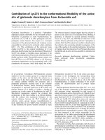
their SD in the two groups are shown in Table 4. The efficacy
of CRP measurements and CRPv in differentiating between
bacterial and non-bacterial febrile illness was evaluated by
ROC analysis. The AUC for CRP and for CRPv were 0.783
(95% confidence interval (CI) = 0.717 to 0.850) and 0.871
(95% CI = 0.817 to 0.924), respectively (Figure 1).
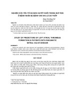
There were 122 patients with a CRP concentration of less
than 100 mg/L, of whom 59 had bacterial and 63 had non-
bacterial febrile illness. An ancillary analysis was conducted
for this subgroup of patients. The mean, median and interquar-
tile ranges are shown in Table 5. The AUC for CRP and CRPv
for this subgroup of patients were 0.689 (95% CI = 0.0595 to
Table 1
Demographic characteristic, medications on admission and co-morbidities in the bacterial and non-bacterial groups
Bacterial
n = 108
Non-bacterial
n = 70
P value
Age, years Mean ± SD 54.6 ± 23.4 33.1 ± 16.1 < 0.001
Women n (%) 54 (50.0%) 27 (38.6%) 0.135
Body mass index, kg/m
2
Mean ± SD 25.4 ± 4.5 23.5 ± 4.1 0.003
Ischemic heart disease n (%) 21 (19.4%) 2 (2.9%) 0.001
Diabetes mellitus n (%) 23 (21.3%) 3 (4.3%) 0.002
Steroids usage n (%) 2 (1.9%) 1 (1.4%) 0.83
SD = standard deviation.
Critical Care Vol 13 No 2 Paran et al.
Page 4 of 8
(page number not for citation purposes)
0.782) and 0.842 (95% CI = 0.77 to 0.914), respectively,
showing an even greater improvement of the AUC when the
CRPv was considered (Figure 2).
A cut-off value of 1.08 mg/L/hour (sensitivity 78%, specificity
77.8%) was chosen using the Youden index (= Sensitivity +
Specificity - 1). Based on these observations, we defined a
two-step model for the diagnosis of bacterial infection:
patients with a CRP of 100 mg/L or higher, or patients with a
CRP less than 100 mg/L and a CRPv of 1.08 mg/L/hour or
higher, which demonstrated a sensitivity of 88% and a specif-
icity of 70% for the diagnosis of bacterial infection as the ori-
gin of fever.
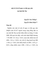
PCT was also measured in a subgroup of 48 patients, 31 of
whom had bacterial infections and 17 of whom had non-bac-
terial febrile illness. The median CRP, CRPv and PCT were
significantly higher for the patients with bacterial infections
than for the patients with non-bacterial febrile illness (Table 6).
The efficacy of CRP measurements, CRPv and the PCT in dif-
ferentiating between bacterial and non-bacterial febrile illness
was evaluated by ROC analysis for this subgroup of patients.
The AUC for CRP, CRPv and PCT were 0.824 (95% CI =
0.707 to 0.942), 0.844 (95% CI = 0.714 to 0.974) and 0.693
(95% CI = 0.54 to 0.846), respectively – significantly better
for CRP and CRPv as indicated by the non overlapping 95%
CIs (Figure 3).
Discussion
The results of the current investigation demonstrated that it is
possible to improve the differentiation between acute bacterial
and non-bacterial febrile illnesses in the setting of the emer-
gency room by using the parameter of CRPv instead of the
absolute concentration of CRP alone. This diagnostic
improvement proved to be significant and involved no addi-
tional cost due to the fact that it needed only the acquisition of
additional information. This benefit, however, might be relevant
only in patients for whom the time of fever onset is known.
Table 2
Infectious diagnosis according to site of infection
Bacterial infection (n = 108) Non-bacterial febrile illness (n = 70)
Pneumonia (n = 38)* Unspecified viral infection (n = 19)
Urinary tract infection (n = 34)* URTI/bronchitis (n = 16)
Skin, soft tissue (n = 13) Gastroenteritis/colitis (n = 16)
Pharyngitis (n = 12), sinusitis (n = 1)* EBV, CMV (n = 9), herpes zoster (n = 1)
Gastroenteritis/colitis (n = 10) Viral meningitis (n = 3)
Abdominal infection (n = 2) Hepatitis (n = 2)
Autoimmune disorders (n = 4)
*Two patients had concomitant infections: one had pneumonia and a urinary tract infection, and one had pneumonia and sinusitis. CMV =
cytomegalovirus; EBV = Epstein Barr virus; URTI = upper respiratory tract infection.
Table 3
Pathogens isolated in cultures or demonstrated by serology
Blood cultures (n) Urine cultures (n) Soft tissue and skin
abscess cultures (n)
Stool cultures (n) Throat cultures (n) Serology (n)
Escherichia Coli (7) Escherichia coli (12) Streptococcus
pyogenes (3)
Shigella sonnei (2) Group C
Streptococcus (2)
Mycoplasma
pneumonia (1)
Acinetobacter
baumannii (1)
Pseudomonas
aeruginosa (1)
Methicillin-sensitive
Staphylococcus
aureus (1)
Campylobacter jejuni
(2)
Streptococcus
pyogenes (3)
Cytomegalovirus (4)
Klebsiella
pneumoniae (1)
Enterococcus faecalis
(2)
Pseudomonas
aeruginosa (1)
Epstein-Barr virus (5)
Methicillin-sensitive
Staphylococcus
aureus (1)
Klebsiella oxytoca (1) Coxsackievirus B4
enterovirus (1)
Enterococcus faecalis
(1)
Klebsiella
pneumoniae (1)
Hepatitis B virus (1)
Herpes simplex virus
(1)
Eight patients had two positive cultures at the same time.
Available online />Page 5 of 8
(page number not for citation purposes)
To the best of our knowledge, the usefulness of a biomarker
velocity, such as CRP, has not been tested in the context of
differentiation between acute bacterial and non-bacterial
febrile illnesses. Our results are therefore significant because
they show the feasibility of using this diagnostic property of
CRP in relation to the duration of the acute febrile disease.
The rationale for using the value of CRPv stems from the
assumption that severe infections might be associated with a
cytokine storm. In fact, cytokine storms are frequently seen in
the context of acute and severe bacterial infections [13-17].
We hypothesized that a more rapid increment in the concen-
tration of these cytokines might translate into a more rapid syn-
thesis of the presently used biomarker, CRP.
The finding that patients with acute bacterial infections
present with higher CRP concentrations than those who have
non-bacterial febrile illnesses has been often shown in the
past [1-8]. We now add the observation that patients with
acute bacterial infections present to the emergency room ear-
lier than those with acute non-bacterial infections. The rela-
tively late arrival of individuals with acute non-bacterial febrile
illness might be associated with a lower concentration of
cytokines and therefore a lesser feeling of being unwell than
individuals who have acute and severe bacterial infections.
Combining these two facts (higher CRP concentrations and
earlier presentations), one can understand the behavior of the
Figure 1
Receiver operator curve for C-reactive protein and C-reactive protein velocity for the diagnosis of bacterial infectionReceiver operator curve for C-reactive protein and C-reactive protein velocity for the diagnosis of bacterial infection. CRP = C-reactive protein;
CRPv = C-reactive protein velocity; WBC = white blood cells.
Table 4
Comparison of different parameters between patients with bacterial infection and those with non-bacterial infection
Variable Duration of fever
(hours)
CRP (mg/L) CRP-velocity (mg/:/
hour)
WBC (10
3
/ml
3
) Neutrophils (10
3
/ml
3
)
Bacterial
(n = 108)
Mean 36.5 63.77 3.61 13.94 11.74
Interquartile range 6.0 to 46.8 37.4 to 156.0 1.97 to 7.41 9.85 to 17.55 7.47 to 15.41
Non-bacterial
(n = 70)
Mean 68.7 15.2 0.41 8.35 5.77
Interquartile range 17.8 to 90 9.8 to 51.6 0.18 to 1.49 5.88 to 9.75 3.44 to 7.31
P value < 0.001 < 0.001 < 0.001 < 0.001 < 0.001
CRP = C-reactive protein; WBC = white blood cells.
Critical Care Vol 13 No 2 Paran et al.
Page 6 of 8
(page number not for citation purposes)
biomarker; namely, a steeper rise in patients with acute bacte-
rial infections as opposed to a slower increment in those with
non-bacterial febrile illnesses. In addition, the CRPv might
have special relevance in individuals who present with concen-
trations that are not very high (i.e., less than 100 mg/L).
Indeed, sensitivities and specificities of up to 90% have been
shown for bacterial infections in individuals who present with
CRP concentrations of 100 mg/L or more [2,11,12]. The
improved results observed in our sub-analysis on patients with
CRP concentrations of less than 100 mg/L support the notion
that CRPv might be especially useful for them.
Different biomarkers have been previously tested for the pur-
pose of differentiating between bacterial and non-bacterial
febrile illnesses. These biomarkers include CRP [1-8], PCT
[7,18-22], the absolute neutrophil count and IL-6 [5,6,16].
Although the populations in these studies were not similar to
ours, the results reported in the literature were generally infe-
rior to those we reported. Moreover, we measured PCT levels
in a subgroup of 48 consecutive patients and found CRPv to
be a better marker compared with PCT in distinguishing bac-
terial from non-bacterial febrile illnesses. Recent study on the
combination of six different biomarkers (CRP, PCT, neu-
Figure 2
Receiver operator curve for C-reactive protein and C-reactive protein velocity for the diagnosis of bacterial infection for patients with C-reactive pro-tein less than 100 mg/LReceiver operator curve for C-reactive protein and C-reactive protein velocity for the diagnosis of bacterial infection for patients with C-reactive pro-
tein less than 100 mg/L. CRP = C-reactive protein; CRPv = C-reactive protein velocity.
Table 5
Comparison of different parameters between patients with bacterial and non-bacterial febrile illness presenting with a C-reactive
protein concentration less than 100 mg/L
Variable Duration of fever
(hours)
CRP (mg/L) CRP-velocity (mg/L/
hour)
WBC (10
3
/ml
3
) Neutrophils (10
3
/ml
3
)
Bacterial
(n = 59)
Mean 27.2 30.58 2.6 13.1 11.0
Interquartile range 4.0 to 24.5 18.6 to 80.6 1.1 to 6.0 8.4 to 16.6 6.79 to 14.87
Non-bacterial
(n = 63)
Mean 67.7 12.16 0.35 8.3 5.7
Interquartile range 17 to 88 8.3 to 36.8 0.16 to 1.04 5.9 to 9.9 3.5 to 7.3
P value < 0.001 < 0.001 < 0.001 < 0.001 < 0.001
CRP = C-reactive protein; WBC = white blood cells.
Available online />Page 7 of 8
(page number not for citation purposes)
trophils, macrophage migration inhibitory factor, soluble uroki-
nase-type plasminogen activator receptor and soluble
triggering receptor expressed on myeloid cells-1) for the
detection of bacterial versus non-bacterial febrile illness in
patients with systemic inflammatory response syndrome
reported an AUC of 0.88 [23]. In comparison, our newly
defined parameter of CRPv showed similar efficacy without
using any other test and without any further costs other than
adding another simple question to history taking.
One limitation of the present study is that a clear bacterial eti-
ology could not be obtained in all individuals in the bacterial
infection group. This is especially relevant for the ones with
pneumonia and gastroenteritis/colitis. In all cases we con-
sulted infectious disease specialists. We agree that some
cases of bacterial infections might have been viral and vice
versa, but this limitation is known in similar studies. Another
limitation is that every person has certain background inflam-
matory activity that is ignored in our calculations. However, this
background inflammation is generally in the range of CRP of
less than 10 mg/L [24], so this problem probably has a minor
effect. Moreover, patients with a bacterial infection without
clinically apparent fever were not included in this study.
Hence, our conclusions are only relevant to patients with
apparent fever. Finally, the main concern about the interpreta-
tion of the CRPv is its dependence on the time that has
elapsed between the onset of fever and the measurement of
CRP.
Figure 3
Receiver operator curve for C-reactive protein, C-reactive protein velocity and procalcitonin for the diagnosis of bacterial infection in a subgroup of 48 patients, 31 of whom had bacterial infectionsReceiver operator curve for C-reactive protein, C-reactive protein velocity and procalcitonin for the diagnosis of bacterial infection in a subgroup of
48 patients, 31 of whom had bacterial infections. CRP = C-reactive protein; CRPv = C-reactive protein velocity; PCT = procalcitonin.
Table 6
The median and interquartial range of C-reactive protein, C-reactive protein velocity and procalcitonin for the bacterial and non-
bacterial groups in a subgroup of 48 patients
Non-bacterial (n = 17) Bacterial (n = 31) P value
Median Interquartile range Median Interquartile range
PCT (ng/ml) 0.05 0.05 to 0.24 0.21 0.05 to 0.53 0.025
CRP (mg/L) 29.0 15.5 to 52.1 99.9 41.3 to 143.7 < 0.001
CRPv (mg/L/hour) 0.44 0.26 to 1.32 2.67 1.39 to 5.16 < 0.001
CRP = C-reactive protein; CRPv = C-reactive protein velocity; PCT = procalcitonin.
Critical Care Vol 13 No 2 Paran et al.
Page 8 of 8
(page number not for citation purposes)
Conclusions
In conclusion, we demonstrate that by adding the value of
CRPv and not only looking at the absolute CRP concentration,
it is possible to improve the differentiation between acute bac-
terial and non-bacterial febrile illnesses. CRPv is cost free and
could be applied as a useful diagnostic tool to identify individ-
uals with bacterial infection.
Competing interests
The authors declare that they have no competing interests.
Authors' contributions
PH, SB, YP, DY, and GC participated in the study design and
coordination. OR, RBA, MK and HS analysed the data. IZ, TR,
and YO carried out the laboratory assays. YP, SB, and DJ
drafted the manuscript.
Acknowledgements
Esther Eshkol is thanked for editorial assistance.
References
1. Hengst JM: The role of C-reactive protein in the evaluation and
management of infants with suspected sepsis. Adv Neonatal
Care 2003, 3:3-13.
2. Gabay C, Kushner I: Acute phase proteins and other systemic
responses to inflammation. N Engl J Med 1999, 340:1376.
3. Yeung CY, Lee HC, Lin SP, Fang SB, Jiang CB, Huang FY,
Chuang CK: Serum cytokines in differentiating between viral
and bacterial enterocolitis. Ann Trop Paediatr 2004,
24:337-343.
4. Nuutila J, Lilius EM: Distinction between bacterial and viral
infections. Curr Opin Infect Dis 2007, 20:304-310.
5. Toikka P, Irjala K, Juvén T, Virkki R, Mertsola J, Leinonen M,
Ruuskanen O: Serum procalcitonin, C-reactive protein and
interleukin-6 for distinguishing bacterial and viral pneumonia
in children. Pediatr Infect Dis J 2000, 19:598-602.
6. Gaïni S, Koldkjaer OG, Pedersen C, Pedersen SS: Procalcitonin,
lipopolysaccharide-binding protein, interleukin-6 and C-reac-
tive protein in community-acquired infections and sepsis: a
prospective study. Crit Care 2006, 10:R53.
7. Simon L, Gauvin F, Amre DK, Saint-Louis P, Lacroix J: Serum pro-
calcitonin and C-reactive protein levels as markers of bacterial
infection: a systematic review and meta-analysis. Intensive
Care Med 2001, 27:211-215.
8. Flood RG, Badik J, Aronoff SC: The utility of serum C-reactive
protein in differentiating bacterial from nonbacterial pneumo-
nia in children: a meta-analysis of 1230 children. Pediatr Infect
Dis J 2008, 27:95-99.
9. White HD, Chew DP: Acute myocardial infarction. Lancet 2008,
372:570-584.
10. Kasper DL, Braunwald E, Fauci AS, Hauser SL, Longo DL, Jame-
son JL, Loscalzo J: Harrison's principles of internal medicine 17th
edition. New York: McGraw-Hill Medical Publishing Division;
2008.
11. Morley JJ, Kushner I: Serum C-reactive protein levels in disease.
Ann N Y Acad Sci 1982, 389:406-418.
12. Flanders SA, Stein J, Shochat G, Sellers K, Holland M, Maselli J,
Drew WL, Reingold AL, Gonzales R: Performance of a bedside
C-reactive protein test in the diagnosis of community-acquired
pneumonia in adults with acute cough. Am J Med 2004,
116:529-535.
13. Pinsky MR, Vincent JL, Deviere J, Alegre M, Kahn RJ, Dupont E:
Serum cytokine levels in human septic shock: relation to mul-
tiple-system organ failure and mortality. Chest 1993,
103:565-575.
14. Oberholzer A, Souza SM, Tschoeke SK, Oberholzer C, Abou-
hamze A, Pribble JP, Moldawer LL: Plasma cytokine measure-
ments augment prognostic scores as indicators of outcome in
patients with severe sepsis. Shock 2005, 23:488-493.
15. Bozza FA, Salluh JI, Japiassu AM, Soares M, Assis EF, Gomes RN,
Bozza MT, Castro-Faria-Neto HC, Bozza PT: Cytokine profiles as
markers of disease severity in sepsis: a multiplex analysis.
Crit Care 2007, 11:R49.
16. Buck C, Bundschu J, Gallati H, Bartmann P, Pohlandt F: Inter-
leukin-6: a sensitive parameter for the early diagnosis of neo-
natal bacterial infection. Pediatrics 1994, 93:54-58.
17. Kellum JA, Kong L, Fink MP, Weissfeld LA, Yealy DM, Pinsky MR,
Fine J, Krichevsky A, Delude RL, Angus DC: GenIMS Investiga-
tors. Understanding the inflammatory cytokine response in
pneumonia and sepsis: results of the Genetic and Inflamma-
tory Markers of Sepsis (GenIMS) Study. Arch Intern Med 2007,
167:1655-1663.
18. Gendrel D, Bohuon C: Procalcitonin as a marker of bacterial
infection. Pediatr Infect Dis J 2000, 19:679-687. quiz 688.
19. Delèvaux I, André M, Colombier M, Albuisson E, Meylheuc F,
Bègue RJ, Piette JC, Aumaître O: Can procalcitonin measure-
ment help in differentiating between bacterial infection and
other kinds of inflammatory processes? Ann Rheum Dis 2003,
62:337-340.
20. Chan YL, Tseng CP, Tsay PK, Chang SS, Chiu TF, Chen JC: Pro-
calcitonin as a marker of bacterial infection in the emergency
department: an observational study. Crit Care 2004, 8:R12-20.
Epub 2003 Nov 20.
21. George A, Coulson C, De R: Procalcitonin: a bacterial specific
marker of infection. Clin Otolaryngol 2007, 32:310.
22. Monneret G, Doche C, Durand DV, Lepape A, Bienvenu J:
Procal-
citonin as a specific marker of bacterial infection in adults.
Clin Chem Lab Med 1998, 36:67-68.
23. Kofoed K, Andersen O, Kronborg G, Tvede M, Petersen J, Eugen-
Olsen J, Larsen K: Use of plasma C-reactive protein, procalci-
tonin, neutrophils, macrophages migration inhibitory factor,
soluble urokinase-type plasminogen activator receptor, and
soluble triggering receptor expressed on myeloid cells-1 in
combination to diagnose infections: a prospective study. Crit
Care 2007, 11:R38.
24. Luna MC: C-reactive protein in pneumonia – let me try again.
Chest 2004, 125:1192-1195.
Key messages
• CRPv distinguishes febrile bacterial infections from
non-bacterial febrile illnesses better than CRP alone.
• CRPv distinguishes febrile bacterial infections from
non-bacterial febrile illnesses better than CRP alone
especially in patients with CRP levels less than 100 mg/
L at presentation.
• CRPv correlates with other acute-phase proteins such
as IL-1, IL-6, and TNF-.
• CRPv is feasible in the setup of the emergency
department.