Báo cáo y học: "Prediction of hospital outcome in septic shock: a prospective comparison of tissue Doppler and cardiac biomarkers" pdf
Bạn đang xem bản rút gọn của tài liệu. Xem và tải ngay bản đầy đủ của tài liệu tại đây (367.53 KB, 11 trang )
RESEARC H Open Access
Prediction of hospital outcome in septic shock: a
prospective comparison of tissue Doppler and
cardiac biomarkers
David J Sturgess
1,2*
, Thomas H Marwick
1,3
, Chris Joyce
1,4
, Carly Jenkins
1,3
, Mark Jones
5
, Paul Masci
1
, David Stewart
4
,
Bala Venkatesh
1,2,4
Abstract
Introduction: Diastolic dysfunction as demonstrated by tissue Doppler imaging (TDI), particularly E/e’ (peak early
diastolic transmitral/peak early diastolic mitral annular velocity) is common in critical illness. In septic shock, the
prognostic value of TDI is undefined. This study sought to evaluate and compare the prognostic significance of TDI
and cardiac biomarkers (B-type natriuretic peptide (BNP); N-terminal proBNP (NTproBNP); troponin T (TnT)) in septic
shock. The contribution of fluid management and diastolic dysfunction to elevation of BNP was also evaluated.
Methods: Twenty-one consecutive adult patients from a multidisciplinary intensive care unit underwent
transthoracic echocardiography and blood collection within 72 hours of developing septic shock.
Results: Mean ± SD APACHE III score was 80.1 ± 23.8. Hospital mortality was 29%. E/e’ was significantly higher in
hospital non-survivors (15.32 ± 2.74, survivors 9.05 ± 2.75; P = 0.0002). Area under ROC curves were E/e’ 0.94, TnT
0.86, BNP 0.78 and NTproBNP 0.67. An E/e’ threshold of 14.5 offered 100% sensitivity and 83% speci ficity.
Adjustment for APACHE III, cardiac disease, fluid balance and grade of diastolic function, demonstrated E/e’ as an
independent predictor of hospital mortality (P = 0.019). Multiple linear regression incorporating APACHE III, gender,
cardiac disease, fluid balance, noradrenaline dose, C reactive protein, ejection fraction and diastolic dysfunction
yielded APACHE III (P = 0.033), fluid balance (P = 0.001) and diastolic dysfunction (P = 0.009) as independent
predictors of BNP concentration.
Conclusions: E/e’ is an independent predictor of hospital survival in septic shock. It offers better discrimination
between survivors and non-survivors than cardiac biomarkers. Fluid balance and diastolic dysfunction were
independent predictors of BNP concentration in septic shock.
Introduction
Septic shock in adults refers to a state of acute circula-
tory fai lure characterized by persistent arterial hypoten-
sion unexplained by other causes [1]. Although this
clinical syndrome is heterogeneous with regard to fac-
tors such as causal micro-organism, patient predisposi-
tion, co-morbidity and response to therapy, a key
element and unifying feature is the manifestation of car-
diovascular dysfunction. Although the underlying cause
of death in septic shock is often multifactorial, refractory
hypotension and cardiovascular collapse are frequently
observed in the terminal phases of the condition [2].
Whilst impaired systolic function has been identified as
the major culprit, the contribution of d iastol ic dysfunc-
tion (and hence ventricular filling) to cardiovascular
morbidity and mortality in septic shock is not fully
understood. Inves tigation of left ventricular (LV) diasto-
lic function at the bedside is challenging, but techniques
such as echocardiography and biomarkers such as
B-type natriuretic peptide (BNP) are increasingly sup-
ported by current literature [3-5]. In particular, recent
application of non-invasive, bedside technologies, such
as tissue Doppler imaging (TDI), offer fresh insight [6].
TDI is an echocardiographic technique that measures
myocardial velocities [7], which are low frequency,
* Correspondence:
1
School of Medicine, The University of Queensland, Princess Alexandra
Hospital, Ipswich Road, Brisbane, 4102, Australia
Sturgess et al. Critical Care 2010, 14:R44
/>© 2010 Sturgess et a l.; licensee BioMed Central Ltd. This is an open access article distributed under the terms of the Creative Commons
Attribution License ( which permits unrestricted use, distribution, and reproduction in
any medium, provided the original work is properly cited.
high-amplitude signals filtered from conventional Dop-
pler imaging [8]. TDI has gained acceptance amongst
cardiologists for the evaluation of diastolic function,
particularly as a measure of ventricular relaxation and
ventricular filling pressure [9]. However, there are
scant data regarding its use in critical care. TDI has
demonstrated prognostic utility in a range of cardio-
vascular diseases [10], including following myocardial
infarction [11,12], heart failure [13-16], abnormal LV
function at dobutamine echocardiography [17], non-
valvular atrial fibrillation [18], hypertension [19], and
end-stage renal disease [20].
Previously, we demonstrated that evidence of diastolic
dysfunction on TDI is common in critically ill patients
[21]. The significance of this was re cently highlighted by
Ikonomidis and colleagues, who demonstrated that TDI
may be prognostically useful in the general ICU popula-
tion [22]. To date, the prognostic significance of this
technique has not been specifically evaluated in septic
shock.
Cardiac biomarkers including BNP [23,24], N-terminal
proBNP (NTproBNP) [25] and troponin [26] potentially
offer prognostic information in the critically ill. To date,
no comparison has been made between TDI and cardiac
biomarkers (BNP, NTproBNP and troponin) with regard
to prediction of hospital outcome in septic shock.
This study sought to evaluate and compare the prog-
nostic significance of TDI variabl es and cardiac biomar-
kers in septic shock. An auxiliary aim was to evaluate
the potential contribution of LV diastolic dysfunction
and fluid management to elevation of plasma BNP con-
centrations in septic shock.
Materials and methods
This prospective observational study was approved by
the Princess Alexandra Hospital Human Research Ethics
Committee (project 2005/213), and the Guardianship
and Administration Tribunal of Queensland (project
2006/07) and informed consent was obtained from the
patient or legally authorized representative where
appropriate.
Patients
Twenty-one consecutive adult patients with septic shock
were recruited from the ICU during an 11-month period
(May 2005 to March 2006). Eligible patients were
enrolled within 72 hours of admission to the ICU with
septic shock or development of septic shock while in
the ICU. Septic shock was defined as severe sepsis with
persistent hypotension (ie. with a mean arterial pressure
(MAP) < 60 mmHg or a reduction in systolic blood
pressure (SBP) > 40 mmHg from baseline) despite ade-
quate volume resuscitation in the absence of other
causes for hypotension [1].
Exclusion criteria included: age younger than 18 years;
presence of moderate to severe valvular heart disease; or
patient or legally authorized representa tive declined
participation.
Patient care followed standard practice. Clinical fluid
resuscitation and mana gement were undertaken in a
fashion consistent with surviving sepsis guidelines [27].
More specifically, fluid challenges were undertaken
incrementally while clinical response was observed.
Therapeutic variables considered in determining the
requirement and response to fluid management
included pulse rate, blood pressure (target MAP >
65 mmHg), peripheral perfusion, urine output (target >
0.5 ml/kg/hr), a nd central venous pressure (CVP).
Research measurements were not released to the treat-
ing clinician.
Clinical and outcome data
Clinical data included height,weight,ventilationmode
and settings, heart rate, rhythm, arterial blood pressure
(SBP; diastolic blood pressure (DBP); MAP) and CVP.
Body surface area (BSA) was calculated [28]. ICU fluid
balance was recorded for the study day (fluid balance).
Vasopressor/inotropic infusion rates and, ICU and hos-
pital length of s tay and outcome were recorded. Illness
severity was quantified using (Acute Physiology and
Chronic Health Evaluation) APACHE III and Sequential
Organ Failure Assessment (SOFA) scores. Patients were
considered to have a history of cardiac disease if they
had prior or current ischemic heart disease (angina or
myocardial infarction) or cardiac surgery.
Echocardiography
Transthoracic echocardiography and Doppler examina-
tions were performed by experienced echocardiogra-
phers (coordinated by Jenkins C) using commercially
available echocardiographic equipment (Acuson Sequoia,
Siemens AG, Muni ch, Germany and Sonos 7500, Philips
Medical Systems, Andover, MA, USA). Measurements
were made off-line, using AccessPoint™ 2000 software
(Freeland Systems, Westfield, IN, USA). Unless other-
wise stated, measurements were made in triplicate at
end expiration.
Two-dimensional echocardiography
LV end-diastolic volume (LVEDV) and LV end-systolic
volume (LVESV) were calculated using the biplane
method of disks (modified Simpson’s rule) from the api-
cal four-chamber and two-chamber views [29] and
indexed to BSA (LVEDVI and LVESVI, respectively). LV
ejection fraction (LVEF) was calculated as (LVEDV -
LVESV)/LVEDV × 100. Systolic dysfunction was defined
as EF below 55%. LV outflow tract diameter (OTD) was
recorded as the maximum measurement from triplicate
zoomed parasternal long axis view.
Sturgess et al. Critical Care 2010, 14:R44
/>Page 2 of 11
Doppler echocardiography
Transmitral flow velocities were recorded with pulsed-
wave Doppler with the sample volume placed at the
mitral valve tips from the apical four-chamber view [30].
Peak passive (E) and active (A) velocities were recorded.
E wave deceleration time (DT) was measured. E to A
ratio (E/A) was calculated.
Doppler interrogation of LV outflow tract velocity was
guided by apical five-c hamber view [30]. Heart rate
(HR), velocity time integral (VTI) an d peak velocity
(Vpeak) were measured. Stroke volume was calculated
as the product of VTI and cross-sectional area of the
LV outflow tract [π.(OTD /2)
2
]. Cardiac output was cal-
culated as the product of stroke volume and HR. Stroke
volume and cardiac output measurements wer e indexed
to body surface area (SVI and CI, respectively).
Tissue Doppler
Myocardial velocit ies were obtained using tissue Doppler
settings, with the pulsed-wave Doppler sample volume at
the septal mitral annulus in the apical four-chamber view.
Peak systolic (s’), early diastolic (e’) and late diastolic (a’)
myocardial velocities were measured. E/e’ was calculated.
When A and/or a’ were indistinguishable due to sinus
tachycardia, E and/or e’ were measured as described by
Nagueh and colleagues [31]. In the presence of atrial
dysrhythmia, transmitral and tissue Doppler velocities
were measured over five consecutive cardiac cycles [18].
As previously described [21], thresholds for abnormal
diastolic TDI were accepted as e’less than 9.6 cm/s
(myocardial relaxation below the lower 95% confidence
limit of normal subjects) [ 32] or E/e’ more than 1 5
(mean LV end-diastolic pressure > 15 mmHg) [33].
Diastolic dysfunction
Guidelines previously published by our group were used to
grade LV diastolic function as normal, impaired relaxation,
pseudonormal or restrictive [34]. Age-dependent thresh-
olds for deceleration time (< 40 years < 220 ms; 40 to 60
years 140 to 250 ms; > 60 years 140 to 275 ms) were used
to determine impaired relaxation (DT above normal limit)
and restrictive patterns (DT below normal limit). In order
to distinguish between normal a nd pseudonorma l pat-
terns, we incorporated E/e’ (normal < 8; pseudonormal >
15). Where E/e’ was inconclus ive (8 to 15), increased left
atrial area (> 20 cm) was used as a marker of raised LV
filling pressure (pseudonormal pattern). Patients categor-
ized other than no rmal were considered to have diastolic
dysfunction.
Biochemical assay
Plasma BNP concentration was measured using a Biosite
Triage® immunoassay (Biosite Diagnostics, San Diego,
CA, USA), Plasma Troponin T (TnT; Elecsys® Troponin
T, 3
rd
generation immunoassay; Roche Diagnostics Aus-
tralia Pty Ltd, Castle Hill, NSW, Australia) and
NTProBNP concentration (Elecsys® proBNP, Roche
Diagnostics Australia Pty Ltd, Castle Hill, NSW, Austra-
lia) were run on Roche Elecsys® analyzers (Roche Diag-
nostics Australia Pty Ltd, Castle Hill, NSW, Australia).
Plasma C reactive protein (CRP) concentration was
measured using an immunotubidometric assay (UniCel®
DxI 800 Access® Immunoassay System, Beckman Coul-
ter Australia Pty. Ltd., Gladesville, NSW, Australia).
Laboratory thresholds were used to determine elevation
of biomarkers: BNP (normal < 100 ng/L), NTproBNP (0
to 50 years < 450; 50 to 75 years < 900; > 75 years <
1800 ng/L), TnT (< 0.03 μg/L) and CRP (< 5.0 mg/L).
Blinding
Coded echocardiographic and Doppler recordings were
analyzed at least one month after acquisition by a single
observer blinded to clinical and biochemical data.
Biochemical assay was performed on coded samples by
technicians blinded to clinical and echocardiographic data.
Statistics
Analysis was performed by SPSS, version 14.0 for Win-
dows (SPSS Inc., Chicago, IL, USA). Descriptive mea-
sures were used to evaluate the distribution of variables.
Differences between groups were assessed using Fisher’s
exact test for categorical data. Continuous data were
assessed using Levene’s test for equality of variance
before applying Student’s t-test for independent samples.
BNP and NTproBNP concentrations were log-trans-
formed to achieve normality before application of linear
regression techniques. Discrimination between hospital
survivors and non-survivors was evaluated by receiver
operating characteristic (ROC) curve analysis.
Cox proportional hazards regression was used for time
to event outcomes (hospital survival) from the time of
echocardiography. Adjustment was made for the poten-
tial influence of cardiac disease, fluid balance and grade
of diastolic dysfunction upon E/e’.
Multiple-linear regression analyses were undertaken to
determine contributions to BNP concentration (lnBNP).
Potential predictor variables include d APACH E III score
(first ICU day), gender, [35], cardiac disease [35], intrave-
nous fluid therapy [36], noradrenaline dose [37], CRP [38],
LVEF [39], and LV diastolic dysfunction [40]. A backwards
elimination procedure was then used to discard predictor
variables with P < 0.1 in multiple regressio n models one
by one until a final ‘best’ model was achieved.
In final analyses, a P-value less than 0.05 was regarded
as significant. Unless stated otherwise, results are
reported as mean ± standard deviation (SD) (range).
Sample size
Subgroup analysis of septic patients from data previously
published by our group yielded a mean ± SD E/e’ of
Sturgess et al. Critical Care 2010, 14:R44
/>Page 3 of 11
11.4 ± 5 (rang e: 3.59 to 23.15) and hospital mortality of
30% [21]. It was determined that a sample of 20 patients
would allow detection of a mean difference in E/e’ of 4
or more between survivors and non-survivors (80%
power; a = 0.05) [41].
Results
Patient characteristics
Twenty-one consecutive septic shock patients were stu-
died (Table 1). Fifteen participants (71%) were studied
within 24 hours of developing septic shock. Variables
recorded on the study day are presented in Table 2.
Sixteen patients (76%) were mechanically ventilated at
the time of the initial assessment. The requirement for
mechanical ventilation did not distinguish survivors
from non-survivors (P = 0.15). Of the mechanically ven-
tilated patients, positive end-expiratory pressure (PEEP)
requirements were not different between survivors and
non-survivors (7.05 ± 3 cmH
2
Ovs7.9±5.1cmH
2
O,
respectively; P = 0.67).
Eleven patients (52%) were in normal sinus rhythm
and two (9.5%) were paced. Noradrenaline infusion was
running at the time of initial assessment in seventeen
patients (81%; Mean ± SD infusion rate 0.124 ± 0.12
micrograms/kg/min). In addition to noradrenaline, one
patient was receiving adrenaline and one was receiving
dopamine. Mean ± SD fluid balance on the day of study
was 1780 ± 1848 mL (range: -1734 to 5320).
The diagnosis of cardiac disease (Table 1) was based
on previous history (non-acute) in seven out of nine
patients. Of the remaining patients, one developed sepsis
secondary to wound infection eight days following aortic
root and valve replacement (no significant coronary
artery disease; survived to hospital discharge), whereas
the other developed pneumonia sixteen days following
emergency coronary artery bypass grafting for acute
myocardial infarction (non-survivor). No patients had a
previous history of heart failure. Fourteen patients had
been receiving treatment for hypertension prior to the
development of septic shock. Six patients had been pre-
viously diagnosed with diabetes mellitus (n = 5; all type
2) or glucose intolerance prior to the development of
septic shock.
Table 1 Patient characteristics
Total number of patients 21
Male:Female ratio 13:8
Age, years 65 ± 17 (24-86)
Height, cm 167 ± 7 (156-180)
Weight, kg 80 ± 18 (42-130)
Body surface area, m
2
1.88 ± 0.25 (1.4-2.5)
APACHE III score (Day 1 ICU) 80.1 ± 23.8 (46-141)
SOFA score (Day 1 ICU) 11 ± 2.8 (6-16)
ICU length of stay, days 12.5 ± 12.3 (1-54)
Hospital length of stay, days 29.6 ± 29.3 (1-125)
ICU mortality, n (%) 4 (19%)
Hospital mortality, n (%) 6 (29%)
28-day mortality, n (%) 6 (29%)
Source of infection
Abdominal, n (%) 8 (38%)
Pulmonary, n (%) 7 (33%)
Neurologic, n (%) 2 (9.5%)
Necrotizing fasciitis, n (%) 2 (9.5%)
Catheter related sepsis, n (%) 1 (5%)
Mediastinitis, n (%) 1 (5%)
Cardiac disease, n (%) 9 (43%)
Angina, n (%) 3 (14%)
Myocardial infarction, n (%) 6 (28%)
Cardiac surgery, n (%) 5 (24%)
APACHE, Acute Physiology and Chronic Health Evaluation; SOFA, Sequential
Organ Failure Assessment.
Table 2 Variables measured on study day
Variable Mean ± SD (Range)
Day of study
APACHE III score 82.9 ± 29.6 (28-141)
SOFA score 11.6 ± 3.6 (5-19)
Echocardiography
LVEDVI, mL/m
2
65.8 ± 22.4 (31.9-121.8)
LVESVI, mL/m
2
37.5 ± 18.5 (13.9-83.2)
SVI, mL/m
2
26.6 ± 14.5 (8.3-67.9)
EF, % 43 ± 14 (11-63)
VTI, cm 19.08 ± 5.06 (12.7-29.3)
Vpeak, m/s 1.042 ± 0.234 (0.71-1.48)
CI, L/min/m
2
3.14 ± 1.16 (1.9-6.32)
E, m/s 0.94 ± 0.27 (0.54-1.5)
DT, s 0.201 ± 0.054 (0.097-0.311)
A, m/s 0.63 ± 0.22 (0.22-1.17)
E/A 1.7 ± 1.1 (0.7-5.3)
e’, cm/s 9.3 ± 3.4 (4.8-18.8)
a’, cm/s 9.9 ± 3.3 (5.3-17.7)
E/e’ 10.93 ± 3.98 (4.29-18.56)
s’, cm/s 11.7 ± 4.2 (4.3-18.4)
Biochemistry
BNP, ng/L 714 ± 882 (49-2930)
NTproBNP, ng/L 1115 ± 1234 (28-4139)
CRP, mg/L 223 ± 96 (11-394)
TnT, μg/L 0.158 ± 0.21 (0-0.71)
a’, peak active (late) diastolic septal mitral annulus velocity; A, peak active
(late) diastolic transmitral flow velocity; APACHE, Acute Physiology and
Chronic Health Evaluation; BNP, B-type natriuretic peptide; CI, cardiac output
index; CRP, C reactive protein; DT, E wave deceleration time; e’, peak early
diastolic septal mitral annulus velocity; E, peak early diastolic transmitral flow
velocity; E/A, ratio of E to A; E/e’, ratio of E to e’; EF, ejection fraction; LVEDVI,
left ventricular end-diastolic volume index; LVESVI, left ventricular end-systolic
volume index; NTproBNP, N-terminal proBNP; s’, peak systolic septal mitral
annulus velocity; SD, standard deviation; SOFA, Sequential Organ Failure
Assessment; SVI, stroke volume index; TnT, troponin T; Vpeak, peak left
ventricular outflow tract velocity; VTI, left ventricular outflow tract velocity
time integral.
Sturgess et al. Critical Care 2010, 14:R44
/>Page 4 of 11
Echocardiography
Systolic dysfunction (EF < 55%) was evident in 14
patien ts (67%). Transthoracic measurement of e’ and E /
e’ was feasible in 20 of 21 patients. Fusion of E and A
waves was observed in four examinations (19%). Fusion
of e’and a’ waves was observed in three (15%). At initial
assessment, e’ was less than 9.6 cm/s in 11 (55%)
patients. At this time, E/e’ wasmorethan15inthree
(15%), 8 to 15 in thirteen (65%) and less than 8 in four
(20%) patients. TDI variables (including e’,a’,s’ and E/
e’) were not significantly different between ventilated
and non-ventilated patients. Diastolic function was
graded as normal in nine (43%), impaired relaxation in
three (14%), pseudonormal in seven (33%) and restric-
tive in two patients (10%). Thus, diastolic dysfunction
was present in 57% of patients (n = 12).
Biochemistry
BNP was elevated in fifteen patients (71%), NTproBNP
in six (28%) and TnT in fourteen (67%).
Hospital outcome
Significant differences were observed between hospital
survivors and non-survivors (Table 3) with respect to
E/e’ (survivor 9.05 ± 2.75, non-survivor 15.32 ± 2.74; P
=0.0002),e’ (survivor 10.4 ± 3.4 cm/s, non-survi vor 6.8
±1.9cm/s;P = 0.025) and s’ (survivor 13 ± 3.7 cm/s,
non-survivor8.6±4.1cm/s;P = 0.03). The area under
the ROC curve (c statistic) for each of these variables
was 0.94 for E/e’, 0.86 for e’and 0.83 for s ’.AnE/e’
threshold value of 14.5 offered sensitivity of 100% and
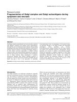
specificity of 83% (Figure 1). The c statistic of 0.86 for
TnT, 0.78 for BNP and 0.67 for NTproBNP. No differ-
ence in LVEF (systolic function) was observed (survivor
43 ± 15%, non-survivor 43 ± 14%; P = 0.91).
Prediction of hospital survival
Univariate Cox regression analysis (Table 3) yielded sig-
nificant associations between survival to hospital dis-
charge and E/e’ (P = 0.005), e’ (P = 0.04), s’ (P = 0.048)
and TnT (P = 0.03). Adjustment for APACHE III score,
history of cardiac disease, fluid balance and grade of dia-
stolic function, reveal ed E/e’ as an independent predic-
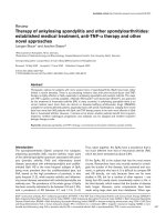
tor of hospital mortality (P = 0.019). A Kaplan-Meier
plot of the association between E/e’ and survival to hos-
pital discharge is shown in Figure 2.
Plasma BNP concentration
From an initial model containing APACHE III score,
gender, cardiac disease, fluid balance, noradrenaline dose,
CRP, EF and diastolic dysfunction, the backward elimina-
tion procedure yielded a ‘best’ model containing gender
(P = 0.089), APACHE III score (P = 0.033), fluid balance
( P = 0.001) and diastolic dysfunction (P = 0.009). This
final model accounted for 71.3% of variation in lnBNP
concentration (adjusted R square 0.713).
Discussion
The cardinal finding of this study is that E/e’ offers
independent and better prognostic prediction of hospital
outcome in septic shock as c ompared with c ardiac bio-
markers (BNP, NTproBNP, TnT). We also observed
that conventional meas ures of systolic function, such as
EF and SVI (Table 3) did not discriminate between hos-
pital survivors and non-survivors. This s tudy also
demonstrates that fluid balance and diastolic dysfunc-
tion are independent predictors of BNP concentration in
septic shock patients.
Diastolic function and tissue Doppler imaging in
septic shock
We have demonstrated an association between TDI
indices of diastolic function and outcome in septic shock.
The significance of this important new finding is high-
lighted by the superiority of these variables over the mea-
sures o f ca rdiac systolic function and cardiac biomarkers
incorporated into this study. Despite demonstrating value
in a range of cardiovascular diseases [42], and more
recently in a study of general ICU patients by Ikonomidis
and colleagues [22], the prognostic potential of TDI in
septic shock per se has not previously been reported.
Our demonstration of an association between diastolic
function and mortality in septic shock complements
previous data. In a radionuclide cineangiographic study,
Parker and colleagues documented that non-survivors
did not demonstrate LV dilation (’preload adaptation’)
and therefore were unable to maintain stroke volume
and cardiac output [43,44]. Also, Munt and colleagues
demonstrated DT as an independent predictor of mor-
tality in severe sepsis [45]. In addition to our TDI find-
ings, we o bserved a trend toward an association
between DT and hospital mortality (P = 0.07). Although
no clear functional relation has been demonstrated, sep-
sis-induced diastolic dysfunction is likely to be asso-
ciated with a range of histologic abnormalities such as
inflammatory infiltrate, interstitial edema, apoptosis, and
necrosis [46,47].
The peak early diastolic mitral annular velocity (E)’,as
measured by TDI, reflects LV relaxation [48,49].
Although this variable appears not to be as preload
insensitive as originally proposed [49,50], it i s increas-
ingly valued as a quantitative index of LV diastolic func-
tion.Thisisbecauseitdoesnot pseudo-normalize in
the same way as transmitral flow [51]. Also, the E/e’
ratio has been proposed as an estimate of LV filling
pressure that corrects E velocity for the influence of
myocardial relaxation [33,52].
Sturgess et al. Critical Care 2010, 14:R44
/>Page 5 of 11
Table 3 Comparison of hospital survivors and nonsurvivors
Variable Survivors (Mean ± SD) Non-survivors (Mean ± SD) P* Cox Regression§
Baseline characteristics
n (%) 15 (71%) 6 (29%)
Gender, M:F 8:7 5:1 0.2 ‡ .3
Age, years 64 ± 16 67 ± 20 0.73 0.66
Height, cm 166.5 ± 8 169 ± 6.7 0.56 0.56
Weight, kg 79.1 ± 20.3 81.5 ± 14.4 0.79 0.95
BSA, m
2
1.86 ± 0.27 1.93 ± 0.2 0.56 0.74
Cardiac disease, n (%) 5 (24%) 4 (19%) 0.18 ‡ 0.17
APACHE III score (Day 1 ICU) 78.5 ± 24.9 84 ± 22.7 0.65 0.65
SOFA score (Day 1 ICU) 10.3 ± 2.6 12.3 ± 2.7 0.15 0.19
Study day
Time from onset of septic shock, days 2 ± 0.8 1.7 ± 0.5 0.34 0.3
APACHE III score 80.8 ± 33.2 88.2 ± 19.1 0.62 0.77
SOFA score 10.6 ± 3.6 13.3 ± 3.2 0.14 0.23
Mechanical ventilation, n (%) 10 (48%) 6 (29%) 0.15 ‡ 0.38
Clinical monitoring
HR, beats/min 87 ± 15 85 ± 10 0.77 0.7
SBP, mmHg 115 ± 16 109 ± 14 0.39 0.32
DBP, mmHg 54 ± 7 48 ± 9 0.21 0.16
MAP, mmHg 72 ± 8 68 ± 10 0.38 0.3
CVP, mmHg 13.8 ± 3 14.5 ± 6 0.72 0.63
Fluid and vasopressor management
Fluid balance, mL 1375 ± 1679 2792 ± 2009 0.11 0.08
Noradrenaline dose, μg/kg/min 0.115 ± 0.123 0.147 ± 0.111 0.58 0.47
Echocardiography
LA area, cm
2
22.49 ± 5.1 24.28 ± 4.45 0.46 0.35
LVEDVI, mL/m
2
61.2 ± 19.6 83 ± 26.6 0.08 0.14
LVESVI, mL/m
2
36 ± 20 43 ± 12 0.52 0.66
SVI, mL/m
2
25.1 ± 10.3 30.2 ± 22.8 0.62 † 0.62
EF, % 43 ± 15 43 ± 14 0.91 0.84
OTD, cm 2.054 ± 0.243 2.23 ± 0.17 0.13 0.17
VTI 19.63 ± 4.54 17.42 ± 6.71 0.41 0.4
Vpeak 1.084 ± 0.203 0.914 ± 0.297 0.16 0.1
CI, L/min/m
2
3.13 ± 0.96 3.18 ± 1.76 0.93 0.98
E, m/s 0.89 ± 0.24 1.04 ± 0.33 0.27 0.24
DT, s 0.215 ± 0.055 0.168 ± 0.032 0.07 0.07
E A fusion, n (%) 2 (9.5%) 2 (9.5%) 0.32 ‡ 0.4
A, m/s 0.6 ± 0.18 0.73 ± 0.33 0.32 0.3
E/A 1.7 ± 1.2 1.5 ± 0.6 0.75 0.7
e’, cm/s 10.4 ± 3.4 6.8 ± 1.9 0.025 0.04
a’, cm/s 9.9 ± 3.4 9.9 ± 3.2 0.99 0.96
e’a’ fusion, n (%) 1 (5%) 2 (9.5%) 0.18 ‡ 0.15
E/e’ 9.05 ± 2.75 15.32 ± 2.74 0.0002 0.005
s’, cm/s 13 ± 3.7 8.6 ± 4.1 0.03 0.048
Diastolic dysfunction, n (%) 8 (38%) 4 (19%) 0.66 ‡ 0.55
Biochemistry
BNP, ng/L 448 ± 607 1289 ± 1155 0.14 † 0.07 ¶
NTproBNP, ng/L 841 ± 818 1801 ± 1853 0.27 † 0.2 ¶
Sturgess et al. Critical Care 2010, 14:R44
/>Page 6 of 11
Two recent studies have utilized TDI in the evaluation
of septic ICU pati ents. McLean and colleagues used E/e’
as an estimate of LV filling pressure in their prognostic
study of BNP in patients with severe sepsis and septic
shock [53]. They reported E/e’ to be non-significantly
lower in non-survivors (survivors 14.8 ± 7. 4, non-survi-
vors 12.1 ± 4.6; P = 0.452). However, their study incor-
porated a number of pat ients with severe sepsis (lower
severity of illness compared with the current study), and
did not report fluid management, which is an important
determinant of survival in sepsis [54]. Bouhemad and
colleagues [55] used TDI to demonstrate isolated and
rev ersible impairm ent of ventricular relaxation in septic
shock patients with increased plasma troponin I concen-
tration but associations with mortality were not
assessed.
Cardiac biomarkers including BNP [23,24], NTproBNP
[25] and troponin [26] have been offered as potential
prognostic tools in the critically ill. Our study demon-
strates the superiority of TDI over these biomarkers.
This is potentially explained by the magnitude of poten-
tial confounders on plasma biomarker concentrations in
the critically ill [3]. Furthermore, TDI offers more direct
evaluation of myocardial function.
B-type natriuretic peptide
In general, BNP is a peptide hormone secreted by the
ventricular myocardial in response to wall stress [3]. Its
principal clinical use is the diagnosi s of heart failure [56].
However, elevated BNP appears to lack validity as a bio-
marker of myocardial dysfunction in sepsis. Potential
explanations include inflammation [38], altered clearance
Figure 1 Receiver operating characteristic curve comparing E/e’ with BNP, and TnT as discriminators of hospital mortality. BNP, B-type
natriuretic peptide; E/e’, ratio of peak early diastolic transmitral flow velocity to peak early diastolic septal mitral annulus; TnT, Troponin T.
Table 3: Comparison of hospital survivors and nonsurvivors (Continued)
TnT, μg/L 0.114 ± 0.174 0.268 ± 0.251 0.12 0.03
CRP, mg/L 228 ± 85 207 ± 135 0.68 0.51
*Comparison performed using Student’s T test for independent groups (equal variance assumed) unless otherwise indicated. †Equal variances not assumed
(Levene’s test P < 0.05). ‡ Fisher’s exact test (2 tail). §Univariate Cox regression analysis of variables as predictors of hospital survival. ¶Variables log-transformed
prior to Cox regr ession analysis. A, peak active (late) diastolic transmitral flow velocity; a ’, peak active (late) diastolic septal mitral annulus velocity; APACHE III,
Acute Physiology and Chronic Health Evaluation III; BNP, B-type natriuretic peptide; BSA, body surface area; CI, cardiac output indexed to body surface area; CRP,
C reactive protein; CVP, centr al venous pressure; DBP, diastolic blood pressure; DT, E wave deceleration time; E, peak early diastolic transmitral flow velocity;
e’, Peak early diastolic septal mitral annulus velocity; E/A, ratio of E to A; E/e’, patio of E to e’; EF, ejection fraction; F, female; HR, heart rate; LV, left ventricle or
ventricular; LVEDVI, left ventricular end-diastolic volume indexed to body surface area; LVESVI, left ventricular end-systolic volume indexed to body surf ace area;
M, male; MAP, mean arterial pressure; NTproBNP, N-terminal proBNP; OTD, LV outflow tract diameter; s’, peak systolic septal mitral annulus velocity; SBP, systolic
blood pressure; SD, standard deviation; SOF A, Sequential Organ Failure Assessment score; SVI, left ventricular stroke volume indexed to body surface area; TnT,
Troponin T; Vpeak, peak LV outflow tract velocity; VTI, LV outflow tract velocity time integral.
Sturgess et al. Critical Care 2010, 14:R44
/>Page 7 of 11
[57], altered intrathoracic pressures/mechanical ventila-
tion [58], vasoa ctive and ino tropic drugs [37], fluid man-
agement [36,59], and diastolic dysfunction [40].
On the basis of previous laboratory data [ 36] and our
own clinical research [60], we incorporated an auxiliary
aim of the current study of evaluating the potential
influence of fluid management on plasma BNP concen-
trations in se ptic shock. Also, the relation between dia-
stolic function and plasma BNP concentration had not
been evaluated in septic shock. We are the first to
demonstrate fluid balance and diastolic dysfunction as
independent predictors of plasma BNP concentration in
septic shock.
Limitations
In keeping with international guidelines for hemodynamic
monitoring in shock, our unit does not routinely use pul-
monary artery catheters [61], and LV filling pressure is not
pursued as a therapeutic target. Although incorporation of
pulmonary artery catheter data might have yielded inter-
esting comparisons, it was unnecessary to achieve or sta-
ted aims and might have impaired the feasi bility of our
study. We propose that the resultant observational data
forms a robust reflection of clinical practice in the context
of contemporary sepsis management. Based on our find-
ings, add itional research incorporating pulmonary artery
catheterization might now be justified.
We have reported TDI measurements taken at the
septal mitral annulus. This technique was based on
results reported by Ommen and colleagues demonstrat-
ing good prediction of LV end-diastolic pressure [33].
Although the feasibility of this approach in critical care
is appealing, the mean of measurements sampled around
the perimeter of the mitral valve would be less suscepti-
ble to regional wall motion abnormalities, if present [9].
The potential influence of mechanical ventilation, right
ventricular function and inotropes/vasopressors upon
tissue Doppler variables is unclear. Our observational
study has not been designed to clarify these potential
interactions but based on the current findings, further
research in these areas is justified.
In clinical studies, it is challenging to standardize data
collection at a fixed time from onset of sepsis. We studied
patients within 72 hours of development of septic shock
(admission to ICU or onset in IC U). The strength of this
design is that it potentially optimizes the comparison of
TDI with cardiac biomarkers, particularly BNP [23], as
predictors of outcome. Due to an inability to predict the
development of sep sis, we were unable to define the pre-
morbid diastolic function of the study participants.
Potential clinical significance and directions for future
research
The findings of this study are of potential clinical
importance. First, TDI might prove useful in risk strati-
fication. This may help identify septic shock patients
requiring more intensive therapy based upon their dia-
stolic performance. Secondly, the association between
diastolic dysfunction and mortality might offer a novel
therapeutic target. Further research incorporat ing thera-
pies targeted t oward improved cardiac relaxation (lusi-
tropy) must be pursued.
Figure 2 Kaplan Meier plot of association between E/e’ and hospital survival. Cases are censored at hospital discharge. E/e’, ratio of peak
early diastolic transmitral flow velocity to peak early diastolic septal mitral annulus.
Sturgess et al. Critical Care 2010, 14:R44
/>Page 8 of 11
Conclusions
In this preliminary study, we have found that after
adjustment for sev erity of illness, cardiac disease, fluid
management and grade of diastolic dysfunction, E/e’ is
an independent predictor of hospital survival in septi c
shock patients. In addition, E/e’ offers better discrimina-
tion between hospital survivors and non-survivors than
cardiac biomarkers (BNP, NTproBNP, TnT). Fluid bal-
ance and diastolic dysfunction are independent predic-
tors of BNP concentration in septic shock.
Key messages
• E/e’ is an independent predictor of hospital survi-
val in septic shock patients.
• E/e’ offers better discrimination between hospital
surviv ors and non-survivors than cardiac biomarkers
(BNP, NTproBNP, TnT).
• Fluid balance and diastolic dysfunction are inde-
pendent predictors of BNP concentration in septic
shock.
Abbreviations
A: peak active (late) diastolic transmitral flow velocity; a’: peak active (late)
diastolic septal mitral annulus velocity; APACHE III: Acute Physiology and
Chronic Health Evaluation III; BNP: B-type natriuretic peptide; BSA: body
surface area; CI: cardiac output indexed to body surface area; CRP: C reactive
protein; CVP: central venous pressure; DBP: diastolic blood pressure; DT: E
wave deceleration time; E: peak early diastolic transmitral flow velocity; e’:
Peak early diastolic septal mitral annulus velocity; E/A: ratio of E to A; E/e’:
patio of E to e’; EF: ejection fraction; HR: heart rate; lnBNP: multiple-linear
regression analyses of BNP concentration; LV: left ventricle or ventricular;
LVEDV: left ventricular end-diastolic volume; LVESV: left ventricular end-
systolic volume; LVEDVI: LVEDV indexed to body surface area; LVESVI: LVESV
indexed to body surface area; MAP: mean arterial pressure; NTproBNP:
N-terminal proBNP; OTD: LV outflow tract diameter; PEEP: positive end
expiratory pressure; ROC: receiver operating characteristic; s’: peak systolic
septal mitral annulus velocity; SBP: systolic blood pressure; SD: standard
deviation; SOFA: Sequential Organ Failure Assessment score; SVI, left
ventricular stroke volume indexed to body surface area; TDI: Tissue Doppler
imaging; TnT: Troponin T; Vpeak: peak LV outflow tract velocity; VTI: LV
outflow tract velocity time integral.
Acknowledgements
Mr Goce Dimeski (Chemical Pathology, Princess Alexandra Hospital,
Queensland Health Pathology Service, Ipswich Road, Brisbane, Australia)
assisted with advice regarding biochemical assay techniques. Dr Elaine Beller
(School of Population Health, The University of Queensland, Princess
Alexandra Hospital, Brisbane, Australia) assisted with advice regarding final
statistical analysis. This study was conducted with the support of grants from
the Australian and New Zealand College of Anaesthetists. This study was
performed at the Department of Intensive Care and The University of
Queensland, Princess Alexandra Hospital, Ipswich Road, Brisbane, 4102,
Australia.
Author details
1
School of Medicine, The University of Queensland, Princess Alexandra
Hospital, Ipswich Road, Brisbane, 4102, Australia.
2
Department of Intensive
Care, The Wesley Hospital, Coronation Drive, Brisbane, 4066, Australia.
3
Department of Echocardiography, Princess Alexandra Hospital, Ipswich
Road, Brisbane, 4102, Australia.
4
Department of Intensive Care, Princess
Alexandra Hospital, Ipswich Road, Brisbane, 4102, Australia.
5
School of
Population Health, The University of Queensland, Princess Alexandra
Hospital, Ipswich Road, Brisbane, 4102, Australia.
Authors’ contributions
D Sturgess conceived of the study, coordinated study design and
implementation and drafted the manuscript. TM participated in study design
and helped to draft the manuscript. C Joyce participated in study design
and helped to draft the manuscript. C Jenkins participated in the design of
the study, performed and coordinated echocardiography. MJ participated in
the design of the study and provided statistical advise. PM participated in
study design, provided laboratory equipment and advice regarding
biochemical assays. D Stewart assisted in recruitment of participa nts and
collection of data. BV participated in its design and helped to draft the
manuscript. All authors read and approved the final manuscript.
Competing interests
The authors declare that they have no competing interests.
Received: 24 September 2009 Revised: 20 January 2010
Accepted: 24 March 2010 Published: 24 March 2010
References
1. Levy MM, Fink MP, Marshall JC, Abraham E, Angus D, Cook D, Cohen J,
Opal SM, Vincent JL, Ramsay G: 2001 SCCM/ESICM/ACCP/ATS/SIS
International Sepsis Definitions Conference. Crit Care Med 2003,
31:1250-1256.
2. Krishnagopalan S, Kumar A, Parrillo JE: Myocardial dysfunction in the
patient with sepsis. Curr Opin Crit Care 2002, 8:376-388.
3. Sturgess DJ, Marwick TH, Joyce CJ, Venkatesh B: B-Type Natriuretic Peptide
Concentrations and Myocardial Dysfunction in Critical Illness. Anaesth
Intensive Care 2006, 34:151-163.
4. Parekh N, Maisel AS: Utility of B-natriuretic peptide in the evaluation of
left ventricular diastolic function and diastolic heart failure. Curr Opin
Cardiol 2009, 24:155-160.
5. Dahlstrom U: Can natriuretic peptides be used for the diagnosis of
diastolic heart failure? Eur J Heart Fail 2004, 6:281-287.
6. Sturgess DJ, Marwick TH, Venkatesh B: Diastolic (Dys)Function in Sepsis.
Yearbook of Intensive Care and Emergency Medicine Berlin Heidelberg:
Springer-VerlagVincent JL 2007, 444-454.
7. Isaaz K, Thompson A, Ethevenot G, Cloez JL, Brembilla B, Pernot C: Doppler
echocardiographic measurement of low velocity motion of the left
ventricular posterior wall. Am J Cardiol 1989, 64:66-75.
8. Dokainish H: Tissue Doppler imaging in the evaluation of left ventricular
diastolic function. Curr Opin Cardiol 2004, 19:437-441.
9. Nagueh SF, Appleton CP, Gillebert TC, Marino PN, Oh JK, Smiseth OA,
Waggoner AD, Flachskampf FA, Pellikka PA, Evangelisa A:
Recommendations for the evaluation of left ventricular diastolic function
by echocardiography. Eur J Echocardiogr 2009, 10:165-193.
10. Wang M, Yip GW, Wang AY, Zhang Y, Ho PY, Tse MK, Lam PK,
Sanderson JE: Peak early diastolic mitral annulus velocity by tissue
Doppler imaging adds independent and incremental prognostic value. J
Am Coll Cardiol 2003, 41:820-826.
11. Hillis GS, Moller JE, Pellikka PA, Gersh BJ, Wright RS, Ommen SR, Reeder GS,
Oh JK: Noninvasive estimation of left ventricular filling pressure by e/e’
is a powerful predictor of survival after acute myocardial infarction. JAm
Coll Cardiol 2004, 43:360-367.
12. Moller JE, Sondergaard E, Poulsen SH, Seward JB, Appleton CP, Egstrup K:
Color M-mode and pulsed wave tissue Doppler echocardiography:
powerful predictors of cardiac events after first myocardial infarction. J
Am Soc Echocardiogr 2001, 14:757-763.
13. Troughton RW, Prior DL, Frampton CM, Nash PJ, Pereira JJ, Martin M,
Fogarty A, Morehead AJ, Starling RC, Young JB, Thomas JD, Lauer MS,
Klein AL: Usefulness of tissue doppler and color M-mode indexes of left
ventricular diastolic function in predicting outcomes in systolic left
ventricular heart failure (from the ADEPT study). Am J Cardiol 2005,
96:257-262.
14. Olson JM, Samad BA, Alam M: Prognostic value of pulse-wave tissue
Doppler parameters in patients with systolic heart failure. Am J Cardiol
2008, 102:722-725.
15. Dini FL, Conti U, Fontanive P, Andreini D, Panicucci E, De Tommasi SM:
Prognostic value of N-terminal pro-type-B natriuretic peptide and
Doppler left ventricular diastolic variables in patients with chronic
systolic heart failure stabilized by therapy. Am J Cardiol 2008,
102:463-468.
Sturgess et al. Critical Care 2010, 14:R44
/>Page 9 of 11
16. Dokainish H, Zoghbi WA, Lakkis NM, Ambriz E, Patel R, Quinones MA,
Nagueh SF: Incremental predictive power of B-type natriuretic peptide
and tissue Doppler echocardiography in the prognosis of patients with
congestive heart failure. J Am Coll Cardiol 2005, 45:1223-1226.
17. Marwick TH, Case C, Leano R, Short L, Baglin T, Cain P, Garrahy P: Use of
tissue Doppler imaging to facilitate the prediction of events in patients
with abnormal left ventricular function by dobutamine
echocardiography. Am J Cardiol 2004, 93:142-146.
18. Okura H, Takada Y, Kubo T, Iwata K, Mizoguchi S, Taguchi H, Toda I,
Yoshikawa J, Yoshida K: Tissue doppler derived index of left ventricular
filling pressure, e/e’, predicts survival in patients with non- valvular atrial
fibrillation. Heart 2006.
19. Wang M, Yip GW, Wang AY, Zhang Y, Ho PY, Tse MK, Yu CM, Sanderson JE:
Tissue Doppler imaging provides incremental prognostic value in
patients with systemic hypertension and left ventricular hypertrophy. J
Hypertens 2005, 23:183-191.
20. Wang AY, Wang M, Lam CW, Chan IH, Zhang Y, Sanderson JE: Left
ventricular filling pressure by Doppler echocardiography in patients with
end-stage renal disease. Hypertension 2008, 52:107-114.
21. Sturgess DJ, Marwick TH, Joyce CJ, Jones M, Venkatesh B: Tissue Doppler in
critical illness: a retrospective cohort study. Crit Care 2007, 11:R97.
22. Ikonomidis I, Nikolaou M, Dimopoulou I, Paraskevaidis I, Lekakis J, Mavrou I,
Tzanela M, Kopterides P, Tsangaris I, Armaganidis A, Kremastinos DT:
Association of left ventricular diastolic dysfunction with elevated NT-
proBNP in general intensive care unit patients with preserved ejection
fraction: A complementary role of Tissue Doppler imaging parameters
and NT-proBNP levels for adverse outcome. Shock 2010, 33:141-8.
23. Charpentier J, Luyt CE, Fulla Y, Vinsonneau C, Cariou A, Grabar S,
Dhainaut JF, Mira JP, Chiche JD: Brain natriuretic peptide: A marker of
myocardial dysfunction and prognosis during severe sepsis. Crit Care
Med 2004, 32:660-665.
24. Post F, Weilemann LS, Messow CM, Sinning C, Munzel T: B-type natriuretic
peptide as a marker for sepsis-induced myocardial depression in
intensive care patients. Crit Care Med 2008, 36:3030-3037.
25. Varpula M, Pulkki K, Karlsson S, Ruokonen E, Pettila V: Predictive value of N-
terminal pro-brain natriuretic peptide in severe sepsis and septic shock.
Crit Care Med 2007, 35:1277-1283.
26. Spies C, Haude V, Fitzner R, Schroder K, Overbeck M, Runkel N,
Schaffartzik W: Serum cardiac troponin T as a prognostic marker in early
sepsis. Chest 1998, 113:1055-1063.
27. Dellinger RP, Levy MM, Carlet JM, Bion J, Parker MM, Jaeschke R, Reinhart K,
Angus DC, Brun-Buisson C, Beale R, Calandra T, Dhainaut JF, Gerlach H,
Harvey M, Marini JJ, Marshall J, Ranieri M, Ramsay G, Sevransky J,
Thompson BT, Townsend S, Vender JS, Zimmerman JL, Vincent JL,
International Surviving Sepsis Campaign Guidelines Committee; American
Association of Critical-Care Nurses; American College of Chest Physicians;
American College of Emergency Physicians; Canadian Critical Care Society;
European Society of Clinical Microbiology and Infectious Diseases, et al:
Surviving Sepsis Campaign: international guidelines for management of
severe sepsis and septic shock: 2008. Crit Care Med 2008, 36:296-327.
28. Mosteller RD: Simplified calculation of body-surface area. N Engl J Med
1987, 317
:1098.
29. Lang RM, Bierig M, Devereux RB, Flachskampf FA, Foster E, Pellikka PA,
Picard MH, Roman MJ, Seward J, Shanewise JS, Solomon SD, Spencer KT,
Sutton MS, Stewart WJ, Chamber Quantification Writing Group; American
Society of Echocardiography’s Guidelines and Standards Committee;
European Association of Echocardiography: Recommendations for chamber
quantification: a report from the American Society of Echocardiography’s
Guidelines and Standards Committee and the Chamber Quantification
Writing Group, developed in conjunction with the European Association
of Echocardiography, a branch of the European Society of Cardiology. J
Am Soc Echocardiogr 2005, 18:1440-1463.
30. Quinones MA, Otto CM, Stoddard M, Waggoner A, Zoghbi WA:
Recommendations for quantification of Doppler echocardiography: a
report from the Doppler Quantification Task Force of the Nomenclature
and Standards Committee of the American Society of Echocardiography.
J Am Soc Echocardiogr 2002, 15:167-184.
31. Nagueh SF, Mikati I, Kopelen HA, Middleton KJ, Quinones MA, Zoghbi WA:
Doppler estimation of left ventricular filling pressure in sinus
tachycardia. A new application of tissue doppler imaging. Circulation
1998, 98:1644-1650.
32. Garcia-Fernandez MA, Azevedo J, Moreno M, Bermejo J, Perez-Castellano N,
Puerta P, Desco M, Antoranz C, Serrano JA, Garcia E, Delcan JL: Regional
diastolic function in ischaemic heart disease using pulsed wave Doppler
tissue imaging. Eur Heart J 1999, 20:496-505.
33. Ommen SR, Nishimura RA, Appleton CP, Miller FA, Oh JK, Redfield MM,
Tajik AJ: Clinical utility of Doppler echocardiography and tissue Doppler
imaging in the estimation of left ventricular filling pressures: A
comparative simultaneous Doppler-catheterization study. Circulation
2000, 102:1788-1794.
34. Mottram PM, Marwick TH: Assessment of diastolic function: what the
general cardiologist needs to know. Heart 2005, 91:681-695.
35. McLean AS, Huang SJ, Nalos M, Tang B, Stewart DE: The confounding
effects of age, gender, serum creatinine, and electrolyte concentrations
on plasma B-type natriuretic peptide concentrations in critically ill
patients. Crit Care Med 2003, 31:2611-2618.
36. Inoha S, Inamura T, Nakamizo A, Matushima T, Ikezaki K, Fukui M: Fluid
loading in rats increases serum brain natriuretic peptide concentration.
Neurol Res 2001, 23:93-95.
37. Hanford DS, Glembotski CC: Stabilization of the B-type natriuretic peptide
mRNA in cardiac myocytes by alpha-adrenergic receptor activation:
potential roles for protein kinase C and mitogen-activated protein
kinase. Mol Endocrinol 1996, 10:1719-1727.
38. Shor R, Rozenman Y, Bolshinsky A, Harpaz D, Tilis Y, Matas Z, Fux A, Boaz M,
Halabe A: BNP in septic patients without systolic myocardial dysfunction.
Eur J Intern Med 2006, 17:536-540.
39. McDonagh TA, Robb SD, Murdoch DR, Morton JJ, Ford I, Morrison CE,
Tunstall-Pedoe H, McMurray JJ, Dargie HJ: Biochemical detection of left-
ventricular systolic dysfunction. Lancet 1998, 351:9-13.
40. Lubien E, DeMaria A, Krishnaswamy P, Clopton P, Koon J, Kazanegra R,
Gardetto N, Wanner E, Maisel AS: Utility of B-natriuretic peptide in
detecting diastolic dysfunction: comparison with Doppler velocity
recordings. Circulation 2002, 105:595-601.
41. Dupont WD, Plummer WD: PS power and sample size program available
for free on the internet. Controlled Clin Trials
1997, 18:274.
42. Yu CM, Sanderson JE, Marwick TH, Oh JK: Tissue Doppler imaging a new
prognosticator for cardiovascular diseases. J Am Coll Cardiol 2007,
49:1903-1914.
43. Parker MM, Shelhamer JH, Bacharach SL, Green MV, Natanson C,
Frederick TM, Damske BA, Parrillo JE: Profound but reversible myocardial
depression in patients with septic shock. Ann Intern Med 1984, 100:483-490.
44. Parrillo JE: Pathogenetic mechanisms of septic shock. N Engl J Med 1993,
328:1471-1477.
45. Munt B, Jue J, Gin K, Fenwick J, Tweeddale M: Diastolic filling in human
severe sepsis: an echocardiographic study. Crit Care Med 1998,
26:1829-1833.
46. Fernandes CJ Junior, Akamine N, Knobel E: Myocardial Depression in
Sepsis. Shock 2008, 30:14-17.
47. Rudiger A, Singer M: Mechanisms of sepsis-induced cardiac dysfunction.
Crit Care Med 2007, 35:1599-1608.
48. Sohn DW, Chai IH, Lee DJ, Kim HC, Kim HS, Oh BH, Lee MM, Park YB,
Choi YS, Seo JD, Lee YW: Assessment of mitral annulus velocity by
Doppler tissue imaging in the evaluation of left ventricular diastolic
function. J Am Coll Cardiol 1997, 30:474-480.
49. Vignon P, Allot V, Lesage J, Martaille JF, Aldigier JC, Francois B, Gastinne H:
Diagnosis of left ventricular diastolic dysfunction in the setting of acute
changes in loading conditions. Crit Care 2007, 11:R43.
50. Jacques DC, Pinsky MR, Severyn D, Gorcsan J: Influence of alterations in
loading on mitral annular velocity by tissue Doppler echocardiography and
its associated ability to predict filling pressures. Chest 2004, 126:1910-1918.
51. Marwick TH: Clinical applications of tissue Doppler imaging: a promise
fulfilled. Heart 2003, 89:1377-1378.
52. Nagueh SF, Middleton KJ, Kopelen HA, Zoghbi WA, Quinones MA: Doppler
tissue imaging: a noninvasive technique for evaluation of left ventricular
relaxation and estimation of filling pressures. J Am Coll Cardiol 1997,
30:1527-1533.
53. McLean AS, Huang SJ, Hyams S, Poh G, Nalos M, Pandit R, Balik M, Tang B,
Seppelt I: Prognostic values of B-type natriuretic peptide in severe sepsis
and septic shock. Crit Care Med 2007, 35:1019-1026.
54. Rivers E, Nguyen B, Havstad S, Ressler J, Muzzin A, Knoblich B, Peterson E,
Tomlanovich M: Early goal-directed therapy in the treatment of severe
sepsis and septic shock. N Engl J Med 2001, 345:1368-1377.
Sturgess et al. Critical Care 2010, 14:R44
/>Page 10 of 11
55. Bouhemad B, Nicolas-Robin A, Arbelot C, Arthaud M, Feger F, Rouby JJ:
Isolated and reversible impairment of ventricular relaxation in patients
with septic shock. Crit Care Med 2008, 36:766-774.
56. Maisel AS, Krishnaswamy P, Nowak RM, McCord J, Hollander JE, Duc P,
Omland T, Storrow AB, Abraham WT, Wu AH, Clopton P, Steg PG,
Westheim A, Knudsen CW, Perez A, Kazanegra R, Herrmann HC,
McCullough PA, Breathing Not Properly Multinational Study Investigators:
Rapid measurement of B-type natriuretic peptide in the emergency
diagnosis of heart failure. N Engl J Med 2002, 347:161-167.
57. Pirracchio R, Deye N, Lukaszewicz AC, Mebazaa A, Cholley B, Mateo J,
Megarbane B, Launay JM, Peynet J, Baud F, Payen D: Impaired plasma
B-type natriuretic peptide clearance in human septic shock. Crit Care
Med 2008, 36:2542-2546.
58. Shirakami G, Magaribuchi T, Shingu K, Suga S, Tamai S, Nakao K, Mori K:
Positive end-expiratory pressure ventilation decreases plasma atrial and
brain natriuretic peptide levels in humans. Anesth Analg 1993,
77:1116-1121.
59. McLean AS, Poh G, Huang SJ: The effects of acute fluid loading on
plasma B-type natriuretic peptide levels in a septic shock patient.
Anaesth Intensive Care 2005, 33:528-530.
60. Sturgess DJ, Pascoe RLS, Scalia G, Venkatesh B: A Comparison of
Transcutaneous Doppler corrected flow time, b-type natriuretic peptide
and central venous pressure as predictors of fluid responsiveness in
septic shock: a preliminary evaluation. Anaesth Intensive Care 2010,
38:336-341.
61. Antonelli M, Levy M, Andrews PJ, Chastre J, Hudson LD, Manthous C,
Meduri GU, Moreno RP, Putensen C, Stewart T, Torres A: Hemodynamic
monitoring in shock and implications for management: International
Consensus Conference, Paris, France, 27-28 April 2006. Intensive Care Med
2007, 33:575-590.
doi:10.1186/cc8931
Cite this article as: Sturgess et al.: Prediction of hospital outcome in
septic shock: a prospective comparison of tissue Doppler and cardiac
biomarkers. Critical Care 2010 14:R44.
Submit your next manuscript to BioMed Central
and take full advantage of:
• Convenient online submission
• Thorough peer review
• No space constraints or color figure charges
• Immediate publication on acceptance
• Inclusion in PubMed, CAS, Scopus and Google Scholar
• Research which is freely available for redistribution
Submit your manuscript at
www.biomedcentral.com/submit
Sturgess et al. Critical Care 2010, 14:R44
/>Page 11 of 11