Báo cáo y học: " Modeling the signaling endosome hypothesis: Why a drive to the nucleus is better than a (random) walk" ppt
Bạn đang xem bản rút gọn của tài liệu. Xem và tải ngay bản đầy đủ của tài liệu tại đây (2.07 MB, 15 trang )
BioMed Central
Page 1 of 15
(page number not for citation purposes)
Theoretical Biology and Medical
Modelling
Open Access
Research
Modeling the signaling endosome hypothesis: Why a drive to the
nucleus is better than a (random) walk
Charles L Howe*
Address: Departments of Neuroscience and Neurology, Mayo Clinic College of Medicine, Guggenheim 442-C, 200 1st Street SW, Rochester, MN
55905, USA
Email: Charles L Howe* -
* Corresponding author
Abstract
Background: Information transfer from the plasma membrane to the nucleus is a universal cell biological
property. Such information is generally encoded in the form of post-translationally modified protein
messengers. Textbook signaling models typically depend upon the diffusion of molecular signals from the
site of initiation at the plasma membrane to the site of effector function within the nucleus. However, such
models fail to consider several critical constraints placed upon diffusion by the cellular milieu, including the
likelihood of signal termination by dephosphorylation. In contrast, signaling associated with retrogradely
transported membrane-bounded organelles such as endosomes provides a dephosphorylation-resistant
mechanism for the vectorial transmission of molecular signals. We explore the relative efficiencies of signal
diffusion versus retrograde transport of signaling endosomes.
Results: Using large-scale Monte Carlo simulations of diffusing STAT-3 molecules coupled with
probabilistic modeling of dephosphorylation kinetics we found that predicted theoretical measures of
STAT-3 diffusion likely overestimate the effective range of this signal. Compared to the inherently nucleus-
directed movement of retrogradely transported signaling endosomes, diffusion of STAT-3 becomes less
efficient at information transfer in spatial domains greater than 200 nanometers from the plasma
membrane.
Conclusion: Our model suggests that cells might utilize two distinct information transmission paradigms:
1) fast local signaling via diffusion over spatial domains on the order of less than 200 nanometers; 2) long-
distance signaling via information packets associated with the cytoskeletal transport apparatus. Our model
supports previous observations suggesting that the signaling endosome hypothesis is a subset of a more
general hypothesis that the most efficient mechanism for intracellular signaling-at-a-distance involves the
association of signaling molecules with molecular motors that move along the cytoskeleton. Importantly,
however, cytoskeletal association of membrane-bounded complexes containing ligand-occupied
transmembrane receptors and downstream effector molecules provides the ability to regenerate signals
at any point along the transmission path. We conclude that signaling endosomes provide unique
information transmission properties relevant to all cell architectures, and we propose that the majority of
relevant information transmitted from the plasma membrane to the nucleus will be found in association
with organelles of endocytic origin.
Published: 19 October 2005
Theoretical Biology and Medical Modelling 2005, 2:43 doi:10.1186/1742-4682-2-
43
Received: 01 September 2005
Accepted: 19 October 2005
This article is available from: />© 2005 Howe; licensee BioMed Central Ltd.
This is an Open Access article distributed under the terms of the Creative Commons Attribution License ( />),
which permits unrestricted use, distribution, and reproduction in any medium, provided the original work is properly cited.
Theoretical Biology and Medical Modelling 2005, 2:43 />Page 2 of 15
(page number not for citation purposes)
Background
The transmission of signals from the extracellular surface
of the plasma membrane to the nucleus is a complex proc-
ess that involves a large repertoire of trafficking-related
and signal-transducing proteins. A highly dynamic and
carefully orchestrated series of molecular events has
evolved to ensure that signals emanating from outside the
cell are communicated to the nuclear transcriptional
apparatus with fidelity and signal integrity. The classic
model for the execution of this molecular symphony is a
cascade of protein:protein interactions resulting in the
spread of an amplified wave of protein phosphorylation
that eventually culminates in a cadence of transcription
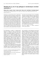
factor activity. For example, as illustrated in Figure 1, epi-
dermal growth factor (EGF) binds to it receptor tyrosine
kinase (EGFR) on the surface of a cell, resulting in the
transmission of a wave of tyrosine, serine, and threonine
phosphorylation events that leads to the activation and
nuclear translocation of several transcription factors,
including STAT-3 (signal transducer and activator of tran-
scription-3) and ERK1/2 (extracellular signal-related
kinase-1/2; also known as mitogen-activated protein
kinase, MAPK). This cascading wave model depends
inherently upon the notion that activated transcription
factors diffuse through the cytoplasm, enter the nucleus,
and execute a program of transcriptional activation. Con-
ceptually, this model is easy to grasp – but does it accu-
rately reflect the biology and the physical constraints of
cellular architecture? The answer appears to be "No", as a
significant body of work over the past decades has chal-
lenged the fundamental validity of the diffusion model
[1-3] and has offered elegant alternative models for the
transmission of intracellular signals [4,5].
Neurons exhibit a unique architecture that places severe
physical limitations on the possible mechanisms for
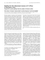
translocation of signals. As shown in Figure 2A, projection
neurons extend axons into target fields over distances that
dwarf the dimensions of the cell body. And yet, the Neu-
rotrophic Factor Hypothesis of neurodevelopment
requires that target-derived soluble trophic factors induce
signals in the presynaptic terminal of axons that result in
transcriptional and translational changes in the nucleus
and neuronal cell body (Figure 2B) [6]. While it is possi-
ble that a signal generated at the plasma membrane of the
presynaptic terminal diffuses along the length of the axon
Simplified diagram showing the activation of STAT-3 and Erk1/2 downstream from EGF binding to EGFRFigure 1
Simplified diagram showing the activation of STAT-3 and Erk1/2 downstream from EGF binding to EGFR. In the general model
of signal transduction, the cascading chain of phosphorylation events culminating in activation of transcription factors such as
STAT-3 and Erk1/2 depends upon the diffusion of these molecules from the site of signal initiation at the plasma membrane to
the site of transcriptional regulation within the nucleus.
Theoretical Biology and Medical Modelling 2005, 2:43 />Page 3 of 15
(page number not for citation purposes)
in order to elicit an effect at the nucleus – it is not at all
probable [5]. For some projection neurons the length of
the axon is five orders of magnitude greater than the diam-
eter of the neuron cell body, and the axoplasm therefore
constitutes 1000-fold more volume than the cytoplasm of
an average cell. The Signaling Endosome Hypothesis pos-
its that an active, directed process of signal transmission is
required to overcome the physical constraints of axonal
distances and volumes [7]. Specifically, this hypothesis
states that the most efficient mechanism for signaling-at-
a-distance involves the packaging of a secreted growth fac-
tor signal into a discrete, coherent, membrane-bounded
organelle that is moved along the length of the axon via a
cytoskeleton-based transport machine (Figure 3) [7].
Indeed, a substantial body of research supports the signal-
ing endosome hypothesis within the context of neuro-
trophin signaling in neurons [8-12]. However, while the
unique geometry of neurons provides a teleological basis
for the existence of signaling endosomes, it is far more
interesting to posit that the signaling endosome hypo-
A) Neurons throughout the nervous system send axonal projections over distances ranging from microns to metersFigure 2
A) Neurons throughout the nervous system send axonal projections over distances ranging from microns to meters. For large
or anatomically specialized animals such as the giraffe or the whale, more than 5 meters may separate the neuron cell body
from the distal axon terminal. B) During development, neurons establish trophic interactions with target tissues. As an organ-
ism develops, the strength and maintenance of these trophic interactions determine whether neurons survive or die. Soluble
protein trophic factors released by the target tissue (1) bind to transmembrane receptors on the presynaptic axon terminal
(2), inducing receptor activation and the induction of intracellular signaling cascades (3). These signals must travel from the site
of initiation to the distant cell body (4) in order to enter the nucleus and elicit transcriptional changes that determine the sur-
vival of the cell. This long-distance information transfer is a universal theme in neurodevelopment.
Theoretical Biology and Medical Modelling 2005, 2:43 />Page 4 of 15
(page number not for citation purposes)
thesis represents a general biological mechanism for sig-
nal transduction and signal compartmentalization [4].
Such a generalized hypothesis might state that the most
efficient mechanism for communicating signals from the
plasma membrane to the nucleus is the compartmentali-
zation of signal transducers into quantal endocytic mem-
brane-associated signaling packets that are retrogradely
transported along microtubules through the cytoplasm.
By utilizing the intrinsic directionality and nucleus-
directed organization of the cellular microtubule network,
signaling endosomes provide a noise-resistant mecha-
nism for the vectorial transport of plasma membrane-
derived signals to the nucleus.
A number of findings support the concept that signaling
from internal cellular membranes is a general phenome-
non that is relevant to understanding receptor tyrosine
kinase signaling in many cellular systems. For example,
EGFR, as discussed above, is internalized via clathrin-
coated vesicles following EGF-binding and receptor acti-
vation [13-15]. In the past, trafficking through this com-
partment was considered part of a normal degradative
process that removes activated receptors from the plasma
membrane and thereby truncates and controls down-
stream signaling [16]. But while this certainly remains a
critical function of endocytosis, recent experiments dem-
onstrate that EGFR remains phosphorylated and active
following internalization [17], and that downstream sign-
aling partners such as Ras colocalize with these internal-
ized, endosome-associated receptors [18-23]. Moreover,
the signals emanating from these internalized EGFR are
biologically meaningful, as cell survival is directly sup-
ported by such signaling [24]. Likewise, Bild and col-
leagues recently observed that STAT-3 signaling initiated
by EGFR activation localized to endocytic vesicles that
moved from the plasma membrane to the nucleus, and
they found that inhibition of EGFR endocytosis prevented
STAT-3 nuclear translocation and abrogated STAT-3-
mediated gene transcription [25]. However, while evi-
dence supports the existence of signaling endosomes, it
does not rule out simultaneous diffusion-based signal
transduction.
We have previously provided evidence that neurotrophin-
induced Erk1/2 signaling from retrogradely transported
endosomes is more efficient than diffusion over distances
ranging from 1.3 microns to 13 microns [7]. We also sug-
gested that the phosphorylation signal associated with sig-
naling endosomes is regenerative [7], consistent with our
previous observations regarding the characterization of
purified signaling endosomes from neurotrophin-stimu-
lated cells [26]. Figure 4 provides additional analysis in
support of the regenerative capacity of signaling endo-
somes. Such signal regeneration is in stark contrast to the
terminal dephosphorylation experienced by diffusing sig-
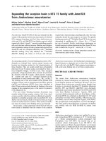
The signaling endosome hypothesis of long-distance axonal signal transmissionFigure 3
The signaling endosome hypothesis of long-distance axonal
signal transmission. Soluble protein trophic factors released
by the target (1) bind to transmembrane receptors on the
presynaptic axon terminal (2), inducing receptor activation
and internalization via clathrin-coated membranes or other
endocytic structures (3). These endocytic vesicles give rise to
transport endosomes that bear the receptor and associated
signaling molecules as well as molecular motors (shown in
turquoise) (4) that utilize microtubules (shown in pink)
within the axon to carry the endosome toward the cell body
(5). Upon arrival at the neuron cell body the endosome-asso-
ciated signals may either initiate additional local signals or
may directly translocate (6) into the nucleus to elicit tran-
scriptional changes (7).
Theoretical Biology and Medical Modelling 2005, 2:43 />Page 5 of 15
(page number not for citation purposes)
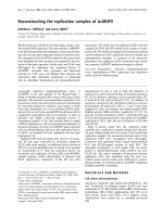
Growth factor receptors are internalized into clathrin-coated vesicles (CCVs) following ligand binding and receptor activation (1–5)Figure 4
Growth factor receptors are internalized into clathrin-coated vesicles (CCVs) following ligand binding and receptor activation
(1–5). These CCVs are uncoated (6) and mature into early endosomes (EE) (7) that may serve as transport endosomes [48].
The concentration of growth factor in transport endosomes is high enough to guarantee effectively 100% receptor occupancy.
Hence, if the endosome-associated receptor encounters a phosphatase, the phosphorylation signal is rapidly regenerated.
Theoretical Biology and Medical Modelling 2005, 2:43 />Page 6 of 15
(page number not for citation purposes)
The Microtubular HighwayFigure 5
The Microtubular Highway. Evidence of the directionality of dynein-mediated retrograde transport.
Theoretical Biology and Medical Modelling 2005, 2:43 />Page 7 of 15
(page number not for citation purposes)
nal transducers, and is a key element in favor of the sign-
aling endosome hypothesis [4,7]. However, our previous
observations depended upon the comparison of the Ein-
stein-Stokes diffusion equation-derived root-mean-square
effective distance for Erk1/2 and the average transport
velocity for nerve growth factor [7]. Such a comparison
overlooks a critical feature of signaling endosome trans-
port and a critical failure of diffusion: directionality. Dif-
fusion is inherently directionless, while the movement of
signaling endosomes along microtubules is inherently
directional and vectorial (see Figure 5 "The Microtubular
Highway"). Likewise, simple modeling of the root-mean-
square effective diffusion distance against transport veloc-
ity ignores dephosphorylation and the regenerative capac-
ity of endosome-associated signals. Herein, we report that
brute-force Monte Carlo (random walk) simulations of
STAT-3 diffusion and dephosphorylation kinetics indi-
cates that facilitated transport of endosomal-based signals
is more efficient than diffusion over even very small cellu-
lar distances. Therefore, we conclude that signaling from
endosomes represents a general biological principle rele-
vant to all cell types and to all signal transduction path-
ways.
Results and discussion
Assumptions – Transport Velocity
For modeling, a dynein-based transport rate of 5 microns
per second is assumed, based on a report by Kikushima
and colleagues [27]. This value was used for ease of calcu-
lation: with a cell radius of 7.5 microns and a nuclear
radius of 2.5 microns, a 5 µm per second transport rate
moves the signaling endosome from the plasma mem-
brane to the nucleus in one second. Actual transport rates
likely range from 1–10 µm per second in cytosol or axo-
plasm [7].
Assumptions – Diffusion Coefficient
The crystal structure of STAT-3B [28], deposited in the
Protein Data Bank as PDB 1BG1 [29], indicates unit cell
dimensions of 17.4 × 17.4 × 7.9 nm. With the caveat that
this structure is bound to an 18-base nucleic acid, the vol-
ume of a STAT-3B molecule is 2400 nm
3
. Assuming a
spherical molecule, STAT-3B therefore has a molecular
radius of approximately 8 nm. Likewise, the molecular
weight of STAT-3 is 100000 Daltons, and therefore one
molecule of STAT-3 weighs 1.7 × 10
-19
g. The Einstein-
Stokes equation for the coefficient of diffusion is:
D = (1/8)(k·T)/(π·γ·η)
where k is Boltzmann's constant, T is absolute tempera-
ture in degrees Kelvin, γ is the radius of the molecule, and
η is the viscosity of an isotropic medium. The viscosity of
axoplasm is approximately 5 centipoise [30], a value that
also approximates cytoplasm [31,32]. Hence,
k = 1.3805 × 10
-20
m
2
·g·(1/(s
2
·K))
T = 310 K
γ = 8 × 10
-9
m
m = 1.7 × 10
-19
g
η = 5 g/(m·s)
Therefore, the coefficient of diffusion for a molecule of
STAT-3 is:
D = 4.3 µm
2
per second
Likewise, the instantaneous velocity v
x
, the step length δ,
and the step rate τ, were derived as:
v
x
= ((k·T)/m)
0.5
= 5 m/s
δ = (1/4)(k·T)/(v
x
·π·γ·η) = 1.7 × 10
-12
m
τ = v
x
/δ = 2.9 × 10
12
sec
-1
It is important to note that our mass estimation may sub-
stantially underestimate the actual mass of the functional
STAT-3 molecular complex, described by Sehgal and col-
leagues as two populations with masses ranging from
200–400 kDa ("Statosome I") to 1–2 MDa ("Statosome
II") [33,34]. Such a massive molecular complex certainly
has important biological implications for STAT-3 diffu-
sion. However, because no crystal structure exists for these
higher molecular weight statosomes from which to calcu-
late the molecular radius, and in order to calculate the
"best-case scenario" for effective diffusion distance, we
have calculated the STAT-3 diffusion coefficient on the
basis of a 100 kDa monomeric molecule. The actual diffu-
sion coefficient for STAT-3 may be 30% of the value calcu-
lated above (assuming 2 MDa mass and a four-fold
increase in molecular radius to account for molecular
packing of the statosome) and the root-mean-square dis-
placement may be 50% of the value calculated below. The
impact of these variables awaits further investigation.
Assumptions – Diffusion Modeling
We modeled diffusion using a random walk algorithm in
two dimensions. The choice of dimensionality was con-
strained by the intensive computational burden associ-
ated with three-dimensional algorithms, as discussed
below (see Methods). At every iteration of the random
walk two pseudo-random numbers (see Methods) were
generated and used to determine the direction of move-
ment in the x-y plane. Using the instantaneous velocity v
x
, the step length δ, and the step rate τ, defined above, we
conclude that a diffusing molecule of STAT-3 will ran-
Theoretical Biology and Medical Modelling 2005, 2:43 />Page 8 of 15
(page number not for citation purposes)
domly walk 3 × 10
12
steps per second, and each step will
be 1.7 × 10
-12
meters long. Thus, the root-mean-square
displacement for STAT-3 diffusion in one second is 2.9
µm. The random walk was modeled on one second of bio-
logical time using a loop of 3 × 10
12
iterations. During
each iteration the molecule randomly moved ± 1.7 × 10
-
12
meters in the x-plane and ± 1.7 × 10
-12
meters in the y-
plane.
Assumptions – Dephosphorylation Kinetics
The decay of a phospho-protein is an exponential func-
tion mapped between the plasma membrane and the
nucleus [5,35]:
α
2
= (K
p
)(L
2
/D)
And the probability function for dephosphorylation is:
p(x)/p(m) = (e
αx
– e
-αx
)/x(e
α
– e
-α
)
Where α is a dimensionless measure of dephosphoryla-
tion probability, K
p
is the first-order rate constant for the
activity of the relevant phosphatase, L is the cell diameter,
D is the diffusion coefficient, x is the distance from the cell
center, and m is the distance from cell center to plasma
membrane normalized to a value of one. α scales such
that for α = 10, half of all phospho-molecules become
dephosphorylated within approximately 0.075 units of
distance from the plasma membrane to the cell center
(e.g. 750 nm for a cell with 10 µm radius) [5]. In general,
K
p
, the first-order rate constant of phosphatase activity,
varies between 0.1 per second and 10 per second [4,35-
37]. For our model K
p
= 5 was assumed, yielding α = 8.1.
With regard to an estimate of enzymatic activity relevant
to dephosphorylation of STAT-3, Todd and colleagues
report a second-order rate constant of 40000/M·s for
dephosphorylation of Erk1/2 [38], which gives:
k
cat
/k
m
= 40000/M·s
Furthermore, Denu and colleagues report that diphos-
phosphorylated Erk1/2 peptides exhibit k
m
values of
approximately 100 µM in vitro [39]. Therefore:
k
cat
= 4/s
Since k
cat
measures the number of substrate molecules
turned over per enzyme per second, a k
cat
of 4 per second
means that, on average, each molecule of enzyme (phos-
phatase) converts (dephosphorylates) 4 substrate mole-
cules every second. Assuming a degree of molecular
similarity between Erk dephosphorylation and STAT-3
dephosphorylation, and for ease of calculation, we set k
cat
= 5 per second. It is important to note that this assump-
tion may not be valid, but has been necessarily adopted in
the absence of better biophysical data in order to illustrate
the potential circumscription of diffusion by dephospho-
rylation.
Assumptions – Dephosphorylation Modeling
The random walk employed for modeling STAT-3 diffu-
sion depends upon the massively iterative generation of
random numbers to describe the movement of the walk-
ing molecule in two-dimensional space. Since significant
computational time was already invested in our diffusion
calculations for the generation of extremely long period
pseudo-random numbers, we opted to model STAT-3
dephosphorylation as a stochastic event using the follow-
ing logic: for any given randomly walking molecule, the
probability of encountering a phosphatase is independent
of both all other molecules and all other steps in the walk.
Therefore, during one second of biological time, equiva-
lent to 3 × 10
12
steps in the random walk, and assuming
that k
cat
= 5 dephosphorylations per second, there will be
1.67 × 10
-12
dephosphorylation events per step. This can
be effectively modeled as a probability test by generating
a pseudo-random number on (0,1) at each step of the ran-
dom walk and asking whether this number is less than
1.67 × 10
-12
. If the test is positive, the molecule is consid-
ered to be "dephosphorylated" and the random walk is
truncated. High-speed modeling of time to dephosphor-
ylation for a large number of molecules (i.e. in the
absence of the random walk) led to a probability function
that matched the equations described by Kholodenko [5].
Results – Diffusion-only Model
Figure 6 shows the result of 12 random walks plotted in
two-dimensional space and compared to the pathlength
of a signaling endosome transported on microtubules. For
these simulations, 500 milliseconds of biological time
were modeled, resulting in the transport of the signaling
endosome over 2.5 µm. The random walks were simu-
lated using only the diffusion coefficient criteria (i.e. no
dephosphorylation modeling) over the same time win-
dow. This figure illustrates the tremendous variability in
the path vector for each of the diffusing particles. While
not unexpected or surprising, Figure 6 offers graphic evi-
dence that the model is working appropriately. Average
pathlength analysis is discussed below.
Results – Diffusion and Dephosphorylation Model
Figure 7 shows the result of 22 random walks modeled
over one second of biological time incorporating both the
diffusion coefficient criteria and the dephosphorylation
probability criteria. Again, the random walks are com-
pared to the pathlength for the transported signaling
endosome, which in this case moves across the entire 5
µm distance separating the plasma membrane and the
nucleus. As with Figure 6, there is a large amount of vari-
Theoretical Biology and Medical Modelling 2005, 2:43 />Page 9 of 15
(page number not for citation purposes)
ability in the diffusion paths, but it is clear that the incor-
poration of dephosphorylation into the model
substantially truncates the effective distance over which a
diffusing molecule of STAT-3 travels. As discussed above,
with α = 8.1, 50% of all phosphorylated molecules should
be dephosphorylated within 0.1 distance units of the
plasma membrane. For our model, this means that 50%
of phospho-STAT-3 molecules should be inactivated
Representative trajectories for 12 random walk simulations using only diffusion criteria (red and blue lines), compared to the movement of a signaling endosome within the same 500 millisecond time frame (green line)Figure 6
Representative trajectories for 12 random walk simulations using only diffusion criteria (red and blue lines), compared to the
movement of a signaling endosome within the same 500 millisecond time frame (green line). Parameters: 15 µm cell diameter,
5 µm nucleus diameter, 37°C, 500 msec, coefficient of diffusion as described in the text. Arrows along the plasma membrane
surface denote the sites of signal initiation.
Theoretical Biology and Medical Modelling 2005, 2:43 />Page 10 of 15
(page number not for citation purposes)
within 750 nm of the plasma membrane (α = 8.1; x = 0.9
for p = 0.5; radius = 7.5 µm; hence x = 6.75 µm, or 750 nm
from the plasma membrane). Likewise, only 15% of
phosphorylated STAT-3 molecules remain active at a dis-
tance half-way between the cell center and the plasma
membrane, and, assuming a nucleus of 2.5 µm radius in a
cell with 7.5 µm radius, fewer than 4% of phosphorylated
molecules will cross the entire distance. Our random walk
Representative trajectories for 22 random walk simulations using both diffusion and dephosphorylation criteria (red and blue lines), compared to the movement of a signaling endosome within the same 1 second time frame (green line)Figure 7
Representative trajectories for 22 random walk simulations using both diffusion and dephosphorylation criteria (red and blue
lines), compared to the movement of a signaling endosome within the same 1 second time frame (green line). Parameters: 15
µm cell diameter, 5 µm nucleus diameter, 37°C, 1 sec, coefficient of diffusion and dephosphorylation probability as described in
the text. Arrows along the plasma membrane surface denote the sites of signal initiation.
Theoretical Biology and Medical Modelling 2005, 2:43 />Page 11 of 15
(page number not for citation purposes)
Endpoint analysis of 100 diffusion-only random walks and 100 diffusion plus dephosphorylation random walksFigure 8
Endpoint analysis of 100 diffusion-only random walks and 100 diffusion plus dephosphorylation random walks. Black lines rep-
resent vectors calculated by the final random walk point for each simulation, compared to the distance covered by a retro-
gradely transported signaling endosome in the same amount of time (green lines). The blue line represents the averaged vector
for 100 diffusion-only random walks, while the red line depicts the averaged vector for 100 diffusion plus dephosphorylation
simulations.
Theoretical Biology and Medical Modelling 2005, 2:43 />Page 12 of 15
(page number not for citation purposes)
incorporating the dephosphorylation probability model
captures the salient features of the expected dephosphor-
ylation kinetics.
Results – Endpoint Analysis of Both Models
Finally, Figure 8 illustrates the endpoint analysis for 100
diffusion-only random walks and 100 diffusion plus
dephosphorylation walks. It should be noted that each
random walk required, on average, more than 48 hours of
dedicated processor time. For this analysis, the final coor-
dinate of each diffusing molecule was used to calculate a
vector for the random walk (i.e. distance and direction
from point of origin). Of the 200 vectors calculated under
both models, no diffusing molecule intersected the
nuclear membrane within the computed timeframe. In
contrast, for the one second computations incorporating
both diffusion and dephosphorylation, the retrogradely
transported signaling endosome reaches the nucleus with
the STAT-3 phosphorylation state intact. Finally, the
observed root-mean-square displacement for the 100
dephosphorylation model random walks was 0.96 µm ±
0.1 µm, or less than 20% of the distance from the plasma
membrane to the nucleus. As calculated above using only
the step length and step rate derived from the coefficient
of diffusion parameters, the predicted root-mean-square
displacement for STAT-3 is 2.9 µm. Thus, the observed
effective distance for a phosphorylated STAT-3 molecule is
one-third of the predicted distance, indicating that our
previously published analysis substantially overestimated
the range over which diffusion efficiently transmits intra-
cellular information.
Predictions
Using the observed root-mean-square displacement after
one second of biological time to establish an adjustment
factor (33% of predicted), and assuming that the relation-
ship between observed and predicted values is linear
through time, we generated the plots shown in Figure 9.
Figure 9A shows that the signaling endosome becomes
more efficient at transmitting information from the
plasma membrane over distances greater than 2 microns
(greater than 400 milliseconds of biological time) using
the predicted root-mean-square displacement values for
comparison. However, using the adjusted root-mean-
square displacement values for comparison, the signaling
endosome is more efficient than diffusion within 200
nanometers from the plasma membrane (within 40 milli-
seconds of biological time) (Figure 9B). Therefore, our
model predicts that the facilitated retrograde transport of
signaling endosomes is a more efficient mechanism of
information transfer from the plasma membrane to the
nucleus, and is, in fact, more efficient for the transmission
of phosphorylated STAT-3 signals over any distance
greater than only 200 nanometers.
Caveats and Future Directions
The signaling endosome retrograde transport rate utilized
in our model may overestimate the actual transport veloc-
ity, especially as an average across the entire lifetime of the
endosome-associated signal. The rate we modeled did not
account for the kinetics of endocytosis or of vesicle load-
ing onto the microtubule network. Our previous observa-
tions suggested transport velocities that ranged from 5.6
µm per second to 0.56 µm per second [7], but experiments
addressing real transport rates for a variety of signaling
molecules are required to improve our model. On the
other hand, while we potentially overestimated the retro-
A and B) Diffusion modeling incorporating dephosphoryla-tion kinetics indicates substantial truncation of the root-mean-square (r.m.s.) displacement for STAT-3 diffusion (dashed red line compared to solid red line)Figure 9
A and B) Diffusion modeling incorporating dephosphoryla-
tion kinetics indicates substantial truncation of the root-
mean-square (r.m.s.) displacement for STAT-3 diffusion
(dashed red line compared to solid red line). This has the
effect of reducing the crossing point at which signaling endo-
some transport (solid blue line) overcomes diffusion (ca. 2
µm for theoretical r.m.s. vs. transport reduced to ca. 200 nm
for adjusted r.m.s. vs. transport). B shows same data as A at
higher Y-axis magnification.
Theoretical Biology and Medical Modelling 2005, 2:43 />Page 13 of 15
(page number not for citation purposes)
grade transport rate for the signaling endosome, we also
very likely overestimated the size of the effective diffusion
domain due to the two-dimensional restrictions of our
current model. While the cytoskeletal transport of the sig-
naling endosome is inherently a dimensionally-restricted
vectorial event, diffusion within the cell most certainly
occurs in three dimensions. Our current model predicts a
three-fold reduction in the actual root-mean-square dis-
placement for STAT-3 as compared to the predicted dis-
placement using a two-dimensional random walk model,
and we predict that a model incorporating three dimen-
sions will exhibit even greater curtailment of the effective
spatial domain for diffusion. However, the addition of a
third dimension to the random walk simulations substan-
tially increases computational demand, and therefore this
analysis awaits either a more efficient algorithm or more
computer time. Our current and future goals are to paral-
lelize the random walk algorithm in order to perform
massively parallel diffusion simulations in three dimen-
sions.
Conclusion
Molecular diffusion obviously benefits from the
extremely high molecular velocities of single particles
moving in a vacuum. For gases and other very small mol-
ecules and under conditions of low viscosity or high tem-
perature, diffusion is extremely fast and far-ranging.
However, within the context of biological molecules and
biological viscosities, diffusion is vastly circumscribed [1-
3,40]. Despite the limitations imposed by biological
parameters, diffusion at first glance still appears to be a
viable mechanism for the transmission of information
through cytoplasm. In fact, the "textbook" conception of
signal transduction depends upon the free diffusion of
signaling molecules. However, closer scrutiny finds sev-
eral faults in the diffusion model [1]. For example, diffu-
sion is certainly directionless – even within the context of
a bounded space such as the cell, the majority of molecu-
lar motions taken by a diffusing molecule are non-pro-
ductive with regard to movement of signals toward a
target (such as the nucleus). Likewise, a diffusing molecu-
lar signal is a ready target for interaction with and trunca-
tion by cytoplasmic phosphatases. Certainly, the effective
range over which a diffusing signal maintains informa-
tional integrity depends upon the concentration and
activity of equally randomly diffusing phosphatases, but it
also seems likely that cells maintain levels of phosphatase
sufficient to prevent run-away signal transduction
[41,42]. Thus, diffusion of information is limited by both
lack of direction and inevitable signal elimination. In dis-
tinct contrast, the retrograde movement of quantal signal-
ing units capable of regenerating the information content
of the original stimulus is inherently vectorial. Therefore,
signaling endosomes, despite an overall lower transport
velocity compared to diffusion velocities, exhibit charac-
teristics of an optimized information transmission sys-
tem. We previously sought to determine the effective
range over which Erk1/2 signaling endosomes exhibited
greater efficiency than diffusing Erk1/2 molecules [7].
This work relied upon the direct comparison of the root-
mean-square displacement for phosphorylated Erk1/2
with the retrograde transport velocity of neurotrophin-
induced signaling endosomes. In an effort to refine this
model we incorporated in our present study the addi-
tional element of dephosphorylation kinetics. Thus our
current model addresses both the non-vectorial nature of
diffusion and the inherent susceptibility to signal trunca-
tion by interaction with cellular phosphatases. Using an
iterative random walk modeling scheme we determined
that the root-mean-square displacement predicted by the
coefficient of diffusion for STAT-3 overestimated the root-
mean-square displacement observed in our simulations
by a factor of 3. Incorporating this scaling factor into the
equation for root-mean-square displacement through
time, we found that signaling endosomes become more
effective at the transmission of information when the dis-
tance from the plasma membrane exceeds 200 nanome-
ters. This observation suggests that any cellular situation
that requires the transmission of information in the form
of phosphorylated signaling molecules over distances in
excess of 200 nanometers would benefit from the packag-
ing of such signals into quantal, cytoskeleton-associated
signaling packets such as signaling endosomes.
Our model suggests that cells utilize two distinct informa-
tion transmission paradigms: 1) fast local signaling via
diffusion over spatial domains on the order of less than
200 nanometers; 2) long-distance (>200 nanometers) sig-
naling via information packets associated with the
cytoskeletal transport apparatus. Moreover, while we have
focused explicitly on the role of signaling endosomes
derived from the internalization of plasma membrane
receptor tyrosine kinases and associated downstream sig-
naling partners, our model suggests that any signal that
must move from the outer reaches of the cytoplasm to the
perinuclear region would benefit from an association with
the retrograde transport machine. For example, transcrip-
tion factors may associate directly with molecular motors
and chaperone proteins that protect them from dephos-
phorylation in a nonvesiculated manner that takes advan-
tage of directional retrograde transport in the absence of a
plasma-membrane-derived organelle. Such a mechanism
was recently proposed for the transport of soluble (i.e.
non-membrane-associated) activated Erk1/2 within
injured axons [43]. Thus, our model supports previous
observations suggesting that the signaling endosome
hypothesis is a subset of a more general hypothesis that
the most efficient mechanism for intracellular signaling-
at-a-distance involves the association of signaling mole-
cules with molecular motors that move along the
Theoretical Biology and Medical Modelling 2005, 2:43 />Page 14 of 15
(page number not for citation purposes)
cytoskeleton [4]. The additional benefit provided by the
cytoskeletal association of membrane-bounded com-
plexes that package a ligand-bound transmembrane
receptor with downstream effector molecules is the ability
to regenerate the signal at any point along the transmis-
sion path [7]. We conclude that signaling endosomes pro-
vide unique information transmission properties relevant
to all cell architectures, and we propose that the majority
of relevant information transmitted from the plasma
membrane to the nucleus will be found in association
with organelles of endocytic origin.
Methods
Pseudo-Random Number Generation
It should be self-evident that "built-in" pseudo-random
number generators (RNGs) available in the majority of
operating systems and programming languages are essen-
tially useless for large-scale Monte Carlo simulations [44].
However, during our initial efforts to optimize the
processing time for the one-second simulations we exper-
imented with several common RNGs; all failed to exhibit
sufficiently long periods, a failure that was manifested in
an initial period of random walking followed by capture
in a continuously repeating cyclical path. We also experi-
mented with an implementation of the Mersenne Twister
algorithm, which exhibited a robust period (theoretically
2
19937
-1) and computational demand comparable to
many other standard RNGs [45]. However, our final opti-
mized diffusion-only code utilized a multiply-with-carry
RNG (MWC) described by George Marsaglia [44,46,47].
The MWC algorithm generates extremely long-period
pseudo-random numbers on [0,1], and we utilized this
very efficient RNG for Boolean testing of step direction in
two dimensions. For the combined diffusion and dephos-
phorylation models, we used the Mersenne Twister mod-
ified to generate pseudo-random numbers on (0,1) for the
probabilistic determination of a dephosphorylation event
and the MWC algorithm for step direction determination.
Hardware
We utilized a variety of platforms for development, test-
ing, and implementation of the diffusion models, includ-
ing the IBM Power4 p690 supercomputer (running AIX
5.2) and the SGI Altix 3700 supercomputer (running SGI
Advanced Linux 3.4) at the University of Minnesota
Supercomputing Institute. The serial models described
above were primarily implemented on a single processor
Intel P4 3.0 GHz machine running Red Hat Linux 9.0. The
IBM Power4, the SGI Altix 3700, and a dual processor
Xeon 3.0 GHz Nocona box running Red Hat Enterprise
Linux 3.0 were used for development and testing of paral-
lel implementations. Total wallclock time on all platforms
currently exceeds 10000 hours.
Software
All algorithms were coded in C and compiled with gcc or
xlc (serial implementations) or with pgcc, xlc, or icc
(OpenMP parallel implementations). Our first diffusion
model efforts required more than one week of dedicated
processing time per walk; after several rounds of code
optimization we could obtain one second of simulated
time in approximately 48 hours on the Power4 architec-
ture and the Pentium 4 architecture described above.
Competing interests
The author(s) declare that they have no competing inter-
ests.
Authors' contributions
The author contributed to all phases of the work.
Acknowledgements
The author thanks the University of Minnesota Supercomputing Institute
(MSI)
for access to the IBM Power4 pSeries 690
and to the SGI Altix supercomputers. The author also thanks Dr. Birali
Runesha of the MSI for technical assistance. This work was supported by
Donald and Frances Herdrich and by grant RG3636 from the National Mul-
tiple Sclerosis Society.
References
1. Agutter PS, Malone PC, Wheatley DN: Intracellular transport
mechanisms: a critique of diffusion theory. J Theor Biol 1995,
176:261-272.
2. Agutter PS, Malone PC, Wheatley DN: Diffusion theory in biol-
ogy: a relic of mechanistic materialism. J Hist Biol 2000,
33:71-111.
3. Agutter PS, Wheatley DN: Random walks and cell size. Bioessays
2000, 22:1018-1023.
4. Kholodenko BN: MAP kinase cascade signaling and endocytic
trafficking: a marriage of convenience? Trends Cell Biol 2002,
12:173-177.
5. Kholodenko BN: Four-dimensional organization of protein
kinase signaling cascades: the roles of diffusion, endocytosis
and molecular motors. J Exp Biol 2003, 206:2073-2082.
6. Sofroniew MV, Howe CL, Mobley WC: Nerve growth factor sig-
naling, neuroprotection, and neural repair. Annu Rev Neurosci
2001, 24:1217-1281.
7. Howe CL, Mobley WC: Signaling endosome hypothesis: A cel-
lular mechanism for long distance communication. J Neurobiol
2004, 58:207-216.
8. Miaczynska M, Pelkmans L, Zerial M: Not just a sink: endosomes
in control of signal transduction. Curr Opin Cell Biol 2004,
16:400-406.
9. Guzik BW, Goldstein LS: Microtubule-dependent transport in
neurons: steps towards an understanding of regulation, func-
tion and dysfunction. Curr Opin Cell Biol 2004, 16:443-450.
10. Zweifel LS, Kuruvilla R, Ginty DD: Functions and mechanisms of
retrograde neurotrophin signalling. Nat Rev Neurosci 2005,
6:615-625.
11. Weible MW, Hendry IA: What is the importance of multivesic-
ular bodies in retrograde axonal transport in vivo? J Neurobiol
2004, 58:230-243.
12. Howe CL, Mobley WC: Long-distance retrograde neurotrophic
signaling. Curr Opin Neurobiol 2005, 15:40-48.
13. Carpenter G: The EGF receptor: a nexus for trafficking and
signaling. Bioessays 2000, 22:697-707.
14. Sorkin A: Internalization of the epidermal growth factor
receptor: role in signalling. Biochem Soc Trans 2001, 29:480-484.
15. Sorkin A, Von Zastrow M: Signal transduction and endocytosis:
close encounters of many kinds. Nat Rev Mol Cell Biol 2002,
3:600-614.
Theoretical Biology and Medical Modelling 2005, 2:43 />Page 15 of 15
(page number not for citation purposes)
16. Le Roy C, Wrana JL: Clathrin- and non-clathrin-mediated
endocytic regulation of cell signalling. Nat Rev Mol Cell Biol 2005,
6:112-126.
17. Sorkin A, McClure M, Huang F, Carter R: Interaction of EGF
receptor and grb2 in living cells visualized by fluorescence
resonance energy transfer (FRET) microscopy. Curr Biol 2000,
10:1395-1398.
18. Wiley HS, Burke PM: Regulation of receptor tyrosine kinase sig-
naling by endocytic trafficking. Traffic 2001, 2:12-18.
19. Roy S, Wyse B, Hancock JF: H-Ras signaling and K-Ras signaling
are differentially dependent on endocytosis. Mol Cell Biol 2002,
22:5128-5140.
20. Pol A, Calvo M, Enrich C: Isolated endosomes from quiescent
rat liver contain the signal transduction machinery. Differen-
tial distribution of activated Raf-1 and Mek in the endocytic
compartment. FEBS Lett 1998, 441:34-38.
21. Jiang X, Sorkin A: Coordinated traffic of Grb2 and Ras during
epidermal growth factor receptor endocytosis visualized in
living cells. Mol Biol Cell 2002, 13:1522-1535.
22. Chiu VK, Bivona T, Hach A, Sajous JB, Silletti J, Wiener H, Johnson RL,
Cox AD, Philips MR: Ras signalling on the endoplasmic reticu-
lum and the Golgi. Nat Cell Biol 2002, 4:343-350.
23. Burke P, Schooler K, Wiley HS: Regulation of epidermal growth
factor receptor signaling by endocytosis and intracellular
trafficking. Mol Biol Cell 2001, 12:1897-1910.
24. Wang Y, Pennock S, Chen X, Wang Z: Endosomal signaling of epi-
dermal growth factor receptor stimulates signal transduc-
tion pathways leading to cell survival. Mol Cell Biol 2002,
22:7279-7290.
25. Bild AH, Turkson J, Jove R: Cytoplasmic transport of Stat3 by
receptor-mediated endocytosis. Embo J 2002, 21:3255-3263.
26. Howe CL, Valletta JS, Rusnak AS, Mobley WC: NGF signaling from
clathrin-coated vesicles: evidence that signaling endosomes
serve as a platform for the Ras-MAPK pathway. Neuron 2001,
32:801-814.
27. Kikushima K, Yagi T, Kamiya R: Slow ADP-dependent accelera-
tion of microtubule translocation produced by an axonemal
dynein. FEBS Lett 2004, 563:119-122.
28. Becker S, Groner B, Muller CW: Three-dimensional structure of
the Stat3beta homodimer bound to DNA. Nature 1998,
394:145-151.
29. Berman HM, Westbrook J, Feng Z, Gilliland G, Bhat TN, Weissig H,
Shindyalov IN, Bourne PE: The Protein Data Bank. Nucleic Acids
Res 2000, 28:235-242.
30. Haak RA, Kleinhans FW, Ochs S: The viscosity of mammalian
nerve axoplasm measured by electron spin resonance. J Phys-
iol 1976, 263:115-137.
31. Luby-Phelps K: Cytoarchitecture and physical properties of
cytoplasm: volume, viscosity, diffusion, intracellular surface
area. Int Rev Cytol 2000, 192:189-221.
32. Luby-Phelps K, Lanni F, Taylor DL: The submicroscopic proper-
ties of cytoplasm as a determinant of cellular function. Annu
Rev Biophys Biophys Chem 1988, 17:369-396.
33. Guo GG, Patel K, Kumar V, Shah M, Fried VA, Etlinger JD, Sehgal PB:
Association of the chaperone glucose-regulated protein 58
(GRP58/ER-60/ERp57) with Stat3 in cytosol and plasma
membrane complexes. J Interferon Cytokine Res 2002, 22:555-563.
34. Ndubuisi MI, Guo GG, Fried VA, Etlinger JD, Sehgal PB: Cellular
physiology of STAT3: Where's the cytoplasmic monomer? J
Biol Chem 1999, 274:25499-25509.
35. Kholodenko BN, Brown GC, Hoek JB: Diffusion control of pro-
tein phosphorylation in signal transduction pathways. Bio-
chem J 2000, 350 Pt 3:901-907.
36. Haugh JM, Lauffenburger DA: Physical modulation of intracellu-
lar signaling processes by locational regulation. Biophys J 1997,
72:2014-2031.
37. Zhao Y, Zhang ZY: The mechanism of dephosphorylation of
extracellular signal-regulated kinase 2 by mitogen-activated
protein kinase phosphatase 3. J Biol Chem 2001,
276:32382-32391.
38. Todd JL, Tanner KG, Denu JM: Extracellular regulated kinases
(ERK) 1 and ERK2 are authentic substrates for the dual-spe-
cificity protein-tyrosine phosphatase VHR. A novel role in
down-regulating the ERK pathway. J Biol Chem 1999,
274:13271-13280.
39. Denu JM, Zhou G, Wu L, Zhao R, Yuvaniyama J, Saper MA, Dixon JE:
The purification and characterization of a human dual-spe-
cific protein tyrosine phosphatase. J Biol Chem 1995,
270:3796-3803.
40. Wheatley DN: Diffusion theory, the cell and the synapse. Bio-
systems 1998, 45:151-163.
41. Hunter T: Signaling 2000 and beyond. Cell 2000, 100:113-127.
42. Blume-Jensen P, Hunter T: Oncogenic kinase signalling. Nature
2001, 411:355-365.
43. Perlson E, Hanz S, Ben-Yaakov K, Segal-Ruder Y, Seger R, Fainzilber
M: Vimentin-dependent spatial translocation of an activated
MAP kinase in injured nerve. Neuron 2005, 45:715-726.
44. L'Ecuyer P: Random Number Generation. In Handbook of Com-
putational Statistics Edited by: Gentle JE, Haerdle W and Mori Y. ,
Springer-Verlag; 2004:35-70.
45. Matsumoto M, Nishimura T: Mersenne Twister: A 623-dimen-
sionally equidistributed uniform pseudo-random number
generator. ACM Transactions on Modeling and Computer Simulation
1998, 8:3-30.
46. L'Ecuyer P, Blouin F, Couture R: A search for good multiple
recursive random number generators. ACM Transactions on
Modeling and Computer Simulation 1993, 3:87-98.
47. Goresky M, Klapper A: Efficient multiply-with-carry random
number generators with maximal period. ACM Transactions on
Modeling and Computer Simulation 2003, 13:1-12.
48. Delcroix JD, Valletta JS, Wu C, Hunt SV, Kowal AS, Mobley WC:
NGF Signaling in sensory neurons: evidence that early endo-
somes carry NGF retrograde signals. Neuron 2003, 39:69-84.
49. Valetti C, Wetzel DM, Schrader M, Hasbani MJ, Gill SR, Kreis TE,
Schroer TA: Role of dynactin in endocytic traffic: effects of
dynamitin overexpression and colocalization with CLIP-170.
Mol Biol Cell 1999, 10:4107-4120.
50. Quintyne NJ, Gill SR, Eckley DM, Crego CL, Compton DA, Schroer
TA: Dynactin is required for microtubule anchoring at cen-
trosomes. J Cell Biol 1999, 147:321-334.
51. King SJ, Brown CL, Maier KC, Quintyne NJ, Schroer TA: Analysis of
the dynein-dynactin interaction in vitro and in vivo. Mol Biol
Cell 2003, 14:5089-5097.
52. Burkhardt JK, Echeverri CJ, Nilsson T, Vallee RB: Overexpression
of the dynamitin (p50) subunit of the dynactin complex dis-
rupts dynein-dependent maintenance of membrane
organelle distribution. J Cell Biol 1997, 139:469-484.
53. Lakadamyali M, Rust MJ, Babcock HP, Zhuang X: Visualizing infec-
tion of individual influenza viruses. Proc Natl Acad Sci U S A 2003,
100:9280-9285.
54. Heerssen HM, Pazyra MF, Segal RA: Dynein motors transport
activated Trks to promote survival of target-dependent neu-
rons. Nat Neurosci 2004, 7:596-604.
55. Watson FL, Heerssen HM, Moheban DB, Lin MZ, Sauvageot CM,
Bhattacharyya A, Pomeroy SL, Segal RA: Rapid nuclear responses
to target-derived neurotrophins require retrograde trans-
port of ligand-receptor complex. J Neurosci 1999, 19:7889-7900.
56. Yano H, Lee FS, Kong H, Chuang J, Arevalo J, Perez P, Sung C, Chao
MV: Association of Trk neurotrophin receptors with compo-
nents of the cytoplasmic dynein motor. J Neurosci 2001,
21:RC125.
57. Bhattacharyya A, Watson FL, Pomeroy SL, Zhang YZ, Stiles CD, Segal
RA: High-resolution imaging demonstrates dynein-based
vesicular transport of activated Trk receptors. J Neurobiol
2002, 51:302-312.
58. Ehlers MD, Kaplan DR, Price DL, Koliatsos VE: NGF-stimulated
retrograde transport of trkA in the mammalian nervous sys-
tem. J Cell Biol 1995, 130:149-156.