Báo cáo y học: "Stochastic modeling of oligodendrocyte generation in cell culture: model validation with time-lapse data" pdf
Bạn đang xem bản rút gọn của tài liệu. Xem và tải ngay bản đầy đủ của tài liệu tại đây (279.68 KB, 8 trang )
BioMed Central
Page 1 of 8
(page number not for citation purposes)
Theoretical Biology and Medical
Modelling
Open Access
Research
Stochastic modeling of oligodendrocyte generation in cell culture:
model validation with time-lapse data
Ollivier Hyrien
1
, Ibro Ambeskovic
2
, Margot Mayer-Proschel
2
, Mark Noble
2
and Andrei Yakovlev*
1
Address:
1
Department of Biostatistics and Computational Biology, University of Rochester, 601 Elmwood Avenue, Rochester, New York 14642,
USA and
2
Department of Biomedical Genetics, University of Rochester, 601 Elmwood Avenue, Rochester, New York 14642, USA
Email: Ollivier Hyrien - ; Ibro Ambeskovic - ; Margot Mayer-
Proschel - ; Mark Noble - ;
Andrei Yakovlev* -
* Corresponding author
Abstract
Background: The purpose of this paper is two-fold. The first objective is to validate the
assumptions behind a stochastic model developed earlier by these authors to describe
oligodendrocyte generation in cell culture. The second is to generate time-lapse data that may help
biomathematicians to build stochastic models of cell proliferation and differentiation under other
experimental scenarios.
Results: Using time-lapse video recording it is possible to follow the individual evolutions of
different cells within each clone. This experimental technique is very laborious and cannot replace
model-based quantitative inference from clonal data. However, it is unrivalled in validating the
structure of a stochastic model intended to describe cell proliferation and differentiation at the
clonal level. In this paper, such data are reported and analyzed for oligodendrocyte precursor cells
cultured in vitro.
Conclusion: The results strongly support the validity of the most basic assumptions underpinning
the previously proposed model of oligodendrocyte development in cell culture. However, there
are some discrepancies; the most important is that the contribution of progenitor cell death to cell
kinetics in this experimental system has been underestimated.
Background
The theory of branching stochastic processes has proved a
powerful tool for cell kinetics in general and for analyzing
clonal growth of cultured cells in particular. The ongoing
development of mathematical aspects of this theory is fre-
quently stimulated by or directed towards applied prob-
lems. A comprehensive account of the theory and some
biological applications are given in books by Harris [1],
Sevastyanov [2], Mode [3], Athreya and Ney [4], Jagers [5],
Assmussen and Hering [6], Yakovlev and Yanev [7], Gut-
torp [8], Kimmel and Axelrod [9] and Haccou et al. [10].
Since the choice of a particular model is frequently deter-
mined by its tractability, the Bellman-Harris branching
process and its modifications have been traditionally con-
sidered as a fairly general framework for cell kinetics stud-
ies. The multi-type version of this process is defined as
Published: 17 May 2006
Theoretical Biology and Medical Modelling 2006, 3:21 doi:10.1186/1742-4682-3-21
Received: 06 April 2006
Accepted: 17 May 2006
This article is available from: />© 2006 Hyrien et al; licensee BioMed Central Ltd.
This is an Open Access article distributed under the terms of the Creative Commons Attribution License ( />),
which permits unrestricted use, distribution, and reproduction in any medium, provided the original work is properly cited.
Theoretical Biology and Medical Modelling 2006, 3:21 />Page 2 of 8
(page number not for citation purposes)
follows. Let , i,k = 1, , K be the number of cells of
the k
th
type at time t given that the clonal growth starts
with a single (initiator) cell of type i at time t = 0. The vec-
tor Z
(i)
(t) = ( , , ) is said to be a Bellman-
Harris branching stochastic process with K types of cells if
the following conditions are met. Each cell of type k, 1 ≤ k
≤ K, transforms into j
1
, , j
K
, daughter cells of types 1, ,
K, respectively, with probability p
k
(j
1
, , j
K
). The time to
transformation is a non-negative random variable (r.v.)
with cumulative distribution function (c.d.f.) F
k
(.). The
usual independence assumptions are adopted.
The problem of quantitative inference from clonal data on
cell development in tissue culture has been addressed in
our publications [11-21]. These papers employ a multi-
type Bellman-Harris branching process to model the pro-
liferation of oligodendrocyte/type-2 astrocyte progenitor
cells and their transformation into terminally differenti-
ated oligodendrocytes. This model is widely applicable to
other in vitro cell systems. The precursor cell that gives rise
directly to oligodendrocytes was first discovered by Raff,
Miller and Noble in 1983 [22], when it was named as an
oligodendrocyte/type-2 astrocyte (O-2A) progenitor for
the two cell types it could generate in vitro. This cell is also
known as an oligodendrocyte precursor cell (OPC), and
will be referred to henceforth as an O-2A/OPC. Such cells
appear to be present in various regions of the perinatal rat
CNS, and cells with similar properties also have been iso-
lated from the human CNS [23].
The O2A/OPC-oligodendrocyte lineage has provided a
remarkably useful system for studying general problems
in cellular and developmental biology. In the context of
our present studies, three advantages of this lineage are
that it is possible to analyze progenitor cells grown at the
clonal level, that progenitor cells and oligodendrocytes
can be readily distinguished visually, and that the genera-
tion of oligodendrocytes is associated with exit from the
cell cycle. In the culture system we use in our experiments,
the dividing O-2A/OPCs only make either more progeni-
tor cells or oligodendrocytes; no other branching in the
process of their development is possible. This makes it
possible to conduct well-controlled experiments that gen-
erate quantitative information on cell division and differ-
entiation at the clonal level and at the level of individual
cells.
The earlier proposed model was designed to describe the
development of cell clones derived from O-2A/OPCs
under in vitro conditions. Cells of this type are partially
committed to further differentiation into oligodendro-
cytes but they retain the ability to proliferate. It is believed
that the main function of progenitor cells in vivo is to pro-
vide a quick proliferative response to an increased
demand for cells in the population. Terminally differenti-
ated oligodendrocytes represent a final cell type; they are
responsible for maintaining tissue-specific functions and
they do not divide under normal conditions. Both cell
types are susceptible, in variable degrees, to death.
A substantial amount of new biological knowledge has
emerged from applications of our model to experimental
data, with a particular focus on understanding the regula-
tion of differentiation at the clonal level. As all differenti-
ation processes require that cells make a decision between
differentiating and not differentiating, it is important to
understand how this process is controlled at the level of
the individual dividing precursor cell. Early studies had
indicated that individual O-2A/OPCs would divide a lim-
ited number of times before all clonally related cells dif-
ferentiated synchronously and symmetrically under the
control of a cell-intrinsic biological clock. Subsequent
biological studies showed that the cell-intrinsic regulator
of differentiation promoted asymmetric and asynchro-
nous differentiation among clonally related cells unless
promoters of oligodendrocyte generation were present. It
was only through our modeling studies, however, that the
popular clock model of oligodendrocyte generation in
vitro was disproved by testing a more general (hierarchi-
cal) model against experimental data [11,15].
In the earliest version of our model [11,15], it was
assumed that the initial population of progenitors is a
mixture of subpopulations with different numbers of
"critical" cycles. In each of these subpopulations the prob-
ability of division is 1 until the critical number is reached
and drops sharply to a fixed value p < 0.5 afterwards. The
number of critical cycles is not directly observed, and one
can only verify this basic assumption by fitting the model
to experimental data on the evolution (over time) of
clones consisting of two distinct types of cells. However, if
one considers the whole population of cells, there is a
more gradual decline in the division probability from 1 to
p, suggesting that an alternative model is also plausible, in
which there is a single population of progenitor cells with
a gradually decreasing division probability [17]. While
both models are in almost equally good agreement with
clonal data, the latter model has a more parsimonious
structure, which is also perfectly consistent with the time-
lapse data to be reported in the present paper.
The basic stochastic model of proliferation and differenti-
ation of O-2A/OPCs was based on the following assump-
tions:
A1. The process starts with a single progenitor cell of type
1 at time 0.
Zt
k
i()
()
Zt
i
1
()
() Zt
K
i()
()
Theoretical Biology and Medical Modelling 2006, 3:21 />Page 3 of 8
(page number not for citation purposes)
A2. After completion of its mitotic cycle, every progenitor
cell of type l ≥ 1 either divides to produce two new progen-
itor cells of age 0 and type l + 1 with probability p
l
, or
transforms into a differentiated cell of type l = 0 (oli-
godendrocyte) with probability 1 - p
l
.
A3. The time to division of a progenitor cell of type l ≥ 1
is a non-negative r.v. T
l,1
with c.d.f. F
1
(x), while the time to
differentiation of a progenitor cell of type l ≥ 1 is a non-
negative r.v. T
l,2
with c.d.f. F
2
(x).
A4. Differentiated oligodendrocytes neither divide nor
differentiate further, but they may die; their lifespan T
0
has
c.d.f. L(x) = Pr(T
0
≤ x).
A5. Whenever counts of dead oligodendrocytes are uti-
lized for estimation purposes, the model needs to be
extended further to include the following assumption:
every dead oligodendrocyte disappears (disintegrates)
from the field of observation after a random lapse of time
T
-1
distributed in accordance with c.d.f. H(x) = Pr(T
-1
≤ x).
The time to the disintegration event is expected to be quite
long, as there are no macrophages present in the culture to
clear away cell debris.
A6. The cells do not migrate out of the field of observa-
tion.
A7. Of the two cell types, oligodendrocytes appear to be
more susceptible to death. Therefore, it was assumed that
progenitor cells do not die during the period of observa-
tion.
A8. The assumption of independence of cell evolutions is
adopted. This assumption is critical for making the math-
ematical treatment of the resultant branching stochastic
process tractable.
The probabilities p
l
can be described by an arbitrary func-
tion of the mitotic cycle label l that satisfies the natural
constraints: 0 ≤ p
l
≤ 1 for all l ≥ 1. In [17], these probabil-
ities are specified as p
l
= min{p + qr
l
, 1}, where p, q and r
are free positive parameters with p representing the limit-
ing probability of division of progenitor cells as the
number of cycles tends to infinity. In our analysis of the
time lapse data in the next section we proceeded from this
choice as well. All the distributions introduced above were
specified by a two-parameter family of gamma distribu-
tions, which is the most popular choice in cell kinetics
studies [7].
Assumption A3 was introduced in [19,20] to allow the
mitotic cycle duration and the time to differentiation to
follow dissimilar distribution functions. The authors pro-
ceeded from the following line of reasoning. In the classi-
cal Bellman-Harris process, either the event of division or
the event of differentiation is allowed to occur upon com-
pletion of the mitotic cycle. Let the r.v.s X and Y represent
the time to division and the time to differentiation,
respectively. Then the postulates of the Bellman-Harris
process imply that the joint distribution of X and Y is sin-
gular along the diagonal X = Y. A natural alternative is to
assume that the r.v.s X and Y have dissimilar continuous
distributions. This alternative is biologically plausible
because the proliferation and differentiation of cells
involve different molecular mechanisms. The analysis of
clonal growth of cultured O-2A/OPCs has corroborated
this hypothesis [19,20], and the time-lapse data presented
in the next section provide additional evidence in favor of
its validity.
In [18], the mitotic cycle duration and the time to differ-
entiation of O-2A/OPCs were assumed to follow the same
distribution, that is, F
1
(x) = F
2
(x) for all x, but we allowed
the distribution of the time to division and differentiation
of initiator cells to be potentially different from that of
cells in subsequent generations. Our time-lapse data pro-
vide the opportunity to look more closely at variations in
the mitotic cycle duration across cell generations and their
consistency with this basic model assumption. The design
of our previous studies generated cell counts in independ-
ent cell clones at different times after plating. We also used
longitudinal data on cell counts produced by observations
of the same cell clone at different time points [20]. How-
ever, much more information can be extracted from data
yielded by time-lapse video recording of individual cell
evolutions, and we take advantage of this experimental
technique to verify the most basic elements of the earlier
proposed model.
Results and discussion
This study is designed to validate the most basic assump-
tions behind our model of oligodendrocyte development
in cell culture. In what follows, we describe our experi-
mental findings in the context of the model presented in
Section 1. Each element of the model structure is dis-
cussed separately.
Mitotic cycle
We estimated the distributions of the mitotic cycle dura-
tion (MCD) for each generation of progenitor cells. The
corresponding Kaplan-Meier estimates are shown on Fig-
ure 1. They suggest that the MCD becomes larger as the
number of divisions undergone by a progenitor cell
increases. However, the log-rank test does not declare
these differences to be statistically significant in all pair-
wise comparisons of the MCD distributions for different
generations starting with Generation 3. The fact that the
MCD distribution in Generation 1 is distinct from those
for other generations is consistent with our previous
Theoretical Biology and Medical Modelling 2006, 3:21 />Page 4 of 8
(page number not for citation purposes)
clonal analyses [18]. The most plausible explanation for
this phenomenon is that the initiator progenitor cells
sampled in vivo are already actively proliferating and,
therefore, it is the residual time needed to complete their
current mitotic cycle that one observes in cell culture. The
second mitotic cycle of the progenitor cells also tends to
be shorter than subsequent cycles in both experimental
settings (with and without thyroid hormone) but no
explanation for this tendency can be offered at present.
The mean MCDs averaged over the generations were esti-
mated as 27.86 hours (standard error (SE) = 0.7 hours)
and 22.13 hours (SE = 1.59 hours) in the presence and
absence of thyroid hormone, respectively. These estimates
are in close agreement with those obtained from clonal
data in our past studies [11-17,20]. However, they are dif-
ferent from those reported in [18,19]. This discrepancy is
attributable to dissimilar activities of the cytokine PDGF-
AA in the culture medium [18]. The effect of thyroid hor-
mone on the MCD distribution is statistically significant
(p < 0.0001).
We designed a parametric bootstrap goodness-of-fit test
based on the Kolmogorov-Smirnov statistic to test the
shape of the MCD distribution. Our study was limited to
Generations 1–3 because censoring (by other events such
as cell differentiation and death) is too heavy in later gen-
erations. A two-parameter gamma distribution provided a
good fit for all generations in the absence of thyroid hor-
mone and for Generations 1 and 3 in the presence of thy-
roid hormone. The only exception was the second
generation in the presence of thyroid hormone. In the lat-
ter (worst) case, the theoretical gamma distribution and
its empirical estimate (kernel estimate with a Gaussian
kernel) still coincide quite closely (Figure 2A) so we see no
immediate need to replace this approximation with a
more flexible parametric family of distributions. For com-
parison, Figure 2B shows another example where the
goodness-of-fit hypothesis was not rejected by the statisti-
cal test.
Probabilities of division, death and differentiation
The probabilities (rates) of death and differentiation
increase with generation while the probability of division
shows the opposite trend. Notice that the rates of death
and differentiation are per cell. The death rate for O-2A/
OPCs increases from 0.23 in Generation 2 to 0.57 in Gen-
eration 7 in the absence of thyroid hormone and from
0.05 in Generation 2 to 0.11 in Generation 5 in its pres-
ence. Therefore, the survival rate of O-2A/OPCs increases
in the presence of thyroid hormone. The probability of
differentiation increases from 0.07 in Generation 2 to
0.21 in Generation 7 in the absence of thyroid hormone
and from 0.18 in Generation 2 to 0.72 in Generation 5 in
its presence. This is consistent with the effect of thyroid
hormone inferred from our previous analyses of clonal
data.
Figure 3 shows the estimated conditional probability of
division, given that the cell does not die before division or
differentiation, as a function of the number of genera-
tions. In [17], we used the function p
l
= min{p + qr
l
, 1}, l
≥ 1, to approximate this probability. The same function
was used to fit the data in Figure 3 by the non-linear least
squares method. Because of conditioning on the event of
cell survival, the probability of differentiation equals 1 - p
l
.
It is clear from Figure 3 that the approximation works
well.
Time to differentiation
The overall mean time to differentiation (averaged over
the generations) is 31.6 hours (SE = 1.6 hours) for O-2A/
OPCs cultured in the presence of thyroid hormone and
31.8 hours (SE = 1.59 hours) in its absence. The time-
lapse data confirm that the time to division and the time
to differentiation have dissimilar distributions, a conjec-
ture we made earlier from the results of clonal data analy-
sis. The distribution of the differentiation time does not
vary significantly across generations (p > 0.28). The addi-
tion of thyroid hormone has no effect on this distribution.
Time to death
The overall mean time to death of O-2A/OPCs (averaged
over the generations) is equal to 28.1 hours (SE = 2.48
hours) and 19.7 hours (SE = 1.17 hours) with and without
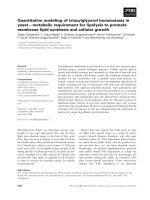
Kaplan-Meier survival curves for the mitotic cycle time across generationsFigure 1
Kaplan-Meier survival curves for the mitotic cycle time
across generations. Generation 1 – dotted line, Generation 2
– dash-dotted line, Generation 3 – dashed line, Generation 4
– solid line. Top panel presents data without thyroid hor-
mone; bottom panel shows data with thyroid hormone in the
culture medium.
0 10 20 30 40 50 60 70 80
0
0.2
0.4
0.6
0.8
1
Survival function
0 10 20 30 40 50 60 70 80
0
0.2
0.4
0.6
0.8
1
Survival function
Time (hours)
A
B
1
Theoretical Biology and Medical Modelling 2006, 3:21 />Page 5 of 8
(page number not for citation purposes)
thyroid hormone, respectively. These values are very close
to the mean mitotic cycle durations recorded in the corre-
sponding experimental settings. The distribution of the
time to death for O-2A/OPCs does not vary significantly
across generations (p > 0.08). Addition of thyroid hor-
mone extends the time to death for O-2A/OPCs (p <
0.0018), which is consonant with its positive effect on cell
survival.
The presence of thyroid hormone extends the life-time of
oligodendrocytes (p < 0.0001) as well. The mean time to
death of an oligodendrocyte is 19.7 hours in the absence
of thyroid hormone but 78.0 hours in its presence. As far
as oligodendrocytes are concerned, the estimated overall
mean time to death tends to be smaller than our estimates
reported in [18] because of the effect of data censoring
caused by a limited period of observation [29]. The time
to death of oligodendrocytes was not significantly differ-
ent across generations no matter whether the cells were
cultured with or without thyroid hormone (p = 0.3 and p
= 0.27).
Correlations
We computed correlation coefficients between the times
to division for all sister cells and for the corresponding
mother-daughter correlations. Because the cells pertaining
to the first and second generations had significantly
shorter mitotic cycles than those in subsequent genera-
tions, we included only the third and later generations in
this analysis. The sample correlation coefficients are
shown in Table 1. It is clear that the mother-daughter type
of correlation is irrelevant to this cell lineage. However,
there is a tangible positive correlation between the mitotic
cycles of sister cells. Both observations are consistent with
the data reported by Powell [24] for bacteria.
One should expect the mean number of cells not to be
affected by this type of correlation, while the variance can
only be larger than that in the independent case [1,25].
This was confirmed by our simulation of a population of
dividing cells obeying the postulates of the bifurcating
autoregressive process. This process [26] reduces to the
Bellman-Harris branching process when sister cells have
uncorrelated MCD. In this study, we assumed that the log-
arithms of mitotic cycle times for sister cells have bivariate
normal distributions with equal means (25 hours) and
equal variances (40 hours), and a fixed positive correla-
tion coefficient denoted by ρ. The bivariate log-normal
distribution was chosen as a convenient parametric family
for modeling correlations between random variables,
while keeping the positivity constraint on cell cycle
lengths. The choice of this distribution (instead of the tra-
ditional gamma distribution) is of little consequence to
the net results of the study. Table 2 displays the standard
deviation of the number of cells in this process for ρ = 0
(independent case) and ρ = 0.5, the latter being a reason-
able value in accordance with Table 1.
The standard deviations of the bifurcating autoregressive
process with correlations among sister cells, and the Bell-
man-Harris process without correlations among sister
cells, were estimated from 50000 simulated runs of each
process. The observed effect of correlations among sister
cells on the standard deviation of the number of cells is
rather weak (Table 2). In terms of parameter estimation,
this effect translates into a change in the mean MCD of
less than 1.5% and a change in the standard deviation of
the MCD of less than 3.4%.
Table 1: Sample correlation coefficients and their asscciated p-values.
correlation type mother-daughter p-value sister-sister p-value
without thyroid hormone 0.06 0.6 0.62 <0.0001
with thyroid hormone 0.19 0.5 0.49 0.028
(A) The only case where the null hypothesis is rejected when a gamma distribution density is fitted to observed times to mitotic division; (B) An example where the null hypothesis is not rejected when a gamma distribution density is fitted to observed times to divisionFigure 2
(A) The only case where the null hypothesis is rejected when
a gamma distribution density is fitted to observed times to
mitotic division; (B) An example where the null hypothesis is
not rejected when a gamma distribution density is fitted to
observed times to division.
0 20 40 60 80
0
0.005
0.01
0.015
0.02
0.025
0.03
0.035
0.04
0.045
Time (hours)
Density
0 20 40 60 80
0
0.005
0.01
0.015
0.02
0.025
0.03
0.035
0.04
0.045
Time (hours)
A B
Theoretical Biology and Medical Modelling 2006, 3:21 />Page 6 of 8
(page number not for citation purposes)
Some extensions of the Bellman-Harris branching process
have been proposed to allow for dependences between
cellular attributes across generations. For example, the
bifurcating autoregressive process [26] is designed to
model sister-sister and mother-daughter correlations in
terms of the MCD. It should be noted that this model
describes populations of cells that could divide but nei-
ther die nor differentiate. Further improvements of the
model and associated methods of statistical inference are
being pursued [27]. To the best of our knowledge, the util-
ity of the bifurcating autoregressive process and its various
extensions have so far been considered only in the context
of time-lapse data. This is not surprising because such data
provide abundant information on individual cell evolu-
tions and allow the necessary correlations to be estimated
directly.
The situation is not the same when modeling cell develop-
ment at the clonal (population) level. Except for a few spe-
cial examples, all stochastic models in cell population
kinetics, Markovian or otherwise, disallow for interactions
between individual cell evolutions. The same applies
indiscriminately to all other stochastic models of discrete
entities introduced in mathematical biology, from sto-
chastic models of carcinogenesis or infectious diseases to
applications of stochastic processes in ecology and
demography. There seems to be no viable alternative to
the assumption of independence in all such models as
long as they are intended to describe the events of interest
at the population level so that their underlying stochastic
processes are only partially observed. The main reason for
this claim is that stochastic dependencies, such as correla-
tions among sister cells, are basically unobservable at the
cell population level and this is exactly the point at which
the issue of non-identifiability becomes insurmountable.
This, however, does not apply to functional dependencies
that may manifest themselves in dynamics of the expected
values a typical example is a density dependence such that
the net proliferation rate slows down when a set point is
reached. A functional dependency of this type may still be
identifiable if its structure is parsimonious enough. It
should also be noted that, except in some very special
cases, branching processes with stochastically dependent
cell evolutions are mathematically intractable and we are
unaware of a single publication presenting a sufficiently
general framework for such processes within which the
requisite basic formulae have been derived. Computer
simulations with all their inherent problems are the only
option in such cases.
The aforesaid, however, does not diminish the usefulness
of branching stochastic processes in biological applica-
tions. All indirect quantitative inferences from real biolog-
ical data are conditional on the validity of the assumed
model. In other words, we interpret the results of data
analysis in terms of model parameters as if all the adopted
premises were absolutely valid. In this sense, the assump-
tion of independent evolutions is no different from any
other constraint on model structure. It is commonplace to
say that all models are wrong but some of them are useful.
However, this truism imparts very precisely the essence of
mathematical modeling and its place in natural sciences.
On the other hand, biomathematicians should do the best
they can to make a mathematical model as realistic as pos-
sible, subject to certain constraints on its tractability and
identifiability. While alternative variants of a given model
most typically emerge when it is in conflict with experi-
mental data, the quest for generality is always warranted
in model building. From this perspective, our estimates of
sister-sister correlations and the associated simulation
study are of practical significance because they show that
the observed level of positive correlation among sister
cells has only a small effect on the standard deviation of
the number of cells at any instant. In accordance with the-
Table 2: The standard deviation of a binary splitting Bellman-
Harris branching process (no correlation) and the corresponding
bifurcating autoregressive process (sister-sister correlation).
Time (days) 3 4 5 6 7 8 9 10 11
ρ = 0.0 0.02 0.3 0.7 0.5 1.1 1.2 1.7 2.3 3.0
ρ = 0.5 0.02 0.3 0.8 0.6 1.3 1.4 2.1 2.7 3.5
Conditional (given that the cell does not die before the event of interest) probabilities of division (×) and differentiation (circles) of O-2A/OPCs with (lower panel) and without (upper panel) thyroid hormoneFigure 3
Conditional (given that the cell does not die before the event
of interest) probabilities of division (×) and differentiation
(circles) of O-2A/OPCs with (lower panel) and without
(upper panel) thyroid hormone. The solid lines correspond
to the fitted probabilities of division and differentiation, and
each error bar indicates two standard errors for the empiri-
cal proportion.
1 2 3 4 5 6 7 8
0
0.2
0.4
0.6
0.8
1
without thyroid hormone
1 2 3 4 5 6 7 8
0
0.2
0.4
0.6
0.8
1
with thyroid hormone
generation
Theoretical Biology and Medical Modelling 2006, 3:21 />Page 7 of 8
(page number not for citation purposes)
oretical considerations, this correlation does not affect the
expected values at all. These observations provide a
rationale for using the method of moments for estimation
purposes because, when based only on the mean values
and standard deviations, this method appears to be well
guarded against correlations between sister cells.
Other interesting observations
We noticed two unusual events. The first is where the two
daughters do not separate fully from each other following
division from a mother. For a brief time it looks as if they
will separate (1–3 h), but a cytoplasmic bridge between
them persists so that eventually they pull back together.
This event may be due to spindle dysfunction of the same
general kind that leads to tetraploidy in cell culture.
The second event is where the two daughters separate but
after a brief period (3–5 h) they track back to each other
and appear to merge again into one cell. This type of
behavior seems to bear similarities to the reversible
incomplete cell separation induced by Cdk1 inhibitors
that has recently been reported for primary mammalian
cells [28]. In that case, the incomplete separation seems to
be a consequence of failure of chromatin segregation. As
our cells are cultured in serum-free defined medium in the
absence of any chemical Ckd1 inhibitors it is unlikely that
these observations are related, although the phenotypic
behavior seems very similar.
Conclusion
This study strongly supports the validity of the assump-
tions introduced in Section 1. However, it also indicates
that the death of progenitor cells is an important element
to be incorporated into the model. Before our time-lapse
experiments were conducted, we used to believe that the
death of O-2A/OPCs was negligible. This belief was based
on the results of scoring dead progenitor cells in clonal
experiments, which is far less accurate than time-lapse
video recording. This experimental evidence was the rea-
son why we did not incorporate the death of O-2A/OPCs
into the model.
Previous clonal studies also suggested that the death of
oligodendrocytes normally begins on day 7 after plating
and its rate increases with time. However, our time-lapse
experiments indicate that death begins earlier than we
originally thought. While this earlier and more pro-
nounced oligodendrocyte death may be attributable to
subtle differences in the growth conditions used in these
differing experimental sets, due attention should be given
to this discrepancy in future studies.
The time-lapse experiments reported in this paper provide
quantitative insight into the correlation structure of reali-
zations of the underlying branching process. Virtually no
correlation was observed between the mitotic times of
mother and daughter cells. In contrast, the correlation
between sister cells is positive and quite high. Among the
statistical techniques available for estimating numerical
parameters from partially observed branching stochastic
processes, moment-based techniques such as the pseudo-
maximum likelihood, the least squares, the generalized
method of moments or the quasi-likelihood estimators
are methods of choice. It follows from our simulation
study that the variance of the number of cells appears to
be insensitive to the sister-sister correlation of this magni-
tude, thereby suggesting that the method of moments, as
long as it is based on the first two moments, is robust to
possible violations of the independence assumption.
The analysis of time-lapse observations reported here sug-
gests certain improvements in the earlier proposed sto-
chastic model of oligodendrocyte generation in vitro. This
issue invites special investigation and will be addressed in
future publications. We hope that many investigators will
benefit from the data presented in their efforts to develop
useful stochastic models for quantitative analysis of other
cell lineages.
Methods
1. Experimental protocol
Oligodendrocyte progenitor cells were isolated from optic
nerves of 6 days old rat pups using standard isolation pro-
tocols as described in [29] and seeded at a density of 20 k
per T-25 flask in DMEM SATO- [30] with 10 ng/ml PDGF-
aa. Prior to the start of imaging, 24 h later, the cells were
treated with either thyroid hormone T3/T4 (1:1000) to
promote oligodendrocyte differentiation [31] or the vehi-
cle (10 mM NaOH). They were then brightfield-imaged
on a Nikon TE300 inverted scope equipped with a heated
and motorized stage, an atmosphere regulator, and shut-
ter control. The motors controlling the stage and the shut-
ter control were connected to a central control unit, which
was in turn connected to a PowerMac G4 computer run-
ning IPLab 3.6 software. Using the software, (x,y,z) coor-
dinates of 36 fields were recorded and each field was
sequentially imaged every 15 min for 138 hours. Once the
imaging process was completed, the images were assem-
bled into QuickTime movies using the IPLab software. For
analysis, 30 clones were analyzed per experimental condi-
tion (60 clones were thus recorded in total), and the time
to five kinds of events was recorded for each cell within a
clone: division, differentiation, death, exit from the field
of view, and the event of censoring due to a limited period
of observation. The data were then summarized using
clonal trees, where a tree would start with a single cell (a
"clone") and would branch out into its progeny, and their
fate over time was noted.
Theoretical Biology and Medical Modelling 2006, 3:21 />Page 8 of 8
(page number not for citation purposes)
2. Statistical methods
Most of the data generated by time-lapse experiments are
represented by time-to-event observations. A special fea-
ture of such data is the presence of censoring effects that
need to be accommodated in the statistical inference
using methods of survival analysis [32]. The Kaplan-Meier
estimator was used to estimate the cumulative time-to-
event distribution functions and the corresponding haz-
ard rates. The log-rank test was applied for two-sample
comparisons in the presence of right-hand censoring.
Since the numbers of observations per generation were
not large, we designed a Monte Carlo version of the Kol-
mogorov test to assess the goodness-of-fit of the gamma
distribution chosen to model the MCD distribution. The
parameters of the gamma distribution were estimated by
the method of maximum likelihood. The test proceeded
by first generating bootstrap samples from the fitted
gamma distribution. Then the Kolmogorov test statistic
was computed for each simulated sample, as well as for
the actual sample. The decision rule was similar to the one
described in [33].
Authors' contributions
All the authors contributed equally to this paper. M.M-P.
and M.N. were responsible for the biological aspects of
this work, including the time-lapse video recording exper-
iments. I.A. conducted the experiments. O.H. and A.Y.
were responsible for all aspects of data analysis.
Acknowledgements
This research is supported by NIH/NINDS grant NS39511 (Yakovlev), and
by NIEHS grant P30 ES01247 (Gasiewicz). The authors are grateful to Drs.
N. Yanev (Institute of Mathematics, Bulgaria) and A. Zorin (University of
Rochester) for fruitful discussions. We would like to express our gratitude
to the three anonymous reviewers for their thoughtful comments and sug-
gestions.
References
1. Harris T: The Theory of Branching Processes Berlin: Springer; 1963.
2. Sevastyanov BA: Branching Processes Moscow: Nauka; 1973. (in Rus-
sian)
3. Mode CJ: Multitype Branching Processes New York: Elsevier; 1971.
4. Athrea KB, Ney PE: Branching Processes Berlin: Springer; 1972.
5. Jagers P: Branching Processes with Biological Applications London: Wiley;
1957.
6. Assmussen S, Hering H: Branching Processes Boston: Birkhauser; 1983.
7. Yakovlev AY, Yanev NM: Transient Processes in Cell Proliferation Kinetics
Berlin-Heidelberg-New York: Springer-Verlag; 1989.
8. Guttorp P: Statistical Inference for Branching Processes New York:
Wiley; 1991.
9. Kimmel M, Axelrod DE: Branching Processes in Biology New York:
Springer; 2002.
10. Haccou P, Jagers P, Vatutin VA: Branching Processes: Variation, Growth
and Extinction of Populations Cambridge: Cambridge University Press;
2005.
11. Yakovlev AY, Boucher K, Mayer-Proschel M, Noble M: Quantitative
insight into proliferation and differentiation of oligodendro-
cyte type 2 astrocyte progenitor cells in vitro. Proc Natl Acad
Sci USA 1998, 95:14164-14167.
12. Yakovlev AYu, Mayer-Proschel M, Noble M: A stochastic model of
brain cell differentiation in tissue culture. J Math Biol 1998,
37:49-60.
13. Boucher K, Yakovlev AY, Mayer-Proschel M, Noble M: A stochastic
model of temporally regulated generation of oligodendro-
cytes in vitro. Math Biosci 1999, 159:47-78.
14. von Collani E, Tsodikov A, Yakovlev A, Mayer-Proschel M, Noble M:
A random walk model of oligodendrocyte generation in vitro
and associated estimation problems. Math Biosci 1999,
159:189-204.
15. Yakovlev A, von Collani E, Mayer-Proschel M, Noble M: Stochastic
formulations of a clock model for temporally regulated gen-
eration of oligodendrocytes in vitro. Mathematical and Computer
Modelling 2000, 32:125-137.
16. Zorin AV, Yakovlev AY, Mayer-Proschel M, Noble M: Estimation
problems associated with stochastic modeling of prolifera-
tion and differentiation of O-2A progenitor cells in vitro.
Math Biosci 2000, 67:109-121.
17. Boucher K, Zorin AV, Yakovlev AY, Mayer-Proschel M, Noble M: An
alternative stochastic model of generation of oligodendro-
cytes in cell culture. J Math Biol 2001, 43:22-36.
18. Hyrien O, Mayer-Proschel M, Noble M, Yakovlev AY: Estimating
the life-span of oligodendrocytes from clonal data on their
development in cell culture. Math Biosci 2005, 193:255-274.
19. Hyrien O, Mayer-Proschel M, Noble M, Yakovlev AY: A stochastic
model to analyze clonal data on multi-type cell populations.
Biometrics 2005, 61:199-207.
20. Hyrien O, Mayer-Proschel M, Noble M, Yakovlev A: The statistical
analysis of longitudinal clonal data on oligodendrocyte gen-
eration. WSEAS Trans Biol Biomed 2006, 3:238-243.
21. Hyrien O: Pseudo likelihood estimation for discretely
observed multitype Bellman-Harris branching processes. J
Statistical Planning and Inference 2006 in press.
22. Raff MC, Miller RH, Noble M: A glial progenitor cell that devel-
ops in vitro into an astrocyte or an oligodendrocyte depend-
ing on the culture medium. Nature 1983, 303(5916):390-396.
23. Scolding NJ, Rayner PJ, Compston DA: Identification of A2B5-
positive putative oligodendrocyte progenitor cells and
A2B5-positive astrocytes in adult human white matter. Neu-
roscience 1999, 89:1-4.
24. Powell EO: Some features of the generation times of individ-
ual bacteria. Biometrika 1955, 42:16-44.
25. Crump KS, Mode CJ: An age-dependent branching process with
correlations among sister cells. J Appl Prob 1969, 6:205-210.
26. Cowan R, Staudte R: The bifurcative autoregression model in
cell lineage studies. Biometrics 1986, 42:769-783.
27. Huggins R, Basawa IV: Extensions of the bifurcative autoregres-
sive model for cell lineage studies. J Appl Prob 1999,
36:1225-1233.
28. Potapova TA, Daum JR, Pittman BD, Hudson JR, Jones TN, Satinover
DL, Stukenberg PT, Gorbsky GJ: The reversibility of mitotic exit
in vertebrate cells. Nature 2006, 440:954-958.
29. Raff MC, Williams BP, Miller RH: The in vitro differentiation of a
bipotential glial progenitor cell. EMBO J 1984, 3:1857-1864.
30. Sato S, Quarles RH, Brady RO, Tourtellotte WW: Elevated neutral
protease activity in myelin from brains of patients with mul-
tiple sclerosis. Ann Neurol 1984, 15:264-267.
31. Barres BA, Lazar MA, Raff MC: A novel role for thyroid hor-
mone, glucocorticoids and retinoic acid in timing oli-
godendrocyte development. Development 1994,
120(5):1097-1108.
32. Kalbfleisch JD, Prentice RL: The Statistical Analysis of Failure Time Data
Second edition. New Jersey: Wiley; 2002.
33. Hall P, Titterington DM: The effect of simulation order on level
accuracy and power of Monte Carlo tests. J Roy Statistical Soc,
Ser B 1989, 51:459-467.