Clinical Dermatology - part 4 ppt
Bạn đang xem bản rút gọn của tài liệu. Xem và tải ngay bản đầy đủ của tài liệu tại đây (2.87 MB, 38 trang )
104 CHAPTER 8
ination, chest X-ray, ESR and biochemical tests mon-
itoring the function of various organs are indicated.
However, the most important test is urine analysis,
checking for proteinuria and haematuria, because
vasculitis can affect the kidney subtly and so lead to
renal insufficiency.
Skin biopsy will confirm the diagnosis of small
vessel vasculitis. The finding of circulating immune
complexes, or a lowered level of total complement
(CH50) or C4, will implicate immune complexes
as its cause. Tests for hepatitis virus, cryoglobulins,
rheumatoid factor and antinuclear antibodies may
also be needed.
Direct immunofluorescence can be used to identify
immune complexes in blood vessel walls, but is seldom
performed because of false-positive and false-negative
results, as inflammation may destroy the complexes
in a true vasculitis and induce non-specific deposition
in other diseases. Henoch–Schönlein vasculitis is con-
firmed if IgA deposits are found in the blood vessels of
a patient with the clinical triad of palpable purpura,
arthritis and abdominal pain.
Treatment
The treatment of choice is to identify the cause and
eliminate it. In addition, antihistamines and bed rest
sometimes help. Colchicine 0.6 mg twice daily or
dapsone 100 mg daily may be worth a trial, but require
monitoring for side-effects (Formulary 2, p. 352). Pati-
ents whose vasculitis is damaging the kidneys or other
internal organs may require systemic corticosteroids or
immunosuppressive agents such as cyclophosphamide.
Polyarteritis nodosa
Cause
This necrotizing vasculitis of large arteries causes skin
nodules, infarctive ulcers and peripheral gangrene.
skin signs include angioedema. General features include
malaise and arthralgia.
Course
The course of the vasculitis varies with its cause,
its extent, the size of blood vessel affected, and the
involvement of other organs.
Complications
Vasculitis may simply be cutaneous; alternatively,
it may be systemic and then other organs will be
damaged, including the kidney, central nervous sys-
tem, gastrointestinal tract and lungs.
Differential diagnosis
Small vessel vasculitis has to be separated from other
causes of purpura (p. 145) such as abnormalities
of the clotting system and sepsis (with or without
vasculitis). Vasculitic purpuras are raised (palpable).
Occasionally, the vasculitis may look like urticaria if
its purpuric element is not marked. Blanching such an
urticarial papule with a glass slide may reveal subtle
purpura.
Investigations
Investigations should be directed toward identifying
the cause and detecting internal involvement. Ques-
tioning may indicate infections; myalgias, abdominal
pain, claudication, mental confusion and mononeuritis
may indicate systemic involvement. A physical exam-
LEARNING POINT
Leucocytoclastic vasculitis of the skin may
indicate that the kidneys are being damaged.
Be sure to check the urine.
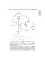
Fig. 8.14 Urticarial vasculitis: a combination of urticaria
and bruising.
CD3C08 21/5/05 11:48 AM Page 104
REACTIVE ERYTHEMAS AND VASCULITIS 105
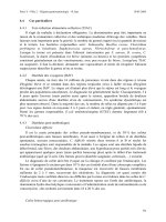
polyarteritis nodosa), or also affect the kidneys, heart
muscle, nerves and joints (Fig. 8.15). Patients may
be febrile, lose weight and feel pain in the muscles,
joints or abdomen. Some develop peripheral neuro-
pathy, hypertension and ischaemic heart disease.
Renal involvement, with or without hypertension,
is common.
Course
Untreated, systemic polyarteritis nodosa becomes
chronic. Death, often from renal disease, is common,
even in treated patients.
Immune complexes may initiate this vasculitis, and
sometimes contain hepatitis B or C virus or antigen.
Other known causes are adulterated drugs, B-cell
lymphomas and immunotherapy.
Presentation
Tender subcutaneous nodules appear along the line of
arteries. The skin over them may ulcerate or develop
stellate patches of purpura and necrosis. Splinter
haemorrhages and a peculiar net-like vascular pat-
tern (livedo reticularis) aid the clinical diagnosis.
The disorder may be of the skin only (cutaneous
Malaise,
weight loss
Myocardial
infarction
Nephritis
Nodules
Livedo
Arthritis
Abdominal
pains
Stellate
purpura
Ulcers
Peripheral
gangrene
Fig. 8.15 Clinical features of
polyarteritis nodosa.
CD3C08 21/5/05 11:48 AM Page 105
106 CHAPTER 8
organs can be affected, including the eye, joints, heart,
nerves, lung and kidney. Antineutrophil antibodies are
present in most cases and are a useful but non-specific
diagnostic marker. Cyclophosphamide is the treatment
of choice, used alone or with systemic steroids.
Further reading
Cousin, F., Philips, K., Favier, B., Bienvenu, J. &
Nicolas, J.F. (2001) Drug-induced urticaria. Euro-
pean Journal of Dermatology 11, 181–187.
Cuellar, M.L. & Espinoza, L.R. (2000) Laborat-
ory testing in the evaluation and diagnosis of
vasculitis. Current Rheumatology Reports 2, 417–
422.
Grattan, C., Powell, S. & Humphreys, F. (2001)
Management and diagnostic guidelines for urticaria
and angio-oedema. British Journal of Dermatology
144, 708–714.
Greaves, M.W. (2001) Antihistamines. Dermatologic
Clinics 19, 53–62.
Joint Task Force on Practice Parameters (2000) The
diagnosis and management of urticaria: a practice
parameter. I. Acute urticaria/angioedema. II. Chronic
urticaria/angioedema. Annals of Allergy, Asthma
and Immunology 85, 521–544.
Lotti, T., Ghersetich, I., Comacci, C. & Jorizzo, J.L.
(1998) Cutaneous small vessel vasculitis. Journal
of the American Academy of Dermatology 39,
667–687.
Schachner, L.A. (2000) Erythema multiforme.
Pediatric Dermatology 17, 75–83.
Sharma, J.K., Miller, R. & Murray, S. (2000) Chronic
urticaria: a Canadian perspective on patterns and
practical management strategies. Journal of
Cutaneous Medicine and Surgery 4, 89–93.
Wakelin, S.H. (2001) Contact urticaria. Clinical and
Experimental Dermatology 26, 132–136.
Differential diagnosis
Embolism, panniculitis and infarctions can cause a sim-
ilar clinical picture. Wegener’s granulomatosis, allergic
granulomatosis, temporal arteritis, and the vasculitis
that accompanies systemic lupus erythematosus and
rheumatoid arthritis should be considered.
Investigations
The laboratory findings are non-specific. An elevated
ESR, neutrophil count, and gammaglobulin level are
common. Investigations for cryoglobulins, rheumatoid
factor, antinuclear antibody, antineutrophil antibod-
ies and hepatitis C and B surface antigen are worth-
while, as are checks for disease in the kidneys, heart,
liver and gut. Low levels of complement suggest active
disease. The use of biopsy to confirm the diagnosis of
large vessel vasculitis is not always easy as the arterial
involvement may be segmental, and surgery itself
difficult. Histological confirmation is most likely
when biopsies are from a fresh lesion. Affected vessels
show aneurysmal dilatation or necrosis, fibrinoid
changes in their walls, and an intense neutrophilic
infiltrate around and even in the vessel wall.
Treatment
Systemic steroids and cyclophosphamide improve
chances of survival. Low-dose systemic steroids alone
are usually sufficient for the purely cutaneous form.
Wegener’s granulomatosis
In this granulomatous vasculitis of unknown cause,
fever, weight loss and fatigue accompany nasorespirat-
ory symptoms such as rhinitis, hearing loss or sinusitis.
Only half of the patients have skin lesions, usually
symmetrical ulcers or papules on the extremities. Other
CD3C08 21/5/05 11:48 AM Page 106
107
ally bind the skin (p. 11 and p. 15). This type of
mechanism has not yet been proven for dermatitis
herpetiformis; but the characteristic deposition of
immunoglobulin (Ig) A in the papillary dermis, and
an association with a variety of autoimmune dis-
orders, both suggest an immunological basis for the
disease.
Blisters are accumulations of fluid within or under
the epidermis. They have many causes, and a correct
clinical diagnosis must be based on a close study of
the physical signs.
The appearance of a blister is determined by the
level at which it forms. Subepidermal blisters occur
between the dermis and the epidermis. Their roofs are
relatively thick and so they tend to be tense and intact.
They may contain blood. Intraepidermal blisters appear
within the prickle cell layer of the epidermis, and so
have thin roofs and rupture easily to leave an oozing
denuded surface: this tendency is even more marked
with subcorneal blisters, which form just beneath the
stratum corneum at the outermost edge of the viable
epidermis, and therefore have even thinner roofs.
Sometimes the morphology or distribution of a bul-
lous eruption gives the diagnosis away, as in herpes
simplex or zoster. Sometimes the history helps too, as
in cold or thermal injury, or in an acute contact derm-
atitis. When the cause is not obvious, a biopsy should
be taken to show the level in the skin at which the blis-
ter has arisen. A list of differential diagnoses, based on
the level at which blisters form, is given in Fig. 9.1.
The bulk of this chapter is taken up by the three
most important immunobullous disordersapemphigus,
pemphigoid and dermatitis herpetiformis (Table 9.1)
aand by the group of inherited bullous disorders known
as epidermolysis bullosa. Our understanding of both
groups has advanced in parallel, as several of the skin
components targeted by autoantibodies in the immuno-
bullous disorders are the same as those inherited in an
abnormal form in epidermolysis bullosa.
Bullous disorders of immunological origin
In pemphigus and pemphigoid, the damage is done
by autoantibodies directed at molecules that norm-
9 Bullous diseases
Location of bullae
Diseases
Subcorneal bulla
Intra-epidermal bulla
Sub-epidermal bulla
Bullous impetigo
Miliaria crystallina
Staphylococcal
scalded skin syndrome
Acute eczema
Viral vesicles
Pemphigus
Miliaria rubra
Incontinentia pigmenti
Bullous pemphigoid
Cicatricial pemphigoid
Pemphigoid gestationis
Dermatitis herpetiformis
Linear IgA disease
Bullous erythema multiforme
Bullous lichen planus
Bullous lupus erythematosus
Porphyria cutanea tarda
Toxic epidermal necrolysis
Cold or thermal injury
Epidermolysis bullosa
Fig. 9.1 The differential diagnosis of bullous diseases based
on the histological location of the blister.
CD3C09 21/5/05 11:47 AM Page 107
108 CHAPTER 9
Presentation
Pemphigus vulgaris is characterized by flaccid blisters
of the skin (Fig. 9.2) and mouth (Fig. 9.3) and, after the
blisters rupture, by widespread painful erosions. Most
patients develop the mouth lesions first. Shearing
Pemphigus
Pemphigus is severe and potentially life-threatening.
There are two main types. The most common is
pemphigus vulgaris, which accounts for at least
three-quarters of all cases, and for most of the deaths.
Pemphigus vegetans is a rare variant of pemphigus
vulgaris. The other important type of pemphigus,
superficial pemphigus, also has two variants: the
generalized foliaceus type and localized erythema-
tosus type. A few drugs, led by penicillamine, can
trigger a pemphigus-like reaction, but autoanti-
bodies are then seldom found. Finally, a rare type of
pemphigus (paraneoplastic pemphigus) has been
described in association with a thymoma or an under-
lying carcinoma; it is characterized by unusually severe
mucosal lesions.
Cause
All types of pemphigus are autoimmune diseases in
which pathogenic IgG antibodies bind to antigens
within the epidermis. The main antigens are des-
moglein 3 (in pemphigus vulgaris) and desmoglein 1
(in superficial pemphigus). Both are cell-adhesion
molecules of the cadherin family (see Table 2.5),
found in desmosomes. The antigen–antibody reaction
interferes with adhesion, causing the keratinocytes
to fall apart.
Table 9.1 Distinguishing features of the three main immunobullous diseases.
Site of General Blisters in Nature of Circulating Fixed
Age blisters health mouth blisters antibodies antibodies Treatment
Pemphigus Middle age Trunk, Poor Common Superficial IgG to IgG in Steroids
flexures and flaccid intercellular intercellular Immunosuppressives
and scalp adhesion space
proteins
Pemphigoid Old Often Good Rare Tense and IgG to IgG at Steroids
flexural blood-filled basement basement Immunosuppressives
membrane membrane
region
Dermatitis Primarily Elbows, knees, Itchy Rare Small, IgG to the IgA granular Gluten-free diet
herpetiformis adults upper back, excoriated endomysium deposits in Dapsone
buttocks and grouped of muscle papillary Sulphapyridine
dermis
Fig. 9.2 Pemphigus vulgaris: widespread erosions that have
followed blisters.
CD3C09 21/5/05 11:47 AM Page 108
BULLOUS DISEASES 109
Course
The course of all forms of pemphigus is prolonged, even
with treatment, and the mortality rate of pemphigus
vulgaris is still at least 15%. Superficial pemphigus
is less severe. With modern treatments, most patients
with pemphigus can live relatively normal lives, with
occasional exacerbations.
Complications
Complications are inevitable with the high doses of
steroids and immunosuppressive drugs that are needed
to control the condition. Indeed, side-effects of treat-
ment are now the leading cause of death. Infections
of all types are common. The large areas of denuda-
tion may become infected and smelly, and severe oral
ulcers make eating painful.
Differential diagnosis
Widespread erosions may suggest a pyoderma,
impetigo, epidermolysis bullosa or ecthyma. Mouth
ulcers can be mistaken for aphthae, Behçet’s disease
or a herpes simplex infection.
Investigations
Biopsy shows that the vesicles are intraepidermal, with
rounded keratinocytes floating freely within the blister
cavity (acantholysis). Direct immunofluorescence
(p. 39) of adjacent normal skin shows intercellular
epidermal deposits of IgG and C3 (Fig. 9.5). The serum
from a patient with pemphigus contains antibodies that
bind to the desmogleins in the desmosomes of normal
epidermis, so that indirect immunofluorescence (p. 39)
can also be used to confirm the diagnosis. The titre of
these antibodies correlates loosely with clinical activ-
ity and may guide changes in the dosage of systemic
steroids.
stresses on normal skin can cause new erosions to
form (a positive Nikolsky sign). In the vegetans variant
(Fig. 9.4), heaped up cauliflower-like weeping areas
are present in the groin and body folds. The blisters in
pemphigus foliaceus are so superficial, and rupture so
easily, that the clinical picture is dominated more by
weeping and crusting erosions than by blisters. In the
rarer pemphigus erythematosus, the facial lesions are
often pink, dry and scaly.
Fig. 9.3 Painful sloughy mouth ulcers in pemphigus vulgaris.
Fig. 9.4 Pemphigus vegetans in the axilla, some intact
blisters can be seen.
LEARNING POINT
Pemphigus is more attacking than pemphigoid
and needs higher doses of steroids to control it.
CD3C09 21/5/05 11:47 AM Page 109
110 CHAPTER 9
grouped or located in body folds. Bullous impetigo is
caused by Staphylococcus aureus.
Scalded skin syndrome (p. 192)
A toxin elaborated by some strains of S. aureus makes
the skin painful and red; later it peels like a scald. The
staphylococcus is usually hidden (e.g. conjunctiva,
throat, wound, furuncle).
Miliaria crystallina (p. 161)
Here sweat accumulates under the stratum corneum
leading to the development of multitudes of uniformly
spaced vesicles without underlying redness. Often this
occurs after a fever or heavy exertion. The vesicles
look like droplets of water lying on the surface, but the
skin is dry to the touch. The disorder is self-limiting
and needs no treatment.
Subcorneal pustular dermatosis
As its name implies, the lesions are small groups of
pustules rather than vesicles. However, the pustules
pout out of the skin in a way that suggests they were
once vesicles (like the vesico-pustules of chickenpox).
Treatment
Because of the dangers of pemphigus vulgaris, and the
difficulty in controlling it, patients should be treated
in a specialized unit. Resistant and severe cases need
very high doses of systemic steroids, such as prednis-
olone (Formulary 2, p. 348) 80–320 mg/day, and the
dose is dropped only when new blisters stop appear-
ing. Immunosuppressive agents, such as azathioprine
or cyclophosphamide and, recently, mycophenylate
mofetil, are often used as steroid-sparing agents. New
and promising approaches include plasmapheresis and
intravenous immunoglobulin as used in other auto-
immune diseases. Treatment needs regular follow-up
and is usually prolonged. In superficial pemphigus,
smaller doses are usually needed, and the use of top-
ical corticosteroids may help too.
Other causes of subcorneal and
intraepidermal blistering
Bullous impetigo
(p. 190)
This is a common cause of blistering in children. The
bullae are flaccid, often contain pus and are frequently
Predominant
immunosorbent
Epidermis
Lamina lucida
Lamina densa
Anchoring fibrils
Dermis
Pemphigus
Bullous pemphigoid
Cicatricial pemphigoid
Pemphigoid gestationis
Linear IgA bullous disease
Dermatitis herpetiformis
Epidermolysis bullosa acquisita
Bullous lupus erythematosus
IgG IgG IgG C3 IgA IgA IgG IgG
Fig. 9.5 Immunofluorescence (red) in
bullous diseases.
CD3C09 21/5/05 11:47 AM Page 110
BULLOUS DISEASES 111
However, their titre does not correlate with clinical
disease activity. The IgG antibodies bind to two main
antigens: most commonly to BP230 (within the cel-
lular part of the hemidesmosome, p. 15), and less
often to BP180 (a transmembrane molecule with one
end within the hemidesmosome and the other bound
to the lamina lucida). Complement is then activated
(p. 24), an inflammatory cascade starts and mast cells
degranulate, liberating a variety of inflammatory
mediators.
Presentation
Pemphigoid is a chronic, usually itchy, blistering dis-
ease, mainly affecting the elderly. The tense bullae
can arise from normal skin but usually do so from
urticarial plaques (Fig. 9.6). The flexures are often
affected; the mucous membranes usually are not. The
Nikolsky test is negative.
Course
Pemphigoid is usually self-limiting and treatment can
often be stopped after 1–2 years.
The cause of this rare disease is unknown, but oral
dapsone (Formulary 2, p. 351) usually suppresses it.
Acute dermatitis (Chapter 7)
Severe acute eczema, especially of the contact allergic
type, can be bullous. Plants such as poison ivy, poison
oak or primula are common causes. The varied size of
the vesicles, their close grouping, their asymmetry, their
odd configurations (e.g. linear, square, rectilinear)
and a history of contact with plants are helpful guides
to the diagnosis.
Pompholyx (p. 89)
In pompholyx, highly itchy small eczematous vesicles
occur along the sides of the fingers, and sometimes
also on the palms and soles. Some call it ‘dyshidrotic
eczema’, but the vesicles are not related to sweating
or sweat ducts. The disorder is very common, but its
cause is not known.
Viral infections (Chapter 14)
Some viruses create blisters in the skin by destroying
epithelial cells. The vesicles of herpes simplex and
zoster are the most common examples.
Transient acantholytic dermatosis
(Grover’s disease)
Itchy vesicles appear on the sun-damaged skin of the
trunk, usually of middle-aged males. The cause is not
known and the condition can be persistentadespite
its name.
Subepidermal immunobullous disorders
These can be hard to separate on clinical grounds
and only the two most important, pemphigoid and
dermatitis herpetiformis, are described in detail here.
Several others are mentioned briefly.
Pemphigoid
Pemphigoid is an autoimmune disease. Serum from
about 70% of patients contains antibodies that bind
in vitro to normal skin at the basement membrane zone.
Fig. 9.6 Numerous large tense blisters in an elderly person
suggest pemphigoid.
CD3C09 21/5/05 11:47 AM Page 111
112 CHAPTER 9
suppressive agents may also be required. The dosage
is reduced as soon as possible, and patients end up on
a low maintenance regimen of systemic steroids,
taken on alternate days until treatment is stopped. For
unknown reasons, tetracyclines and niacinamide help
some patients.
Pemphigoid gestationis (herpes gestationis)
This is pemphigoid occurring in pregnancy, or in the
presence of a hydatidiform mole or a choriocarcinoma.
As in pemphigoid, most patients have linear deposits
of C3 along the basement membrane zone (Fig. 9.5),
although IgG is detected less often. The condition
usually remits after the birth but may return in future
pregnancies. It is not caused by a herpes virus: the
name herpes gestationis should be discarded now so
that the disease is not confused with herpes genitalis.
Treatment is with systemic steroids. Oral contracept-
ives should be avoided.
Cicatricial pemphigoid (Fig. 9.8)
Like pemphigoid itself, cicatricial pemphigoid is an
autoimmune skin disease showing IgG and C3 depo-
sition at the basement membrane zone (Fig. 9.5). The
antigens are often as in pemphigoid, but other anti-
gens are sometimes targeted such as laminin 5 (in
anchoring filaments). The condition differs from pem-
phigoid in that its blisters and ulcers occur mainly
on mucous membranes such as the conjunctivae,
the mouth and genital tract. Bullae on the skin itself
are uncommon. Lesions heal with scarring: around
the eyes this may cause blindness, especially when the
palpebral conjunctivae are affected (Fig. 9.8). The
condition tends to persist and treatment is relatively
ineffective, although very potent local steroids, sys-
temic steroids and immunosuppressive agents are
Complications
Untreated, the disease causes much discomfort and
loss of fluid from ruptured bullae. Systemic steroids
and immunosuppressive agents carry their usual com-
plications if used long-term (Formulary 2, p. 348 and
p. 346, respectively). The validity of a possible associ-
ation with internal malignancy is still debated.
Differential diagnosis
Pemphigoid may look like other bullous diseases, espe-
cially epidermolysis bullosa acquisita, bullous lupus
erythematosus, dermatitis herpetiformis, pemphigoid
gestationis, bullous erythema multiforme and linear
IgA bullous disease. Immunofluorescence helps to
separate it from these (Fig. 9.5).
Investigations
The histology is that of a subepidermal blister, often
filled with eosinophils. Direct immunofluorescence
shows a linear band of IgG and C3 along the base-
ment membrane zone. Indirect immunofluorescence,
using serum from the patient, identifies IgG antibodies
that react with the basement membrane zone in some
70% of patients (Fig. 9.7).
Treatment
In the acute phase, prednisolone or prednisone
(Formulary 2, p. 348) at a dosage of 40– 60 mg/day is
usually needed to control the eruption. Immuno-
Fig. 9.7 Indirect immunofluorescence using serum from
a patient with pemphigoid, showing basement zone
immunofluorescence.
LEARNING POINTS
1 Death is uncommon and the disease is self-
limiting.
2 Some elderly people get fatal side-effects
from their systemic steroids. Reduce the dosage
as soon as possible.
CD3C09 21/5/05 11:47 AM Page 112
BULLOUS DISEASES 113
Dermatitis herpetiformis
Dermatitis herpetiformis is a very itchy chronic
subepidermal vesicular disease, in which the vesicles
erupt in groups as in herpes simplexahence the name
‘herpetiformis’.
Cause
Gluten-sensitive enteropathy, demonstrable by small
bowel biopsy, is always present, but most patients
do not suffer from diarrhoea, constipation or mal-
nutrition as the enteropathy is mild, patchy and
involves only the proximal small intestine. Absorption
of gluten, or another dietary antigen, may form cir-
culating immune complexes that lodge in the skin. A
range of antibodies can be detected, notably directed
against reticulin, gliadin and endomysiumaa com-
ponent of smooth muscle. Granular deposits of IgA
and C3 in the superficial dermis under the basement
membrane zone (Fig. 9.5) induce inflammation, which
then separates the epidermis from the dermis. These
deposits clear slowly after the introduction of a
gluten-free diet.
Presentation
The extremely itchy, grouped vesicles (Fig. 9.9) and
urticated papules develop particularly over the elbows
(Fig. 9.10) and knees, buttocks and shoulders. They
are often broken by scratching before they reach any
size. A typical patient therefore shows only grouped
excoriations, sometimes with eczema-like changes
added by scratching.
Course
The condition typically lasts for decades.
Complications
The complications of gluten-sensitive enteropathy
include diarrhoea, abdominal pain, anaemia and, rarely,
malabsorption. Small bowel lymphomas have been
reported, and the use of a gluten-free diet may reduce
this risk. There is a proven association with other
autoimmune diseases, most commonly of the thyroid.
Treatment, notably with dapsone (Formulary 2,
p. 352), can cause side-effects.
usually tried. Good eye hygiene and the removal of
ingrowing eyelashes are important.
Linear IgA bullous disease
This is clinically similar to pemphigoid, but affects
children as well as adults. Blisters arise on urticarial
plaques, and are more often grouped, and on extensor
surfaces, than is the case with pemphigoid. The
so-called ‘string of pearls sign’, seen in some affected
children, is the presence of blistering around the rim
of polycyclic urticarial lesions. The conjunctivae may
be involved. Linear IgA bullous disease is, as its name
implies, associated with linear deposits of IgA and C3
at the basement membrane zone (Fig. 9.5). IgG is
sometimes also found. The disorder responds well to
oral dapsone (Formulary 2, p. 352).
Acquired epidermolysis bullosa
This can also look like pemphigoid, but has two im-
portant extra features: many of the blisters are a
response to trauma and arise on otherwise normal
skin; and milia are a feature of healing lesions. The
target of the autoantibodies is type VII collagen in
anchoring fibrils (see Fig. 9.5). The antigen lies on
the dermal side of the lamina densa, in contrast to
the pemphigoid antigens, which lie on the epidermal
sideaa difference that can be demonstrated when the
basement membrane is split by incubating skin in a
saline solution (the ‘salt-split’ technique). The condi-
tion responds poorly to systemic corticosteroids or
immunosuppressive agents.
Fig. 9.8 Longstanding cicatricial pemphigoid. Adhesions
are now forming between the upper and lower eyelids.
CD3C09 21/5/05 11:47 AM Page 113
114 CHAPTER 9
IgA deposits remain in the skin, and the skin disease
can drag on for many months. Because of this, and
because a gluten-free diet is hard to follow and enjoy,
some patients prefer to combine the diet with dapsone
(Formulary 2, p. 352) or sulphapyridine (sulfapyridine)
at the start, although both can cause severe rashes,
haemolytic anaemia (especially in those with glucose-
6-phosphate dehydrogenase deficiency), leucopenia,
thrombocytopenia, methaemoglobinaemia and peri-
Differential diagnosis
The disorder masquerades as scabies, an excoriated
eczema, insect bites or neurodermatitis.
Investigations
If a vesicle can be biopsied before it is scratched away,
the histology will be that of a subepidermal blister,
with neutrophils packing the adjacent dermal papil-
lae. Direct immunofluorescence of uninvolved skin
shows granular deposits of IgA, and usually C3, in
the dermal papillae and superficial dermis (Fig. 9.5).
Small bowel biopsy is no longer recommended as
routine because the changes are often patchy. Tests
for malabsorption are seldom needed.
Treatment
The disorder responds to a gluten-free diet, which
should be supervised by a dietitian. Adherence to this
can be monitored using the titre of antiendomysial
antibody, which should fall if gluten is strictly avoided.
The bowel changes revert quickly to normal but
LEARNING POINTS
1 Biopsy non-involved skin to demonstrate the
diagnostic granular deposits of IgA in the
dermal papillae.
2 The gluten enteropathy of dermatitis
herpetiformis seldom causes frank
malabsorption.
3 Dapsone works quickly and a gluten-free
diet only very slowly. Combine the two at the
start and slowly reduce the dapsone.
Fig. 9.10 The itchy blisters of dermatitis herpetiformis
favour the points of the elbows and knees, where they
are quickly destroyed by scratching.
Fig. 9.9 The typical small tense grouped itchy blisters of
dermatitis herpetiformis.
CD3C09 21/5/05 11:47 AM Page 114
BULLOUS DISEASES 115
sign is positive (p. 109). The mucous membranes may
be affected, including the mouth, eyes, and even the
bronchial tree.
Course
The condition usually clears if the offending drug is
stopped. New epidermis grows out from hair follicles
so that skin grafts are not usually needed. The dis-
order may come back if the drug is taken again.
Complications
Toxic epidermal necrolysis is a skin emergency and
can be fatal. Infection, and the loss of fluids and elec-
trolytes, are life-threatening, and the painful denuded
skin surfaces make life a misery. Corneal scarring may
remain when the acute episode has settled.
Differential diagnosis
The epidermolysis of the staphylococcal scalded skin
syndrome (p. 192) looks like toxic epidermal necrolysis
clinically, but only the stratum corneum is lost. Whereas
toxic epidermal necrolysis affects adults, the staphy-
lococcal scalded skin syndrome is seen in infancy
or early childhood. Histology differentiates the two.
Pemphigus may also look similar, but starts more
slowly and is more localized. Severe graft-vs host reac-
tions can also cause this syndrome. Some believe that
toxic epidermal necrolysis can evolve from Stevens–
Johnson syndrome because some patients have the
clinical features of both.
pheral neuropathy. Regular blood checks are therefore
necessary.
Other causes of subepidermal blisters
Porphyria cutanea tarda
(p. 287)
The bullae and erosions occur on the backs of the
hands and on other areas exposed to sunlight.
Blisters in diabetes and renal disease
A few diabetics develop unexplained blisters on their
legs or feet. The backs of the hands of patients with
chronic renal failure may show changes rather like
those of porphyria cutanea tarda (pseudoporphyria).
Frusemide (furosemide) can contribute to blister
formation.
Bullous lupus erythematosus
Vesicles and bullae may be seen in severe active sys-
temic lupus erythematosus (p. 119). This disorder is
uncommon and carries a high risk of kidney disease.
Non-cutaneous manifestations of systemic lupus ery-
thematosus do not respond to dapsone; however, the
bullae do.
Bullous erythema multiforme
Bullous erythema multiforme in the form of the
Stevens–Johnson syndrome is discussed in Chapter 8.
Toxic epidermal necrolysis (Lyell’s disease)
Cause
Toxic epidermal necrolysis is usually a drug reaction,
most commonly to sulphonamides, barbiturates,
carbamazepine or allopurinol (Chapter 22), but can
also be a manifestation of graft-vs host disease. Some-
times it is unexplained.
Presentation
The skin becomes red and intensely painful, and then
begins to come off in sheets like a scald. This leaves an
eroded painful glistening surface (Fig. 9.11). Nikolsky’s
Fig. 9.11 The burn-like appearance of toxic epidermal
necrolysis.
CD3C09 21/5/05 11:47 AM Page 115
116 CHAPTER 9
epidermolysis bullosa is not inherited and was dis-
cussed earlier in this chapter.
Simple epidermolysis bullosa
Several subtypes are recognized, of which the most
common are the Weber–Cockayne (mainly affecting
the hands and feet) and the Dowling–Meara (featur-
ing herpetiform blisters on the trunk) types. Most are
inherited as autosomal dominant conditions and are
caused by abnormalities in genes responsible for pro-
duction of the paired keratins (K5 and K14) expressed
Investigations
Biopsy helps to confirm the diagnosis. The split is
subepidermal in toxic epidermal necrolysis, and the
entire epidermis may be necrotic. A frozen section
provides a quick answer if there is genuine difficulty in
separating toxic epidermal necrolysis from the scalded
skin syndrome (p. 192). There are no tests to tell
which drug, if any, caused the disease.
Treatment
If toxic epidermal necrolysis is caused by a drug, this
must be stopped (Chapter 22); otherwise, treatment
relies mainly on symptomatic management. Intensive
nursing care and medical support are needed, includ-
ing the use of central venous lines, intravenous fluids
and electrolytes. Many patients are treated in units
designed to deal with extensive thermal burns. Air
suspension beds increase comfort. The weight of
opinion has turned against the use of systemic corti-
costeroids but, if they are given, it should be for short
periods only, right at the start. Intravenous IgG seems
more promising.
Epidermolysis bullosa
There are many types of epidermolysis bullosa: the
five main ones are listed in Table 9.2. All are charac-
terized by an inherited tendency to develop blisters
after minimal trauma, although at different levels in
the skin (Fig. 9.12). The more severe types have a
catastrophic impact on the lives of sufferers. Acquired
Table 9.2 Simplified classification of epidermolysis bullosa.
Type Mode of inheritance Level of split Mutations in
Simple epidermolysis bullosa Usually autosomal dominant Intraepidermal Keratins 5 and 14
Junctional epidermolysis bullosa Autosomal recessive Lamina lucida Components of the
(epidermolysis bullosa letalis) hemidesmosome-anchoring
filaments (e.g. laminins,
integrins and bullous
pemphigoid 180 molecule)
Dystrophic epidermolysis bullosa Autosomal dominant Beneath lamina densa Type VII collagen
Dystrophic epidermolysis bullosa Autosomal recessive Beneath lamina densa Type VII collagen
Acquired epidermolysis bullosa Not inherited Dermal side of lamina densa Nil
Epidermal
basal cell
Lamina lucida
Lamina densa
Anchoring
fibrils
Collagen
Dystrophic
and acquired
Junctional
Simple
Fig. 9.12 Levels of blister formation in epidermolysis
bullosa at the dermo-epidermal junction.
CD3C09 21/5/05 11:47 AM Page 116
BULLOUS DISEASES 117
Autosomal dominant dystrophic
epidermolysis bullosa
In this type blisters appear in late infancy. They are
most common on friction sites (e.g. the knees, elbows
and fingers), healing with scarring and milia formation.
The nails may be deformed or even lost. The mouth is
not affected. The only treatment is to avoid trauma
and to dress the blistered areas.
Autosomal recessive dystrophic
epidermolysis bullosa
In this tragic form of epidermolysis bullosa, blisters
start in infancy. They are subepidermal and may be
filled with blood. They heal with scarring, which can
be so severe that the nails are lost and webs form
between the digits (Fig. 9.14). The hands and feet may
become useless balls, having lost all fingers and toes.
The teeth, mouth and upper part of the oesophagus are
all affected; oesophageal strictures may form. Squamous
cell carcinomas of the skin are a late complication.
Treatment is unsatisfactory. Phenytoin, which reduces
the raised dermal collagenase levels found in this
variant, and systemic steroids are disappointing. It is
especially important to minimize trauma, to prevent
contractures and web formation between the digits,
and to combat anaemia and secondary infection.
Referral to centres with expertise in management of
these patients is strongly recommended.
in basal keratinocytes (see Fig. 2.4). Linkage studies
show that the genetic defects responsible for the most
common types of simple epidermolysis bullosa lie on
chromosomes 17 and 12.
Blisters form within or just above the basal cell
layers of the epidermis and so tend to heal without
scarring. Nails and mucosae are not involved. The
problems are made worse by sweating and ill-fitting
shoes. Blistering can be minimized by avoiding trauma,
wearing soft well-fitting shoes and using foot powder.
Large blisters should be pricked with a sterile needle
and dressed. Their roofs should not be removed. Local
antibiotics may be needed.
Junctional epidermolysis bullosa
The abnormalities in the basal lamina include loss
of anchoring filaments and defective laminins (p. 15;
see Fig. 2.9). This rare and often lethal condition is
evident at birth. The newborn child has large raw areas
and flaccid blisters, which are slow to heal (Fig. 9.13).
The peri-oral and peri-anal skin is usually involved,
as are the nails and oral mucous membrane. There is
no effective systemic treatment. Hopes for the future
include adding the normal gene to epidermal stem
cells, and then layering these onto the denuded skin.
Dystrophic epidermolysis bullosa
There are many subtypes, all of which probably result
from abnormalities of collagen VII, the major struc-
tural component of anchoring fibrils.
Fig. 9.13 Junctional epidermolysis bullosa: minor trauma
has caused large blisters and erosions which will heal slowly
or not at all.
Fig. 9.14 Autosomal recessive dystrophic
epidermolysis bullosa: note large blood-filled blister.
Scarring has led to fixed deformity of the fingers and
loss of nails.
CD3C09 21/5/05 11:47 AM Page 117
118 CHAPTER 9
Nousari, H.C. & Anhalt, G.J. (1999) Pemphigus and
bullous pemphigoid. Lancet 354, 667–672.
Schmidt, E. & Zillikens, D. (2000) Autoimmune and
inherited subepidermal blistering diseases: advances
in the clinic and the laboratory. Advances in Der-
matology 16, 113–157.
Wojnarowska, F., Kirtschig, G., Highet, A.S. et al.
(2002) Guidelines for the management of bullous
pemphigoid. British Journal of Dermatology 147,
214–221.
Further reading
Cotell, S., Robinson, N.D. & Chan, L.S. (2000)
Autoimmune blistering skin diseases. American
Journal of Emergency Medicine 18, 288–299.
Fleming, T.E. & Korman, N.J. (2000) Cicatricial
pemphigoid. Journal of the American Academy of
Dermatology 43, 571–591.
CD3C09 21/5/05 11:47 AM Page 118
119
way, whereas others including oral contraceptives, anti-
convulsants, minocycline and captopril, precipitate
the disease just occasionally.
Presentation
Typically, but not always, the onset is acute. SLE is
an uncommon disorder, affecting women more often
than men (in a ratio of about 8 : 1). The classic rash
of acute SLE is an erythema of the cheeks and nose in
the rough shape of a butterfly (Figs 10.1 and 10.2),
with facial swelling. Occasionally, a few blisters may
be seen. Some patients develop widespread discoid
papulosquamous plaques very like those of discoid
LE; others, about 20% of patients, have no skin dis-
ease at any stage.
Other dermatological features include peri-ungual
telangiectasia (see Fig. 10.7), erythema over the digits,
hair fall (especially at the frontal margin of the scalp),
and photosensitivity. Ulcers may occur on the pal-
ate, tongue or buccal mucosa.
Course
The skin changes may be transient, continuous or
recurrent; they correlate well with the activity of the
systemic disease. Acute SLE may be associated with
fever, arthritis, nephritis, polyarteritis, pleurisy, pneu-
monitis, pericarditis, myocarditis and involvement of
the central nervous system. Internal involvement can
be fatal, but the overall prognosis now is for about
three-quarters of patients to survive for 15 years. Renal
involvement suggests a poorer prognosis.
Complications
The skin disease may cause scarring or hyperpigmenta-
tion, but the main dangers lie with damage to other
The cardinal feature of these conditions is inflamma-
tion in the connective tissue which leads to dermal
atrophy or sclerosis, to arthritis, and sometimes to
abnormalities in other organs. In addition, antibodies
form against normal tissues and cellular components;
these disorders are therefore classed as autoimmune.
Many have difficulty in remembering which antibody
features in which condition: Table 10.1 should help
here.
The main connective tissue disorders present as a
spectrum ranging from the benign cutaneous variants
to severe multisystem diseases (Table 10.2).
Lupus erythematosus
Lupus erythematosus (LE) is a good example of such
a spectrum, ranging from the purely cutaneous type
(discoid LE), through patterns associated with some
internal problems (disseminated discoid LE and sub-
acute cutaneous LE), to a severe multisystem disease
(systemic lupus erythematosus, SLE; Table 10.2).
Systemic lupus erythematosus
Cause
This is unknown, but hereditary factors, e.g. com-
plement deficiency and certain HLA types, increase
susceptibility. Particles looking like viruses have been
seen in endothelial cells, and in other tissues, but their
role is not clear. Patients with LE have autoantibodies
to DNA, nuclear proteins and to other normal antigens,
and this points to an autoimmune cause. Exposure
to sunlight and artificial ultraviolet radiation (UVR),
pregnancy and infection may precipitate the disease
or lead to flare-ups. Some drugs, such as hydralazine
and procainamide trigger SLE in a dose-dependent
10 Connective tissue disorders
CD3C10 21/5/05 11:47 AM Page 119
Table 10.1
Some important associations with non-organ-specific autoantibodies.
Antibody directed against
Nucleoprotein
(ANA or ANF)* Double
(IF pattern in stranded Ro (SSA)
Nuclear
Topoiso-
brackets)
DNA and La (SSB) Sm (ENA) Cardiolipin
RNP Centromere Histones Jo-1
merase (Scl-70)
Discoid LE
+ive in up to 35%
Rarely +ive
(homogenous
and speckled)
Subacute LE
+ive in up to 80%
+ive in 60%
(homogenous
and speckled)
Systemic LE
+ive in up to 100%
+ive in May be
+ive +ive in 30%
+ive in subset with
+ive in 6%
+ive in drug-
(homogenous 50–70% (e.g. 20%) if
recurrent abortions,
induced cases
and speckled) (esp with ANF
−ive
thrombosis, livedo
nephritis)
and skin necrosis
Dermatomyositis
+ive in up to
Occasionally
+ive in 20%
80% (speckled)
+ive
Systemic
+ive in up to 90%
+ive in up
+ive in 20%
sclerosis (speckled and
to 50%
nucleolar)
Mixed
+ive in 100%
+ive in high
High titre is
+ive in 6%
connective (speckled)
titre
aup to
diagnostic
tissue
100%
disorder
* Antibodies tested against human substrates (e.g. Human Hep. 2 cells).
CD3C10 21/5/05 11:47 AM Page 120
CONNECTIVE TISSUE DISORDERS 121
organs and the side-effects of treatment, especially
systemic steroids.
Differential diagnosis
SLE is a great imitator. Its malar rash can be confused
with sunburn, polymorphic light eruption (p. 238)
and rosacea (p. 156). The discoid lesions are distinct-
ive, but are also seen in discoid LE and in subacute
cutaneous LE. Occasionally, they look like psoriasis or
lichen planus (p. 64). The hair fall suggests telogen
effluvium (p. 168). Plaques on the scalp may cause
a scarring alopecia. SLE should be suspected when a
characteristic rash is combined with fever, malaise
and internal disease (Table 10.3).
Investigations
Conduct a full physical examination, looking for
internal disease. Biopsy of skin lesions is worthwhile
because the pathology and immunopathology are dis-
tinctive. There is usually some thinning of the epidermis,
Table 10.2 Classification of connective tissue disease.
Localized disease Intermediate type Aggressive multisystem disease
Discoid lupus erythematosus Subacute lupus erythematosus Systemic lupus erythematosus
Juvenile dermatomyositis Adult dermatomyositis
Morphoea CREST syndrome Systemic sclerosis
Fig. 10.1 In systemic lupus
erythematosus (SLE) (left) the eruption
is often just an erythema, sometimes
transient, but occupying most of the
‘butterfly’ area. In discoid LE (right)
the fixed scaling and scarring plaques
may occur in the butterfly area (dotted
line), but can occur outside it too.
Fig. 10.2 Erythema in the butterfly area, suggestive of SLE.
CD3C10 21/5/05 11:47 AM Page 121
122 CHAPTER 10
other drugs (e.g. antihypertensive therapy or anticon-
vulsants) may also be needed. Antimalarial drugs may
help some patients with marked photosensitivity, as
may sunscreens. Intermittent intravenous infusions of
gamma globulin show promise. Long-term and regular
follow-up is necessary.
Subacute cutaneous lupus erythematosus
This is less severe than acute SLE, but is also often
associated with systemic disease. Its cause is unknown,
but probably involves an antibody-dependent cellular
cytotoxic attack on basal cells by K cells bridged by
antibody to Ro (SS-A) antigen.
Presentation
Patients with subacute cutaneous LE are often photo-
sensitive. The skin lesions are sharply marginated
liquefaction degeneration of epidermal basal cells, and
a mild perivascular mononuclear cell infiltrate. Direct
immunofluorescence is helpful: IgG, IgM, IgA and
C3 are found individually or together in a band-like
pattern at the dermo-epidermal junction of involved
skin and often uninvolved skin as well. Relevant
laboratory tests are listed in Table 10.4.
Treatment
Systemic steroids are the mainstay of treatment, with
bed rest needed during exacerbations. Large doses of
prednisolone (Formulary 2, p. 348) are often needed
to achieve control, as assessed by symptoms, signs,
erythrocyte sedimentation rate (ESR), total comple-
ment level and tests of organ function. The dosage
is then reduced to the smallest that suppresses the
disease. Immunosuppressive agents, such as azathio-
prine (Formulary 2, p. 346), cyclophosphamide and
Table 10.3 Criteria for the diagnosis
of SLE (must have at least four).
Malar rash
Discoid plaques
Photosensitivity
Mouth ulcers
Arthritis
Serositis
Renal disorder
Neurological disorder
Haematological disorder
Immunological disorder
Antinuclear antibodies (ANA)
Table 10.4 Investigations in SLE.
Test Usual findings
Skin biopsy Degeneration of basal cells, epidermal thinning, inflammation around appendages
Skin immunofluorescence Fibrillar or granular deposits of IgG, IgM, IgA and/or C3 alone in basement
membrane zone
Haematology Anaemia, raised ESR, thrombocytopenia, decreased white cell count
Immunology Antinuclear antibody, antibodies to double-stranded DNA, false positive tests
for syphilis, low total complement level, lupus anticoagulant factor
Urine analysis Proteinuria or haematuria, often with casts if kidneys involved
Tests for function of other organs As indicated by history but always test kidney and liver function
LEARNING POINTS
1 Do not wait for the laboratory to confirm
that your patient has severe SLE: use systemic
steroids quickly if indicated by clinical
findings.
2 A person with aching joints and small
amounts of antinuclear antibodies probably
does not have SLE.
3 Once committed to systemic steroids, adjust
their dosage on clinical rather than laboratory
grounds.
CD3C10 21/5/05 11:47 AM Page 122
CONNECTIVE TISSUE DISORDERS 123
Presentation
Plaques show erythema, scaling, follicular plugging
(like a nutmeg grater), scarring and atrophy, telan-
giectasia, hypopigmentation and a peripheral zone of
hyperpigmentation. They are well demarcated and lie
mostly on sun-exposed skin of the scalp, face and ears
(Figs 10.1 and 10.3). In one variant (chilblain LE)
dusky lesions appear on the fingers and toes.
Course
The disease may spread relentlessly, but in about half
of the cases the disease goes into remission over the
course of several years. Scarring is common and hair
may be lost permanently if there is scarring in the scalp
(Fig. 10.4). Whiteness remains after the inflammation
has cleared, and hypopigmentation is common in dark-
skinned people. Discoid LE rarely progresses to SLE.
Differential diagnosis
Psoriasis is hard to tell from discoid LE when its plaques
first arise but has larger thicker scales, and later it is
usually symmetrical and affects sites different from
scaling psoriasiform plaques, sometimes annular,
lying on the forehead, nose, cheeks, chest, hands and
extensor surfaces of the arms. They tend to be sym-
metrical and are hard to tell from discoid LE, or SLE
with widespread discoid lesions.
Course
As in SLE, the course is prolonged. The skin lesions
are slow to clear but, in contrast to discoid LE, do so
with little or no scarring.
Complications
Systemic disease is frequent, but not usually serious.
Children born to mothers who have, or have had,
this condition are liable to neonatal LE with transient
annular skin lesions and permanent heart block.
Differential diagnosis
The morphology is characteristic, but lesions can
be mistaken for psoriasis or widespread discoid LE.
Annular lesions may resemble tinea corporis (p. 216)
or figurate erythemas (p. 133).
Investigations
Patients with subacute cutaneous LE should be
evaluated in the same way as those with acute SLE,
although deposits of immunoglobulins in the skin and
antinuclear antibodies in serum are present less often.
Many have antibodies to the cytoplasmic antigen Ro
(SS-A).
Treatment
Subacute cutaneous LE does better with antimalarials,
such as hydroxychloroquine (Formulary 2, p. 352),
than acute SLE. Oral retinoids (Formulary 2, p. 349)
are also effective in some cases. Systemic steroids may
be needed too.
Discoid lupus erythematosus
This is the most common form of LE. Patients with
discoid LE may have one or two plaques only, or
many in several areas. The cause is also unknown but
UVR is one factor.
Fig. 10.3 Red scaly fixed plaques of discoid LE. This degree
of scaling is not uncommon in the active stage. Follicular
plugging is seen on the nose.
CD3C10 21/5/05 11:47 AM Page 123
124 CHAPTER 10
(Fig. 10.5). Direct immunofluorescence shows deposits
of IgG, IgM, IgA and C3 at the basement membrane
zone. Biopsies for direct immunofluorescence are best
taken from older untreated plaques. Blood tests are
usually normal but occasionally serum contains anti-
nuclear antibodies (Table 10.5).
Treatment
Discoid LE needs potent or very potent topical corti-
costeroids (Formulary 1, p. 333). In this condition, it
is justifiable to use them on the face, as the risk of scar-
ring is worse than that of atrophy. Topical steroids
should be applied twice daily until the lesions disappear
or side-effects, such as atrophy, develop; weaker pre-
parations can then be used for maintenance. If discoid
LE does not respond to this, intralesional injections of
triamcinolone (2.5 or 10 mg/mL) may help. Stubborn
and widespread lesions often do well with oral anti-
malarials such as hydroxychloroquine (Formulary 2,
p. 352), but rarely these cause irreversible eye dam-
age. The eyes should therefore be tested before and at
intervals during treatment. Sun avoidance and screens
are also important. Oral retinoids (Formulary 2, p. 349)
and thalidomide have proved helpful in stubborn cases
but a specialist, with experience of their use, should
prescribe these controlled treatments and supervise
management.
those of discoid LE. Discoid LE is more common on
the face and ears, and in sun-exposed areas, whereas
psoriasis favours the elbows, knees, scalp and sacrum.
Discoid LE is far more prone than psoriasis to scar
and cause hair loss. Jessner’s lymphocytic infiltration
is best viewed as a dermal form of discoid LE.
Investigations
Most patients with discoid LE remain well. However,
screening for SLE and internal disease is still worth-
while. A skin biopsy is most helpful if taken from an
untreated plaque where appendages are still present
Fig. 10.4 Discoid LE of the scalp leading to permanent hair
loss. Note the marked follicular plugging.
Follicular plug of keratin
Thick
stratum
corneum
Perivascular and
peri-appendageal
T-lymphocyte
infiltrate
Thin
epidermis
Destruction
of hair
follicle
Destruction
of basal
cells
Fig. 10.5 The histology of discoid LE.
CD3C10 21/5/05 11:47 AM Page 124
CONNECTIVE TISSUE DISORDERS 125
Dermatomyositis
Dermatomyositis is a subset of polymyositis with
distinctive skin changes. There are adult and juvenile
types (Table 10.2). The cause is unknown but an auto-
immune mechanism seems likely. Autoantibodies to
striated muscle are found. When starting after the age
of 40, dermatomyositis may signal an internal malig-
nancy. Presumably, the epitopes of some tumour anti-
gens are so similar to those of muscle antigens that
antibodies directed against the tumour cross-react
with muscle cells and initiate the disease in a few
adults with internal malignancy. Serological evidence
for acute toxoplasmosis in polymyositis-dermato-
myositis was found in one series.
Presentation
The skin signs are characteristic. Typical patients have
a faint lilac discoloration around their eyes (sometimes
called ‘heliotrope’ because of the colour of the flower).
This is associated with malar erythema and oedema
(Fig. 10.6) and, sometimes, less striking erythema of
the neck and presternal area. Most patients also develop
lilac slightly atrophic papules over the knuckles of
their fingers (Gottron’s papules), streaks of erythema
over the extensor tendons of the hand, peri-ungual
telangiectasia and ragged cuticles (Fig. 10.7). The skin
signs usually appear at the same time as the muscle
symptoms but, occasionally, appear months or even
years earlier. Sometimes, the skin signs appear in
isolation. Many, but not all, patients have weakness
of proximal muscles. Climbing stairs, getting up from
chairs and combing the hair become difficult.
Course
In children the disorder is often self-limiting, but in
adults it may be prolonged and progressive. Raynaud’s
phenomenon, arthralgia, dysphagia and calcinosis may
Table 10.5 Some factors distinguishing the different types of LE.
Antinuclear antibodies Sun sensitivity Internal organ involvement
Systemic LE ++++ +++ ++
Subacute LE + ++++ +
Discoid LE +/−+−
Fig. 10.6 Acute dermatomyositis: oedematous purple face
with erythema on presternal area. Severe progressive muscle
weakness, but no underlying tumour was found.
Fig. 10.7 Erythema and telangiectasia of the nail folds are
important clues to systemic connective tissue disorders. This
patient has dermatomyositis. Note Gottron’s papules over
the knuckles.
follow. The rash may become scaly and, rarely, itchy;
eventually that on the light-exposed areas and overly-
ing involved muscles develops poikiloderma (p. 252).
Features of mixed connective disease (see below) may
CD3C10 21/5/05 11:47 AM Page 125
126 CHAPTER 10
treatment is adjusted according to clinical response
and CPK level. As in SLE, intravenous gamma globulin
infusions seem promising. Long-term and regular
follow-up is necessary.
Systemic sclerosis
In this disorder the skin becomes hard as connective
tissues thicken. Early in the condition, T-helper cells
dominate the inflammatory infiltrate in the dermis and
cause fibroblasts to proliferate and produce more
hyaluronic acid and type I collagen (p. 16). In addition
there is intimal thickening of arterioles and arteries.
These processes are not confined to the skin, but involve
many other organs, including the gut, lungs, kidneys
and heart, leading to their dysfunction and to death.
The cause of systemic sclerosis is unknown but
many, apparently unrelated, pieces of the complex
jigsaw are now beginning to come together. A systemic
sclerosis-like syndrome is a feature of the chronic
graft-vs host disease sometimes seen after bone marrow
transplantation (p. 286) and prolonged, untreated
porphyria cutanea torda (p. 287). Similar syndromes
have been reported following ingestion of adulterated
rapeseed oil in Spain and dimerised l-tryptophan for
insomnia and treatment with the antitumour agent,
bleomycin. Environmental factors may also be rel-
evant in isolated cases; changes like those of systemic
sclerosis have affected workers exposed to polyvinyl
chloride monomers, trichlorethylene and epoxy resins
and in those subjected for years to severe vibration.
Presentation
Most patients suffer from Raynaud’s phenomenon
(p. 135) and sclerodactyly. Their fingers become immob-
ile, hard and shiny. Some become hyperpigmented
and itchy early in their disease. Peri-ungual telangiec-
tasia is common.
develop. The presence of calcinosis suggests a good
prognosis.
Complications
Myositis may lead to permanent weakness and immo-
bility, and inflammation to contractures or cutaneous
calcinosis. Some die from progessive and severe
myopathy.
Differential diagnosis
Other connective tissue disorders may look similar,
particularly mixed connective tissue disease (p. 129)
and SLE. In LE, the finger lesions favour the skin
between the knuckles whereas in dermatomyositis
the knuckles are preferred. Toxoplasmosis may cause
a dermatomyositis-like syndrome. Myopathy can be
a side-effect of systemic steroids, so weakness is not
always caused by the disease itself.
Investigations
About 30% of adults with dermatomyositis also
have an underlying malignancy. Their dermatomy-
ositis coincides with the onset of the tumour and may
improve if it is removed. Adult dermatomyositis or
polymyositis therefore requires a search for such an
underlying malignancy. The levels of muscle enzymes
such as aldolase and creatinine phosphokinase (CPK)
are often elevated. Electromyography (EMG) detects
muscle abnormalities, and biopsy of an affected
muscle shows inflammation and destruction. Sur-
prisingly, the ESR is often normal and antinuclear
antibodies may not be detected. Toxoplasmosis
should be excluded by serology.
Treatment
Systemic steroids, often in high doses (e.g. prednisolone
60 mg/day for an average adult; Formulary 2, p. 348),
are the cornerstone of treatment and protect the
muscles from destruction. A maintenance regimen
may be needed for several years. Immunosuppressive
agents, such as azathioprine (Formulary 2, p. 346),
also help to control the condition and to reduce the
high steroid dose. Cyclosporin (Formulary 2, p. 347)
and methotrexate (Formulary 2, p. 348) have proved
useful alternatives in stubborn cases. Maintenance
LEARNING POINT
Hunt for internal malignancy in the middle
aged and elderly, but not in juvenile cases.
CD3C10 21/5/05 11:47 AM Page 126
CONNECTIVE TISSUE DISORDERS 127
in chronic graft-vs host reactions after bone marrow
transplants.
Investigations
The diagnosis is made clinically because histological
abnormalities are seldom present until the physical
signs are well established. Laboratory tests should
include a fluorescent antinuclear antibody test and
the evaluation of the heart, kidney, lungs, joints and
muscles. Barium studies are best avoided as obstruc-
tion may follow poor evacuation. Other contrast media
are available. X-rays of the hands, measurement
of muscle enzymes and immunoglobulin levels, and
a blood count, ESR and test for the scleroderma-
associated antibody Scl-70 are also worthwhile.
Treatment
This is unsatisfactory. The calcium channel blocker
nifedipine may help Raynaud’s phenomenon (p. 135).
Systemic steroids, salicylates, antimalarials and long-
term penicillin are used, but are not of proven value.
d-penicillamine has many side-effects, especially on
Course
As the disease progresses, sclerosis spreads to the
face, scalp and trunk. The nose becomes beak-like,
and wrinkles radiate around the mouth (Fig. 10.8–
10.10). Most have abnormalities of the gut including
dysphagia, oesophagitis, constipation, diarrhoea and
malabsorption. Fibrosis of the lungs leads to dyspnoea,
and fibrosis of the heart to congestive failure. The
kidneys are involved late, but this has a grave prognosis
from malignant hypertension.
Complications
Most complications are caused by the involvement of
organs other than the skin, but ulcers of the fingertips
and calcinosis are distressing (Fig. 10.11). Hard skin
immobilizes the joints and leads to contractures.
Differential diagnosis
Other causes of Raynaud’s phenomenon are given
in Table 11.5. The differential diagnosis includes
chilblains (p. 132) and erythromelalgia (p. 132). The
sclerosis should be distinguished from that of wide-
spread morphoea, porphyria cutanea tarda, mixed
connective tissue disease, eosinophilic fasciitis, dia-
betic sclerodactyly and an acute arthritis with swollen
fingers. Rarely the disease is mimicked by progeria,
scleromyxoedema, amyloidosis or carcinoid syndrome.
Changes like those of progressive systemic sclerosis
affect workers exposed to polyvinyl chloride mono-
mers or to severe chronic vibration, and are also seen
Fig. 10.8 Systemic sclerosis: radial furrowing around the
mouth.
Fig. 10.9 Mat-like telangiectasia seen in a patient with
systemic sclerosis.
CD3C10 21/5/05 11:47 AM Page 127
128 CHAPTER 10
CREST syndrome
This is a variant of systemic sclerosis with a relatively
good prognosis associated often with serum anti-
bodies to nuclear centromeres. The mnemonic stands
for Calcinosis, Raynaud’s phenomenon, oEsophageal
dysmotility, Sclerodactyly and Telangiectasia. Telan-
giectasia is peri-ungual on the fingers and flat, mat-
like or rectangular on the face. Many patients with
this syndrome develop a diffuse progressive systemic
sclerosis after months or years.
Eosinophilic fasciitis
Localized areas of skin become indurated, sometimes
after an upper respiratory tract infection or prolonged
severe exercise. Hypergammaglobulinaemia and eo-
sinophilia are present and a deep skin biopsy, which
includes muscle, shows that the fascia overlying the
muscle is thickened. Despite its name, and despite a
profound eosinophilia in the peripheral blood, the
renal function. Physiotherapy is helpful; photo-
pheresis is experimental. Recently, there have been
promising reports of the efficacy of ultraviolet A-1
(340– 400 nm) phototherapy for affected skin in
systemic sclerosis.
Accentuated creases
on forehead
Pinched beak-like nose
Loss of pulp substance,
peri-ungual telangiectasias,
painful digital ulcer and
sclerotic skin (flecks of
calcium extruding)
Claw-like deformity
of sclerotic hand
Diffuse
hyperpigmentation
Mat-like
telangiectasia
Small mouth with
radial furrowing
around
Shiny, indurated,
tethered skin
Fig. 10.10 Signs of systemic sclerosis.
Fig. 10.11 Loss of fingertip pulp, and extrusion of chalky
material.
CD3C10 21/5/05 11:47 AM Page 128