Báo cáo y học: "Vascular pedicle width in acute lung injury: correlation with intravascular pressures and ability to discriminate fluid status" pot
Bạn đang xem bản rút gọn của tài liệu. Xem và tải ngay bản đầy đủ của tài liệu tại đây (1.08 MB, 10 trang )
RESEARCH Open Access
Vascular pedicle width in acute lung injury:
correlation with intravascular pressures and
ability to discriminate fluid status
Todd W Rice
1*
, Lorraine B Ware
1
, Edward F Haponik
2
, Caroline Chiles
3
, Arthur P Wheeler
1
, Gordon R Bernard
1
,
Jay S Steingrub
4
, R Duncan Hite
2
, Michael A Matthay
5
, Patrick Wright
6
, E Wesley Ely
1
,
the NIH NHLBI ARDS Network
Abstract
Introduction: Conservative fluid management in patients with acute lung injury (ALI) increases time alive and free
from mechanical ventilation. Vascular pedicle width (VPW) is a non-invasive measurement of intravascular volume
status. The VPW was studied in ALI patients to determine the correlation between VPW and intravascular pressure
measurements and whether VPW could predict fluid status.
Methods: This retrospective cohort study involved 152 patients with ALI enrolled in the Fluid and Catheter
Treatment Trial (FACTT) from five NHLBI ARDS (Acute Respiratory Distress Syndrome) Network sites. VPW and
central venous pressure (CVP) or pulmonary artery occlusion pressure (PAOP) from the first four study days were
correlated. The relationships between VPW, positive end-expiratory pressure (PEEP), cumulative fluid balance, and
PAOP were also evaluated. Receiver operator characteristic (ROC) curves were used to determine the ability of VPW
to detect PAOP <8 mmHg and PAOP ≥18 mm Hg.
Results: A total of 71 and 152 patients provided 118 and 276 paired VPW/PAOP and VPW/CVP measurements,
respectively. VPW correlated with PAOP (r = 0.41; P < 0.001) and less well with CVP (r = 0.21; P = 0.001). In linear
regression, VPW correlate d with PAOP 1.5-fold better than cumulative fluid balance and 2.5-fold better than PEEP.
VPW discriminated achievement of PAOP <8 mm Hg (AUC = 0.73; P = 0.04) with VPW ≤67 mm demonstrating
71% sensitivity (95% CI 30 to 95%) and 68% specificity (95% CI 59 to 75%). For discriminating a hydrostatic
component of the edema (that is, PAOP ≥18 mm Hg), VPW ≥72 mm demonstrated 61.4% sensitivity (95% CI 47 to
74%) and 61% specificity (49 to 71%) (area un der the curve (AUC) 0.69; P = 0.001).
Conclusions: VPW correlates with PAOP better than CVP in patients with ALI. Due to its only moderate sensitivity
and specificity, the ability of VPW to discriminate fluid status in patients with acute lung injury is limited and
should only be considered when intravascular pressures are unavailable.
Introduction
The NIH NHLBI ARDS Network Fluid and Catheter
Treatment Trial (FACTT) demonstrated that fluid man-
agement for patients with acute lung injury (ALI) using
a protocol guided by intravascular pressure measure-
ments from a central venous catheter (CVC) resulted in
similar clinical outcomes compared to fluid management
directed by measurements from a pulmonary artery
catheter (PAC) [1]. The PAC group expe rienced signifi-
cantly more nonfatal complications, mostly in the form
of arrhythmias. These results, combined with previous
studies demonstrating either lack of benefit or increased
harm, have led many experts to discourage the routine
use of the PAC in patients with ALI [2,3]. Regardless of
thetypeofcatheter,aconservative fluid management
strategy in ALI patients increased the number of days
alive and free from mechanical ventilation [4]. Central
venous pressure (CVP) or pulmonary artery occlusion
* Correspondence:
1
Division of Allergy, Pulmonary, and Critical Care Medicine, Vanderbilt
University School of Medicine, T-1218 MCN Nashville, TN 37221, USA
Full list of author information is available at the end of the article
Rice et al. Critical Care 2011, 15:R86
/>© 2011 Rice et al.; licensee BioMed Central Ltd This is an open access article distributed under the terms of the Creative Common s
Attribution License ( es/by/2.0), which permits unrestricted use, distribution, and reproduction in
any medium, provided the origina l work is properly cited.
pressure (PAOP) was used to generate instructions and
function as target s for the fluid m anagement strategies
in this trial. It remains unknown if such invasive mea-
surements are required for management of critically ill
patients or if non-invasive measurements would suffice.
Portable chest x-rays (CXR) are obtained frequently in
patients with ALI. In previous studies, the vascular pedi-
cle width (VPW), either alone or in conjunction with
the cardiothoracic ratio (CTR), which are both easily
measured on most por table CXRs [5], has correlat ed
with intravascular volume status in both critically ill and
non-critically ill patients [6-11]. Despite these data,
monitoring of VPW is not part of standard practice.
The purpose of this study was to investigate the rela-
tionship between non-invasive measures of intravascular
volume status, namely the VPW and CTR and invasive
intravascular pressur e measurement s, na mely CVP and/
or PAOP, in ALI patients enrolled in the FACTT study
at five Acute Respiratory Distress Syndrome (ARDS)
Network sites. In addition, the ability of VPW to discri-
minate when the edema had a hydrostatic component
or when conservative fluid management goals were
achieved was also investigated.
Materials and methods
Patients included in this analysis were a subset of
patients enrolled in the ARDS Network Fluid and
Catheter Treatment Trial (FACTT). All centers enrol-
ling in FACTT obtained local IRB approval and a ll
patients or t heir surrogates provided informed consent.
This data analysis was also specifically considered
exempt by the Vanderbilt Institutional Review Board.
FACTT was a multi-c enter , randomized clinical trial of
two different fluid strategies (conservative vs. liberal)
fact orialized with two different methods of intravascular
pressure measurement (CVP or PAOP). The patients
randomized to receive PAC had both PAOP and CVP
measurements while only CVP measurements were
available for those randomized to management with a
CVC. Neither CVP nor PAOP measurements were
adjusted for positive-end expiratory pressure (PEEP)
levels. FACTT used a standardized fluid management
protocol [4], which attempted to achieve intravascular
pressure targets when patients were not in shock and
had adequate renal and circulatory function. Intravascu-
lar pressure measurements were taken every four hours
for the shorter of seven days or duration of mechanical
ventilation. Two intravascular measurements were
recorded daily; one from 08:00 AM and a second from a
random protocol check time which changed each day.
To be eligible fo r this substudy, patients enrolled in
FACTT must have had both a chest radiograph available
for review and a “matching” intravascular pressure mea-
surement for any day between study days 0 through 4.
Matching intravascular pressure measurement was
defined as a CVP and/or PAOP measurement obtained
within three hours before or after the time of the chest
radiograph. In the case of two recorded intravascular
pressure measurements within the desired time window,
the one closest to the time of the CXR was used. When
two CXRs within the time window for a single pressure
measurement were available, the closest CXR was
utilized.
Chest radiograph interpretation
De-identified digital copies of the chest radiographs were
sent to Vanderbilt for central distributi on to the readers.
In instances where the CXR was not available in digital
format, de-identified hard copies were utilized. All radio-
graphs were interpreted independently by five investiga-
tors; a radiologist (CC), two intensivists experienced at
measuring VPW (EWE, EH), and two intensivists inex-
perienced at measuring VPW (TWR, LBW). The inexper-
ienced intensivists received a half day training session
reading VPW and CTR measurements alongside an
experienced intensivist prior to interpreting the films for
this study. The radiographs were scored by each reader
as satisfactory or unsatisfactory with regard to both posi-
tioning and technique. At least three of the five readers
had to score the radiograph as satisfactory for both posi-
tioning and technique in order for the measurements to
be utilized in the final analysis. Each reade r also indepen-
dently measured the VPW and CTR (see below) for each
radiograph that they scored as satisfactory for both posi-
tioning and technique. The VPW and CTR values were
averaged to obtain a single VPW and CTR measurement
for each radiograph. All of the roentgenographic inter-
pretations were performed in a blinded fashion.
Vascular pedicle width and cardiothoracic ratio
measurements
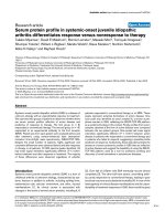
The vascular pedicle width represents the mediastina l
silhouette of the great vessels. First described in detail
by Milne and colleagues two decades ago, VPW is the
distance from whi ch the l eft subclavian artery exits the
aortic arch measured across to the point at which the
super ior vena cava crosses the right mainstem bronchus
(Figure 1) [5]. The vertical lateral border of the superior
vena cava or right brachiocephalic vein was utilized for
the measurement in radiographs where the right border
of the vascular pedicle was indistinct. The cardiothoracic
ratio was calculated by dividing the measurement of the
largest width of the cardiac silhouette by the interior
width of the thoracic cavity at the same vertical location.
Covariates
A number of covariates were collected prospectively
during the FACTT trial that may also have influenced
Rice et al. Critical Care 2011, 15:R86
/>Page 2 of 10
both VPW and/or the intravascular pressure measure-
ments (Table 1). Net fluid balance was collected for the
24 hours prior to enrollment and then every day until
the earlier of extubation, death, or study Day 7. PEEP
was recorded from morning ventilator measurements
daily through study Day 7. Serum albumin was mea-
sured at baseline.
Statistical analysis
Correlati on between VPW measurements from the por-
table chest radiograph with the PAOP represented the
primary endpoint. Secondary endpoints included corre-
lation of VPW and CTR with both PAOP and CVP. The
effect of cumulative fluid balance, PEEP, and serum
albumin on the relationship between VPW and PAOP
represented additional secondary endpoints. A formal
sample size calculation was not undertaken as this study
utilized all available patients with matching CXR and
vascular pressure measurements from the five sites. The
mean VPW and CTR were determined for each indivi-
dual radiograph by averaging the measurements from all
the readers who gave a satisfactory grade to position
and technique for that radiograph. Inter-rater variability
was assessed by calculating the difference between read-
ings for each pair of readers for each measurement.
These differences were then averaged and divided by the
mean value of the reading to obtain the relative percent
variation. VPW and CTR were compared separately to
both CVP and PAOP measurements using scatterplots
with regression equations. R values were determined
using Spearman’s correlations. Multivariate linear
regression analysis was utilized to determine the effect
that cumulative fluid balance, PEEP, and baseline serum
albumin had on the r elationship between VPW and
PAOP. All variables were inclu ded in the model regard-
less of the significance of their associati on. Both the net
fluid balance for the day of the intravascular pressure
measur ement and the cumulative net fluid balance from
24 hours prior to enrollment through the day of the
VPW measurement were included in the multivariate
regression analysis separately. Standardized coefficients
were obtained to compare the relative effect each covari-
ate had on PAOP. Cumulative net fluid balance from 24
hours prior to enrollment through the day of the VPW
measurement had a better correlation than the daily
fluid balance, so it was utilized in the final model. The
PEEP value used in the regression analysis was the
morning (that is, 06:00 to 10:00 AM) value from the day
Figure 1 Representation of the VPW measurement and change
in VPW over time. The VPW is the distance between where the
left subclavian artery exits the aortic arch and where the superior
vena cava crosses the right mainstem bronchus. (a-b) represent
CXRs from the same patient at baseline and Day 3, respectively,
where the VPW has decreased by 13 mm.
Table 1 Multivariate regression of VPW, net fluid balance, PEEP, and albumin with PAOP
Unstandardized coefficients 95% CI for B Standardized coefficients P-value
B Std. error Lower bound Upper bound
Constant -3.34 4.05 -11.39 4.71 0.41
VPW 0.20 0.04 0.11 0.29 0.43 <0.001
Cumulative Net Fluid (L) 0.21 0.08 0.05 0.37 0.26 0.01
PEEP 0.26 0.14 -0.02 0.54 0.19 0.07
Albumin 1.05 0.90 -0.73 2.84 0.11 0.24
Standardized coefficients allow comparison of the covariate correlations to PAOP. For example, VPW correlates with PAOP about 2.5 times as well as PEEP (0.42
vs. 0.19). 95% CI, 95% confidence interval; VPW, vascular pedicle width; PEEP, positive end-expiratory pressure; PAOP, pulmonary artery occlusion pressure.
Rice et al. Critical Care 2011, 15:R86
/>Page 3 of 10
of the CXR. Receiver operating characteristic (ROC)
curves were utilized to determine both the optimal
VPW cutoff for discriminating adequateness of conser-
vative fluid management, defined as a PAOP measure-
ment <8 mmHg and whether some component of
hydrostatic edema may also be present (that is, PAOP
≥18 mm Hg). Sensitivity, specificity, and likelihood
ratios of the VPW cutoff value were calculated using
Confidence Interval Analysis 2.1.0 [12]. The change in
VPW over time was calculated from the first CXR to
the last available CXR in patients with two CXRs at
least 48 hours apart between baseline and study Day 4.
The median change in VPW over time was compared
between conservative and liberal treatment strategy
groups using Mann Whitney U testing. Data were ana-
lyzed using SPSS (Version 15.0; Chicago, IL, USA) and
two-sided P-values ≤0.05 were utilized to determine sta-
tistical significance.
Results
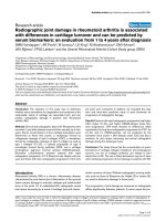
Of the 1,001 patients enrolled in FACTT, 293 were
enrolled at one of the five sites participati ng in this
study. Those 2 93 patients provided 555 CXRs through
study Day 4 for interpretation. Of the available 555
CXRs, 510 (91.9%) were deemed satisfactory for both
technique and position by at least three of the reviewers.
Of the satisfactory CXRs, 118 (f rom 71 patients) were
able to be paired with a “matching” PAOP measurement
(that is, within three hours of the measurement) and
276 (from 152 patients) were able to be paired with a
“matching” CVP measurement (Figure 2). The average
CVP and PAOP for the paired measurements were 11.9
±5.1and16.2±5.4mmHg,respectively.Inthe118
pairs with both measurements available, PAOP and CVP
were highly correlated (CVP = 0.58 + 0.73*PAOP; r =
0.74; P < 0.001). The average VPW and CTR for paired
measurements was 71.8 ± 11.2 mm and 0.56 ± 0.06,
respectively. The correlation between VPW and CTR (r
=0.33;P < 0.001) was also significant, but less strong
than that betwe en PAOP and CVP. The average differ-
ence between readers’ measureme nts were 8 ± 6 mm
for cardiac width, 6 ± 5 mm for thoracic width, and 8 ±
4 mm for VPW. These represent relative percent varia-
tions of 5 ± 4%, 2 ± 2%, and 11 ± 6%, for cardiac, thor-
acic, and VPW measurements, respectively.
VPW, CTR, and intravascular pressure measurement
correlations
The VPW decreased by a median width of 1.8 (inter-
quartile range (IQR): -7.2 to + 3.5) mm over time in
patients assigned to the conservative (n = 72) fluid man-
agement strategy compared to a median increase in
width of 2.3 (IQR: -4.4 to +8.8) mm in those assigned to
the liberal fluid management strategy (n = 77) (P =
0.012). For these sam e patients, conservative fluid man-
agement strategy resulted in a less positive cumulative
fluid balance (742 ± 7,986 vs. 6,553 ± 7,913 cc; P <
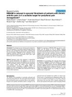
0.001). Figure 3a shows a s catterplot demonstrating the
relationship between VPW and PAOP while Figure 3b
demonstrates the relationship between VPW and CVP.
Although statistically significant, VPW did not highly
correlate with either PAOP (r = 0.41; P < 0.0 01) or CVP
(r = 0.21; P = 0.001). The relationship between VPW
and PAOP is described by the linear regression equa-
tion: VPW = 57 + 0.9*(PAOP) while the equation: VPW
= 66.4 + 0.45*(CVP) describes the correlation with CVP.
Cardiothoracic ratio correlated modestly with PAOP (r
= 0.30; P = 0.001) and demonstrated little correlation
with CVP (r = 0.15; P = 0.01).
VPW, PAOP and covariates
PAOP was positively correlated with VPW (r = 0.41; P <
0.001), cumulative net fluid balance to the time of the
paired measurement (r = 0.31; P = 0.002), and PEEP (r
= 0.22; P = 0.02) but not serum albumin (P =0.23).
VPW did not correlate significantly with cumulative
fluid balance (P = 0.46), PEEP (P = 0.21), or serum albu-
min (P = 0.20). Multivariate regression analysis demo n-
strated that VPW and cumulative fluid balance
independently correlated with PAOP and PEEP trended
toward a correlation with PAOP. Serum albumin did
not correlate with VPW in multivariate analysis. Stan-
dardized coefficients indicate that VPW had a 1.5-fold
stronger correlation with PAOP than cumulative fluid
balance and a 2.5-fold stronger correlation than PEEP
(Table 1).
Optimal VPW for discriminating adequacy of conservative
fluid management or hydrostatic component to the
edema
Only seven (6%) of the 118 PAOP and 19 (7%) of the
276 CVP measurements were within the target range for
conservative fluid management strategy (that is, PAOP
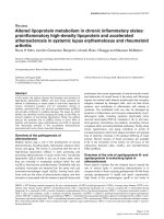
<8 or CVP <4 mm Hg). The ROC curve (Figure 4a)
demonstrates the ability of VPW to discriminate achiev-
ing PAOP <8 mm Hg (AUC = 0.73; 95% CI: 0.59 to
0.87; P =0.04).AVPW≤67 mm had 71.4% sensitivity
(95% CI 30.1 to 95.4%) and 67.6% specificity (95% CI
58.5 to 75.4%) for predicting PAOP <8 mm Hg. Due to
the high percentage of measurements outside the target
range, however, a VPW greater than 67 mm had a nega-
tive predictive value of 97.4% (95% CI 91.0 to 99.3%) for
PAOP ≥8 mm Hg. The positive and negative likelihood
ratios for the VPW cutoff of 67 mm discriminating
PAOP <8 (that is, c onservative fluid strategy target
range) were 2.2 (95% CI: 1.3 to 3.8) and 0.42 (95% CI:
0.13 to 1.3), respectively. VPW was not able to discrimi-
nate achieving the conservative fluid management target
Rice et al. Critical Care 2011, 15:R86
/>Page 4 of 10
using CVP (that is, CVP <4 mmHg) (AUC = 0.57; 95%
CI: 0.43 to 0.70; P = 0.32).
Over a third (44/118) of the PAOP measurements
were ≥18 mm Hg, suggesting a hydrostatic component
to the edema in these patients with lung injury. A VPW
cutoff ≥72 mm best discriminated a PAOP ≥18 mm Hg
(AUC 0.686; 95% CI 0.589 to 0.784; P = 0.001) (Figure
4b). This cutoff demonstrated 61.4% sensitivity (95% CI
46.6 to 74.3%) and 60.8% specificity (95% CI 49.4 to
71.1%). However, the positive predictive va lue was only
48.2% (95% CI 35.7 to 61.0%) and negative predictive
value was 72.6% (95% CI 60.4 to 82.1%).
Discussion
Multiple studies in patients with a spectrum of intra-
vascular volume ranging from ALI to CHF indicate
that the VPW measured from a CXR correlates highly
with intravascular pressure and distinguishes cardio-
genic from non-cardiogenic edema, but this is the first
studytoourknowledgeassessingtheroleofthiseasily
measured anatomic landmark among patients exclu-
sively with ALI (a markedly narrower intravascular
volume range). VPW correlated moderately well with
PAOP and less well with CVP. In multivariate regres-
sion, the correlation between VPW and PAOP was
stronger than that between net cumulative fluid bal-
ance or PEEP and PAOP, while serum albumin did not
independently correlate with PAOP. Furthermore,
VPW decreased over time in the conservative fluid
management strategy arm, but increased in the liberal
fluid management arm. VPW, however, was only mod-
erately able to discrimin ate achievement of the conser-
vative fluid management target of PAOP <8 mmHg
and unable to discriminate achievement of CVP <4
mm Hg. VPW was also only moderately able to discri-
minate whether a hydrostatic component of the edema
mayalsobepresentinthesepatientswithALI.These
new observations provide additional data on the relia-
bility and clinical relevance of this non-invasive radi-
ologic measurement.
Figure 2 Flow diagram showing study enrollment and available CXRs.
Rice et al. Critical Care 2011, 15:R86
/>Page 5 of 10
Although underutilized, determining intravascular
volume status by radiograp hic appearance has classically
revolved around measurement of the VPW and analysis of
patterns of lung parenchymal infiltration [8,13,14]. A
review of acute pulmonary edema recommended the
VPW as a potentially use ful facto r in differentiating car-
diogenic from non-cardiogenic pulmonary edema [15].
Initially characterized in upright posteroanterior CXRs
from non-critically ill patients, the VPW measurement has
subsequently been shown to have similar predictive ability
in ICU patients with anteroposterior supine films [6,9,10].
Several investigations have addressed relationships
between VPW and intravascular volume status [12,16,17].
Other studies have demonstrated the ability of the VPW
to differentiate pulmonary edema due to volume overload
from that due to acute lung injur y [6,9,1 0]. Our op timal
cutoff of a VPW ≥72 mm for distinguishing a hydrostatic
component to the pulmonary edema was similar to the
values of 68 and 70 mm found in previous studies [6,9]. In
addition to confirming the findings of these studies, our
data also suggest that VPW might be able to be used to
identify when hydrostatic edema may be contributing to
ALI and whether conservative fluid management target s
have been reached in cases where intravascular pressure
measurements are not available.
Figure 3 Correlation of V PW w ith PAOP and CVP. (a)
demonstrates that VPW correlates moderately well with PAOP (VPW
= 57 + 0.9*PAOP; r = 0.41; P < 0.001). (b) demonstrates the weak
correlation between VPW and CVP (VPW = 66.4 + 0.45*CVP; r = 0.21;
P = 0.001).
Figure 4 ROC curve for VPW discriminating fluid status by
PAOP. (a) demonstrates that VPW of 67 mm discriminates PAOP <8
mmHg (AUC = 0.73; P = 0.04). (b) demonstrates that VPW of 72
discriminates PAOP ≥18 mmHg (AUC = 0.69; P = 0.001).
Rice et al. Critical Care 2011, 15:R86
/>Page 6 of 10
Application of VPW measurement or the necessity for
uptake into clinical practice has been marginal because
of the decreasing prevalence of placement of invasive
catheters such as pulmonary artery or central venous
catheters as well as unfamiliarity with data related to its
measurement and potential value when invasive tools
are not in place. In the current period of critical care in
which fewer pulmona ry arte ry catheters are placed,
most intravascular measurements are taken on a routine
basis from the conventional catheter measuring a CVP.
Of note, in this investigation, VPW correlated with
PAOP better than CVP.
It is helpful to be facile with factors that can increase
or reduce the VPW. The supine position can increase
the VPW by nearly 20% compared to the upright posi-
tion [5], and thus the “normal” VPW on films taken
when the patient is supine would be 58 to 62 mm. Rota-
tion of the patient to the right artificially increases the
VPW, while rotation to the left decreases the measure-
ment [11]. Importantly, in this study all the patients’
CXRs and intravascular measurements were taken in the
supine or semi-supine position and only films graded as
satisfactory for positioning (that is, not overly rotated on
visual inspection) were included in the analysis. In addi-
tion to patient positioning, some have raised concern
that the disease process might affect the assessment of
VPW. Indeed, the effects of recent trauma, thoracic sur-
gery, or prior radi ation therapy alter components of the
mediastinal silhouet te and compromise the utility of the
VPW [18,19]. On the other hand, respiratory factors
have been shown to have relatively little eff ect on VPW
measurements. Milne observed comparable VPW mea-
surements during both inspiration and expiration [5].
Although mechanical ventilation may have profound
effects upon other radiographic findings such as the pat-
tern and severity of parenchymal infiltrates [20,21],
VPW measurements have been found to be consistent
between spontaneo us and positive pressure breaths [20].
Our data also found only a trend toward a weak correla-
tion between PEEP and VPW measurements. Despite
these potential limitations in measuring the VPW, we
confirmed prior findings that VPW correlates with
PAOP and we found tha t the VPW correlated 1.5 times
better with PAOP than cumulative net fluid balance and
2.5 times better than PEEP. Thus, for patients without
or for clinicians who prefer not to use invasive intravas-
cular pressure measurements, VPW represents a better
surrogate of PAOP than net fluid balance.
One limitation of our study is that w e compare VPW
to two surrogate measures of intravascular volume, CVP
and PAOP, and not a d irect measure of intravascular
volume, such as right (RVEDV) or left ventricular end-
diastolic volume (LVEDV). Although echocardiography
might estimate RVEDP and LVEDP, too few patients
had these available on days with VPW measurements to
investigate this correlation directly. CVP and PAOP do
correlate well with right (RVEDP) and left ventricular
end-diastolic pressure (LVEDP), respectively [22-24].
Although a similar correlation with RVEDV and LVEDV
is widely presumed, this is not the case in a number of
conditions pertinent to acute lung injury, including sep-
sis [25-27], trauma [28], and acute respiratory f ailure
requiring mechanical ventilation [29]. Observations by
Kumar and c olleagues suggest that CVP and PAOP do
not correlate well with RVEDV or LVEDV even in nor-
mal, healthy volunteers [30]. This is likely due to varying
compliance of the ventricles from patient to patient and
heartbeat to heartbeat within the same patient. Because
VPW is an objective, anatomic measurement of vascular
structures, it is likely influenced less than CVP and
PAOP by outside forc es such as mechan ical ventilation,
PEEP, large intrathoracic pressure variations during t he
respiratory cycle, and even varying cardiac compl iances.
As such, VPW may prove to be a more accurate mea-
sure of intravascular volume than either CVP or PAOP
and may correlate better with actual intravascular
volume than these intravascular pressure surrogates.
Although our data lack a direct intravascular volume
measurement, future studies could incorporate one as a
different reference standard. It is noteworthy that even
in this selected population of patients with noncardio-
genic pulmonary edema, that VPW measurements mod-
erately differentiated volume status.
Our study also has other limitations. The patients
enrolled in FACTT are a highly-select ed group of
patients with acute lung injury. This substudy evaluates
data from a subset of the overall FACTT population.
However, alm ost 30% of the enrolled patients were
included, with five geographically diverse centers with
heterogeneous patient popu lations participating.
Although all the data were collected prospectively dur-
ing the conduct of the original study, this substudy
represents a post-hoc, retrospective analysis. As such,
many of the CXR and vascular pressure measurements
did not occur simultaneously. To minimize any potential
bias this might introduce, we limited our analysis to
“matched” measurements and CXRs obtained within
three hours of each other. Furthermore, although a
VPW of 67 mm, was found to best pr edict a PAOP <8
mmHg the relatively few instances that conservative
fluid management resulted in target PAOP or CVP mea-
surements being reached resulted in wide confidence
intervals for the sensitivity and specificity. Similar to the
cutoffs previously defined for differentiating patients
with cardiogenic versus noncardiogenic edema [ 6,9], a
VPW value of 72 or higher in our study, also discrimi-
nated a PAOP of at least 18 mmHg, which could repre-
sent cases where volume overload and hydrostatic
Rice et al. Critical Care 2011, 15:R86
/>Page 7 of 10
edema may be contributing to the hypoxia and patients
who may benefit from diuresis. Despite only having
moderate sensitivity and specificity for predicting either
volume overload or conservative fluid status, given its
non-invasive nature, relative availability, and moderate
sensitivity and sensitivity, we think these data support
the use of VPW in a fluid management strategy when
other measures, such as intravascular pressure measure-
ments, are unavailable. A suggested algorithm is pre-
sented in Figure 5.
This study also has a number of strengths. We aver-
aged the VPW measurements from multiple, indepen-
dent, blinded readers of the CXRs, ranging from a
seasoned radiologist to intensivists with both extensive
and limited prior experience in measuring VPW.
Although inter-rater variability in this study was higher
than that seen in previous studies [6,10], the VPW was
still a significant predictor of intravascular status of the
cohort. This v ariability, likely secondary to the number
of readers and inexperience of two readers, might be
reduced through standardized teaching and more
experience, yielding even more striking results. Despite
the relatively small number of patients, ours still repre-
sents one of the largest studies of VPW measurements
to date. In addition to confirming a relationship between
VPW and intravascular pressure measurements, this
investigation also introduces the novel idea that VPW
can be used to identify when conservative fluid manage-
ment targets have been reache d. The nature of the data
collected allowed us to compare VPW with both PAOP
and CVP and to compare the effect of other possible
confounders, such as cumulative fluid balance, PEEP,
and serum albumin on the relationship.
TheFACTTstudydemonstratedthatpatientswith
ALI treated with a conservative fluid strategy had signif-
icantly more days alive and free from mechanical venti-
lation and alive and out o f the ICU compared t o those
managed with a more liberal fluid management strategy
[4]. Despite these important outco me benefits, wide-
spread implementation of a conservative fluid strategy
in practice has been relatively slow [31]. The reasons for
this delayed acceptance are likely multifactorial, includ-
ing lack of survival benefit and the relative complexity
of the management algorithm, which includes the need
Figure 5 Suggested fluid management algorithm for ALI patients using VPW.
Rice et al. Critical Care 2011, 15:R86
/>Page 8 of 10
for some assessment of intravascular pressure. Invasive
measurements were utilized in the clinical trial, with
similar outcomes resulting from CVP and PAOP mea-
surements [1]. While thi s likely will contrib ute to a
further reduction in the insertion of PACs, obtaining
CVP measurements still requires an invasive procedure
and risk for complications. Although many patients with
ALI have central venous catheters placed for routine
care, the frequency of invasive procedures is decreasing
in clinical practice and 8.1% of patients were excluded
from the parent study due to physicians not intending
to place central venous access [1]. The ability to utilize
non-invasive measures of intravascular volume may
obviate the need for a CVC in some patients and further
reduce the risk of complications. The use of the non-
invasive VPW may enhance implementation and accep-
tance of the conservative fluid strategy into routine clin-
ical practice. It remains to be established whether fluid
adjustments made on the basis of VPW me asurements
achieve similar outcomes as strategies guided by invasive
hemodynamic measurements.
Conclusions
VPW correlated moderately well with PAOP and less
well with CVP in patients with ALI enrolled in a clinical
trial of different fluid management strategies. VPW had
a higher correlation with the historical standard of
PAOP than did cum ulative fluid balance or PEE P.
Although the actual correlation between VPW and
direct intravascular volume measurements remains
unknown, these data confirm previous studies that show
the utility of VPW as a noninvasive measure and the
best radiographic sign of patients’ intravascular volume
status. VPW is measured easily on most CXRs and
might be useful for discriminating when a hydrostatic
component of the edema m ay be contributing or con-
servative fluid management pressure targets have yet to
be reached in patients with ALI when invasive vascular
pressure measurements are unavailable. Routine substi-
tution of VPW for CVP or PAOP in fluid management
of ALI patients canno t be recommended, however, until
a trial using VPW directly to titrate diuretic d osing has
been completed.
Key messages
• In ventilated ICU cohorts of both high and low
intravascular volume status (for example, ALI and
CHF), the VPW has been consistently shown as a
correlate of intravascular volume status.
• In this study restricted to ALI patients, the “non-
invasively obtained” VPW correlated with PAOP bet-
ter than CVP.
• Changes in VPW correlat ed with chan ges in
volume status.
• VPW had a 1.5-fold stronger correlation with
PAOP than cumulative fluid balance and a 2.5-fold
stronger correlation than PEEP.
• Within the narrower range of volume status pre-
sented by restricting this cohort to only ALI, the
ability of VPW to discriminate a hydrostatic compo-
nent of the edema and achievement of fluid manage-
ment goals was limited.
• Given its no n-invasive nature an d availabil ity,
VPW might still be able to be used to direct fluid
management in patients with ALI when intravascular
pressure measurements are unavailable.
Abbreviations
ALI: acute lung injury; ARDS: acute respiratory distress syndrome; AUC: area
under the curve; CTR: cardiothoracic ratio; CVC: central venous catheter; CVP:
central venous pressure; CXR: chest X-ray; FACTT: Fluid and Catheter
Treatment Trial; ICU: intensive care unit; IQR: interquartile range; IRB:
institutional review board; LVEDP: left ventricular end-diastolic pressure;
LVEDV: left ventricular end-diastolic volume; NHLBI: National Heart Lung and
Blood Institute; NIH: National Institutes of Health; PAC: pulmonary artery
catheter; PAOP: pulmonary artery occlusion pressure; PEEP: positive end-
expiratory pressure; ROC: receiver operating characteristic; RVEDP: right
ventricular end-diastolic pressure; RVEDV: right ventricular end-diastolic
volume; VPW: vascular pedicle width; 95% CI: 95% confidence interval.
Acknowledgements
Funding Sources: National Institutes of Health, Heart Lung and Blood
Institute: HL81431 (TWR); HR46054 (TWR, APW, GRB); HL081332(LBW);
HL088263 (LBW); HR 16147 (JSS); HR 16155 (RDH, PW).
Author details
1
Division of Allergy, Pulmonary, and Critical Care Medicine, Vanderbilt
University School of Medicine, T-1218 MCN Nashville, TN 37221, USA.
2
Section on Pulmonary, Critical Care, Allergy, and Immunologic Diseases,
Wake Forest University School of Medicine, Medical Center Blvd, Winston-
Salem, NC 27157, USA.
3
Division of Radiological Sciences, Department of
Radiology, Wake Forest University School of Medicine, Medical Center Blvd,
Winston-Salem, NC 27157, USA.
4
Division of Critical Care Medicine, Baystate
Medical Center, 759 Chestnut St, Springfield, MA 01199, USA.
5
Department of
Medicine and Anesthesia, Cardiovascular Research Institute, University of
California, San Francisco, 505 Parnassus Avenue, Moffitt Hospital, M-917, San
Francisco, CA 94143, USA.
6
Department of Pulmonary and Critical Care
Medicine, Moses Cone Health System, 1200 N Elm St, Greensboro, NC 27403,
USA.
Authors’ contributions
All authors participated in the design of the study and data acquisition.
TWR, LBW, EWE, CC and EH interpreted the CXRs. TWR, EWE and LBW
analyzed and interpreted the data. TWR, EWE and LBW drafted the
manuscript. EWE, LBW, MAM, RDH, JSS and EH revised the manuscript
critically for important intellectual content. All authors read and approved
the final manuscript.
Competing interests
The authors declare that they have no competing interests.
Received: 28 June 2010 Revised: 8 February 2011
Accepted: 7 March 2011 Published: 7 March 2011
References
1. Wheeler AP, Bernard GR, Thompson BT, Schoenfeld D, Wiedemann HP,
deBoisblanc B, Connors AF Jr, Hite RD, Harabin AL: Pulmonary-artery
versus central venous catheter to guide treatment of acute lung injury.
N Engl J Med 2006, 354:2213-2224.
Rice et al. Critical Care 2011, 15:R86
/>Page 9 of 10
2. Antonelli M, Levy M, Andrews PJ, Chastre J, Hudson LD, Manthous C,
Meduri GU, Moreno RP, Putensen C, Stewart T, Torres A: Hemodynamic
monitoring in shock and implications for management. International
Consensus Conference, Paris, France, 27-28 April 2006. Intensive Care Med
2007, 33:575-590.
3. Heresi GA, Arroliga AC, Wiedemann HP, Matthay MA: Pulmonary artery
catheter and fluid management in acute lung injury and the acute
respiratory distress syndrome. Clin Chest Med 2006, 27:627-635.
4. Wiedemann HP, Wheeler AP, Bernard GR, Thompson BT, Hayden D,
deBoisblanc B, Connors AF Jr, Hite RD, Harabin AL: Comparison of two
fluid-management strategies in acute lung injury. N Engl J Med 2006,
354:2564-2575.
5. Milne EN, Pistolesi M, Miniati M, Giuntini C: The vascular pedicle of the
heart and the vena azygos. Part I: The normal subject. Radiology 1984,
152:1-8.
6. Ely EW, Smith AC, Chiles C, Aquino SL, Harle TS, Evans GW, Haponik EF:
Radiologic determination of intravascular volume status using portable,
digital chest radiography: a prospective investigation in 100 patients.
Crit Care Med 2001, 29:1502-1512.
7. Woodring JH, Given CA: Noninvasive estimation of pulmonary capillary
wedge pressure from computed radiography. J Ky Med Assoc 2000,
98:115-120.
8. Milne EN, Pistolesi M, Miniati M, Giuntini C: The radiologic distinction of
cardiogenic and noncardiogenic edema. AJR Am J Roentgenol 1985,
144:879-894.
9. Thomason JW, Ely EW, Chiles C, Ferretti G, Freimanis RI, Haponik EF:
Appraising pulmonary edema using supine chest roentgenograms in
ventilated patients. Am J Respir Crit Care Med 1998, 157:1600-1608.
10. Martin GS, Ely EW, Carroll FE, Bernard GR: Findings on the portable chest
radiograph correlate with fluid balance in critically ill patients. Chest
2002, 122:2087-2095.
11. Aberle DR, Wiener-Kronish JP, Webb WR, Matthay MA: Hydrostatic versus
increased permeability pulmonary edema: diagnosis based on
radiographic criteria in critically ill patients. Radiology 1988, 168:73-79.
12. University of Southampton School of Medicine. Confidence Interval
Analysis (CIA) Software. [ />13. Pistolesi M, Milne EN, Miniati M, Giuntini C: The vascular pedicle of the
heart and the vena azygos. Part II: Acquired heart disease. Radiology
1984, 152:9-17.
14. Miniati M, Pistolesi M, Paoletti P, Giuntini C, Lebowitz MD, Taylor AE,
Milne EN: Objective radiographic criteria to differentiate cardiac, renal,
and injury lung edema. Invest Radiol 1988, 23:433-440.
15. Ware LB, Matthay MA: Clinical practice. Acute pulmonary edema. N Engl J
Med 2005, 353:2788-2796.
16. Haponik EF, Adelman M, Munster AM, Bleecker ER: Increased vascular
pedicle width preceding burn-related pulmonary edema. Chest
1986,
90:649-655.
17. Don C, Burns KD, Levine DZ: Body fluid volume status in hemodialysis
patients: the value of the chest radiograph. Can Assoc Radiol J 1990,
41:123-126.
18. Chiou AC, Abularrage CJ, Olson PN, Hood L, Egeler CE, Griffiths HJ,
Shumway SJ: “Incisura” of the ascending aorta and vascular pedicle
width in the cardiac transplant patient. Ann Thorac Surg 1996,
62:1141-1145.
19. Milne EN, Imray TJ, Pistolesi M, Miniati M, Giuntini C: The vascular pedicle
and the vena azygos. Part III: In trauma–the “vanishing” azygos.
Radiology 1984, 153:25-31.
20. Ely EW, Johnson MM, Chiles C, Rushing JT, Bowton DL, Freimanis RI,
Choplin RH, Haponik EF: Chest X-ray changes in air space disease are
associated with parameters of mechanical ventilation in ICU patients.
Am J Respir Crit Care Med 1996, 154:1543-1550.
21. Langevin PB, Hellein V, Harms SM, Tharp WK, Cheung-Seekit C,
Lampotang S: Synchronization of radiograph film exposure with the
inspiratory pause. Effect on the appearance of bedside chest
radiographs in mechanically ventilated patients. Am J Respir Crit Care Med
1999, 160:2067-2071.
22. Braunwald E, Brockenbrough EC, Frahm CJ, Ross J Jr: Left atrial and left
ventricular pressures in subjects without cardiovascular disease:
observations in eighteen patients studied by transseptal left heart
catheterization. Circulation 1961, 24:267-269.
23. Swan HJ, Ganz W, Forrester J, Marcus H, Diamond G, Chonette D:
Catheterization of the heart in man with use of a flow-directed balloon-
tipped catheter. N Engl J Med 1970, 283:447-451.
24. Flores ED, Lange RA, Hillis LD: Relation of mean pulmonary artery wedge
pressure and left ventricular end-diastolic pressure. Am J Cardiol 1990,
66:1532-1533.
25. Calvin JE, Driedger AA, Sibbald WJ: Does the pulmonary wedge pressure
predict left ventricular preload in critically ill patients? Crit Care Med
1981, 9:437-443.
26. Jardin F, Valtier B, Beauchet A, Dubourg O, Bourdarias JP: Invasive
monitoring combined with two-dimensional echocardiograhpic study in
septic shock. Intensive Care Med 1994, 20:550-554.
27. Sakka SG, Reinhart K, Meier-Hellmann A: Comparison of pulmonary artery
and arterial thermodilution cardiac output in critically ill patients.
Intensive Care Med 1999, 25:843-846.
28. Diebel L, Wilson RF, Heins J, Larky H, Warsow K, Wilson S: End-diastolic
volume versus pulmonary artery wedge pressure in evaluating cardiac
preload in trauma patients. J Trauma 1994, 37:950-955.
29. Lichtwarck-Aschoff M, Zeravik J, Pfeiffer UJ:
Intrathoracic blood volume
accurately reflects circulatory volume status in critically ill patients with
mechanical ventilation. Intensive Care Med 1992, 18:142-147.
30. Kumar A, Anel R, Bunnell E, Habet K, Zanotti S, Marshall S, Neumann A,
Ali A, Cheang M, Kavinsky C, Parrillo JE: Pulmonary artery occlusion
pressure and central venous pressure fail to predict ventricular filling
volume, cardiac performance, or the response to volume infusion in
normal subjects. Crit Care Med 2004, 32:691-699.
31. Leaver SK, Evans TW: Acute Respiratory Distress Syndrome. BMJ 2007,
335:389-394.
doi:10.1186/cc10084
Cite this article as: Rice et al.: Vascular pedicle width in acute lung
injury: correlation with intravascular pressures and ability to
discriminate fluid status. Critical Care 2011 15:R86.
Submit your next manuscript to BioMed Central
and take full advantage of:
• Convenient online submission
• Thorough peer review
• No space constraints or color figure charges
• Immediate publication on acceptance
• Inclusion in PubMed, CAS, Scopus and Google Scholar
• Research which is freely available for redistribution
Submit your manuscript at
www.biomedcentral.com/submit
Rice et al. Critical Care 2011, 15:R86
/>Page 10 of 10