Urological Emergencies in Clinical Practice - part 9 pptx
Bạn đang xem bản rút gọn của tài liệu. Xem và tải ngay bản đầy đủ của tài liệu tại đây (233.09 KB, 20 trang )
obstruction. Because hydronephrosis is a normal finding in the
majority of pregancies, its presence cannot be taken as a sign of
a possible ureteric stone. Ultrasound is an unreliable way of diag-
nosing the presence of stones in pregnant (and in nonpregnant)
women. In a series of pregnant women, ultrasound had a sensi-
tivity of 34% (i.e., it misses 66% of stones) and a specificity of
86% for detecting an abnormality in the presence of a stone (i.e.,
false-positive rate of 14%) (Stothers and Lee 1992).
PRESENTATION OF STONES IN PREGNANCY
Flank pain is the usual presentation, with or without haematuria
(macroscopic or microscopic). Differential diagnoses include
placental abruption, appendicitis, and pyelonephritis, to name
but a few.
WHAT IMAGING STUDY SHOULD BE USED TO ESTABLISH THE
DIAGNOSIS OF A URETERIC STONE IN PREGNANCY?
Exposure of the fetus to ionising radiation can cause fetal
malformations, malignancies in later life (leukaemia), and
mutagenic effects (damage to genes causing inherited disease in
the offspring of the fetus). Fetal radiation doses during various
procedures are shown in Table 8.1.
Radiation doses of <100 mGy are very unlikely to have an
adverse effect on the fetus (Hellawell et al. 2002). In the United
States, the National Council on Radiation Protection (NCRP)
has stated, ‘Fetal risk is considered to be negligible at <50 mGy
when compared to the other risks of pregnancy, and the risk of
malformations is significantly increased above control levels at
doses >150 mGy’ (NCRP 1997). The American College of Obste-
tricians and Gynecologists (ACOG) has stated, ‘X-ray exposure to
152 J. REYNARD
TABLE 8.1. Fetal radiation dose after various radiological investigations
Procedure Fetal dose mGy Risk of inducing fetal
(mean) cancer (up to age 15 years)
KUB x-ray 1.4 —
IVU 6 shot 1.7 1 in 10,000
IVU 3 shot
CT—abdominal 8
CT—pelvic 25
Fluoroscopy for 0.4 1 in 42,000
JJ stent insertion
CT, computed tomography; IVU, intravenous urogram; JJ stent; KUB,
kidney and urinary bladder.
<50 mGy has not been associated with an increase in fetal anom-
alies or pregnancy loss’ (ACOG 1995).
While these recommended maximum radiation levels are well
above those occuring during even computed tomography (CT)
scanning, and a dose of 50 mGy or less is regarded as safe, under-
standably there is a concern that any radiation dose exposes the
fetus to some risk. For this reason every effort should be made
to limit exposure of the fetus to radiation, to use alternative
imaging tests where possible, and to minimise radiation expo-
sure during treatment by JJ stent insertion or ureteroscopy.
However, the pregnant woman may be reassured that the risk to
her unborn child as a consequence of radiation exposure is likely
to be minimal.
Investigations or treatment that involve exposure to ionizing
radiation should not be withheld because of an unjustified fear of
damaging the fetus. The risks associated with irradiating
the fetus have to be balanced against the risks of missing the diag-
nosis of a stone obstructing the ureter and the difficulties and
potential dangers of performing JJ stent insertion or ureteroscopy
without the use of any (ionising radiation) imaging. While
ureteroscopy can be performed without fluoroscopy (Rittenberg
and Bagley 1988), most urologists nowadays perform the major-
ity of their ureteroscopic work under fluoroscopic control, and
may feel uncomfortable doing otherwise in a case that, as it
involves a pregnant woman and an unborn baby, is already high
risk. It is worth remembering that the radiation dose during
fluoroscopy for JJ stent placement is very low (on the order of
0.4 mGy, and up to a maximum of 0.8 mGy) and that the dose used
to assist ureteroscopy is likely to be little more than this.
Plain Radiography and Intravenous Urography (IVU)
These studies have limitations in pregnancy. First, the fetal skele-
ton and the enlarged uterus may obscure ureteric stones, so the
imaging study may not be diagnostic. Second, there may be
delayed excretion of contrast as a consequence of the physiolog-
ical dilatation of the kidney. It can be difficult, if not impossible,
to differentiate this ‘physiological’ delay from that due to an
obstructing stone. Third, there is also the theoretical risk of
fetal toxicity from the contrast material, though none has been
reported.
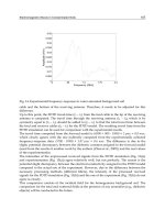
Ultrasound
As stated above, ultrasound is an unreliable way of diagnosing
the presence of stones in pregnant women. Jets of urine expelled
8. URETERIC COLIC IN PREGNANCY 153
by normal peristalsis of the nonobstructed ureter can be seen on
ultrasound scanning (Fig. 8.1), and the absence of such ureteric
jets is said to have a high sensitivity and specificity for diagnos-
ing obstructing stones (Doyle et al. 1995), though others have
reported that ureteric jets may be absent in asymptomatic preg-
nant women (Burke and Washowich 1998).
Computed Tomography Urography (CTU)
Although CT urography is a very accurate method for detecting
ureteric stones and the radiation dose is below 50 mGy, most
radiologist and urologists do not recommend this form of
imaging in pregnant women. Magnetic resonance urography (see
below) provides an alternative form of imaging in this difficult
group of patients.
Magnetic Resonance Urography (MRU)
The American College of Obstetricians and Gynecologists and the
U.S. National Council on Radiation Protection state, ‘Although
154 J. REYNARD
FIGURE 8.1. Jets of urine expelled by normal peristalsis of the non-
obstructed ureter can be seen on ultrasound scanning or on computed
tomography (CT) (as shown here). CT should be avoided if at all possi-
ble in pregnancy.
there is no evidence to suggest that the embryo is sensitive to
magnetic and radiofrequency at the intensities encountered in
MRI, it might be prudent to exclude pregnant women during the
first trimester’ (ACOG 1995, NCRP 1997). Given this advice,
therefore, MRU can potentially be used during the second and
third trimesters, but not during the first trimester.
MRU involves no ionising radiation and can be done with the
administration of contrast (Fig. 8.2). It is very accurate, with one
group reporting a sensitivity for detecting ureteric stones of
100% (Roy et al. 1996). However, MRU is expensive, and not
readily available in most hospitals, particularly after 5 o’clock. As
MR scanners become more widespread, it is likely that this
imaging modality will be used increasingly to establish a diag-
nosis in pregnant women with flank pain.
8. URETERIC COLIC IN PREGNANCY 155
FIGURE 8.2. Magnetic resonance urography.
MANAGEMENT OF URETERIC STONES IN PREGNANT WOMEN
The majority (70–80%) of ureteric stones in pregnant women
pass spontaneously (Stothers et al. 1992. Of those that do not
pass and require temporizing treatment with nephrostomy
tube drainage or JJ stents, many pass spontaneously postpartum.
Opiate-based analgesics are used for pain relief and oral and
intravenous fluids for hydration. Nonsteroidal antiinflammatory
drugs (NSAIDs) should be avoided because they can cause
premature closure of the ductus arteriosus by blocking
prostaglandin synthesis.
The indications for intervention are essentially the same as
in nonpregnant patients and include pain refractory to anal-
gesics, suspected urinary sepsis (high fever, high white count),
high-grade obstruction, and obstruction in a solitary kidney.
Options for intervention are JJ stent urinary diversion,
nephrostomy urinary diversion, or ureteroscopic stone removal.
Which option you use depends on how advanced the pregnancy
is, and on local facilities and expertise. Management of cases
requiring active intervention should aim to minimize radiation
exposure to the fetus, and to minimize the risk of miscarriage
and preterm labour. General anaesthesia can precipitate preterm
labour (Duncan et al. 1986), and with this in mind many urolo-
gists and obstetricians err on the side of temporizing options
such as nephrostomy tube drainage or JJ stent placement, rather
than on operative treatment in the form of ureteroscopic stone
removal.
Nephrostomy urinary diversion is widely available (Fig. 8.3),
can be done rapidly, provides good pain relief, drains infected
urine if present, and has a low risk of inducing miscarriage or
preterm labour (Kavoussi et al. 1992). These advantages must be
weighed against the fact that there is a small risk (in the order
of 1%) of heavy bleeding, requiring embolisation and/or blood
transfusion during nephrostomy insertion, and of septicaemic
shock occurring after insertion (2–4%; Ho and Cowan 2002,
Ramchandani 2001) (see Chapter 10). Furthermore, the nephros-
tomy tube may be required for some months, particularly when
it is inserted at a relatively early stage in the pregnancy. It can
be uncomfortable, may block or become infected, and may need
to be changed several times during the remaining pregnancy.
JJ stents overcome some of the problems of nephrostomy
tube drainage. They can be placed under local anaesthetic or with
light sedation with low doses of pethidine and diazemuls using
either ultrasound guidance or limited periods of fluoroscopy
(Hellawell et al. 2002, Stothers et al. 1992) (see Chapter 10). They
156 J. REYNARD
are an effective way of managing the pain of obstructing stones.
They may be a more comfortable form of urinary diversion than
percutaneous tube drainage, though many patients develop ‘stent
symptoms’ (frequency, urgency, and bladder pain), which can be
so bothersome that in some cases the stent has to be removed
(Hellawell et al. 2002).
8. URETERIC COLIC IN PREGNANCY 157
FIGURE 8.3. Nephrostomy urinary diversion.
In two series totalling 20 pregnant women who underwent
JJ stent placement (all under local anaesthetic or with sedoan-
algesia), at between 6 to 36 weeks’ gestation (mean 31 weeks),
there were no cases of premature labour (Hellawell et al. 2002,
Stothers et al. 1992).
The hypercalciuria of pregnancy may make stent encrusta-
tion and blockage more likely, and as a consequence it has been
suggested that stents should be changed every 6 to 8 weeks to
prevent the occurrence of blockage from encrustation (Kavoussi
et al. 1992). However, in a contemporary series where stent inser-
tion was performed at an average of 28 weeks of gestation for
obstructing ureteric stones, stent replacement was not required
in any patient (Hellawell et al. 2002), and in a slightly older
series, only 1 of 13 stents required replacement because of
ongoing pain (presumably indicating obstruction) (Stothers et al.
1992). It may well be, therefore, that regular stent changes, at
least when using contemporary stents, are not required. Avoid-
ing the need to change JJ stents is clearly desirable, as this is
technically more challenging than replacing a percutaneous
nephrostomy tube (though the difficulty of placement and
replacement depend on the availability of local expertise). There-
fore, one might be more inclined to recommend nephrostomy
tube drainage in very early pregnancy, rather than a JJ stent
where frequent changes of the latter might, at least in theory, be
required throughout the remaining pregnancy (Denstedt and
Razvi 1992).
JJ stents have been reported to become obstructed by
mechanical impingement of the fetal head (Hellawell et al. 2002)
and they may migrate down the ureter and into the bladder and
subsequently be voided per urethra as a consequence of the
dilatation of the ureter that is normally a feature of pregnancy
(Stothers et al. 1992).
Ureteroscopic stone extraction can be performed in preg-
nancy, but again its use depends on available expertise. Distor-
tion of the distal third of the ureter during the latter stages of
pregnancy makes rigid ureteroscopy technically more challeng-
ing, as does the presence of a large stone (European Association
of Urology 2001). For these reasons the less experienced uretero-
scopist may decide that nephrostomy tube drainage or a JJ stent
is a better option later on in pregnancy, with subsequent uretero-
scopic treatment being used if the stone fails to pass within a few
weeks of delivery. In solitary kidneys nephrostomy tube drainage
or a JJ stent may also be safer options rather than attempting
158 J. REYNARD
ureteroscopic stone extraction under the difficult conditions of
late pregnancy.
References
American College of Obstetricians and Gynecologists Committee on
Obstetric Practice. Guidelines for diagnostic imaging during preg-
nancy. ACOG Committee Opinion No. 158. Washington DC: ACOG,
1995.
Burke BJ, Washowich TL. Ureteral jets in normal second- and third
trimester pregnancy. J Clin Ultrasound 1998;26:423–426.
Coe FL, Parks JH, Lindhermer MD. Nephrolithiasis during pregnancy.
N Engl J Med 1978;298:324–326.
Denstedt JD, Razvi H. Management of urinary calculi during pregnancy.
J Urol 1992;148:1072–1075.
Doyle LA, Cronan JJ, Breslaw BH, Ridlen MS. New techniques of ultra-
sound and color Doppler in the prospective evaluation of acute renal
obstruction: do they replace the intravenous urogram? Abdom
Imaging 1995;20:58–63.
Duncan PG, Pope WD, Cohen MM, Green N. Fetal risk of anesthesia and
surgery during pregnancy. Anesthesiology 1986;64:790–794.
European Association of Urology. Guidelines on urolithiasis. ISDN 90-
806179-3-8, March 2001:10.
Hellawell GO, Cowan NC, Holt SJ, Mutch SJ. A radiation perspective for
treating loin pain in pregnancy by double-pigtail stents. Br J Urol Int
2002;90:801–808.
Hendricks SK, Ross SO, Krieger JN. An algorithm for diagnosis and
therapy of management and complications of urolithiasis during
pregnancy. Surg Gynecol Obstet 1991;172:49–54.
Ho S, Cowan NC, Holt SJ et al. Percutaneous nephrostomy (PCN): Pre-
liminary results from a prospective pilot study. Eur J Radiol (ESUR)
2002;12:D3.
Kavoussi LR, Albala DM, Basler JW, et al. Percutaneous management of
urolithiasis during pregnancy. J Urol 1992;148:1069–1071.
National Council on Radiation Protection and Measurement. Medical
radiation exposure of pregnant and potentially pregnant women.
NCRP report No. 54. Bethesda, MD: NCRPM, 1997.
Peake SL, Rowburgh HB, Le Planglois S. Ultrasonic assessment of
hydronephrosis in pregnancy. Radiology 1983;146:167–170.
Quality improvement guidelines for percutaneous nephrostomy.
Ramchandani P, et al. Quality improvement guidelines for percutaneous
nephrostomy. J Vasc Interv Radiol 2001;12:1247–1251.
Rittenberg MH, Bagley DH. Ureteroscopic diagnosis and treatment of
urinary calculi during pregnancy. Urology 1988;32:427–428.
Robert JA. Hydronephrosis of pregnancy. Urology 1976;8:1–4.
Roy C, Saussine C, Le Bras Y, et al. Assessment of painful ureterohy-
dronephrosis during pregnancy by MR urography. Eur Radiol
1996;6:334–338.
Stothers L, Lee LM. Renal colic in pregnancy. J Urol 1992;148:1383–1387.
8. URETERIC COLIC IN PREGNANCY 159
Chapter 9
Management of Urological
Neoplastic Conditions Presenting
as Emergencies
John Reynard and Hashim Hashim
TESTICULAR CANCER
Approximately 10% of cases of testicular cancer present with
metastatic disease in the retroperitoneum (retroperitoneal node
involvement causing back pain), chest (breathlessness, cough),
and neck (enlarged cervical nodes, tracheal compression, and
deviation). Spread to the central nervous system or involvement
of peripheral nerves can result in neurological manifestations
(Fig. 9.1). While most such cases present directly to oncologists,
from time to time the urologist is the first port of call. Such cases
should be referred to the oncologists as a matter of urgency for
high-dose chemotherapy.
MALIGNANT URETERIC OBSTRUCTION
The ureters enter the bladder just a few centimeters from the
bladder neck, and it is not difficult to see how a locally advanced
prostate or bladder cancer can obstruct them (Clarke 2003) (Fig.
9.2). Similarly, the cervix in women is very closely related to the
lower ureters (which is why the latter may be damaged during
hysterectomy) and locally advanced cervical cancer can cause
lower ureteric obstruction, as can a locally advanced rectal
cancer in both sexes (Soper et al. 1988). Other malignancies
(colon, stomach, lymphoma, breast, bronchus) can metastasize
to pelvic and retroperitoneal lymph nodes, causing unilateral or
bilateral malignant ureteric obstruction. In unilateral obstruc-
tion with a normally functioning contralateral kidney, the
obstruction proceeds silently. In bilateral obstruction, oliguria,
leading later to anuria and finally renal failure, is the mode of
presentation.
The emergency presentation is usually one of a patient with
acute renal failure, who may or may not be known to have
cancer. Patients present with a rising creatinine and symptoms
of renal failure including malaise, nausea, vomiting, and in some
cases marked oliguria or anuria as the locally advanced or nodal
metastases obstruct their ureters. This presentation is sometimes
mistaken for urinary retention, particularly if the patient has
some lower abdominal pain. However, when the bladder is
catheterised it contains only a small volume of urine and the high
creatinine level does not fall. In the case of prostate cancer,
digital rectal examination (DRE) reveals a firm (craggy) prostate
that has extended laterally. A locally advanced rectal cancer may
be felt on DRE, and in women vaginal examination may reveal
a hard, craggy mass arising from the cervix.
In terms of clinical examination, it is advisable to perform a
DRE in both men and women. Vaginal examination should be
done in women as should examination of the breasts. General
abdominal examination may reveal other evidence of malignant
disease. Look for cervical and axillary lymph nodes. Measure
the serum creatinine. A renal ultrasound reveals bilateral
hydronephrosis, with an empty bladder. An abdominal computed
9. UROLOGICAL NEOPLASTIC CONDITIONS PRESENTING AS EMERGENCIES 161
FIGURE 9.1. Advanced testicular malignancy with nodal metastases in the
neck causing tracheal deviation.
tomography (CT) scan may demonstrate evidence of retroperi-
toneal and pelvic lymphadenopathy.
Emergency Treatment
In cases of prostate cancer high-dose dexamethsone has been
shown to result in an improvement in urine output and reduction
in serum creatinine within 24 to 48 hours (Hamdy and Williams
1995). Give an 8-mg intravenous bolus followed by 4 mg i.v.
every 6 hours for 3 days, switching to oral dexamathasone
thereafter. A reducing regimen can be used over the course of the
next month.
162 J. REYNARD AND H. HASHIM
FIGURE 9.2. A computed tomography (CT) scan of the bladder showing
the ureters entering posteriorly (outlined with contrast). The ureters
enter the bladder just a few centimeters from the bladder neck and can
easily be obstructed by locally advanced prostate cancer.
Where the patient is uraemic or has a rising serum potas-
sium, more urgent treatment may be required. This can be in the
form of percutaneous nephrostomy tube drainage, or if the
patient is too unwell for this, acute haemodialysis.
In our experience attempts at retrograde JJ stent placement
in the acute situation usually fail (it is impossible to pass a
guidewire past the area of ureteric obstruction). A nephrostomy
tube allows subsequent antegrade JJ stenting, and this may
become the definitive management method, with the stents being
changed every few months. In the case of prostate cancer,
hormone treatment should be started (if not already done so), in
the form of emergency orchidectomy or with antiandrogen
blockade followed by a luteinizing hormone–releasing hormone
(LHRH) agonist.
There are clearly issues related to the long-term prognosis of
such patients. Patients with cervical and prostate cancer can
survive for many months after presenting with ureteric obstruc-
tion, whereas the prognosis in patients with ureteric obstruction
due to other cancers tends to be considerably shorter. Fallon and
colleagues (1980) reported a median survival in prostate cancer
patients treated with nephrostomy drainage for bilateral ureteric
obstruction of 7 months post–nephrostomy insertion, and 55%
of patients survived for over 1 year. For cervical cancer patients
the average survival was 18 months. Bladder cancer patients did
poorly, with a median survival of just 4 months after nephros-
tomy drainage.
SPINAL CORD COMPRESSION IN PATIENTS WITH
UROLOGICAL DISEASE
While cord compression is a relatively uncommon presentation
in patients with malignant disease, it can have a devastating
impact on quality of life. Urologists should be aware of the pre-
sentation and management of cord compression, particularly
since prostate cancer is the second most common cause of malig-
nant spinal cord compression. Local extension of a vertebral
metastasis compresses the spinal cord, leading to venous
obstruction and oedema (at this stage, steroids can decrease the
oedema and reverse the neurological symptoms and prevent
further progression). The majority of cases involve the thoracic
or lumbar spine; the cervical spine is infrequently involved.
All too often patients with spinal cord compression have
warning symptoms and signs, the significance of which is not
appreciated until irreversible damage to the spinal cord has
occurred. Patients are then condemned to spend their remaining
9. UROLOGICAL NEOPLASTIC CONDITIONS PRESENTING AS EMERGENCIES 163
months of life in a wheelchair. In a recent review of 24 patients
presenting with cord compression due to metastatic prostate
cancer (Tazi et al. 2003), 79% had thoracic or lumbar back pain
severe enough to require opiate pain relief, on average for 60 days
(and ranging from 10 to 840 days) before they finally presented
with neurological symptoms such as paralysis. Occasionally cord
compression is the first presenting event in a patient with
metastatic prostate cancer.
Back pain is the most common early presenting symptom. It
is usually gradual in onset and progresses slowly but relentlessly.
The pain may be localised to the area of vertebral metastasis, but
may also involve adjacent spinal nerve roots, causing radicular
pain. Interscapular pain that wakes the patient at night is char-
acteristic of a metastatic deposit. Associated symptoms sugges-
tive of a neurological cause for the pain include pins and needles,
weakness in the arms (cervical cord) or legs (lumbosacral spine),
urinary symptoms such as hesitancy and a poor urinary flow,
constipation, loss of erections, and seemingly bizarre symptoms
such as loss of sensation of orgasm or absent ejaculation. From
time to time the patient may present in urinary retention. It is
all too easy to assume that this is due to malignant prostatic
obstruction if other neurological symptoms and signs are not
sought.
The physical sign of spinal cord compression is a sensory
level, but this tends to occur late in the course of cord compres-
sion. Remember, however, that a normal neurological examina-
tion does not exclude a diagnosis of cord compression. If, on the
basis of the patient’s symptoms you suspect cord compression,
arrange for a magnetic resonance imaging (MRI) without delay.
Imaging in Suspected Cord Compression
While plain x-rays of the cervical, thoracic, and lumbar spine can
show vertebral metastases in over 80% of symptomatic patients,
MRI allows accurate identification and localisation of metastases
and is the imaging modality of choice.
Treatment
In the majority of patients initial treatment consists of pain relief,
cortiscosteroids, and androgen deprivation (if not already
started), followed by radiotherapy.
Dexamethasone is the steroid of choice (Greenberg et al.
1980, Sorensen et al. 1994). It reduces vasogenic oedema. Very
high doses may be required (100 mg bolus of i.v. dexamethasone,
164 J. REYNARD AND H. HASHIM
followed by doses every 6 hours of between 4 to 24 mg). Andro-
gen deprivation therapy may be in the form of either radical
orchidectomy (which produces a rapid response) or maximal
androgen blockade with an antiandrogen combined with an
LHRH agonist.
Surgical decompression (laminectomy) is used in patients
with a life expectancy of >6 months who have had previous radio-
therapy at the involved site, for those whose neurology deterio-
rates during radiotherapy, or for those who have a cord
compression of unknown histology.
Prognosis
Patients who are still able to walk by the time they receive treat-
ment have a high chance (70–90%) of remaining ambulatory
after treatment. Of those patients who present with complete
paralysis prior to onset of treatment, only 20% to 40% will regain
the ability to walk (Tazi et al. 2003). Of those presenting with
urianry retention prior to onset of treatment, only 40% will
regain normal voiding after treatment.
The mean survival of ambulatory patients is longer (on the
order of 18 months) compared with those presenting with para-
plegia (approximately 4 months) (Smith et al. 1993). Those
patients who have not received androgen deprivation prior to the
onset of cord compression survive for longer when compared
with those who are already on hormone treatment at the time
of presentation with cord compression (Huddart et al. 1997, Tazi
et al. 2003).
References
Clarke NW. The management of hormone-relapsed prostate cancer. Br J
Urol Int 2003;92:860–866.
Fallon B, Olney L, Culp DA. Nephrostomy in cancer patients. Br J Urol
1980;52:237–242.
Greenberg HS, Kim JH, Posner JB. Epidural spinal cord compression
from metastatic tumor: results from a new protocol. Ann Neurol
1980;8:361–366.
Hamdy FC, Williams JL. Use of dexamethasone for ureteric obstruction
in advanced prostate cancer: percutaneous nephrostomies can be
avoided. Br J Urol 1995;75:782–785.
Huddart RA, Rajan B, Law M. Spinal cord compression in prostate
cancer: treatment outcome and prognostic factors. Radiother Oncol
1997;44:229–236.
Smith EM, Hampel N, Ruff RL, et al. Spinal cord compression secondary
to prostate carcinoma: treatment and prognosis. J Urol 1993;149:
330–333.
9. UROLOGICAL NEOPLASTIC CONDITIONS PRESENTING AS EMERGENCIES 165
Soper JT, Blaszczyk TM, Oke E, et al. Percutaneous nephrostomy in gyne-
cologic oncology patients. Am J Obstet Gynecol 1988;158:1126–1131.
Sorensen PS, Helweg-Larsen S, Mouridsen H, Hansen HH. Effects of
high-dose dexamethasone in carcinomatous metastatic spinal cord
compression treated with radiotherapy: a randomised trial. Eur J
Cancer 1994;30A.1:22–27.
Tazi H, Manunta A, Rodriguez A, et al. Spinal cord compression in
metastatic prostate cancer. Eur Urol 2003;44:527–532.
166 J. REYNARD AND H. HASHIM
Chapter 10
Common Emergency
Urological Procedures
John Reynard and Nigel Cowan
URETHRAL CATHETERISATION
Indications
Indications for urethral catheterisation include relief of urinary
retention; prevention of urinary retention—a period of post-
operative catheterisation is common employed after many oper-
ations where limited mobility makes normal voiding difficult;
monitoring of urine output, e.g., postoperatively; prevention
of damage to the bladder during caesarean section; bladder
drainage following surgery to the bladder, prostate, or urethra,
e.g., transurethral resection of the prostate (TURP), transurethral
resection of bladder tumour (TURBT), open bladder stone
removal, radical prostatectomy; and bladder drainage following
injuries to the bladder.
Technique
Explain the need for and method of catheterisation to the patient.
Use the smallest catheter—in practical terms usually a 12 Ch,
with a 10-mL balloon. For longer catheterisation periods (weeks)
use a Silastic catheter to limit tissue reaction, thereby reducing
risk of a catheter-induced urethral stricture. If you suspect clot
retention (a history of haematuria prior to the episode of reten-
tion), use a three-way catheter (20 Ch or greater) to allow
evacuation of clots and bladder irrigation to prevent subsequent
catheter blockage.
The technique is aseptic. One gloved hand is sterile, the other
is ‘dirty’. The dirty hand holds the penis or separates the labia to
allow cleansing of the urethral meatus; this hand should not
touch the catheter. Use sterile water or sterile cleaning solution
to ‘prep’ the skin around the meatus.
Apply lubricant jelly to the urethra. Traditionally this con-
tains local anaesthetic [e.g., 2% lignocaine (lidocaine)], which
takes between 3 and 5 minutes to work. However, a randomised,
placebo-controlled trial showed that 2% lignocaine was no more
effective for pain relief than anaesthetic-free lubricant (Birch et
al. 1994), suggesting that it is the lubricant action that prevents
urethral pain. If using local anaesthetic lubricant, warn the
patient that it may ‘sting.’ Local anaesthetic lubricant is con-
traindicated in patients with allergies to local anaesthetics and
in those with urethral trauma, where there is a (theoretical) risk
of complications arising from systemic absorption of lignocaine.
When instilling the lubricant jelly, do so gently, as a sudden,
forceful depression of plunger of syringe can rupture the urethra!
In males, ‘milk’ the gel toward the posterior urethra, while
squeezing the meatus to prevent it from coming back out of the
meatus.
Insert the catheter using the sterile hand, until flow of urine
confirms it is in the bladder. Failure of urine flow may indicate
that the catheter balloon is in the urethra. Intraurethral inflation
of the balloon can rupture the urethra. If no urine flows, attempt
aspiration of urine using a 50-mL bladder syringe (lubricant gel
can occlude eye-holes of catheter). Absence of urine flow indi-
cates either that the catheter is not in the bladder or, if the indi-
cation for the catheterisation is retention, that the diagnosis is
wrong (there will usually be a few millilitres of urine in the
bladder even in cases where the absence of micturition is due to
oliguria or anuria, so complete absence of urine flow usually indi-
cates the catheter is not in the bladder). If the catheter will not
pass into the bladder, and you are sure that the patient is in reten-
tion, proceed with suprapubic catheterisation.
SUPRAPUBIC CATHETERISATION
Indications
Indications are failed urethral catheterisation in urinary reten-
tion; preferred site for long-term catheters.
Long-term urethral catheters commonly lead to acquired
hypospadias in males (ventral splitting of glans penis) and a
patulous urethra in females (leading to frequent balloon expul-
sion and bypassing of urine around the catheter). Hence, the
suprapubic site is preferred for long-term catheters.
Contraindications
Suprapubic catheterisation is best avoided in (1) patients with
clot retention, the cause of which may be an underlying bladder
cancer (the cancer could be spread along the catheter track to
168 J. REYNARD AND N. COWAN
involve the skin); (2) patients with lower midline incisions (bowel
may be ‘stuck’ to the deep aspect of the scar, leading to the poten-
tial for bowel perforation); and (3) pelvic fractures, where the
catheter may inadvertently enter the large pelvic haematoma,
which always accompanies severe pelvic fracture. This can lead
to infection of the haematoma, and the resulting sepsis can be
fatal! Failure to pass a urethral catheter in a patient with a pelvic
fracture usually indicates a urethral rupture (confirmed by ure-
thrography) and is an indication for formal open, suprapubic
cystotomy.
Technique
Prior to insertion of the trocar, be sure to confirm the diagnosis
by (a) abdominal examination (palpate and percuss lower
abdomen to confirm bladder is distended), (b) ultrasound (in
practice usually not available), and (c) aspiration of urine (using
a green needle). Patients with lower abdominal scars may have
bowel interposed between the abdominal wall and bladder and
this can be perforated if the trocar is inserted near the scar and
without prior aspiration of urine! In such cases, ultrasound-
guided catheterisation may be sensible.
Use a wide-bore trocar if you anticipate that the catheter will
be in place for more than 24 hours (small-bore catheters will
block within a few days). Aim to place the catheter about two to
three fingerbreadths above the pubis symphysis. Placement too
close to the symphysis will result in difficult trocar insertion (the
trocar will hit the symphysis). Instill a few millilitres of local
anaesthetic into the skin of the intended puncture site and down
to the rectus sheath. Confirm the location of bladder by drawing
back on the needle to aspirate urine from the bladder. This helps
guide the angle of trocar insertion. Make a 1-cm incision with a
sharp blade through the skin. Hold the trocar handle in your
right hand, and steady the needle end with your left hand (this
hand helps prevent insertion too deeply). Push the trocar in
the same direction in which you previously aspirated urine. As
soon as urine issues from the trocar, withdraw the latter, hold-
ing the attached sheath in place. Push the catheter in as far as it
will go. Inflate the balloon. Peel away the side of the sheath and
remove it.
10. COMMON EMERGENCY UROLOGICAL PROCEDURES 169
BLADDER WASHOUT FOR BLOCKED CATHETER
This may be required after TURP or TURBT. Try to avoid the
problem by ensuring that the nursing staff is familiar with this
potential complication. Nurses should be aware of the impor-
tance of keeping the catheter bag empty and ensuring that there
is always a sufficient supply of irrigant solution. If the urine col-
lection bag becomes full, urine flow ceases and the catheter can
become blocked with clot.
The patient will complain of lower abdominal pain, and the
bladder will be distended (dull to percussion and tense to palpa-
tion). Look at the irrigation channel of the three-way catheter.
There will be no flow of fluid out of the bladder. A small clot may
have blocked the catheter or a chip of prostate may have stuck
in the eye of the catheter.
Attach a bladder syringe to the end of the catheter and pull
back. This may suck out the clot or chip of prostate and flow may
restart. If it does not, draw some irrigant up into the syringe until
it is about half-full and forcefully inject this fluid into the bladder.
This may dislodge (and fragment) a clot that has stuck to the
eye of the catheter. If the problem persists, change the catheter.
The obstructing chip of prostate may appear on the end of the
catheter as it is withdrawn.
If the bladder is full of clot, then it is sometimes possible, by
alternating irrigation and sucking back on the syringe, to remove
the clot, but if there is a large quantity in the bladder, you may
well have to return the patient to the operating room, remove all
the clot by reinserting the resectoscope and applying an Ellik
evacuator, and then find and cauterise the bleeding vessel that
caused the problem in the first place.
The same technique should be used for post-TURBT catheter
blockage as for post-TURP catheter blockage. However, beware
of applying overvigorous pressure to the bladder following resec-
tion of a tumour, since the wall of the bladder will have been
weakened at the site of tumour resection and it is possible to per-
forate the bladder. This is particularly so with the thin bladders
of elderly women.
BLOCKED CATHETERS FOLLOWING BLADDER
AUGMENTATION OR NEOBLADDER
Again, the suture line of these bladders is weak, and overvigor-
ous irrigation with a bladder syringe can rupture the bladder.
Gently fill the bladder with a 100 mL or so of saline, and very
gently wash this fluid around the bladder with the syringe. This
170 J. REYNARD AND N. COWAN
can help to dilute a mucus plug allowing spontaneous flow to be
reestablished.
JJ STENT INSERTION
Indications in Urological Emergencies
Obstructing ureteric stones
Ureteric injury
Malignant obstruction of the ureter
Preparation of the Patient for JJ Stent Insertion
Oral ciprofloxacin 250 mg; lignocaine gel for urethral anaesthesia
and lubrication; sedoanalgesia (diazemuls 2.5–10 mg i.v., pethi-
dine 50–100 mg i.v.). Monitor pulse and oxygen saturation with
a pulse oximeter.
Technique (Hellawell et al. 2002, McFarlane et al. 2001)
A flexible cystoscope is passed into the bladder and rotated
through 180 degrees. This allows greater deviation of the end of
the cystoscope and makes identification of the ureteric orifice
easier. A 0.9-mm hydrophilic guidewire (Terumo Corporation,
Japan) is passed into the ureter under direct vision (Fig. 10.1a).
The guidewire is manipulated into the renal pelvis using C-arm
digital fluoroscopy (Fig. 10.1b). The cystoscope is placed close to
the ureteric orifice and its position relative to bony landmarks in
the pelvis is recorded by frame grabbing a fluoroscopic image.
The flexible cystoscope is then removed and a 4-Ch ureteric
catheter is passed over the guidewire, into the renal pelvis. A
small quantity of nonionic contrast medium is injected into the
renal collecting system, to outline its position and to dilate it. The
Terumo guidewire is replaced with an ultrastiff guidewire (Cook
UK Ltd., Letchworth, UK), and the 4-Ch ureteric catheter is
removed. We use a variety of stent sizes depending on the
patient’s size (6–8 Ch, 20–26 cm) (Boston Scientific Ltd., St.
Albans, UK). The stent is advanced to the renal pelvis under flu-
oroscopic control, checking that the lower end of the stent is not
inadvertently pushed up the ureter by checking the position of
the ureteric orifice on the previously frame-grabbed image (Fig.
10.1c). The guidewire is then removed (Fig. 10.1d).
10. COMMON EMERGENCY UROLOGICAL PROCEDURES 171