Báo cáo y học: "Profiling gene expression in growth-arrested mouse embryos in diapause" potx
Bạn đang xem bản rút gọn của tài liệu. Xem và tải ngay bản đầy đủ của tài liệu tại đây (394.35 KB, 4 trang )
Genome Biology 2004, 6:202
comment
reviews
reports deposited research
interactions
information
refereed research
Minireview
Profiling gene expression in growth-arrested mouse embryos
in diapause
Eiichi Hondo and Colin L Stewart
Address: Cancer and Developmental Biology Laboratory, National Cancer Institute, Division of Basic Science, National Cancer Institute at
Frederick, Frederick, MD 21702, USA.
Correspondence: Colin L Stewart. E-mail:
Abstract
In many mammalian species, embryonic cell proliferation can be reversibly arrested in an
embryonic diapause at the time of embryo implantation. A recent report has identified changes in
embryonic gene expression that are associated with, and may halt, embryonic cell proliferation.
Published: 21 December 2004
Genome Biology 2004, 6:202
The electronic version of this article is the complete one and can be
found online at />© 2004 BioMed Central Ltd
We are all accustomed to the fact that most successful
human pregnancies last approximately nine and a half
months. But in some 100 mammalian species, the duration
of pregnancy can vary among individuals within the same
species, and even between pregnancies in a single individual.
A variety of strategies have evolved for regulating the length
of pregnancy, ranging from the storage and timed release of
sperm for fertilization following mating to delaying develop-
ment of the implanted embryo, as in some bat species. The
most common method, however, is to allow the fertilized egg
to develop to the blastocyst stage and then to arrest embry-
onic cell proliferation and metabolism. This arrest prevents
the blastocyst from implanting into the uterus - an essential
step for subsequent embryonic development. Implantational
delay, or embryonic diapause, results in the blastocyst enter-
ing into a state of metabolic and proliferative quiescence.
Once diapause is interrupted, the blastocycst regains an
active metabolism, cell proliferation is initiated, the blasto-
cyst implants in the uterus and development continues
(Figure 1).
The precise role of diapause in the reproductive strategies of
mammals has not been fully established, but it appears to
have evolved as a strategy to maximize the mammals’ repro-
ductive fitness and increase the probability that offspring
will survive following birth. Diapause, therefore, usually
results in the birth of offspring at a time of abundant food
supply in a seasonal environment (reviewed in [1]). What is
particularly fascinating about diapause is that it is an
inducible, but reversible, mechanism that can halt the prolif-
eration of early embryonic cells - cells that have an intrinsic
ability to proliferate extremely rapidly. The mouse blastocyst
can be considered as being quasi-malignant, because the
surgical transfer of blastocysts to extrauterine sites, such as
underneath the testis or kidney capsule, results in the
embryo forming a rapidly growing teratocarcinoma, which
within three to four weeks weighs several grams [2].
Recently, a study by Hamatani et al. [3] profiled the patterns
of gene expression in diapausing mouse embryos and identi-
fied genes that may mediate this form of naturally induced
growth arrest.
The principal factor regulating embryonic implantation and
diapause is the hormonal state of the maternal uterine envi-
ronment. High levels of progesterone, produced by the ovary,
are required for both embryo implantation and sustaining
post-implantation development. The other ovarian steroid
hormone, estrogen, is first produced at ovulation and is
required (in combination with progesterone) to initiate pro-
liferative and differentiative changes in the uterus in prepara-
tion for the arrival of the blastocyst, thus ensuring a normal
pregnancy. In rodents, a second increase in estrogen levels,
the so-called nidatory rise, initiates blastocyst implantation
[4]. Estrogen induction of embryo implantation is mediated
by the expression of a cytokine, leukemia inhibitory factor
(LIF), in the uterus (Figure 2). LIF is required so that the
uterus is induced to become receptive to the blastocyst,
allowing the embryo to implant: in the absence of LIF blas-
tocysts do not implant [5]. Paradoxically, although LIF was
first identified by the fact that it is required to maintain the
undifferentiated proliferation of mouse embryonic stem (ES)
cells in culture [6,7], LIF is not required in vivo by embryos
for their normal development, even though ES cells are
derived from the inner cell mass (ICM) of the blastocyst [5].
A single intraperitoneal injection of LIF into a LIF-deficient
female carrying LIF-deficient blastocysts is sufficient to
induce blastocyst implantation and subsequent development
of the embryo to birth [8].
In the absence of LIF (as occurs in LIF knockout mice),
unimplanted blastocysts that are recovered from the uterus
between three and four days after implantation should have
occurred look remarkably like embryos undergoing diapause
[5]. It appears that in the absence of LIF mouse blastocysts
may enter, by default, into a state of diapause. Blastocysts
also express the heterodimeric LIF receptor [9], but LIF acti-
vation of these embryonic receptors is not necessary for
implantation, as blastocysts lacking functional LIF receptors
are able to implant and undergo post-implantation develop-
ment [10-12]. Blastocysts lacking gp130, one component of
the LIF receptor, do not survive when undergoing prolonged
diapause, however, because of the gradual loss of ICM cells
[13]. LIF may therefore have multiple roles in regulating
blastocyst implantation, diapause and blastocyst viability in
mice. LIF is required by the uterus for the blastocyst to
implant, and during a normal reproductive cycle embryos do
not require LIF for their own development. If blastocysts
undergo diapause, however, LIF (or another related factor
that binds to gp130) may be required to sustain the ICM cells
202.2 Genome Biology 2004, Volume 6, Issue 1, Article 202 Hondo and Stewart />Genome Biology 2004, 6:202
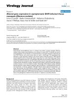
Figure 1
The growth of diapausing blastocysts is reversibly arrested before
implantation into the uterus. (a) A diapausing blastocyst (arrowhead) is
shown in contact with the uterine luminal epithelium (LE). Note the loose
fibroblastic morphology of the stroma (S) underlying the luminal
epithelium. T, the trophoblast of the blastocyst. (b) Implantation after
diapause starts with the luminal epithelium adjacent to the trophoblast of
the blastocyst undergoing apoptosis as the trophoblast cells start to
invade the uterus. After implantation, the stroma has undergone massive
proliferation and differentiation to form the decidua (D).
(a) (b)
D
D
LE
T
S
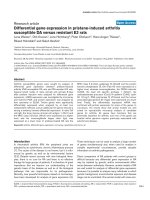
Figure 2
The time course of events leading to embryo implantation in the mouse.
(a) On day 3 after fertilization the uterus is undergoing differentiation and
proliferation under control of the ovarian steroid hormones estrogen and
progesterone. (b) On day 4 the blastocyst is adjacent to the uterus and
the production of leukemia inhibitory factor (LIF) is induced in the
endometrial glands by nidatory estrogen. LIF is released into the uterine
lumen, where it binds to LIF receptors on the luminal epithelium. LIF
binding induces the expression of many genes, including the cell adhesion
factors Coch and CD9, as the uterus becomes receptive to the embryo
allowing the onset of implantation. Other adhesion molecules, such as
E-cadherin, undergo redistribution in their expression. (c) On day 5 the
blastocyst has started to invade the uterus and the stroma is undergoing
decidualization, accompanied by the expression of prostaglandins (PGEs),
which are regulated by Cox-2, and the cytokine interleukin-11 (IL-11),
which is essential for decidualization.
Blastocyst
activation
and implantation
Estrogen and
progesterone
Signals from
non-glandular
epithelium to
blastocyst?
PGEs?
IL-11
Cox-2?
Progesterone
LIF
Nidatory estrogen
stimulates LIF
expression in the
endometrial glands
CD9
E-cadherin
Coch
LIF receptor
gp130
Epithelium
Stroma
Epithelium
Stroma
Epithelium
Stroma
(a) Days 1-3
(b) Days 4-5
(c) Day 5
in conjunction with the arrest of ICM proliferation. This sug-
gests that ES cells derived from the ICM depend on LIF as a
survival factor. What drives ES cell proliferation is, however,
unclear [14].
We clearly know a reasonable amount about how the mother
regulates diapause in the mouse, but what happens in the
blastocyst? Previous studies have shown that the diapausing
blastocysts enter into a state of proliferative and metabolic
quiescence. Cell proliferation ceases, as does amino-acid
uptake and overall metabolism [15,16]. A recent report [3]
has now taken the characterization of the diapausing blasto-
cyst a step further by using microarray technology to identify
genes that are differentially expressed between delayed blas-
tocysts and blastocysts that have been activated following
diapause. In this study, mRNA was isolated from approxi-
mately 100 diapausing and activated embryos, amplified and
screened against the National Institute on Aging (NIA)
mouse microarrays, which are enriched for genes expressed
during different stages of mouse development [17]. The
report by Hamatani et al. [3] identifies 229 genes (out of a
total of around 22,000, so roughly 1% of the genes on the
array) that are differentially expressed between activated and
delayed blastocysts. Delayed blastocysts had 80 genes that
showed increased expression levels compared to 149 genes
that were relatively upregulated in the activated blastocysts.
The genes showing altered levels of expression between the
arrested and activated blastocysts were clustered into six
functional groups: specifically, genes involved in cell-cycle or
cell-proliferation control; energy pathways and carbohydrate
metabolism; signaling; nuclear transport; chromatin remod-
eling; and adhesion. Consistent with the physiological state
of the embryos, delayed blastocysts showed decreased levels
of expression of genes involved in cell-cycle progression,
together with an increase in the levels of expression of genes
that arrest cell proliferation at G
0
or G
1
stages in the cell
cycle - for example, Btg1 and the cell-cycle inhibitor p21Cip1,
which is regulated by p53 [18]. But p53, which can cause
cell-cycle arrest when upregulated, did not show any differ-
ence in mRNA levels between the arrested and activated
blastocysts. Intriguingly, one of the genes upregulated in the
diapausing embryos is the maternally expressed imprinted
gene encoding the Igf2 receptor (Igf2r), which retards cell
proliferation when overexpressed [19]. Activated blastocysts
also showed relative upregulation in the expression of genes
that function in the glucose and pyruvate energy pathways.
Another gene that was identified as upregulated in delayed
embryos was Irs1, encoding insulin receptor 1 substrate, a
docking protein that is involved in the binding and activation
of signal transduction molecules after being phosphorylated
by the insulin receptor kinase. Mutations in the Drosophila
homolog of Irs1, Chico, result in a significant increase in the
lifespan of the mutant flies [20], and Irs1 may therefore be
involved in sustaining blastocyst viability and longevity
during developmental delay. Surprisingly, no alterations in
levels of expression of the LIF receptor or gp130 (which
together comprise the heterodimeric LIF receptor) were
detected in the delayed blastocysts.
The last category of genes investigated by Hamatani et al.
[3] was that involved in mediating cell adhesion and/or
migration. In the mouse, implantation is marked by the
active invasion of the apoptosing luminal epithelium by the
embryonic trophoblast. Six genes whose products are asso-
ciated with tight-junction integrity, cell migration, cell-to-
cell adhesion and focal adhesions were all upregulated in
the activated blastocysts. In addition, the heparin-binding
epidermal growth factor (HB-EGF), which shows a remark-
able localization in expression at the site of blastocyst
attachment [21], was also upregulated in the activated
blastocysts. This finding was particularly intriguing as HB-
EGF interacts with EGF receptors and loss of a functional
EGF receptor in some strains of mouse causes peri-implan-
tation lethality [22]. Although previous results have sug-
gested a critical role for HB-EGF in mediating blastocyst
implantation, recent reports indicated that HB-EGF is not
localized to the cell membrane until after blastocyst attach-
ment [23], and mice lacking HB-EGF are fertile [24], indi-
cating that HB-EGF may not be essential for blastocyst
activation or implantation.
Overall, the expression of many of the genes that differ
between the diapausing and activated blastocysts is consis-
tent with the cellular and physiological events that are
expected to change with activation. No one factor has yet
been identified as being the ‘key’ to regulating diapause,
unless it is among the approximately 30% of the 229 differ-
entially expressed genes to which no function has yet been
assigned. Nevertheless, the list of genes does reveal some
possible candidates that may be essential for mediating
growth arrest in the implanting blastocyst. With the
current availability of knockout lines of mice carrying
mutations in many of the genes identified in this screen it
will be possible to systematically determine which of the
genes may be required for diapause, as has already been
shown for the gp130 receptor. In turn this may provide
both profound and fascinating insights into the molecular
mechanisms regulating cell proliferation and growth
control in the mammalian embryo.
References
1. Renfree MB, Shaw G: Diapause. Annu Rev Physiol 2000, 62:353-375.
2. Stevens LC: The development of transplantable teratocarci-
nomas from intratesticular grafts of pre- and postimplanta-
tion mouse embryos. Dev Biol 1970, 21:364-382.
3. Hamatani T, Daikoku T, Wang H, Matsumoto H, Carter MG, Ko MS,
Dey SK: Global gene expression analysis identifies molecular
pathways distinguishing blastocyst dormancy and activation.
Proc Natl Acad Sci USA 2004, 101:10326-10331.
4. Psychoyos A: Endocrine control of egg implantation. In Hand-
book of Physiology. Volume 2. Edited by Greep RO, Astwood EB. Balti-
more: Williams and Wilkins; 1973:187-215.
5. Stewart CL, Kaspar P, Brunet LJ, Bhatt H, Gadi I, Kontgen F, Abbon-
danzo SJ: Blastocyst implantation depends on maternal
comment
reviews
reports deposited research
interactions
information
refereed research
Genome Biology 2004, Volume 6, Issue 1, Article 202 Hondo and Stewart 202.3
Genome Biology 2004, 6:202
expression of leukaemia inhibitory factor. Nature 1992,
359:76-79.
6. Williams RL, Hilton DJ, Pease S, Willson TA, Stewart CL, Gearing
DP, Wagner EF, Metcalf D, Nicola NA, Gough NM: Myeloid
leukaemia inhibitory factor maintains the developmental
potential of embryonic stem cells. Nature 1988, 336:684-687.
7. Smith AG, Heath JK, Donaldson DD, Wong GG, Moreau J, Stahl M,
Rogers D: Inhibition of pluripotential embryonic stem cell
differentiation by purified polypeptides. Nature 1988, 336:688-
690.
8. Chen JR, Cheng JG, Shatzer T, Sewell L, Hernandez L, Stewart CL:
Leukemia inhibitory factor can substitute for nidatory
estrogen and is essential to inducing a receptive uterus for
implantation but is not essential for subsequent embryogen-
esis. Endocrinology 2000, 141:4365-4372.
9. Nichols J, Davidson D, Taga T, Yoshida K, Chambers I, Smith A:
Complementary tissue-specific expression of LIF and LIF-
receptor mRNAs in early mouse embryogenesis. Mech Dev
1996, 57:123-131.
10. Ware CB, Horowitz MC, Renshaw BR, Hunt JS, Liggitt D, Koblar SA,
Gliniak BC, McKenna HJ, Papayannopoulou T, Thoma B, et al.: Tar-
geted disruption of the low-affinity leukemia inhibitory
factor receptor gene causes placental, skeletal, neural and
metabolic defects and results in perinatal death. Development
1995, 121:1283-1299.
11. Yoshida K, Taga T, Saito M, Suematsu S, Kumanogoh A, Tanaka T,
Fujiwara H, Hirata M, Yamagami T, Nakahata T, et al.: Targeted
disruption of gp130, a common signal transducer for the
interleukin 6 family of cytokines, leads to myocardial and
hematological disorders. Proc Natl Acad Sci USA 1996, 93:407-
411.
12. Dani C, Chambers I, Johnstone S, Robertson M, Ebrahimi B, Saito M,
Taga T, Li M, Burdon T, Nichols J, et al.: Paracrine induction of
stem cell renewal by LIF-deficient cells: a new ES cell regu-
latory pathway. Dev Biol 1998, 203:149-162.
13. Nichols J, Chambers I, Taga T, Smith A: Physiological rationale
for responsiveness of mouse embryonic stem cells to gp130
cytokines. Development 2001, 128:2333-2339.
14. Burdon T, Smith A, Savatier P: Signalling, cell cycle and pluripo-
tency in embryonic stem cells. Trends Cell Biol 2002, 12:432-438.
15. Holmes PV, Dickson AD: Temporal and spatial aspects of
oestrogen-induced RNA, protein and DNA synthesis in
delayed-implantation mouse blastocysts. J Anat 1975, 119:453-
459.
16. Spindler RE, Renfree MB, Gardner DK: Carbohydrate uptake by
quiescent and reactivated mouse blastocysts. J Exp Zool 1996,
276:132-137.
17. Carter MG, Piao Y, Dudekula DB, Qian Y, VanBuren V, Sharov AA,
Tanaka TS, Martin PR, Bassey UC, Stagg CA, et al.: The NIA cDNA
project in mouse stem cells and early embryos. C R Biol 2003,
326:931-940.
18. Kuo ML, Duncavage EJ, Mathew R, den Besten W, Pei D, Naeve D,
Yamamoto T, Cheng C, Sherr CJ, Roussel MF: Arf induces p53-
dependent and -independent antiproliferative genes. Cancer
Res 2003, 63:1046-1053.
19. Hernandez L, Kozlov S, Piras G, Stewart CL: Paternal and mater-
nal genomes confer opposite effects on proliferation, cell-
cycle length, senescence, and tumor formation. Proc Natl Acad
Sci USA 2003, 100:13344-13349.
20. Clancy DJ, Gems D, Harshman LG, Oldham S, Stocker H, Hafen E,
Leevers SJ, Partridge L: Extension of life-span by loss of
CHICO, a Drosophila insulin receptor substrate protein.
Science 2001, 292:104-106.
21. Das SK, Wang XN, Paria BC, Damm D, Abraham JA, Klagsbrun M,
Andrews GK, Dey SK: Heparin-binding EGF-like growth factor
gene is induced in the mouse uterus temporally by the blas-
tocyst solely at the site of its apposition: a possible ligand for
interaction with blastocyst EGF receptor in implantation.
Development 1994, 120:1071-1083.
22. Threadgill DW, Dlugosz AA, Hansen LA, Tennenbaum T, Lichti U,
Yee D, LaMantia C, Mourton T, Herrup K, Harris RC, et al.: Tar-
geted disruption of mouse EGF receptor: effect of genetic
background on mutant phenotype. Science 1995, 269:230-234.
23. Isaacs J, Murphy CR: Heparin-binding EGF-like growth factor is
seen on the extracellular surface of uterine epithelial cells
only after the initial stages of blastocyst attachment. His-
tochem J 2002, 34:339-343.
24. Jackson LF, Qiu TH, Sunnarborg SW, Chang A, Zhang C, Patterson
C, Lee DC: Defective valvulogenesis in HB-EGF and TACE-
null mice is associated with aberrant BMP signaling. EMBO J
2003, 22:2704-2716.
202.4 Genome Biology 2004, Volume 6, Issue 1, Article 202 Hondo and Stewart />Genome Biology 2004, 6:202