REGULATION OF SRF ACTIVITY BY THE ATP-DEPENDENT CHROMATIN REMODELING ENZYME, CHD8
Bạn đang xem bản rút gọn của tài liệu. Xem và tải ngay bản đầy đủ của tài liệu tại đây (1.6 MB, 125 trang )
REGULATION OF SRF ACTIVITY BY THE ATP-DEPENDENT CHROMATIN
REMODELING ENZYME, CHD8
Jennifer Marie Rodenberg
Submitted to the faculty of the University Graduate School
in partial fulfillment of the requirements
for the degree
Doctor of Philosophy
in the Department of Cellular and Integrative Physiology,
Indiana University
December 2008
©2008
Jennifer Marie Rodenberg
ALL RIGHTS RESERVED
iii
I would like to dedicate this dissertation to my adoring husband, Eric, without
whom I would not be where I am today. Through his love and support, he was
my pillar of strength throughout my graduate work, and for this I am eternally
thankful.
iv
ACKNOWLEDGEMENTS
I would like to formally thank all of the members my doctoral research
committee: Dr. B. Paul Herring, Dr. Patricia J. Gallagher, Dr. Fredrick M.
Pavalko and Dr. David Skalnik. Each of these members played a significant role
in helping me to focus the direction of my project and in helping by providing
critical interpretations of the data. I really appreciate all of their guidance that
allowed for the project to move forward, stay focused and stay on schedule. In
addition, I would like to specifically thank my mentor, thesis advisor, and chair of
my dissertation committee, B. Paul Herring, Ph.D. He granted me the
opportunity to work in his lab on a project for my thesis and guided me as a
graduate student. From him I was able to gain a broad scope of laboratory
knowledge and skills, and for this I am very grateful.
Through the duration of my graduate work, several members of the
Herring lab, both former and current, provided me with important scientific and
moral support. These individuals include Hong Fang, April Hoggatt, Omar El-
Mounayri, Ph.D., Ketrija Touw, Feng Yin, Ph.D., Min Zhang and Leo (Jiliang)
Zhou, Ph.D. Other individuals from surrounding labs that played a role in
providing advice and support throughout my graduate work include Emily Blue,
Alesha Castillo, Ph.D., Suzanne Norvell, Ph.D., Rita Gerard-O’Riley, Suzanne
Ponik, Ph.D., Jason Triplett, Ph.D., Ryan Widau, and Suzanne Young, Ph.D.
I would like to acknowledge my entire family. In particular, I would like to
thank my parents, Richard and Marie Stover, who have always been there to
v
lend encouraging words to let me know that they were behind me 100 percent no
matter what. Not only did they help to foster my quest for knowledge throughout
my academic career, but they also provided me with a loving and supportive
home where I grew into the balanced woman that I am today. I am forever
indebted to them for giving me the confidence to pursue my goals, for celebrating
in my successes and for empathizing with my defeats. I could not have asked for
more loving and supportive parents.
Last, but not least, I would like to thank my husband, Eric. Throughout the
duration of my graduate work he was always there to lend intellectual and moral
support. Though I am sure I tried it a few times, his patience and understanding
were always there for me when I needed a helping hand or a bit of guidance. It
really meant a lot to me to have such a wonderful friend and the love of my life
share this experience with me. I cannot begin to thank him enough for giving me
the strength to continue in my studies and achieve my academic goals. He truly
is the greatest husband in the world.
vi
ABSTRACT
Jennifer Marie Rodenberg
REGULATION OF SRF ACTIVITY BY THE ATP-DEPENDENT CHROMATIN
REMODELING ENZYME, CHD8
Under normal conditions, smooth muscle cells do not replicate, or
proliferate, and provide a means of contraction for many internal organs,
including blood vessels and the gut. However, under abnormal or disease
conditions, such as congenital heart disease and cancer, smooth muscle cells
acquire the ability to replicate, to make extracellular matrix proteins and to
migrate. Thus, determining how smooth muscle cells regulate these processes is
crucial to understanding how the cells can switch between normal and diseased
states. Serum response factor (SRF) is a widely expressed protein that plays a
key role in the regulation of smooth muscle differentiation, proliferation and
migration. It is generally accepted that one way that SRF can distinguish
between these functions is through pathway-specific co-factor interactions. A
novel SRF co-factor, chromodomain helicase DNA binding protein 8 (CHD8),
was originally isolated from a yeast two-hybrid assay. CHD8 is widely expressed
in adult tissues including smooth muscle. Data from in vitro binding assays
indicate that the N-terminus of CHD8 can interact directly with the MADS domain
of SRF. Co-immunoprecipitation assays verified the ability of these two proteins
to interact within cells. Adenoviral-mediated shRNA knockdown of CHD8 in
smooth muscle cells resulted in statistically significant 10-20% attenuation of
vii
expression of SRF-dependent, smooth muscle-specific genes. Similar
experiments revealed that knockdown of CHD8 did not affect the SRF-dependent
induction of immediate early genes required to promote proliferation. In contrast,
knockdown of CHD8 in A10 vascular smooth muscle cells resulted in a marked
induction in of apoptosis, characterized by increases in apoptotic markers such
as phospho-H2A.X, cleaved PARP and activated caspase-3. These data suggest
that CHD8 may play a specific role in modulating SRF’s activity toward anti-
apoptotic genes, thereby regulating smooth muscle cell survival.
B. Paul Herring, Ph.D., chair
viii
TABLE OF CONTENTS
List of Tables xi
List of Figures xii
List of Abbreviations xiii
Chapter I: Introduction 1
A. Regulation of Smooth Muscle Differentiation 1
i. Overview of Smooth Muscle 1
ii. Overview of Smooth Muscle Differentiation 2
iii. Mechanisms of Smooth Muscle Differentiation 6
iv. SRF’s Role in Smooth Muscle Differentiation 8
B. SRF’s Regulation of Proliferation, Motility, Apoptosis and
other Processes 18
C. Smooth Muscle Cell Phenotypic Modulation 21
i. Kruppel-like Factors (KLFs) 21
ii. Platelet Derived Growth Factor (PDGF) 25
iii. Matrix Metalloproteinases (MMPs) 26
iv. Transforming Growth Factor Beta-1 (TGFβ1) 27
v. Apoptosis and Senescence in Modulation of Smooth
Muscle Cell Phenotype during Vascular Remodeling 28
D. Chromatin Remodeling Enzymes’ Roles in Development
and Differentiation 31
i. Introduction to Types of Chromatin Remodeling Enzymes 31
ii. ATP-dependent Chromatin Remodeling Enzymes 34
ix
iii. CHD Family and CHD8 36
E. Rationale 42
F. Hypothesis 43
Chapter II: Methods 44
Chapter III: CHD8 binds SRF, promotes the expression of smooth
muscle-specific genes and protects smooth muscle cells
from apoptosis 51
A. Summary 51
B. Introduction 53
C. Results 57
D. Discussion 74
Chapter IV: Discussion and Future Studies 78
References 90
Curriculum Vitae
x
LIST OF TABLES
Table 1.
Changes in apoptotic genes as a result of loss of CHD8 72
xi
LIST OF FIGURES
Figure 1. Schematic of functional domains of serum response factor (SRF) 10
Figure 2. CHD8 is more ubiquitously expressed than duplin, its N-terminal
splice variant 59
Figure 3. SRF and CHD8 interact both in vitro and in vivo 61
Figure 4. Knockdown of CHD8 causes reduced expression of SRF-
dependent genes 64
Figure 5. Knockdown of CHD8 does not affect serum stimulation of
immediate early or late early genes 65
Figure 6.
Loss of CHD8 does not affect TGFβ-dependent stimulation
of myofibroblast differentiation 67
Figure 7.
CHD8 imparts a pro-survival effect on A10 vascular smooth
muscle cells 70
Figure 8.
Loss of CHD8 causes attenuated expression of Birc5/survivin 73
Figure 9. Proposed model of CHD8’s regulation of apoptosis through
mediation of survivin expression 82
xii
CHAPTER I
Introduction
A. Regulation of Smooth Muscle Differentiation
i. Overview of Smooth Muscle
Smooth muscle cells surround the walls of the body’s hollow organs such
as the blood vessels, the gastrointestinal tract, the genitourinary tracts, and the
airways. As a tissue system, smooth muscle provides the contractile force to
regulate flow of materials through the hollow organs. For instance, in the blood
vessels smooth muscle contraction and relaxation helps to regulate both the
blood flow and the blood pressure. Smooth muscle in the gastrointestinal tract is
important for regulating the movement of a food bolus for excretion. Similarly,
smooth muscle that lines the bladder and urinary tract plays a vital role in the
expulsion of urine. The female reproductive tract, where smooth muscle allows
for the contraction and relaxation of the uterus during both menstruation and
childbirth, provides another example of the important function of smooth muscle.
Finally, smooth muscle is critical for the function of the airways, where it helps to
regulate the intake and expulsion of air.
Smooth muscle on the cellular level is extremely complex. The
differentiated smooth muscle cell contains a network of contractile and regulatory
proteins, such as actin and myosin (Owens, 2007). Smooth muscle cells are
1
spindle shaped, and under normal conditions the smooth muscle cell is largely
quiescent. However, this differentiated smooth muscle cell has the uncanny
ability to change from this quiescent, contractile phenotype under normal
conditions into a more proliferative, migratory and synthetic phenotype under
pathological or wound-healing conditions. This pathological state is often termed
“de-differentiated,” as cells lose their spindle shape, have multiple protrusions
and down-regulate expression of many contractile proteins (Owens, 2007).
ii. Overview of Smooth Muscle Differentiation
During mammalian development, or embryogenesis, embryonic stem cells
give rise to all of the body’s tissues and cells. One of the main processes of
embryogenesis is gastrulation, during which cells form the three main germ
layers of the ectoderm, the mesoderm and the endoderm. The ectoderm is the
outermost layer, the mesoderm is the middle layer, and the endoderm in the
innermost layer. As gastrulation and development continue, the multi-potential
cells in the embryo begin to move in various directions and manners to form
different organ systems in the body (Leptin, 2005). As a general rule, the
ectoderm forms the nervous system, the sense organs, the skin (epidermis) and
associated structures and the pituitary gland (Solomon, 1993). Systems such as
the skeleton (bone and cartilage), the muscles (skeletal, smooth and cardiac),
the circulatory system, the excretory system, the reproductive system, the inner
layer of the skin (dermis), the outer layers of the digestive system and the
2
respiratory system are all derived from the mesoderm germ layer (Solomon,
1993). The lining of the digestive tract and the respiratory system arise from the
endoderm. Thus, much of the body’s smooth muscle is developed from the
mesoderm, while a small amount is also derived from the endoderm.
Looking more closely at the development of the vascular smooth muscle,
three main origins of vascular smooth muscle cells become apparent: (a) the
cranial neural crest population, (b) the proepicardium population and (c) the
endothelial progenitor cell population (Hirschi and Majesky, 2004). During
embryogenesis, bone morphogenetic protein (BMP) signaling creates a gradient,
allowing for the formation of the neural crest at the mediolateral border between
the neural plate and the epidermis (Hirschi and Majesky, 2004). These cells then
give rise to various types of cells, including smooth muscle cells. More
specifically, the cranial neural crest cells, located anterior to somite 5, give rise to
the vascular smooth muscle cells of the aortic arch arteries, the pulmonary artery
and the ductus arteriosus (Hirschi and Majesky, 2004).
Whereas the cranial neural crest gives rise to the smooth muscle cells of
the aortic arch and pulmonary artery, the stem cells of the proepicardial organ
(PEO) differentiate into the smooth muscle cells of the coronary vessels (Hirschi
and Majesky, 2004). The PEO structure is transient and establishes contact with
the heart, which leads to epicardial layer formation (Hirschi and Majesky, 2004).
Furthermore, cells in this population undergo the epithelial-to-mesenchymal
transition in response to signals that are released from the myocardium (Hirschi
3
and Majesky, 2004), thereby allowing them to become migratory and to form the
precursors of the smooth muscle cells in the coronary vessels.
The final group of stem cells from which smooth muscle cells are derived
is the endothelial progenitor cell subset. Studies have illustrated that depending
on the conditions present in the environment, these stem cells can form either
endothelial cells or smooth muscle cells. More specifically, smooth muscle cells
are derived from this population in the presence of PDGF-BB (Hirschi and
Majesky, 2004; Yamashita et al., 2000).
In the adult, much of the remodeling and formation of blood vessels has
been thought to occur from the proliferation and migration of smooth muscle cells
that are already present. However, recent studies have suggested that adults
can differentiate smooth muscle cells from various populations (Yamashita et al.,
2000): (a) bone marrow-derived stem cells, (b) hematopoietic stem cells and (c)
circulating stem cells. The bone marrow has been thought to harbor both
hematopoietic stem cells and mesenchymal stem cells. Yet, recently it has been
illustrated that the bone marrow might also contain a population of vascular
smooth muscle progenitor cells (Yamashita et al., 2000). As for the
hematopoietic stem cells, studies show contradictory results as to whether this
population contributes to vascular smooth muscle regeneration after injury (Sata
et al., 2002), or whether this population contributes to the regeneration of
endothelial cells post-injury (Goodell et al., 1996; Hirschi and Majesky, 2004;
Jackson et al., 2001). In addition, various studies have demonstrated that
progenitor cells within the mononuclear fraction of the blood have the potential to
4
give rise to smooth muscle cells, wherein each group was able to show evidence
of smooth muscle alpha actin (SM-α-actin) expression from cultured cells
fractionated from blood (Hillebrands et al., 2001; Hirschi and Majesky, 2004;
Simper et al., 2002).
In general, cellular differentiation can be defined as the process during
development where multi-potential cells acquire the cell-specific attributes that
discriminate them from other cell types (Owens et al., 2004). As Owens, et al.,
describes the actual process of cellular differentiation can be divided into three
main components: (a) activation of specific genes that are required for
differentiation of a cell, (b) control of expression of these specific genes at certain
times and quantities and (c) regulation of overall gene expression via the
microenvironmental signals that regulate the cell’s lineage, which includes
transcription factors and epigenetics (Owens et al., 2004).
In the case of smooth muscle cells, a differentiated smooth muscle cell
can be identified by the expression of specific genes, or smooth muscle markers.
These markers include: smooth muscle alpha and gamma actin (SM-α-actin and
SM-γ-actin), smooth muscle myosin heavy chain (SM-MHC), telokin, SM22α,
calponin, caldesmon, metavinculin, smoothelin and 130 kD myosin light chain
kinase (MLCK). One important characteristic of the promoters of most of these
smooth muscle marker genes is that they contain at least one CArG element,
which is the consensus sequence for the transcription factor serum response
factor (SRF). However, many other factors also play a role in the regulation of
these smooth muscle markers as detailed below.
5
iii. Mechanisms of Smooth Muscle Differentiation
Much is still to be elucidated about how smooth muscle cells differentiate
from the multi-potential cells throughout development. However, several studies
have investigated this differentiation phenomenon, and various factors and
pathways have been described as being involved in the process of deriving a
smooth muscle cell. One well-described example of a factor and its signaling
pathways being implicated in smooth muscle cell differentiation is transforming
growth factor β1 (TGFβ1).
TGFβ1 is a cytokine that signals through multiple membrane receptors
and intracellular pathways. Knockout models of either TGFβ1, TGFβ type II
receptor, activin receptor-like kinase 1 (Alk1), endogelin or SMAD5 are all
embryonic lethal (Bourdeau et al., 1999; Chang et al., 1999; Dickson et al., 1995;
Oh et al., 2000; Oshima et al., 1996; Sinha et al., 2004; Urness et al., 2000). At
least 50% of the TGFβ1 and TGFβ type II receptor null mice die in utero from
defects in the yolk sac vasculature by embryonic day 11.5. Alk1, endoglin and
SMAD5 knockouts succumb to intrauterine death around midgestation due to
hemorrhaging from dilated and fragile vessels. Further highlighting the
importance of TGFβ intracellular signaling specifically in smooth muscle
differentiation, knockout of either endogelin or Alk1 lead to a loss of smooth
muscle cells that coat the dorsal aorta (Li et al., 1999; Oh et al., 2000; Sinha et
al., 2004).
6
TGFβ has the ability to up-regulate the expression of several smooth
muscle markers, including SM22α, SM-α-actin and h
1
-calponin during
myofibroblast differentiation (Bjorkerud, 1991; Sinha et al., 2004). Inactivating
TGFβ1, the TGFβ type II receptor, SMAD2 and SMAD3 in an embryonic stem
cell – embroid body model of smooth muscle differentiation attenuated
expression of many smooth muscle markers including SM MHC, SM- α-actin and
SM22α (Sinha et al., 2004). In addition, the SM-α-actin promoter was found to
be regulated by both SMAD2 and SMAD3; whereas, SM-MHC only required
SMAD2 for proper transcription (Sinha et al., 2004). The authors note that this
difference in requirement of SMADs could be due to the fact that SM-α-actin is
also expressed in the myofibroblast and other cell types, indicating that perhaps
SMAD2 is more important for regulation of smooth muscle cell differentiation
(Sinha et al., 2004). Overall, these findings therefore indicate that TGFβ1
signaling, specifically through SMAD2 and SMAD3, has an important role in the
development of smooth muscle cells.
Another factor implicated in smooth muscle cell differentiation is GATA6.
GATA6 is a member of the GATA family zinc-finger transcription factors. Studies
have shown that GATA6 is the only member of this family to be expressed in
vascular smooth muscle cells (Lepore et al., 2005). Targeted knockout of
GATA6 causes embryonic lethality at embryonic day 6.5 from a defect in visceral
endoderm formation prior to vascular smooth muscle development (Lepore et al.,
2005; Morrisey et al., 1998). Even though GATA6 is not expressed in all types of
smooth muscle, studies have shown that GATA6 weakly activates the SM-MHC
7
promoter and that GATA6 in combination with cysteine-rich protein 2 (CRP2)
activates serum response factor-dependent smooth muscle marker transcription
(Chang et al., 2003; Wada et al., 2002; Wada et al., 2000). These findings all
indicate that GATA6 has a role in regulating the differentiation of smooth muscle
cells; however, this type of regulation may be restricted to vascular smooth
muscle.
iv. SRF’s Role in Smooth Muscle Differentiation
In 1984, Greenberg and colleagues found that cFos transcription in
quiescent cells could be stimulated by serum (Chai and Tarnawski, 2002;
Greenberg and Ziff, 1984). Upon further investigation, it was determined that
serum was able to activate the cFos promoter due to the presence of a specific
DNA element upstream of the transcriptional start site in the promoter. This
element has the general consensus sequence CC(A/T)
6
GG and was named
Serum Response Element (SRE) (Chai and Tarnawski, 2002). Treisman later
identified the transcription factor that bound to this specific sequence as a dimer
and named it Serum Response Factor (SRF) (Chai and Tarnawski, 2002;
Treisman, 1986). The full-length mRNA transcript of SRF contains seven exons;
however, due to alternative RNA splicing, it has been shown that four isoforms of
SRF exist in the mouse: (a) SRF-L, or full-length SRF, (b) SRF-M, which lacks
exon 5, (c) SRF-S, which lacks exons 4 and 5, and (d) SRF-I, which contains
only exons 1,2,6, and 7 (Chai and Tarnawski, 2002; Kemp and Metcalfe, 2000).
8
The resulting SRF proteins are 67, 62, or 48 kDa, depending on the isoform. It is
important to note that the SRF-L isoform corresponds to the dominant isoform
found in the human, and the SRF-M isoform functions as a dominant negative of
the full-length wild type (Belaguli et al., 1999; Chai and Tarnawski, 2002). SRF
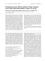
contains three major conserved domains (Figure 1): (a) MADS box, which
serves as a DNA-binding motif, (b) several phosphorylation sites, which allow for
post-translational regulation of SRF, and (c) transactivation domain, which is
located at the C-terminus of SRF.
Expression of SRF in the chicken and mouse provide strong evidence for
a role of SRF in smooth muscle differentiation. In the adult chicken, SRF
expression is only detected in tissues of mesodermal and neuroectodermal origin
(Arsenian et al., 1998; Croissant et al., 1996). During gastrulation in the chicken,
SRF mRNA is localized to the primitive streak, the neural groove, the lateral plate
and the precardiac splanchnic mesoderm, the myocardium and the somites
(Arsenian et al., 1998; Croissant et al., 1996). In an adult mouse the highest
levels of SRF mRNA can be seen in the skeletal, cardiac and smooth muscle
tissues. During development SRF mRNA is highly expressed in the medial
smooth muscle layer of the vessels, the myocardium of the heart and the
myotomal portions of the somites (Arsenian et al., 1998; Belaguli et al., 1997).
As a transcription factor, SRF is involved in multiple pathways that
regulate disparate processes. SRF is critical for the differentiation of skeletal,
cardiac and smooth muscle lineages (Owens et al., 2004; Pipes et al., 2006).
SRF regulates smooth muscle differentiation through its interaction with various
9
10
Figure 1. Schematic of functional domains of serum response factor (SRF). SRF
contains a MADS domain, which serves as a DNA-binding motif; several
phosphorylation sites, which allow for post-translational regulation of SRF; and a
transactivation domain, which is located at the C-terminus of SRF.
coactivators and/or cofactors such as myocardin, the myocardin family members
MKL1 (MRTFA) and MRTFB (Behrens and Lustig, 2004; Wang et al., 2001),
Mhox (Owens et al., 2004), Nkx3.1, Nkx3.2, Nkx2.5 (Carson et al., 2000; Nishida
et al., 2002; Phiel et al., 2001), Barx2, Barx1b (Herring et al., 2001; Nakamura et
al., 2001) and GATA6/CRP2 (Chang et al., 2003).
The members of the myocardin family of SRF coactivators are very
powerful activators of the smooth muscle differentiation program (Pipes et al.,
2006). The expression of myocardin itself is restricted to cardiac and smooth
muscle-specific genes (Wang et al., 2001). By forming a ternary complex with
SRF and CArG elements, myocardin stimulates transcription of CArG-dependent
muscle specific genes (Pipes et al., 2006). The interaction of myocardin with
SRF is crucial because myocardin itself does not bind directly to DNA, whereas
SRF does. The importance of myocardin in vascular smooth muscle
development was demonstrated by analysis of myocardin knockout mice, which
exhibited a lack of differentiated vascular smooth muscle cells and consequently
died during embryonic development (Chen et al., 2003).
In addition, myocardin related transcription factor A (MRTFA), which is
expressed in a wider array of tissues, can also activate smooth muscle gene
expression when over-expressed in fibroblasts, indicating that MRTFA may also
regulate smooth muscle differentiation via interactions with SRF (Al-Aynati et al.,
2004; Pipes et al., 2006; Wang et al., 2002). In support of this proposal siRNA
mediated knockdown of MRTFA in rat vascular smooth muscle cells attenuated
expression of CArG-dependent smooth muscle marker genes (Yoshida et al.,
11
2007). However, MRTFA knockout mice do not exhibit defects in smooth muscle
cells although expression of smooth muscle-specific genes was attenuated in
myoepithelial cells of the mammary gland (Li et al., 2006). The discordance
between these in vivo and in vitro data suggest that myocardin family members
may have partially redundant functions such that one family member may be able
to compensate for the loss of another family member in vivo.
As with MRTFA, myocardin related transcription factor B (MRTFB) is also
more widely expressed than myocardin (Du et al., 2003), and inactivation of both
MRTFA and MRTFB is required to block Rho-A-dependent SRF target gene
activation (Cen et al., 2003; Li et al., 2005). However, unlike MRTFA, despite its
potent transcriptional activation domain, MRTFB is only a weak coactivator (Li et
al., 2005; Wang et al., 2002). In support of the idea that MRTFB also plays a role
in regulating smooth muscle differentiation, MRTFB knockout mice are
embryonic lethal at embryonic day 13.5 to 14.5 (Li et al., 2005; Oh et al., 2005;
Wei et al., 2007). Mice deficient of MRTFB displayed defective brachial arch
arteries and cardiac outflow tracts. MRTFB null mice also demonstrated a lack of
SM-α-actin in vascular smooth structures derived from neural crest cells (Li et al.,
2005; Oh et al., 2005), indicating that MRTFB plays a role in smooth muscle cell
differentiation. However, even though MRTFB is expressed in the embryonic
heart and peripheral vasculature, these structures are normal in MRTFB
knockout mice and only secondary vascular structures such as the brachial arch
arteries and cardiac outflow tracts were affected (Li et al., 2005; Oh et al., 2005).
Additional defects in MRTFB null mice include abnormal liver and portal vascular
12
development (Wei et al., 2007). Nonetheless, since myocardin knockout mice
are still embryonic lethal at embryonic day 10.5 when MRTFB is present, MRTFB
cannot compensate for myocardin at this stage (Li et al., 2005; Oh et al., 2005).
Another factor that affects how SRF regulates the differentiation of smooth
muscle cells is Mhox. Mhox has been shown to dramatically increase SRF
binding to the CArG element in the SM-α-actin promoter (Hautmann et al., 1997;
Owens et al., 2004). This data has indicated that a homeodomain region in near
proximity to one of the CArG elements in the SM-α-actin promoter is important
for regulating SRF’s activity. A further piece of evidence for this idea is that the
over-expression of Mhox stimulated the SM-α-actin promoter in cultured smooth
muscle cells (Owens et al., 2004). In addition, studies have suggested a role for
Mhox in angiotensin II-mediated smooth muscle differentiation. Angiotensin II is
capable of increasing expression of Mhox, which then subsequently enhances
binding of SRF to either of the CArG elements in the SM-α-actin promoter
(Owens et al., 2004).
Nkx, Barx and GATA6 are additional factors that have been shown to
regulate SRF and smooth muscle cell differentiation through forming a ternary
complex with SRF. Both Nkx and Barx are homeodomain-containing proteins
that may function in a similar fashion to Mhox; whereas, GATA6 is a zinc finger
protein. A study by Nishida, et al., illustrated that a trimeric complex of GATA6,
Nkx3.2 and SRF specifically activated smooth muscle markers such as SM22α
and caldesmon, but not the proliferation factor cFos (Nishida et al., 2002; Owens
et al., 2004).
13