THE EFFECT OF OMEGA-3 FATTY ACIDS ON AIRWAY INFLAMMATION, HYPERPNEA-INDUCED BRONCHOCONSTRICTION, AND AIRWAY SMOOTH MUSCLE CONTRACTILITY IN ASTHMA
Bạn đang xem bản rút gọn của tài liệu. Xem và tải ngay bản đầy đủ của tài liệu tại đây (22.69 MB, 335 trang )
THE EFFECT OF OMEGA-3 FATTY ACIDS ON AIRWAY INFLAMMATION,
HYPERPNEA-INDUCED BRONCHOCONSTRICTION,
AND AIRWAY SMOOTH MUSCLE CONTRACTILITY IN ASTHMA
Sally K. Head
Submitted to the faculty of the University Graduate School
in partial fulfillment of the requirements
for the degree
Doctor of Philosophy
in the Department of Cellular and Integrative Physiology,
Indiana University
September 2011
ii
Accepted by the Faculty of Indiana University, in partial
fulfillment of the requirements for the degree of Doctor of Philosophy.
____________________________________
Timothy D. Mickleborough, Ph.D., Chair
____________________________________
Susan J. Gunst, Ph.D.
____________________________________
Maureen A. Harrington, Ph.D.
Doctoral Committee
____________________________________
Michael S. Sturek, Ph.D.
May 31, 2011
____________________________________
Robert S. Tepper, M.D., Ph.D.
____________________________________
Johnathan D. Tune, Ph.D.
iii
ACKNOWLEDGEMENTS
This thesis is the product of the cooperation of multiple Indiana University
departments and campuses. The author would like to thank the Medical Scientist
Training Program and Department of Cellular and Integrative Physiology on the
Indianapolis campus as well as the Department of Kinesiology on the Bloomington
campus for their dedication to this research and the author’s development as a doctoral
candidate. In particular, the author is grateful to her advisor, Dr. Timothy D.
Mickleborough, and the members of her research committee, Drs. Susan J. Gunst,
Maureen A. Harrington, Michael S. Sturek, Robert S. Tepper, and Johnathan D. Tune,
for their guidance. Additionally, the author wishes to thank the subjects who participated
in the studies conducted in Bloomington. This work was supported by the Department of
Cellular and Integrative Physiology, the Department of Kinesiology, the AAU/Bell-
Updyke-Willet Research Fund, the National Institute of General Medical Sciences,
GM077229-02, and the National Institutes of Health, HL29289 and HL074099.
iv
ABSTRACT
Sally K. Head
THE EFFECT OF OMEGA-3 FATTY ACIDS ON AIRWAY INFLAMMATION,
HYPERPNEA-INDUCED BRONCHOCONSTRICTION, AND AIRWAY SMOOTH
MUSCLE CONTRACTILITY IN ASTHMA
Asthma, a chronic inflammatory disease of the airways, affects nearly 25 million
Americans. The vast majority of these patients suffer from exercise-induced
bronchoconstriction (EIB), a complication of asthma. Although traditionally treated
pharmacologically, nutritional strategies provide a promising alternative for managing
EIB as the prevalence of asthma may be due in part to changes in diet.
Our objective was to determine the effects of novel nutritional strategies on
hyperpnea-induced bronchoconstriction (HIB) in asthmatic individuals. HIB uses rapid
breathing to identify EIB in a research or clinical setting. Fish oil, a combination of the
omega-3 fatty acids eicosapentaenoic acid (EPA) and docsahexaenoic acid (DHA), has
been shown to be effective in suppressing EIB. However, its use in combination with
other nutritional supplements, the optimal fish oil formula, and its effect on smooth
muscle contractility have not been fully explored.
An in vivo study (study 1) was conducted in individuals with both asthma and HIB
to determine whether a combination of fish oil and vitamin C was more effective than
either one alone in alleviating HIB. Pulmonary function was significantly improved with
both fish oil and the combination treatment but not with vitamin C alone. In study 2,
individuals with both asthma and HIB were supplemented with DHA alone since the
optimal formula for fish oil has yet to be ascertained; previous in vitro studies have
suggested DHA may be the more potent omega-3 fatty acid in fish oil. However, no
significant changes in pulmonary function or airway inflammation were seen with DHA
supplementation.
v
For study 3, canine airway smooth muscle tissue was treated with fish oil to
determine the in vitro effect of fish oil on smooth muscle contractility. Acute treatment
with fish oil relaxed smooth muscle strips that had been contracted with acetylcholine or
5-hydroxytryptamine. These minor relaxations in smooth muscle tension with fish oil
may represent significant changes at the level of the smaller airways.
These studies have confirmed that fish oil represents a viable treatment modality
for asthmatic individuals with EIB and suggest that fish oil may influence airway smooth
muscle contractility.
Timothy D. Mickleborough, Ph.D., Chair
vi
TABLE OF CONTENTS
LIST OF TABLES ix
LIST OF FIGURES xi
CHAPTER 1: INTRODUCTION 1
Asthma 1
Exercise-Induced Bronchoconstriction 5
Bronchoprovocation Tests to Diagnose Exercise-Induced Bronchoconstriction 5
Pharmacotherapy for Exercise-Induced Bronchoconstriction 8
Diet and Asthma 10
Omega-3 Fatty Acids and Smooth Muscle Contractility 18
Summary and Proposed Experimental Aims 21
CHAPTER 2: THE EFFECT OF FISH OIL, VITAMIN C, AND THEIR
COMBINATION ON HYPEPNEA-INDUCED BRONCHOCONSTRICTION IN
ADULTS WITH ASTHMA 26
Abstract 26
Introduction 27
Methods 30
Results 37
Discussion 79
Acknowledgements 86
Funding 86
CHAPTER 3: THE EFFECT OF THE OMEGA-3 POLYUNSATURATED FATTY
ACID DOCISAHEXAENOIC ACID (DHA) ON HYPERPNEA-INDUCED
BRONCHOCONSTRICTION IN ADULTS WITH ASTHMA 87
Abstract 87
Introduction 88
vii
Methods 90
Results 97
Discussion 119
Acknowledgements 121
Funding 122
CHAPTER 4: THE ASSOCIATION BETWEEN FISH OIL TREATMENT OF
ISOLATED CANINE TRACHEAL SMOOTH MUSCLE TISSUE AND THEIR
CONTRACTILITY 123
Abstract 123
Introduction 124
Methods 126
Results 131
Discussion 165
Acknowledgements 173
Funding 173
CHAPTER 5: DISCUSSION 174
Summary of Findings 174
Clinical Implications 177
Future Directions and Proposed Studies 180
Concluding Remarks 182
APPENDIX A: INSTITUTIONAL REVIEW BOARD DOCUMENTS FOR
CHAPTER 2 184
APPENDIX B: INSTITUTIONAL REVIEW BOARD DOCUMENTS FOR
CHAPTER 3 204
APPENDIX C: RAW DATA FOR CHAPTER 2 228
APPENDIX D: RAW DATA FOR CHAPTER 3 262
viii
APPENDIX E: RAW DATA FOR CHAPTER 4 285
REFERENCES 310
CURRICULUM VITAE
ix
LIST OF TABLES
Table 2-1. Baseline characteristics of the subjects at their first (pre-
supplementation) laboratory visit 38
Table 2-2. Baseline characteristics of the “responders” at their first (pre-
supplementation) laboratory visit 38
Table 2-3. Resting pulmonary function of the subjects in the Fish Oil Group 39
Table 2-4. Resting pulmonary function of the subjects in the Vitamin C Group 39
Table 2-5. Summary of the treatment effects in the Fish Oil Group at each
laboratory visit 40
Table 2-6. Summary of the treatment effects in the Vitamin C Group at each
laboratory visit 41
Table 2-7. Summary of the treatment effects for all subjects at the pre-
supplement and combination treatment tests. 42
Table 2-8. Average intake amounts of selected nutrients for the Fish Oil Group
and the Vitamin C Group 78
Table 3-1. Baseline characteristics of the subjects at their first
(pre-supplementation) laboratory visit 99
Table 3-2. Resting pulmonary function 99
Table 3-3. Summary of the treatment effects 100
Table 3-4. Average intake amounts of selected nutrients 118
Table 4-1. Percent composition of arachidonic acid, eicosapentaentoic acid
(EPA), and docosahexaenoic acid (DHA) in tissues incubated in control, vehicle,
soybean oil, or fish oil media for 4 hours 135
Table 4-2. Percent composition of arachidonic acid, eicosapentaentoic acid
(EPA), and docosahexaenoic acid (DHA) in tissues incubated in control, vehicle,
soybean oil, or fish oil media for 15 hours 135
x
Table 4-3. Comparison of the arachidonic acid, eicosapentaenoic acid (EPA),
and docosahexaenoic acid (DHA) fatty acid composition reported in the current
study and the literature 167
xi
LIST OF FIGURES
Figure 1-1. Mechanism of smooth muscle contraction 2
Figure 1-2. Eucapnic voluntary hyperventilation challenge 8
Figure 1-3. Competing pathways for omega-3 and -6 polyunsaturated fatty acids 11
Figure 1-4. EPA and DHA produce resolvins and protectins. 15
Figure 1-5. Site of action for antioxidant supplementation 17
Figure 1-6. Site of action for fish oil supplementation 18
Figure 1-7. Proposed mechanism of how omega-3 fatty acids reduce airway
inflammation and constriction in hyperpnea-induced bronchoconstriction 25
Figure 2-1. Schematic of study design 31
Figure 2-2. The maximum drop in FEV
1
volume for all subjects at the pre-
supplementation and combination treatment tests following the eucapnic
voluntary hyperventilation challenge 43
Figure 2-3. The maximum percent drop in FEV
1
for all subjects at the pre-
supplementation and combination treatment tests following the eucapnic
voluntary hyperventilation challenge 44
Figure 2-4. The maximum drop in FEV
1
volume for the Fish Oil Group at each
laboratory test 45
Figure 2-5. The maximum percent drop in FEV
1
for the Fish Oil Group at each
laboratory test. 46
Figure 2-6. The maximum drop in FEV
1
volume for the Vitamin C Group at each
laboratory test. 47
Figure 2-7. The maximum drop in FEV
1
volume for the Vitamin C Group at each
laboratory test. 48
Figure 2-8. The mean percent change in FEV
1
volume for all subjects for 20
minutes following the eucapnic voluntary hyperventilation (EVH) challenge 49
xii
Figure 2-9. The area under the FEV
1
curve for the 20 minutes following EVH
(AUC
0-20
) for all subjects 50
Figure 2-10. The mean percent change in FEV
1
volume for the Fish Oil Group for
20 minutes following the eucapnic voluntary hyperventilation (EVH) challenge 51
Figure 2-11. The area under the FEV
1
curve for the 20 minutes following EVH
(AUC
0-20
) in the Fish Oil Group 52
Figure 2-12. The mean percent change in FEV
1
volume for the Vitamin C Group
for 20 minutes following the eucapnic voluntary hyperventilation (EVH) challenge 53
Figure 2-13. The area under the FEV
1
curve for the 20 minutes following EVH
(AUC
0-20
) in the Vitamin C Group 54
Figure 2-14. Maximum drop in FVC following the eucapnic voluntary
hyperventilation challenge for all subjects 55
Figure 2-15. Maximum percent drop in FVC following the eucapnic voluntary
hyperventilation challenge for all subjects 56
Figure 2-16. Maximum drop in FVC following the eucapnic voluntary
hyperventilation challenge for the Fish Oil Group 57
Figure 2-17. Maximum percent drop in FVC following the eucapnic voluntary
hyperventilation challenge for the Fish Oil Group 58
Figure 2-18. Maximum drop in FVC following the eucapnic voluntary
hyperventilation challenge for the Vitamin C Group 59
Figure 2-19. Maximum percent drop in FVC following the eucapnic voluntary
hyperventilation challenge for the Vitamin C Group 60
Figure 2-20. Maximum percent drop in FEF
25-75%
following the eucapnic voluntary
hyperventilation challenge for all subjects 61
Figure 2-21. Maximum drop in FEF
25-75%
following the eucapnic voluntary
hyperventilation challenge for the Fish Oil Group 62
xiii
Figure 2-22. Maximum percent drop in FEF
25-75%
following the eucapnic voluntary
hyperventilation challenge for the Fish Oil Group 63
Figure 2-23. Maximum drop in FEF
25-75%
following the eucapnic voluntary
hyperventilation challenge for the Vitamin C Group 64
Figure 2-24. Maximum percent drop in FEF
25-75%
following the eucapnic voluntary
hyperventilation challenge for the Vitamin C Group 65
Figure 2-25. The fraction of exhaled nitric oxide (F
E
NO) pre- and post-eucapnic
voluntary hyperventilation (EVH) challenge for all subjects 66
Figure 2-26. The fraction of exhaled nitric oxide (F
E
NO) pre- and post-eucapnic
voluntary hyperventilation (EVH) challenge for the Fish Oil Group 68
Figure 2-27. The fraction of exhaled nitric oxide (F
E
NO) pre- and post-eucapnic
voluntary hyperventilation (EVH) challenge for the Vitamin C Group 69
Figure 2-28. Exhaled breath condensate (EBC) pH pre- and post-eucapnic
voluntary hyperventilation (EVH) challenge for the Fish Oil Group 70
Figure 2-29. Exhaled breath condensate pH pre- and post-eucapnic voluntary
hyperventilation (EVH) challenge 71
Figure 2-30. Daily symptom scores for the Fish Oil Group and Vitamin C Group
during each of the study’s phases 73
Figure 2-31. Nightly symptom scores for the Fish Oil Group and Vitamin C Group
during each of the study’s phases 74
Figure 2-32. Average daily bronchodilator use for the Fish Oil Group and Vitamin
C Group during each of the study’s phases 75
Figure 2-33. Morning peak expiratory flow values for the Fish Oil Group and
Vitamin C Group during each of the study’s phases. 76
Figure 2-34. Evening peak expiratory flow values for the Fish Oil Group and
Vitamin C Group during each of the study’s phases 77
xiv
Figure 3-1. Schematic of study design 92
Figure 3-2. Analysis of the treatment periods for a carry-over effect 98
Figure 3-3. Maximum percent drop in FEV
1
following the eucapnic voluntary
hyperventilation challenge 101
Figure 3-4. Maximum drop in FEV
1
following the eucapnic voluntary
hyperventilation challenge 102
Figure 3-5. Maximum percent drop in FVC following the eucapnic voluntary
hyperventilation challenge 103
Figure 3-6. Maximum drop in FVC following the eucapnic voluntary
hyperventilation challenge 103
Figure 3-7. Maximum percent drop in FEF
25-75%
following the eucapnic voluntary
hyperventilation challenge 104
Figure 3-8. Maximum drop in FEF
25-75%
following the eucapnic voluntary
hyperventilation challenge 105
Figure 3-9. The percent change in FEV
1
at 5, 10, 15, and 20 minutes after the
eucapnic voluntary hyperventilation challenge 106
Figure 3-10. The percent change in FVC at 5, 10, 15, and 20 minutes after the
eucapnic voluntary hyperventilation challenge 107
Figure 3-11. The percent change in FEF
25-75%
at 5, 10, 15, and 20 minutes after
the eucapnic voluntary hyperventilation challenge 108
Figure 3-12. The area under the curve of the percent change in FEV
1
for 20
minutes (AUC
0-20
) 109
Figure 3-13. The fraction of exhaled nitric oxide (F
E
NO) pre- and post-eucapnic
voluntary hyperventilation (EVH) challenge. 110
Figure 3-14. Exhaled breath condensate pH pre- and post-eucapnic voluntary
hyperventilation (EVH) challenge 111
xv
Figure 3-15. Exhaled breath condensate 8-isoprostane concentration pre- and
post-eucapnic voluntary hyperventilation (EVH) challenge 112
Figure 3-16. Daily symptom scores for each phase of the study. 113
Figure 3-17. Nightly symptom scores for each phase of the study 114
Figure 3-18. Bronchodilator usage during each phase of the study 115
Figure 3-19. Morning peak expiratory flow (PEF) during each phase of the study 116
Figure 3-20. Evening peak expiratory flow (PEF) during each phase of the study 116
Figure 4-1. Eicosapentaenoic acid (EPA) composition of canine tracheal smooth
muscle tissue incubated in either control or fish oil media for 1 to 6 days 132
Figure 4-2. Docosahexaenoic acid (DHA) composition of canine tracheal smooth
muscle tissue incubated in either control or fish oil media for 1 to 6 days. 133
Figure 4-3. Arachidonic acid composition of canine tracheal smooth muscle
tissue incubated in either control or fish oil media for 1 to 6 days 134
Figure 4-4. The force produced by canine tracheal smooth muscle strips in
response to half-logarithmic doses of acetylcholine following 24 hours of
incubation in control with vehicle or fish oil media 136
Figure 4-5. The effective dose (ED) 50 was not significantly altered by treatment 137
Figure 4-6. The force produced by canine tracheal smooth muscle strips in
response to half-logarithmic doses of acetylcholine following 15 hours of
incubation in control with vehicle or fish oil media 138
Figure 4-7. The force produced by canine tracheal smooth muscle strips in
response to half-logarithmic doses of acetylcholine following 15 hours of
incubation in fish oil or soybean oil media 139
Figure 4-8. The force produced by canine tracheal smooth muscle strips in
response to half-logarithmic doses of acetylcholine following 15 hours of
incubation in control with vehicle or soybean oil media 140
xvi
Figure 4-9. The maximum force generated by the canine tracheal smooth
muscle strips was significantly altered by treatment 141
Figure 4-10. The effective dose (ED) 50 was not significantly altered by
treatment 142
Figure 4-11. The force produced by canine tracheal smooth muscle strips in
response to half-logarithmic doses of acetylcholine following 4 hours of incubation
in control with vehicle or fish oil media 143
Figure 4-12. The force produced by canine tracheal smooth muscle strips in
response to half-logarithmic doses of acetylcholine following 4 hours of incubation
in fish oil or soybean oil media 144
Figure 4-13. The force produced by canine tracheal smooth muscle strips in
response to half-logarithmic doses of acetylcholine following 4 hours of incubation
in control with vehicle or soybean oil media 145
Figure 4-14. The maximum force generated by the canine tracheal smooth
muscle strips was not significantly altered by treatment 146
Figure 4-15. The effective dose (ED) 50 was not significantly altered by
treatment 147
Figure 4-16. The force produced by canine tracheal smooth muscle strips in
response to half-logarithmic doses of acetylcholine following 2 hours of
incubation in control with vehicle or fish oil media 148
Figure 4-17. The force produced by canine tracheal smooth muscle strips in
response to half-logarithmic doses of acetylcholine following 4 hours of incubation
in fish oil or soybean oil media 149
Figure 4-18. The force produced by canine tracheal smooth muscle strips in
response to half-logarithmic doses of acetylcholine following 2 hours of incubation
in control with vehicle or soybean oil media 150
xvii
Figure 4-19. The maximum force generated by the canine tracheal smooth
muscle strips was not significantly altered by treatment 151
Figure 4-20. The effective dose (ED) 50 was not significantly altered by
treatment 152
Figure 4-21. Canine tracheal smooth muscle strips relaxed in response to the
acute exposure of fish oil 153
Figure 4-22. Acute exposure to fish oil did not significantly relax the canine
tracheal smooth muscle response to 10
-7
M acetylcholine 154
Figure 4-23. The force produced by canine tracheal smooth muscle strips in
response to half-logarithmic doses of acetylcholine prior to and following a
4-hour incubation in physiologic saline solution 155
Figure 4-24. The force produced by canine tracheal smooth muscle strips in
response to half-logarithmic doses of 5-hydroxytryptamine prior to and following
a 4-hour incubation in physiologic saline solution 156
Figure 4-25. The force produced by canine tracheal smooth muscle strips in
response to half-logarithmic doses of 5-hydroxytryptamine following 24 hours of
incubation in control with vehicle or fish oil media 157
Figure 4-26. The effective dose (ED) 50 was not significantly altered by
treatment 158
Figure 4-27. The force produced by canine tracheal smooth muscle strips in
response to half-logarithmic doses of 5-hydroxytryptamine following 4 hours of
incubation in control with vehicle or fish oil media 159
Figure 4-28. The force produced by canine tracheal smooth muscle strips in
response to half-logarithmic doses of 5-hydroxytryptamine following 4 hours of
incubation in fish oil or soybean oil media 160
xviii
Figure 4-29. The force produced by canine tracheal smooth muscle strips in
response to half-logarithmic doses of 5-hydroxytryptamine following 4 hours of
incubation in control with vehicle or soybean oil media 161
Figure 4-30. The maximum force generated by the canine tracheal smooth
muscle strips was not significantly altered by treatment 162
Figure 4-31. The effective dose (ED) 50 was not significantly altered by
treatment 163
Figure 4-32. Canine tracheal smooth muscle strips relaxed in response to the
acute exposure of fish oil 164
Figure 4-33. Acute exposure to fish oil did not significantly relax the canine
tracheal smooth muscle response to 10
-7
M 5-hydroxytryptamine 165
Figure 5-34. Proposed mechanism of how omega-3 fatty acids reduce airway
inflammation and constriction in hyperpnea-induced bronchoconstriction 176
1
CHAPTER 1
INTRODUCTION
Asthma
Epidemiology of Asthma
Asthma is a chronic inflammatory disease of the airways characterized by
recurrent wheezing, breathlessness, chest tightness, and coughing (87). The hallmark
features of asthma are airway inflammation, airway hyperresponsiveness, and airway
narrowing (90). In 2009, 24.6 million Americans reported having asthma with 60% of
those 5-17 years of age missing at least one day of school and 34% of those over 18
years of age missing at least one day of work due to asthma symptoms; this translated
to 10.5 million missed school days and 14.2 missed work days (6). Moreover, around
6% in each of the above age groups reported being limited in their activity due to asthma
symptoms (6). In 2007, asthma was reportedly responsible for $19.7 billion in direct and
indirect healthcare costs annually; this includes $6.2 billion spent on prescription drugs
for treating asthma (1). Since asthma is a multifaceted disease, patients often need
multiple medications to optimally control their symptoms. Combination therapies
targeting the acute and chronic symptoms of asthma are increasingly prescribed since
monotherapy is often inadequate (31). Appropriate asthma treatment and management
is thus an important issue due to the substantial burden asthma has placed on American
society in terms of lost productivity and healthcare costs.
Airway Smooth Muscle Contractility
As airway narrowing and hyperresponsiveness are key features of asthma,
airway smooth muscle contraction is an important mechanism. Consequently,
medications that relax airway smooth muscle and thus dilate the airways are among the
most widely prescribed treatments for asthma. These include long- and short-acting β
2
-
agonists, such as salmeterol and albuterol, respectively.
2
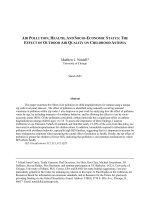
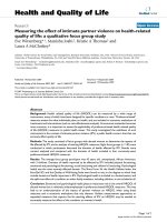
Smooth muscle contraction (figure 1-1) involves membrane depolarization with
subsequent calcium release. Calcium binds to calmodulin which activates myosin light
chain kinase to phosphorylate myosin, the thick filament in muscle. Phosphorylated
myosin binds actin, the thin filament in muscle, to produce contraction. Smooth muscle
relaxation occurs with the reuptake of calcium and de-phosphorylation of myosin by
myosin light chain phosphatase.
Figure 1-1. Mechanism of smooth muscle contraction. Following cell membrane
depolarization, the calcium (Ca
2+
) concentration increases. Myosin light chain kinase
(MLCK), whose activation depends on calcium, phosphorylates myosin. This allows
myosin to bind with actin to produce smooth muscle contraction. Myosin light chain
phosphatase (MLCP) dephosphorylates myosin to cause relaxation.
Airway smooth muscle contractility has been shown to be dependent on the
overlying epithelium. Epithelial functions include creating a barrier between the airways
and the external environment as well as secreting many factors (48). Epithelial
secretions include arachidonic acid metabolites involved in airway smooth muscle tone,
mucus secretion, and inflammation (48). Nitric oxide, growth factors involved in
3
respiratory tissue repair, and proinflammatory cytokines that recruit inflammatory cells to
the airways are also released by the epithelium (48). It has been shown in animal
models that in vitro airway smooth muscle sensitivity to contractile agonists is increased
with the epithelium removed (3, 15). This is important to note as it is known that the
epithelium is damaged or denuded in the airways of asthmatics (48). Although it is thus
likely that this contributes to airway hyperresponsiveness, it is still not clear whether the
epithelial abnormalities are a cause or an effect of asthma (48).
Airway Inflammation
The other key feature of asthma is airway inflammation, which can occur acutely
or chronically, and is the target for many other asthma medications. In fact, guidelines
for managing asthma tend to concentrate on treating airway inflammation (18). Acute
inflammation in asthma includes both an early and a late phase. In the early phase,
mast cells and macrophages in the airways are activated and release proinflammatory
mediators such as histamine, leukotrienes, prostaglandins, and reactive oxygen species
(18). Six to nine hours later, the late phase begins as cytokines released by the mast
cells in the early phase recruit eosinophils, basophils, neutrophils, and macrophages to
the airways (18). Chronic inflammation in asthma is characterized by activated T-cells,
eosinophils, mast cells, macrophages, epithelial cells, fibroblasts, and bronchial smooth
muscle cells in the airways (18). The eosinophils in particular secrete proinflammatory
mediators, cytotoxic mediators, and cytokines which cause many of the features of
asthma, including mucus secretion, smooth muscle contraction, epithelial shedding, and
airway hyperresponsiveness (18).
Various treatment strategies are used to treat the inflammation associated with
asthma. Since the symptoms of wheezing and shortness of breath result from acute
inflammation, β
2
-agonists used to dilate the airways can also be used to treat the effects
of acute inflammation (18). Medications, such as cromolyn and nedocromil, that
4
stabilize mast cells to reduce their early phase secretions are also used to treat
inflammation. Medications that target the proinflammatory products are routinely
prescribed as well. These include enzyme inhibitors, such as zileuton, an inhibitor of 5-
lipoxygenase (the enzyme involved in leukotriene production), and leukotriene receptor
antagonists, such as montelukast and zafirlukast.
Non-Invasive Markers of Airway Inflammation
Non-invasive methods of assessing the adequacy of disease management can
be useful clinically. In addition to changes in pulmonary function and symptoms, the
degree of airway inflammation can demonstrate how effective a particular treatment
regimen is (58). Exhaled breath condensate and exhaled nitric oxide can each be
measured non-invasively to assess airway inflammation in asthma.
Exhaled breath condensate (EBC) pH has been shown to be correlated with
airway inflammation (93). Asthmatics tend to have a lower EBC pH (19). The acidic pH
likely stems from neutrophil and eosinophil products, such as myeloperoxidase and
eosinophil peroxidase, reacting with hydrogen peroxide upon their release to form acids
and increase the concentration of hydrogen ions in the airways (19). Markers in EBC
can also be measured to evaluate airway inflammation. These markers include
inflammatory mediators as well as 8-isoprostane, a marker of oxidative stress (19). 8-
Isoprostane is produced by free radical oxidation of arachidonic acid; its concentration is
increased in asthmatics reflecting increased levels of oxidative stress (67).
The fraction of exhaled nitric oxide (F
E
NO) has been shown to be higher in
asthmatics than in healthy individuals (78). This is thought to be due to the elevated
activation of inducible nitric oxide synthase (iNOS), whose expression can be increased
by proinflammatory cytokines and oxidants (18, 58). It has also been shown that F
E
NO
levels are increased in subjects with exercise-induced bronchoconstriction as compared
to those without it (29).
5
Exercise-Induced Bronchoconstriction
Exercise-induced bronchoconstriction (EIB) is a complication of asthma that
affects 80-90% of people with asthma (87). EIB is characterized by symptoms of
wheezing, reduced exercise tolerance, chest pain, cough, stomachache, and sore throat
occurring during or after exercise that lasts at least five minutes (87). EIB is clinically
diagnosed based on the change in the volume of air exhaled in the first second of a
forced exhalation (FEV
1
) before and after exercise; it is specifically defined as at least a
10% post-exercise drop in FEV
1
(12). EIB is important to consider as it can deter
individuals with asthma from being physically active (43). Moreover, EIB suggests that
an individual’s asthma is not being adequately managed (49). Consequently, EIB testing
can be used to evaluate asthma therapies (49).
Pathophysiology of Exercise-Induced Bronchoconstriction
Currently, there are two major schools of thought on the pathogenesis of EIB, the
hyperosmolarity theory and the airway re-warming theory. According to the
hyperosmolarity theory, the airway surface liquid becomes hypertonic due to water loss
during exercise; the ensuing hyperosmolar environment in the airway cells results in the
release of proinflammatory mediators that cause bronchoconstriction (90). Alternatively,
the less widely accepted airway re-warming theory suggests that hyperventilation during
exercise cools the airway surface cells such that their post-exercise re-warming causes
the surrounding bronchiolar vessels to dilate; this leads to hyperemia with fluid exudation
and proinflammatory mediator release, which subsequently causes bronchoconstriction
(90).
Bronchoprovocation Tests to Diagnose Exercise-Induced Bronchoconstriction
Exercise Testing
Exercise is the actual stimulus for EIB that individuals will encounter outside of
the laboratory or doctor’s office. However, the standard exercise protocol for diagnosing
6
EIB requires patients to breathe dry air while exercising for 6-8 minutes at 85-95% of
their maximal heart rate (8). Therefore, in addition to the need for large and expensive
equipment, not all patients or subjects can complete an exercise protocol (8).
Sport-specific testing is a variation of exercise testing that is important for
athletes who regularly perform at the standard exercise protocol level (55). In this case,
the testing protocol is based on the physical demands of a particular sport; however, by
testing the athlete in his or her workout environment, the ambient conditions cannot be
standardized, which may affect the test’s ability to reliably elicit bronchoconstriction (55).
Methacholine Challenge
Methacholine is a parasympathomimetic drug that causes bronchoconstriction
(76). This widely used method of bronchoprovocation involves the patient inhaling
progressively increasing doses of aerosolized methacholine. There are two different
protocols wherein the patient is instructed to inhale the methacholine with either normal
tidal volume breaths or deep inhalations (76). The patient is deemed to exhibit bronchial
hyperresponsiveness if the dose of methacholine that causes a 20% decline in FEV
1
from the pre-challenge value is less than 4.0 mg/ml (76). Importantly, a negative test
excludes asthma in a symptomatic patient (76); however, a positive test is not specific
for asthma (8). A large number of false positive tests have been reported in athletes
(55). Moreover, a negative methacholine challenge test does not exclude EIB (8).
Mannitol Challenge
Mannitol has only recently been approved by the Food and Drug Administration
in the United States although it has been used regularly as a bronchoprovocation test in
other countries (8). A standardized mannitol test kit provides progressively increasing
doses of mannitol in a dry-powder form for patients to inhale (55). The osmotic gradient
that subsequently develops across the airways leads to the release of inflammatory
mediators that promote bronchoconstriction (8, 55). The test is considered positive for
7
bronchial hyperresponsiveness if the patient demonstrates a 15% or greater decrease in
his or her baseline FEV
1
at a dose less than 635 mg; alternatively, the test is also
considered positive if the patient exhibits a 10% or greater decrease in FEV
1
between
two consecutive doses of mannitol (10). Unfortunately, the mannitol challenge test is no
more sensitive than the methacholine challenge test for identifying EIB (11).
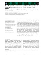
Eucapnic Voluntary Hyperventilation
In a research or clinical setting, EIB can be readily identified with a test involving
hyperpnea, or rapid breathing (9). This test, known as eucapnic voluntary
hyperventilation (EVH), requires subjects or patients to breathe cold, dry air at a high
rate for six minutes (figure1-2) (9). The rate is approximately 85% of the individual’s
maximal voluntary ventilation and is estimated by multiplying the FEV
1
at rest by 30 (9).
EVH is the bronchoprovocation strategy recommended by the International Olympic
Committee to identify athletes with EIB (9). It has been shown that changes in FEV
1
following EVH are comparable to those seen following cold air exercise (81).