- Trang chủ >>
- Y - Dược >>
- Ngoại khoa
lecture notes general surgery- ellis, harold, calne, roy, watson, christopher
Bạn đang xem bản rút gọn của tài liệu. Xem và tải ngay bản đầy đủ của tài liệu tại đây (7.67 MB, 433 trang )
Lecture Notes:
General Surgery
Companion website
The book is supported by a website containing a free bank of interactive questions and answers.
These can be found at:
www.testgeneralsurgery.com
The website includes:
• Interactive Multiple-Choice Questions for each chapter
• Interactive Short Answer Questions for each chapter
Lecture Notes:
General Surgery
Harold Ellis
CBE DM MCh FRCS
Emeritus Professor of Surgery, Guy’s Hospital, London
Sir Roy Calne
MS FRCS FRS
Emeritus Professor of Surgery, Addenbrooke’s Hospital, Cambridge
Christopher Watson
MD BChir FRCS
Reader in Surgery and Honorary Consultant, Addenbrooke’s Hospital,
Cambridge
Twelfth Edition
A John Wiley & Sons, Ltd., Publication
This edition fi rst published 2011© 1965, 1968, 1970, 1972, 1977, 1983, 1987, 1993, 1998, 2002, 2006, 2011 © 2011 Harold Ellis,
Sir Roy Y. Calne, Christopher J. E. Watson
Blackwell Publishing was acquired by John Wiley & Sons in February 2007. Blackwell’s publishing program has been merged
with Wiley’s global Scientifi c, Technical and Medical business to form Wiley-Blackwell.
Registered offi ce: John Wiley & Sons Ltd, The Atrium, Southern Gate, Chichester, West Sussex, PO19 8SQ, UK
Editorial offi ces: 9600 Garsington Road, Oxford, OX4 2DQ, UK
The Atrium, Southern Gate, Chichester, West Sussex, PO19 8SQ, UK
111 River Street, Hoboken, NJ 07030-5774, USA
For details of our global editorial offi ces, for customer services and for information about how to apply for permission to
reuse the copyright material in this book please see our website at www.wiley.com/wiley-blackwell
The right of the author to be identifi ed as the author of this work has been asserted in accordance with the UK Copyright,
Designs and Patents Act 1988.
All rights reserved. No part of this publication may be reproduced, stored in a retrieval system, or transmitted, in any form
or by any means, electronic, mechanical, photocopying, recording or otherwise, except as permitted by the UK Copyright,
Designs and Patents Act 1988, without the prior permission of the publisher.
First published 1965 Fifth edition 1977 Ninth edition 1998
Revised edition 1966 Sixth edition 1983 Reprinted 1999, 2000
Second edition 1968 Reprinted 1984, 1985, 1986 Tenth edition 2002
Third edition 1970 Seventh edition 1987 Reprinted 2003, 2004, 2005
Fourth edition 1972 Reprinted 1989 (twice) Eleventh edition 2006
Reprinted 1974 Eighth edition 1993 Twelfth Edition 2010
Revised reprint 1976 Reprinted 1994, 1996
Greek edition 1968
Portuguese edition 1979
Indonesian edition 1990
Turkish edition 2005
Wiley also publishes its books in a variety of electronic formats. Some content that appears in print may not be available in
electronic books.
Designations used by companies to distinguish their products are often claimed as trademarks. All brand names and
product names used in this book are trade names, service marks, trademarks or registered trademarks of their respective
owners. The publisher is not associated with any product or vendor mentioned in this book. This publication is designed to
provide accurate and authoritative information in regard to the subject matter covered. It is sold on the understanding that
the publisher is not engaged in rendering professional services. If professional advice or other expert assistance is required,
the services of a competent professional should be sought.
Library of Congress Cataloging-in-Publication Data
Ellis, Harold, 1926–
Lecture notes. General surgery / Harold Ellis, Sir Roy Calne, Christopher Watson. – 12th ed.
p. ; cm.
General surgery
Includes bibliographical references and index.
ISBN 978-1-4443-3440-1 (pbk. : alk. paper)
1. Surgery. I. Calne, Roy Yorke. II. Watson, Christopher J. E. (Christopher John Edward) III. Title. IV. Title: General
surgery.
[DNLM: 1. Surgical Procedures, Operative. WO 100]
RD31.E4 2011
617–dc22
2010036446
A catalogue record for this book is available from the British Library.
Set in 8.5/11pt Utopia by Toppan Best-set Premedia Limited
01 2011
Contents
Introduction, vii
Acknowledgements, ix
Abbreviations, xi
1 Surgical strategy, 1
2 Fluid and electrolyte management, 5
3 Preoperative assessment, 10
4 Postoperative complications, 15
5 Acute infections, 26
6 Shock, 31
7 Tumours, 36
8 Burns, 41
9 The skin and its adnexae, 47
10 The chest and lungs, 60
11 The heart and thoracic aorta, 69
12 Arterial disease, 80
13 Venous disorders of the lower limb, 98
14 The brain and meninges, 105
15 Head injury, 114
16 The spine, 126
17 Peripheral nerve injuries, 137
18 The oral cavity, 143
19 The salivary glands, 153
20 The oesophagus, 158
21 The stomach and duodenum, 167
22 Mechanical intestinal obstruction, 183
23 The small intestine, 194
24 Acute appendicitis, 199
25 The colon, 204
26 The rectum and anal canal, 218
27 Peritonitis, 230
28 Paralytic ileus, 236
29 Hernia, 239
30 The liver, 250
31 The gallbladder and bile ducts, 266
32 The pancreas, 276
33 The spleen, 289
34 The lymph nodes and lymphatics, 292
35 The breast, 295
36 The neck, 308
37 The thyroid, 311
38 The parathyroids, 323
39 The thymus, 328
40 The suprarenal glands, 330
41 The kidney and ureter, 335
42 The bladder, 353
43 The prostate, 358
44 The male urethra, 367
45 The penis, 370
46 The testis and scrotum, 374
47 Transplantation surgery, 384
Index, 391
Companion website
The book is supported by a website containing a free bank of interactive questions and answers.
These can be found at:
www.testgeneralsurgery.com
The website includes:
• Interactive Multiple-Choice Questions for each chapter
• Interactive Short Answer Questions for each chapter
Introduction
The ideal medical student at the end of the clinical
course will have written his or her own textbook
– a digest of the lectures and tutorials assiduously
attended and of the textbooks meticulously read.
Unfortunately, few students are perfect, and most
approach the qualifying examinations depressed
by the thought of the thousands of pages of excel-
lent and exhaustive textbooks wherein lies the
wisdom required of them by the examiners.
We believe that there is a serious need in these
days of widening knowledge and expanding syl-
labus for a book that will set out briefl y the impor-
tant facts in general surgery that are classifi ed,
analysed and as far as possible rationalized for the
revision student. These lecture notes represent
our own fi nal - year teaching; they are in no way a
substitute for the standard textbooks but are our
attempts to draw together in some sort of logical
way the fundamentals of general surgery.
Because this book is written at student level,
principles of treatment only are presented, not
details of surgical technique.
The need after only 4 years for a new, 12th,
edition refl ects the rapid changes which are taking
place in surgical practice. We are confi dent that
our constant updating will ensure that this volume
will continue to serve the requirements of our
medical students. We advise you to read this book
in conjunction with Clinical Cases Uncovered –
Surgery , which provides illustrated case studies,
MCQs, EMQs and SAQs, cases that correspond to
the chapters in this volume.
H . E .
R.Y.C.
C.J.E.W.
Acknowledgements
We are grateful to our colleagues – registrars,
housemen and students – who have read and
criticized this text during its production, and
to many readers and reviewers for their con-
structive criticisms. In particular, we are indebted
to Simon Dwerryhouse (Chapters 20 and 21 );
Justin Davies (Chapters 22 , 23 and 25 ); Gordon
Wishart (Chapters 35 , 37 and 38 ); Neville
Jamieson (Chapters 30 – 33 and 40 ); Kathryn Nash
(Chapters 21 and 30 ); and Andrew Doble (Chapters
41 – 46 ).
Finally, we would like to acknowledge the con-
tinued help given by the staff at Wiley Blackwell,
in particular to Jane Fallows, who has created
some new diagrams and brought colour to others,
Rebecca Huxley, who oversaw the production, and
Lindsey Williams, who has meticulously steered
this edition from text to fi nished product.
.
Abbreviations
ABPI ankle brachial pressure index
ACE angiotensin - converting enzyme
ACTH adrenocorticotrophic hormone
ADH antidiuretic hormone
AFP α - fetoprotein
AIDS acquired immune defi ciency syndrome
ALP alkaline phosphatase
ALT alanine transaminase
APACHE Acute Physiology And Chronic Health
Evaluation
APUD amine precursor uptake and
decarboxylation
ASA American Society of Anesthesiologists
AST aspartate transaminase
ATN acute tubular necrosis
BCG bacille Calmette – Gu é rin
CABG coronary artery bypass graft
CEA carcinoembryonic antigen
CNS central nervous system
CRP C - reactive protein
CSF cerebrospinal fl uid
CT computed tomography
DCIS ductal carcinoma in situ
DIC disseminated intravascular coagulopathy
DMSA dimercaptosuccinic acid
DOPA dihydroxyphenyl alanine
DTC differentiated thyroid cancer
DTPA diethylene triamine pentaacetic acid
ECG electrocardiograph
EMG electromyography
ER oestrogen receptor
ERCP endoscopic retrograde
cholangiopancreatography
ESBL extended spectrum beta - lactamase
ESR erythrocyte sedimentation rate
ESWL extracorporeal shock wave lithotripsy
EUS endoscopic ultrasound
FAP familial adenomatous polyposis
FEV
1
forced expiratory volume in 1 second
GCS Glasgow coma scale
GFR glomerular fi ltration rate
GGT gamma glutamyl transferase
GLA gamma linolenic acid
GTN glyceryl trinitrate
HAART highly active anti - retroviral treatment
HbA1c glycosylated haemoglobin
HCC hepatocellular carcinoma
HER2 human epidermal growth factor
receptor 2
HHT hereditary haemorrhagic telangiectasia
HHV human herpes virus
HIV human immunodefi ciency virus
HLA human leucocyte antigen
HPOA hypertrophic pulmonary osteoarthropathy
HPV human papilloma virus
HRT hormone replacement therapy
HTIG human tetanus immunoglobulin
ICP intracranial pressure
ICSI intracytoplasmic sperm injection
IFN - γ interferon γ
IPMN intraductal papillary mucinous tumour
IVC inferior vena cava
IVF in vitro fertilization
IVU intravenous urogram
JVP jugular venous pressure
KSHV Kaposi sarcoma herpes virus
‘ KUB ’ kidneys, ureters and bladder
LAD left anterior descending artery
LCIS lobular carcinoma in situ
LHRH luteinizing hormone - releasing hormone
MAG3 m ercapto - a cetyl tri g lycine
MCN mucinous cystic neoplasm
MEN multiple endocrine neoplasia
MHC major histocompatibility complex
MIBG meta - iodobenzylguanidine
MIBI methoxyisobutylisonitrile
MR magnetic resonance
MRCP magnetic resonance
cholangiopancreatography
MRSA meticillin - resistant Staphylococcus aureus
NAFLD non - alcoholic fatty liver disease
NPI Nottingham Prognostic Index
NSAIDs non - steroidal anti - infl ammatory drugs
NSGCT non - seminomatous germ cell tumour
NST ‘ no special type ’
OCP oral contraceptive pill
OPG orthopantomogram
PET positron emission tomography
PNET primitive neuroectodermal tumour
POSSUM Physiological and Operative Severity Score
for the enUmeration of Mortality and
morbidity
PSA prostate - specifi c antigen
PTA percutaneous transluminal angioplasty
PTC percutaneous transhepatic
cholangiography
PTCA percutaneous transluminal coronary
angioplasty
xii Abbreviations
PTFE polytetrafl uoroethylene
PTH parathormone
SGOT serum glutamic oxaloacetic transaminase
(synonymous with AST)
SGPT serum glutamic pyruvic transaminase
(synonymous with ALT)
SIADH syndrome of inappropriate antidiuretic
hormone
SLE systemic lupus erythematosus
SLN sentinel lymph node
T3 tri - iodothyronine
T4 tetra - iodothyronine, thyroxine
TACE transarterial chemoembolization
TCC transitional cell carcinoma
TED thromboembolism deterrent
TIA transient ischaemic attack
TIPS transjugular intrahepatic portosystemic
shunt
TNF tumour necrosis factor
TOE transoesophageal echocardiography
TPA tissue plasminogen activator
TPN total parenteral nutrition
TSH thyroid - stimulating hormone
TUR transurethral resection
UW University of Wisconsin
VAC vacuum - assisted closure
VATS video - assisted thoracoscopic surgery
VIP vasoactive intestinal polypeptide
VRE vancomycin - resistant Enterococcus
β - HCG β - human chorionic gonadotrophin
1
Surgical s trategy
Learning objectives
✓ To understand the principles of taking a clear history,
performing an appropriate examination, presenting the
fi ndings and formulating a management plan for surgical
diagnosis.
✓ To understand the common nomenclature used in surgery.
to become a good clinician. Remember that the
patient will be apprehensive and often will be in
pain and discomfort. Attending to these is the fi rst
task of a good doctor.
The h istory
The history should be an accurate refl ection of
what the patient said, not your interpretation
of it. Ask open questions such as ‘ When were
you last well? ’ and ‘ What happened next? ’ , rather
than closed questions such as ‘ Do you have
chest pain? ’ . If you have a positive fi nding, do not
leave the subject until you know everything
there is to know about it. For example, ‘ When
did it start? ’ ; ‘ What makes it better and what
makes it worse? ’ ; ‘ Where did it start and where
did it go? ’ ; ‘ Did it come and go or was it constant? ’ .
If the symptom is one characterized by bleeding,
ask about what sort of blood, when, how much,
were there clots, was it mixed in with food/
faeces, was it associated with pain? Remember
that most patients come to see a surgeon because
of pain or bleeding (Table 1.1 ). You need to be
able to fi nd out as much as you can about these
presentations.
Keep in mind that the patient has no knowledge
of anatomy. He might say ‘ my stomach hurts ’ , but
this may be due to lower chest or periumbilical
pain – ask him to point to the site of the pain. Bear
in mind that he may be pointing to a site of
referred pain, and similarly do not accept ‘ back
pain ’ without clarifying where in the back – the
Students on the surgical team, in dealing with
their patients, should recognize the following
steps in their patients ’ management.
1 History taking . Listen carefully to the patient ’ s
story.
2 Examination of the patient .
3 Writing notes .
4 Constructing a differential diagnosis . Ask the
question ‘ What diagnosis would best explain
this clinical picture? ’
5 Special investigations . Which laboratory and
imaging tests are required to confi rm or refute
the clinical diagnosis?
6 Management . Decide on the management of
the patient. Remember that this will include
reassurance, relief of pain and, as far as
possible, allaying the patient ’ s anxiety.
History and e xamination
The importance of developing clinical skills
cannot be overemphasized. Excessive reliance on
special investigations and extensive modern
imaging (some of which may be quite painful and
carry with them their own risks and complica-
tions) is to turn your back on the skills necessary
Lecture Notes: General Surgery, 12th edition. © Harold Ellis,
Sir Roy Y. Calne and Christopher J. E. Watson. Published 2011 by
Blackwell Publishing Ltd.
2 Surgical strategy
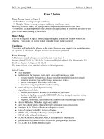
tures of the cloaca such as the bladder, uterus
and fallopian tubes (Figure 1.1 ). Testicular pain
may also be periumbilical, refl ecting the intra -
abdominal origin of these organs before their
descent into the scrotum – never be fooled by the
child with testicular torsion who complains of
pain in the centre of his abdomen.
The e xamination
Remember the classical quartet in this order:
1 inspection;
2 palpation;
3 percussion;
4 auscultation.
sacrum, or lumbar, thoracic or cervical spine, or
possibly loin or subscapular regions. When refer-
ring to the shoulder tip, clarify whether the patient
means the acromion; when referring to the shoul-
der blade, clarify whether this is the angle of the
scapula. Such sites of pain may suggest referred
pain from the diaphragm and gallbladder,
respectively.
It is often useful to consider the viscera in terms
of their embryology. Thus, epigastric pain is gen-
erally from foregut structures such as stomach,
duodenum, liver, gallbladder, spleen and pan-
creas; periumbilical pain is midgut pain from
small bowel and ascending colon, and includes
the appendix; suprapubic pain is hindgut pain,
originating in the colon, rectum and other struc-
Table 1.1 Example of important facts to determine in patients with pain and rectal bleeding
Pain Rectal bleeding
Exact site Estimation of amount (often inaccurate)
Radiation Timing of bleeding
Length of history Colour – bright red, dark red, black
Periodicity Accompanying symptoms – pain, vomiting (haematemesis)
Nature – constant/colicky Associated shock – faintness, etc.
Severity Blood mixed in stool, lying on surface, on paper, in toilet pan
Relieving and aggravating factors
Accompanying features (e.g. jaundice, vomiting,
haematuria)
Figure 1.1 Location of referred pain
for the abdominal organs.
T8,9
Liver
Gallbladder
Spleen
Stomach
Duodenum
Pancreas
Heart and
aorta
Large bowel
Bladder
Prostate ( )
Uterus and
adnexa ( )
Small bowel
Appendix
Caecum
Ascending
colon to
mid-
transverse
Testis
Renal
tract
T10
T11
T12
L1
Surgical strategy 3
infl ammatory disease but the next person
might interpret it as a prolapsed intervertebral
disc. Use the correct surgical terminology
(Table 1.2 ).
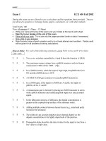
Illustrate your examination unambiguously
with drawings – use anatomical reference points
and measure the diameter of lumps accurately.
When drawing abdominal fi ndings use a hexago-
nal representation (Figure 1.2 ). A continuous line
implies an edge; shading can represent an area of
tenderness or the site where pain is experienced.
If you can feel all around a lump, draw a line to
indicate this; if you can feel only the upper margin,
show only this. Annotate the drawings with your
fi ndings (Figure 1.2 ). At the end of your notes,
write a single paragraph summary, and make a
diagnosis, or write down a differential diagnosis.
Outline a management plan and state what inves-
tigations should be done, indicating which you
have already arranged. Sign your notes and print
your name, position and the time and date legibly
underneath.
Case p resentation
The purpose of presenting a case is to convey to
your colleagues the salient clinical features, diag-
nosis or differential diagnosis, management and
investigations of your patient. The presentation
Learn the art of careful inspection, and keep
your hands off the patient until you have done so.
Inspect the patient generally, as to how he lies and
how he breathes. Is he tachypnoeic because of a
chest infection or in response to a metabolic
acidosis? Look at the patient ’ s hands and feel
his pulse.
Only after careful inspection, proceed to palpa-
tion. If you are examining the abdomen, ask the
patient to cough. This is a surrogate test of rebound
tenderness and indicates where the site of infl am-
mation is within the peritoneal cavity. Remember
to examine the ‘ normal ’ side fi rst, the side that is
not symptomatic, be it abdomen, hand, leg or
breast. Look at the patient while you palpate. If
there is a lump, decide which anatomical plane it
lies in. Is it in the skin, in the subcutaneous tissue,
in the muscle layer or, in the case of the abdomen,
in the underlying cavity? Is the lump pulsatile,
expansile or mobile?
Writing y our n otes
Always write up your fi ndings completely and
accurately. Start by recording the date and the
time of the interview. Write all the negative as
well as positive fi ndings. Avoid abbreviations
since they may mean different things to different
people; for instance, PID – you may mean pelvic
Figure 1.2 Example of how to record
abdominal examination fi ndings.
Previous perforated
duodenal ulcer repair
Kidney
transplant
Bowel sounds normal
PR: No tenderness, no mass
Normal coloured stool
Irregular
enlarged
liver edge
Tender ++
4 Surgical strategy
should not be merely a reading of the case notes,
but should be succinct and to the point, contain-
ing important positive and negative fi ndings. Do
not use words such as ‘ basically ’ , ‘ essentially ’ or
‘ unremarkable ’ , which are padding and meaning-
less. Avoid saying that things are ‘ just ’ palpable –
Table 1.2 Common prefi xes and suffi xes used in surgery
Prefi x Related organ/structure
angio - blood vessels
arthro - a joint
cardio - heart
coelio - peritoneal cavity
cholecysto - gallbladder
colo - and colon - colon
colpo - vagina
cysto - urinary bladder
gastro - stomach
hepato - liver
hystero - uterus
laparo - peritoneal cavity
mammo - and masto - breast
nephro - kidney
oophoro - ovary
orchid - testicle
rhino - nose
thoraco - chest
Suffi x Procedure
- centesis surgical puncture, often accompanied by drainage, e.g. thoracocentesis
- desis fusion, e.g. arthrodesis
- ectomy surgical removal, e.g. colectomy
- oscopy visual examination, usually through an endoscope, e.g. laparoscopy
- ostomy creating a new opening (mouth) on the surface, e.g. colostomy
- otomy surgical incision, e.g. laparotomy
- pexy surgical fi xation, e.g. orchidopexy
- plasty to mould or reshape, e.g. angioplasty; also to replace with prosthesis, e.g. arthoplasty
- rrhapy surgically repair or reinforce, e.g. herniorrhaphy
either you can feel it or you cannot. Make up
your mind. At the end of a good presentation, the
listener should have an excellent word picture of
the patient and his/her problems, what needs
to be watched and what plans you have for
management.
2
Fluid and e lectrolyte
m anagement
Learning objective
✓ To understand the distribution and composition of body fl uids,
and how these may change following surgery.
tion. Only 2% of the total body potassium is in the
extracellular fl uid. There is also a difference in
protein concentration within the extracellular
compartment, with the interstitial fl uid having
a very low concentration compared with the
high protein concentration of the intravascular
compartment.
Knowledge of fl uid compartments and their
composition becomes very important when con-
sidering fl uid replacement. In order to fi ll the
intravascular compartment rapidly, a plasma sub-
stitute or blood is the fl uid of choice. Such fl uids,
with high colloid osmotic potential, remain within
the intravascular space, in contrast to a saline
solution, which rapidly distributes over the entire
extravascular compartment, which is four times as
large as the intravascular compartment. Thus, of
the original 1 L of saline, only 250 mL would
remain in the intravascular compartment. Five
per cent dextrose, which is water with a small
amount of dextrose added to render it isotonic,
will redistribute across both intracellular and
extracellular spaces.
Fluid and e lectrolyte
l osses
In order to calculate daily fl uid and electrolyte
requirements, the daily losses should be meas-
ured or estimated. Fluid is lost from four routes:
the kidney, the gastrointestinal tract, the skin and
The management of a patient ’ s fl uid status is vital
to a successful outcome in surgery. This requires
preoperative assessment, with resuscitation if
required, and postoperative replacement of
normal and abnormal losses until the patient can
resume a normal diet. This chapter will review the
normal state and the mechanisms that maintain
homeostasis, and will then discuss the aberrations
and their management.
Body fl uid c ompartments
(Figure 2.1 )
In the ‘ average ’ person, water contributes 60% to
the total body weight: 42 L for a 70 kg man. Forty
per cent of the body weight is intracellular fl uid,
while the remaining 20% is extracellular. This
extracellular fl uid can be subdivided into intravas-
cular (5%) and extravascular, or interstitial (15%).
Fluid may cross from compartment to compart-
ment by osmosis, which depends on a solute gra-
dient, and fi ltration, which is the result of a
hydrostatic pressure gradient.
The electrolyte composition of each compart-
ment differs. Intracellular fl uid has a low sodium
and a high potassium concentration. In contrast,
extracellular fl uid (intravascular and interstitial)
has a high sodium and low potassium concentra-
Lecture Notes: General Surgery, 12th edition. © Harold Ellis,
Sir Roy Y. Calne and Christopher J. E. Watson. Published 2011 by
Blackwell Publishing Ltd.
6 Fluid and electrolyte management
the respiratory tract. Losses from the last two
routes are termed insensible losses.
Normal fl uid l osses
(Table 2.1 )
The k idney
In the absence of intrinsic renal disease, fl uid
losses from the kidney are regulated by aldoster-
one and antidiuretic hormone (ADH). These two
hormone systems regulate the circulating volume
Figure 2.1 Distribution of fl uid and electrolytes within the body.
Intracellular
fluid
20% body
weight
40% body
weight
Distribution of body water
Distribution of principal cations
Extracellular
fluid
Interstitial fluid
15% body weight
Intravascular fluid
5% body weight
Mg
2+
Ca
2+
Ca
2+
K
+
Na
+
Mg
+
Na
+
K
+
For a 75 kg man, 45 kg (45 litres)
is water, of which 30 litres is
intracellular fluid, 12 litres is
interstitial fluid and 3 litres
is intravascular fluid (plasma)
Table 2.1 Normal daily fl uid losses
Fluid loss Volume (mL) Na
+
(mmol) K
+
(mmol)
Urine 2000 80 – 130 60
Faeces 300
Insensible 400
Total 2700
and its osmolarity, and are thus crucial to home-
ostasis. Aldosterone responds to a fall in glomeru-
lar perfusion by salt retention. ADH responds to
Fluid and electrolyte management 7
occur if predominantly acid or alkaline fl uid is
lost, as occurs with pyloric stenosis and with a
pancreatic fi stula, respectively.
Large occult losses occur in paralytic ileus and
intestinal obstruction. Several litres of fl uid may
be sequestered in the gut, contributing to the
hypovolaemia. Resolution of an ileus is marked by
absorption of the fl uid and the resultant hypervol-
aemia produces a diuresis.
Insensible l osses
Hyperventilation, as may happen with pain or
chest infection, increases respiratory losses. Losses
from the skin are increased by pyrexia and sweat-
ing, with up to 1 L of sweat per hour in extreme
cases. Sweat contains a large amount of salt.
Effects of s urgery
ADH is released in response to surgery, conserving
water. Hypovolaemia will cause aldosterone
secretion and salt retention by the kidney.
Potassium is released by damaged tissues, and the
potassium level may be further increased by blood
transfusion, each unit containing in excess of
20 mmol/L. If renal perfusion is poor, and urine
output sparse, this potassium will not be excreted
and instead accumulates, the resultant hyperka-
laemia causing life - threatening arrhythmias. This
is the basis of the recommendation that supple-
mentary potassium may not be necessary in the
fi rst 48 hours following surgery or trauma.
Prescribing fl uids for
the s urgical p atient
The majority of patients require fl uid replacement
for only a brief period postoperatively until they
resume a normal diet. Some require resuscitation
preoperatively, and others require replacement
of specifi c losses such as those from a fi stula.
In severely ill patients, and those with impaired
gastrointestinal function, long - term nutritional
support is necessary.
Replacement of n ormal l osses
Table 2.1 shows the normal daily fl uid losses.
Replacement of this lost fl uid in a typical adult is
the increased solute concentration by retaining
water in the renal tubules. Normal urinary losses
are around 1500 – 2000 mL/day. The kidneys
control water and electrolyte balance closely, and
can function in spite of extensive renal disease,
and abuse from doctors prescribing intravenous
fl uids. However, damaged kidneys leave the
patient exquisitely vulnerable to inappropriate
water and electrolyte administration.
The g astrointestinal t ract
The stomach, liver and pancreas secrete a large
volume (see Table 2.3 ) of electrolyte - rich fl uid into
the gut. After digestion and absorption, the waste
material enters the colon, where the remaining
water is reabsorbed. Approximately 300 mL is lost
into the faeces each day.
Insensible l osses
Inspired air is humidifi ed in its passage to the
alveoli, and much of this water is lost with expira-
tion. Fluid is also lost from the skin, and the total
of these insensible losses is around 700 mL/day.
This may be balanced by insensible production of
fl uid, with around 300 mL of ‘ metabolic ’ water
being produced endogenously.
Abnormal fl uid l osses
The k idney
Most of the water fi ltered by the glomeruli is rea-
bsorbed in the renal tubules so impaired tubular
function will result in increased water loss.
Resolving acute tubular necrosis (Chapter 41 ,
p. 349 ), diabetes insipidus and head injury may
result in loss of several litres of dilute urine. In
contrast, production of ADH by tumours (the syn-
drome of inappropriate ADH, or SIADH) causes
water retention and haemodilution.
The g astrointestinal t ract
Loss of water by the gastrointestinal tract is
increased in diarrhoea and in the presence of an
ileostomy, where colonic water reabsorption is
absent.
Vomiting, nasogastric aspiration and fi stulous
losses result in loss of electrolyte - rich fl uid.
Disturbance of the acid – base balance may also
8 Fluid and electrolyte management
daily requirements. The composition of these
special losses varies (Table 2.3 ) but, as a rough
guide, replacement with an equal volume of
normal saline should suffi ce. Extra potassium
supplements may be required when losses are
high, such as in diarrhoea. Biochemical analysis of
the electrolyte content of fi stula drainage may be
useful.
Resuscitation
Estimation of the fl uid defi cit in patients is impor-
tant in order to enable accurate replacement.
Thirst, dry mucous membranes, loss of skin turgor,
tachycardia and postural hypotension, together
with a low jugular venous pressure, suggest a loss
of between 5% and 15% of total body water. Fluid
losses of under 5% body water are diffi cult to
detect clinically; over 15%, there is marked circu-
latory collapse.
As an example, consider a 70 kg man presenting
with a perforated peptic ulcer. On examination he
is noted to have dry mucous membranes, a tachy-
cardia and slight postural fall in arterial blood
pressure. If the loss is estimated at 10% of the total
body water, itself 60% of body weight, the volume
defi cit is 10% × 60% of 70 kg, or 10% of 42 L = 4.2 L.
achieved by the administration of 3 L of fl uid,
which may comprise 1 L of normal saline
(150 mmol NaCl) together with 2 L of water (as 5%
dextrose) (Table 2.2 ). Potassium may be added to
each 1 L bag (20 mmol/L). Alternatively, com-
pound sodium lactate (Hartmann ’ s solution) has
been advocated as the more effective fl uid replace-
ment in the postoperative period since it is similar
in composition to plasma (Table 2.2 ). Adjustments
to this regimen should be based on regular clinical
examination, measurement of losses (e.g. urine
output), daily weights (to assess fl uid changes)
and regular blood samples for electrolyte determi-
nation. For example, if the patient is anuric, 1 L/
day of hypertonic dextrose without potassium
may suffi ce, which has the added advantage of
reducing catabolism with the breakdown of
protein and accumulation of urea.
Replacement of s pecial l osses
Special losses include nasogastric aspirates, losses
from fi stulae, diarrhoea and stomas and covert
losses such as occur with an ileus. Loss of plasma
in burns is considered elsewhere (Chapter 8 ). All
fl uid losses should be measured carefully when
possible, and this volume added to the normal
Table 2.2 Electrolyte content of intravenous fl uids
Intravenous infusion Na
+
(mmol/L) Cl
−
(mmol/L) K
+
(mmol/L) HCO
3
−
(mmol/L) Ca
2 +
(mmol/L)
Normal saline (0.9%
saline)
150 150 – – –
4% dextrose/ 0.18%
saline
30 30 – – –
Hartmann ’ s
(compound sodium
lactate)
131 111 5 29 2
Normal plasma values 134 – 144 95 – 105 3.4 – 5.0 22 – 30 2.2 – 2.6
Table 2.3 Daily volume and composition of gastrointestinal fl uids
Fluid Volume (mL) Na
+
(mmol/L) K
+
(mmol/L) Cl
−
(mmol/L) H
+
/HCO
3
−
(mmol/L)
Gastric 2500 30 – 80 5 – 20 100 – 150 H
+
40 – 60
Bile 500 130 10 100 HCO
3
−
30 – 50
Pancreatic 1000 130 10 75 HCO
3
−
70 – 110
Small bowel 5000 130 10 90 – 130 HCO
3
−
20 – 40
Fluid and electrolyte management 9
Parenteral f eeding
For patients with intestinal fi stulae, prolonged
ileus or malabsorption, nutrition cannot be sup-
plemented through the gastrointestinal tract, and
therefore parenteral feeding is necessary. This is
usually administered via a catheter in a central
vein because of the high osmolarity of the solu-
tions used; there is a high risk of phlebitis in
smaller veins with lower blood fl ow. However,
peripheral parenteral nutrition with less hyperos-
molar solutions can be used for short - term
feeding. The principle is to provide the patient
with protein in the form of amino acids, carbohy-
drate in the form of glucose, and fat emulsions
such as Intralipid. Energy is derived from the car-
bohydrate and fat (30 – 50% fat), which must be
given when amino acids are given, usually in a
ratio of 1000 kJ/g protein nitrogen. Trace ele-
ments, such as zinc, magnesium and copper, as
well as vitamins such as vitamin B
12
and ascorbic
acid, and the lipid - soluble vitamins A, D, E and K,
are usually added to the fl uid, which is infused
as a 2.5 L volume over 24 hours. Daily weights as
well as biochemical estimations of electrolytes
and albumin are useful guides to continued
requirements.
The ability of a patient to benefi t from intrave-
nous feeding depends on the general state of
metabolism and residual liver function. Nutritional
support should be continued in the postoperative
period until gastrointestinal function returns and
the patient is restored to positive nitrogen balance
from the perioperative catabolic state. Restoration
of a positive nitrogen balance is often apparent to
the nurses and doctors as a sudden occurrence,
when the patient starts smiling and asks for food.
Occasionally, in chronic malnutrition with intes-
tinal fi stulae or in patients who have lost most of
the small bowel, parenteral feeding may be neces-
sary on a long - term basis.
Complications of total parenteral nutrition
(TPN) include sepsis, thrombosis, hyponatraemia,
hyperglycaemia and liver damage. To minimize
sepsis, the central venous catheter is tunnelled
with a subcutaneous Dacron cuff at the exit site to
reduce the risk of line infection. Thrombosis may
occur on any indwelling venous catheter, and, in
patients requiring long - term TPN, this is a major
cause of morbidity. Hyperglycaemia is common,
particularly following pancreatitis, and may
necessitate infusion of insulin.
As this loss is largely isotonic (gastric juices and
the peritoneal infl ammatory response), infusion
of a balanced crystalloid solution (e.g. Hartmann ’ s
solution) is appropriate. A general rule of thumb
is to replace half of the estimated loss quickly, and
then reassess before replacement of the rest. The
best guide to the success of resuscitation is the
resumption of normal urine output; therefore,
hourly urine output should be measured. Central
venous pressure monitoring will help in the
adjustment of the rate of infusion.
Nutrition
Many patients undergoing elective and emer-
gency surgery are reasonably well nourished and
do not require special supplementation pre - or
postoperatively. Recovery from surgery is usually
swift, and the patient resumes a normal diet
before he/she has become seriously malnour-
ished. There are, however, certain categories of
patients in whom nutrition prior to surgery is
poor, and this may be a critical factor in determin-
ing the outcome of an operation by lowering their
resistance to infection and impairing wound
healing. Such patients include those with chronic
intestinal fi stulae, malabsorption, chronic liver
disease, neoplasia and starvation, and those
who have undergone chemo - and radiotherapy.
Wherever possible in such patients, nutritional
support should be instituted before surgery, as
postoperative recovery will be much quicker.
Enteral f eeding
If the gastrointestinal tract is functioning satisfac-
torily, oral intake can be supplemented by a basic
diet introduced through a fi ne nasogastric tube
directly into the stomach. The constituents of the
diet are designed to be readily absorbable protein,
fat and carbohydrate. Such a diet can provide
8400 kJ with 70 g protein in a volume of 2 L. The
commonest complication is diarrhoea, which is
usually self - limiting.
If a prolonged postoperative recovery is antici-
pated, or a large preoperative nutritional defi cit
needs to be corrected, consideration should be
given to insertion of a feeding jejunostomy at the
time of surgery. This has the advantage of avoiding
a nasogastric tube.
3
Preoperative a ssessment
Learning objectives
✓ To be aware of the principles of preoperative assessment.
✓ To be able to identify and manage likely complicating factors
prior to surgery.
tify factors that may be a problem during or
following surgery. Some problems may be readily
identifi able and treated in advance; for example,
a history of vomiting or intestinal obstruction
would indicate that fl uid volume replacement is
necessary, and this can be done swiftly prior to
surgery. A long history of a condition that is sched-
uled for elective surgical treatment may afford
time in which the patient ’ s comorbid conditions
can be improved before surgery.
Past m edical h istory
• Diabetes – whether controlled by insulin, oral
hypoglycaemics or diet. Severe diabetes may
be complicated by gastroparesis with a risk of
aspiration on induction of anaesthesia.
• Respiratory disease – what is the nature of the
chest problem, and is the breathing as good as
it can be or is the patient in the middle of an
acute exacerbation?
• Cardiac disease – has the patient had a recent
myocardial infarct, or does he/she have mild
stable angina? What is his exercise tolerance?
• Rheumatoid arthritis – may be associated with
an unstable cervical spine so a cervical spine
X - ray is indicated.
• Rheumatic fever or valve disease or presence of
a prosthesis – necessitating prophylactic
antibiotics.
• Sickle cell disease – a haemoglobin
electrophoresis should be checked in all
patients of African – Caribbean descent.
Homozygotes are prone to sickle crises under
general anaesthetic, and postoperatively if they
become hypoxic.
The preoperative assessment involves an overall
analysis of the patient ’ s condition and prepara-
tion of the patient for the proposed procedure.
This involves taking a careful history, confi rming
that the indication for surgery still exists (e.g. that
the enlarged lymph node that was to be removed
for biopsy has not spontaneously regressed), and
that the patient is as fi t as possible for the proce-
dure. Do not accept someone else ’ s diagnosis – it
might be wrong. In particular, verify the proposed
side of surgery and mark the side; write the opera-
tion name next to the arrow.
Fitness for a procedure needs to be balanced
against urgency – there is no point contemplating
a referral to a diabetologist for better diabetic
control for someone with a ruptured aortic aneu-
rysm in need of urgent repair. The assessment
process can be considered in terms of factors spe-
cifi c to the patient and to the operation.
Patient a ssessment
In assessing a patient ’ s fi tness for surgery, it is
worth going through the clerking process with this
in mind.
History of p resenting c omplaint
An emergency presentation may warrant an emer-
gency procedure, so the assessment aims to iden-
Lecture Notes: General Surgery, 12th edition. © Harold Ellis,
Sir Roy Y. Calne and Christopher J. E. Watson. Published 2011 by
Blackwell Publishing Ltd.
Preoperative assessment 11
appropriate precautions taken; such patients
are a high risk for transmission of hepatitis B,
hepatitis C and human immunodefi ciency
virus (HIV).
Drugs
Most drugs should be continued on admission. In
particular, drugs acting on the cardiovascular
system should usually be continued and given on
the day of surgery. The following are examples
of drugs that should give cause for concern
and prompt discussion with the surgeon and
anaesthetist:
• Warfarin – when possible it should be
stopped before surgery. If continued
anticoagulation is required, then convert to a
heparin infusion.
• Aspirin and clopidogrel cause increased
bleeding and should also be stopped whenever
possible at least 10 days before surgery.
• Oral contraceptive pill is associated with an
increased risk of deep vein thrombosis and
pulmonary embolism; it should be stopped at
least 6 weeks preoperatively. The patient
should be counselled on appropriate
alternative contraception since an early
pregnancy might be damaged by teratogenic
effects of some of the drugs used in the
perioperative period.
• Steroids – patients who are steroid dependent
will need extra glucocorticoid in the form of
hydrocortisone injections to tide them over the
perioperative stress.
• Immunosuppression – patients are more prone
to postoperative infection.
• Diuretics – both thiazide and loop diuretics
cause hypokalaemia. It is important to
measure the serum potassium in such patients
and restore it to the normal range prior to
surgery.
• Monoamine oxidase inhibitors are not widely
used nowadays, but do have important
side - effects such as hypotension when
combined with general anaesthesia.
Allergies
It is important to determine clearly the nature of
any allergy before condemning a potentially
useful drug to the list of allergies. For example,
diarrhoea following erythromycin usually refl ects
its action on the motilin receptor rather than a
Past s urgical h istory
• Nature of previous operations – what has been
done before? What is the current anatomy?
What problems were encountered last time?
Ensure a copy of the previous operation note is
available.
• Complications of previous surgery , e.g. deep
vein thrombosis, MRSA wound infection or
wound dehiscence.
Past a naesthetic h istory
• Diffi cult intubation – usually recorded in the
previous anaesthetic note, but the patient may
also have been warned of previous problems.
• Aspiration during anaesthesia – may suggest
delayed gastric emptying (e.g. owing to
diabetes), suggesting that a prolonged fast and
airway protection (cricoid pressure) are
indicated prior to induction.
• Scoline apnoea – defi ciency of
pseudocholinesterase resulting in sustained
paralysis following the ‘ short - acting ’ muscle
relaxant suxamethonium (Scoline). It is usually
inherited (autosomal dominant) and so there
may be a family history.
• Malignant hyperpyrexia – a rapid excessive rise
in temperature following exposure to
anaesthetic drugs due to an uncontrolled
increase in skeletal muscle oxidative
metabolism and associated with muscular
contractions and rigidity, sometimes
progressing to rhabdomyolysis; it carries a high
mortality (at least 10%). Most of the cases are
due to a mutation in the ryanodine receptor
on the sarcoplasmic reticulum, and
susceptibility is inherited in an autosomal
dominant pattern, so a family history should
be sought.
‘ Social ’ h abits
• Smoking – ideally patients should stop
smoking before any general anaesthetic to
improve their respiratory function and reduce
their thrombogenic potential.
• Alcohol – a history suggestive of dependency
should be sought, and management of the
perioperative period instituted using
chlordiazepoxide to avoid acute alcohol
withdrawal syndrome.
• Substance abuse – in particular a history of
intravenous drug usage should be sought and