Bệnh cảnh lâm sàng hiếm gặp của hội chứng Conn
Bạn đang xem bản rút gọn của tài liệu. Xem và tải ngay bản đầy đủ của tài liệu tại đây (128.72 KB, 9 trang )
Unusual presentation of the Conn's syndrome: a
case report
Maryam Al-Rajhi1, Sahasranamaiyer Narayanan2
Department of Medicine, Al- Amiri Hospital, Kuwait.
Received: 20 Dec 2010 Revised: 10 Mar 2011 Accepted: 8 May 2011
Introduction
The primary aldosteronism (PA) resulting from an adrenocortical adenoma
(Conn's syndrome) is a common and a curable cause of secondary hypertension
[5]. The combination of hypertension, hypokalemia, and metabolic alkalosis is
important for a diagnosis of PA. In few cases, hypokalemia caused by PA can be
severe enough to cause rhabdomyolysis.
Tăng aldosterol tiên phát (PA) do u tuyến thượng thận (hội chứng Conn) là một
nguyên nhân gây bệnh cao huyết áp thứ phát phổ biến và có thể chữa được [5]. Sự
kết hợp của tăng huyết áp, giảm kali huyết, và nhiễm kiềm chuyển hóa là triệu
chứng chính để chẩn đoán PA. Trong vài trường hợp PA có hạ kali máu nặng và có
thể gây tiêu cơ vân.
Here we report a case of rhabdomyolysis caused by severe hypokalemia, which in
turn was resulted in the PA. The patient was cured after performing
adrenalectomy
Dưới đây chúng tôi báo cáo một trường hợp tiêu cơ vân do hạ kali máu nặng, mà
lần lượt được dẫn đến các PA. Bệnh nhân đã được chữa khỏi sau khi thực hiện cắt
tuyến thượng thận
Case report
A 26-year-old-woman was admitted to the hospital because of five days history of
generalized muscle weakness involving predominantly the lower limbs. That was
associated with myalgia, muscle cramp and paresthesia. She had history of
episodic muscle weakness of the lower limbs over the past few months. There was
no history of infection or trauma.
Bệnh nhân nữ 26 tuổi đã được nhập viện vì yếu cơ, chủ yếu chi dưới năm ngày
nay, kết hợp với đau cơ, co cứng cơ và dị cảm. Có tiền sử yếu chi dưới trong vài
tháng qua. Không có tiền sử nhiễm trùng hoặc chấn thương.
She also had not experienced diarrhea or vomiting, and any use of diuretics,
herbal supplements, liquorice, laxatives or other medications. She did not suffer
from renal disease or hypertension and had no symptoms suggestive of thyroid
disease. She had regular menstrual cycles.
Cô cũng không có tiêu chảy hoặc nôn mửa, và không sử dụng thuốc lợi tiểu, bổ
sung thảo dược, cam thảo, thuốc nhuận tràng hoặc thuốc khác. Cô không bị bệnh
thận hoặc tăng huyết áp và không có triệu chứng gợi ý bệnh tuyến giáp. Cô có chu
kỳ kinh nguyệt đều đặn.
On physical examination, her weight was 58Kg and height 160cm (BMI = 22.65),
blood pressure 170/115 mmHg, body temperature 36.8 °C, pulse rate 70
beats/min, and respiratory rate 18/min. There was weakness of both lower limbs
(4/5) with normal sensations. The power and sensation of both upper limbs were
normal and normal cardiovascular and respiratory rates. The abdominal
examination did not reveal hepatosplenomegaly or bruits on auscultation with no
features of Cushing's syndrome.
Khám thực thể, cân nặng của cô là 58kg và chiều cao 160cm (BMI = 22,65), huyết
áp 170/115 mmHg, nhiệt độ cơ thể là 36,8 ° C, nhịp tim 70 nhịp / phút, và tốc độ
hô hấp 18 / phút. Có yếu của cả hai chi dưới (4/5) với cảm giác bình thường. Sức
mạnh và cảm giác của cả hai chi trên, tim mạch và hô hấp bình thường. Các khám
bụng không thấy gan lách to. Không có biểu hiện của hội chứng Cushing.
Laboratory investigations were as follow: potassium 1.5 mEq/L [normal range
(NR): 3.6-5.1 mEq/L] pH7.53, PaO2 10.4 kPa, PCO2 5.27 kPa, HCO3 32.5
mEq/L, creatinine phosphokinase (CPK) 19395 IU/L [NR: 20-270 IU/L],
creatinine 0.52 mg/dl [NR:0.599-1.09 mg/dl], sodium 141 mEq/L [NR: 134-144
mEq/L], corrected calcium 2.21 mEq/L [NR: 2.1-2.6 mEq/L], magnesium 0.91
mEq/L [NR: 0.4-2.88 mEq/L], and phosphate 0.57mEq/L [NR: 0.87 – 1.45
mEq/L]. The transaminases were slightly raised (AST 68 IU/L, ALT 109 IU/L). The
bilirubin and coagulation indices and thyroid function test were normal (TSH 3.81
IU/mL). Urinary potassium was 33.5 mEq/24h [NR: 2-300 mEq].
Các xét nghiệm như sau: kali 1,5 mEq / L [bình thường (NR): 3,6-5,1 mEq / L]
pH7.53, PaO2 10,4 kPa, pCO2 5.27 kPa, HCO3 32,5 mEq / L, CPK: 19.395 IU / L
[bình thường: 20-270 IU / L], creatinine 0,52 mg / dl [NR: 0,599-1,09 mg / dl],
Na141 mEq / L [NR: 134-144 mEq / L], canxi 2,21 mEq / L [NR: 2,1-2,6 mEq /
L], magiê 0,91 mEq / L [NR: 0,4-2,88 mEq / L], và phosphate 0.57mEq / L [NR:
0,87-1,45 mEq / L]. Các transaminase đã tăng nhẹ (AST 68 IU / L, ALT 109 IU /
L). Xét nghiệm bilirubin và các chỉ số đông máu và chức năng tuyến giáp bình
thường (TSH 3,81 IU / mL). Kali niệu là 33,5 mEq / 24h [NR: 2-300 mEq].
The ECG showed presence of U waves and no hypertensive changes. She was
treated with boluses of IV potassium chloride plus oral potassium supplements, for
a total of 700 mmol over 4 days. Initially her hypertension was treated with
Amlodypine 5mg once a day. In spite of repeated IV and oral potassium
supplements, her potassium level remained low and the ECG showed changes of
hypokalemia.
ECG cho thấy sự hiện diện của sóng U và không có thay đổi huyết áp. Bà được
điều trị với liều bolus kali clorua tiêm tĩnh mạch cộng với bổ sung kali bằng miệng,
với tổng số 700 mmol trên 4 ngày. Ban đầu tăng huyết áp của cô đã được điều trị
bằng Amlodypine 5mg mỗi ngày một lần. Mặc dù tiêm tĩnh mạch lặp đi lặp lại và
bổ sung kali uống, nồng độ kali cô vẫn ở mức thấp và ECG cho thấy những thay
đổi của hạ kali máu.
The PA was suspected because of persistent hypokalemia in association with
hypertension and metabolic alkalosis. Therefore, after correction of her potassium
level, she was examined for the plasma aldosterone concentration (PAC) and
direct rennin concentration (DRC). Results revealed a low DRC level of 1.5 mUI/L
(NR: 2.8-39.9 mUI/L), along with a very high PAC level of 2669 pmol/L (NR: 22-
477 pmol/L). The aldosterone/rennin ratio was 1779. 24-hour urinary aldosterone
excretion after 3 days of salt overload was 182.9 microg. (NR: 3.9- 55.5 microg).
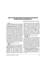
The CT abdomen revealed a left adrenal gland mass measuring about 2.5x1.2 cm
(Fig.1).
Bệnh nhân bị nghi ngờ Tăng aldosterol nguyên phát vì hạ kali máu dai dẳng gắn
với tăng huyết áp và nhiễm kiềm chuyển hóa. Vì vậy, sau khi điều chỉnh các mức
kali của mình, cô đã được kiểm tra cho nồng độ aldosterone huyết tương (PAC) và
sự tập trung rennin trực tiếp (DRC). Kết quả cho thấy một mức độ thấp của DRC
1,5 MUI / L (NR: 2,8-39,9 MUI / L), cùng với một mức độ rất cao của PAC 2669
pmol / L (NR: 22-477 pmol / L). Tỷ lệ aldosterone / rennin là 1779. aldosterone
trong nước tiểu 24h sau 3 ngày kể từ ngày quá tải muối là 182,9 microg. (NR: 3.9-
55.5 microg). CT bụng thấy một khối ở tuyến thượng thận trái đo khoảng 2.5x1.2
cm (Hình 1).
The patient was on spironolactone treatment for three weeks preoperatively,
which improved her weakness and hypokalemia. Hence, she underwent a
successful laparoscopic left adrenalectomy. On gross examination, the mass was
golden-yellow in colour without evidence of necrosis or hemorrhage (Fig.2).
Histopathalogical examination of the mass showed features of adrenal adenoma,
without necrosis, vascular or capsular invasion (Fig.3). The findings were
consistent with Conn's syndrome. Following surgery, the patient was
normotensive, and the serum potassium was 3.7 mmol/L without antihypertensive
medications. She was discharged without medication, and follow up continued at
the outpatient clinic. She remained normotensive and normokalemic for the last 4-
5 months after discharge.
Bệnh nhân đã được điều trị spironolactone trong ba tuần trước mổ, có cải thiện tình
trạng yếu cơ và hạ kali máu. Do đó, cô đã được cắt tuyến thượng thận trái qua nội
soi thành công. Khi thăm khám đại thể khối có màu vàng không có bằng chứng
hoại tử hoặc xuất huyết (Hình 2). Xét nghiệm mô bệnh học của khối u cho thấy các
đặc điểm của u tuyến thượng thận, mà không có nang, không có hoại tử, hay xâm
lấn mạch máu (hình 3). Những phát hiện này là phù hợp với hội chứng Conn. Sau
phẫu thuật, bệnh nhân có huyết áp bình thường, và kali trong huyết thanh là 3,7
mmol / L mà không cần dùng thuốc hạ huyết áp. Cô đã được ra viện mà không cần
dùng thuốc, và được theo dõi tiếp tục tại phòng khám ngoại trú. Bà duy trì huyết áp
bình thường và kali máu bình thường trong 4-5 tháng qua, sau khi xuất viện.
Discussion
PA is a common cause of secondary hypertension, and characterised by
hypertension, hypokalemia, suppressed plasma renin, and increased aldosterone
excretion. The rhabdomyolysis as a presentation feature of primary aldosteronism
is an extremely rare association. There are about sixteen related reported cases in
the literature [1] and to the best of our knowledge this is the first reported case in
Kuwait.
tăng aldosterol nguyên phát là một nguyên nhân phổ biến của bệnh lý tăng huyết
áp thứ phát, và được đặc trưng bởi tăng huyết áp, hạ kali máu, ức chế renin huyết
tương, tăng bài tiết aldosterone. Tiêu cơ vân là một đặc điểm biểu hiện hiếm gặp
của tăng aldosterol tiên phát. Có khoảng mười sáu ca được báo cáo trong các tài
liệu [1] và sự hiểu biết của chúng tôi đây là trường hợp báo cáo đầu tiên ở Kuwait.
The rhabdomyolysis is characterized by muscle necrosis and the release of its
contents into the circulation, including myoglobin, potassium, phosphate, urate
and creatinine kinase. There are many causes of rhabdomyolysis, including crush
injury, excessive exercise, metabolic and endocrine disorders, infections, drugs,
toxins like alcohol or statins, and excessive heat exposure [2]. Hypokalemia is a
recognised cause of rhabdomyolysis. The symptoms of malaise, muscle weakness,
fatigability, and myalgia occur when serum concentration of potassium is below 3
mEq/L. However, muscle enzyme elevations are usually seen when potassium
concentrations fall below 2.5 mEq/L[2].The important biochemical findings in
rhabdomyolysis are hyperkalemia and a high anion gap acidosis as a consequence
of the release of organic acids from necrotic muscles.
Tiêu cơ vân là đặc trưng của hoại tử cơ bắp và sự giải phóng hoạt chất trung gian
của nó vào hệ tuần hoàn, bao gồm myoglobin, kali, phosphate, urate và kinase
creatinine. Có nhiều nguyên nhân gây tiêu cơ vân, bao gồm chấn thương nghiền
nát, tập thể dục quá mức, rối loạn chuyển hóa và nội tiết, nhiễm trùng, thuốc, chất
độc như rượu hoặc thuốc statin, và tiếp xúc với nhiệt độ quá cao [2]. Hạ kali máu
là một nguyên nhân được công nhận của tiêu cơ vân. Các triệu chứng của tình
trạng bất ổn, yếu cơ, hay mệt mỏi, và đau cơ xảy ra khi nồng độ trong huyết thanh
của kali là dưới 3 mEq / L. Tuy nhiên, tăng men cơ thường thấy khi nồng độ kali
giảm xuống dưới 2,5 mEq / L [2] .Các kết quả sinh hóa quan trọng trong cơ vân là
tăng kali máu và một khoảng trống anion cao nhiễm toan như một hệ quả của việc
phát hành các axit hữu cơ từ cơ hoại tử.
In our case, hypokalemic metabolic alkalosis and hypertension raised the
suspicion of primary aldosteronism. The aldosterone-producing adenoma (APA)
and bilateral idiopathic hyperaldosteronism (IHA) are the most common subtypes
of PA. The APA is a small nodule (< 2cm) that mostly occurs in the left adrenal
gland and commonly found in females, and usually present with severe
hypertension and more profound hypokalemia. It is also more common in younger
patients (between the ages of 30-50), with higher plasma and urinary levels of
aldosterone [3]. Biochemical abnormalities of the APA include hypokalemia,
metabolic alkalosis, and a relative hypernatremia.
Trong trường hợp của chúng tôi, nhiễm kiềm chuyển hóa hạ kali-máu và tăng
huyết áp nâng những nghi ngờ về tăng aldosterol tiên phát. Các u biểu mô tuyến
sản suất-aldosterol (APA) và cường aldosteron nguyên phát hai bên (IHA) là các
phân nhóm phổ biến nhất của tăng aldosterol nguyên phát. APA là một nốt nhỏ
(<2cm) mà chủ yếu xảy ra ở tuyến thượng thận trái và tìm thấy phổ biến ở phụ nữ,
và thường có tăng huyết áp nặng và hạ kali máu nghiêm trọng hơn. Nó cũng phổ
biến hơn ở những bệnh nhân trẻ (tuổi từ 30-50), với mức aldosterone huyết tương
và nước tiểu cao hơn[3]. Bất thường về sinh hóa của APA bao gồm hạ kali huyết,
nhiễm kiềm chuyển hóa, và tăng natri huyết tương
Although spontaneous hypokalemia in a patient with hypertension is a strong
indicator of aldosteronism, only a minority of patients (9-30%) have a potassium
level that is in the lownormal range [4]. Therefore, hypokalemia is not the
criterion used for diagnosis of the PA. It is recommended to test for the PA in the
following groups: patients with hypertension and hypokalemia, treatment-resistant
hypertension (i.e on 3 antihypertensive medications with poor control), severe
hypertension 100
≥
160mmHg systolic or
≥
( mmHg diastolic), hypertension with
adrenal insidentaloma, and the onset of hypertension under the age of 20 [5].
Screening is done by measuring the PAC level and PRC.
Mặc dù hạ kali máu tự phát ở bệnh nhân tăng huyết áp là một chỉ dấu rõ rệt của
cường aldosterol, chỉ có một số ít bệnh nhân (9-30%) có mức độ kali đó là trong
phạm vi thấp hoặc bình thường [4]. Vì vậy, hạ kali máu không phải là tiêu chí
được sử dụng để chẩn đoán của tăng aldosterol nguyên phát. Chúng tôi đề nghị để
kiểm tra sự tăng aldosterol nguyên phát trong các nhóm sau: bệnh nhân tăng huyết
áp và hạ kali máu, tăng huyết áp kháng trị (tức là trên 3 loại thuốc hạ áp với sự
kiểm soát kém), tăng huyết áp nặng 100- 160mmHg hoặc huyết áp tâm thu
(mmHg tâm trương), tăng huyết áp với thượng thận aloma insident, và những cơn
tăng huyết áp ở độ tuổi dưới 20 [5]. Sàng lọc được thực hiện bằng cách đo mức
PAC và PRC.
When the PAC is greater than 15ng/mL, the PRC is less than 1ng/mL/h – as seen
in this caseand the ratio of the two (PAC/PRC) is greater than 20, with the
sensitivity and specificity of approximately 75%. This test is valid as long as a
patient is not taking aldosterone antagonists, such as spironolactone, epleranone,
or renin inhibitors [6]. The screening test is not a diagnostic tool, and PA must be
confirmed by demonstrating inappropriate aldosterone secretion. Confirmatory
testing can be done by any of the four procedures: oral sodium loading and
measurement of urinary aldosterone, intravenous sodium chloride loading and
measurement of PAC, fludrocortisone suppression, or captoprill challenge [6].
Khi PAC lớn hơn 15ng / mLPRC là ít hơn hơn so với 1ng / ml / h - như trong
trường hợp này và tỷ lệ của hai (PAC / PRC) là lớn hơn 20, với độ nhạy và độ đặc
hiệu vào khoảng 75 %. Thử nghiệm này là hợp lệ miễn là bệnh nhân không dùng
thuốc đối kháng aldosterone, như spironolactone, epleranone, hoặc thuốc ức chế
renin [6]. Các xét nghiệm kiểm tra không phải là một công cụ chẩn đoán, và tăng
aldosterol nguyên phát phải được xác nhận bằng cách chứng minh có sự bài tiết
aldosterone không phù hợp. Xét nghiệm khẳng định có thể được thực hiện bởi một
trong bốn quy trình: dùng natri đường uống và đo lường aldosterone tiết niệu, dùng
natri clorua đường tĩnh mạch và đo lường PAC, ức chế fludrocortisone, hoặc thử
thách captoprill [6].
In our case we used the oral sodium loading test which showed a non-suppressed
urinary aldosterone excretion. Once PA has been diagnosed biochemically, it is
important to determine the subtype to help in directing the therapy. Unilateral
adrenalectomy in patients with the APA or unilateral adrenal hyperplasia results
in normalization of hypokalemia and hypertension, while in bilateral IHA,
unilateral or bilateral adrenalectomy rarely corrects the hypertension [7], and
medical therapy is therefore the treatment of choice.
Trong trường hợp của chúng tôi, chúng tôi sử dụng xét nghiệm dung nạp natri
đường miệng cho thấy một aldosterone niệu bài tiết không bị đàn áp. Một khi PA
đã được chẩn đoán sinh hóa, điều quan trọng là phải xác định được phân nhóm
giúp cho việc xác định phương pháp điều trị. Do đó cắt tuyến thượng thận một bên
ở những bệnh nhân APA hoặc tăng sản thượng thận đơn phương giúp cải thiện tình
trạng hạ kali máu và tăng huyết áp , trong khi ở IHA hai bên, cắt tuyến thượng thận
một hoặc 2 bên hiếm khi cải thiện được tình trạng tăng huyết áp [7], và điều trị nội
khoa là hướng điều trị được lựa chọn.
Adrenal CT is not a correct choice in distinguishing between the APA and IHA. It
cannot reliably visualize microadenomas or distinguish incidentalomas from the
APAs, which makes the adrenal venous sampling (AVS) to be the most accurate
mean to differentiate between unilateral from bilateral forms of PA [8]. The AVS is
essential for appropriate therapy in many patients with PA who have a high
probability of having an APA and want to pursue surgical management [6], but it
is expensive and invasive. The procedure itself has a relatively low success rate
because of the difficulty in cannulating the right adrenal vein (which is smaller
than the left and empties directly into the IVC rather than the renal vein) [8].
CT Thượng thận không phải là một sự lựa chọn đúng trong việc phân biệt giữa các
APA và IHA. Nó không thể hiển thị một cách tin cậy các khối u nhỏ hoặc phân
biệt khối u thượng thận ngẫu nhiên (incidentalomas) với APAs, do vậy lấy mẫu
tĩnh mạch thượng thận (AVS) có giá trị chính xác nhất để phân biệt giữa PA một
bên và hai bên [8]. AVS là điều cần thiết cho điều trị thích hợp trong nhiều bệnh
nhân với PA có một xác suất bị APA và muốn được phẫu thuật [6], nhưng nó tốn
kém và xâm lấn. quy trình chính nó có một tỷ lệ thành công tương đối thấp do
những khó khăn trong đặt ca nuyn tĩnh mạch thượng thận phải ( nhỏ hơn so với
bên trái và đổ trực tiếp vào tĩnh mạch chủ dưới thay vì tĩnh mạch thận) [8].
The most important factors that determine the successful characterization of both
adrenal veins in a patient with PA are the experience, dedication and presence of
repetition of the radiologist performing the procedure [9]. A more practical
approach is the selective use of AVS as recommended by Young [5] which is based
on patient preferences, age, adrenal morphologic appearance on CT, clinical
comorbid conditions, and clinical probability of finding an APA. For patients
younger than 40, in whom a solitary adenoma is >1cm with normal contralateral
adrenal gland, a unilateral adrenalectomy may be done without venous sampling
and in the absence of comorbid conditions. Therefore, the AVS was bypassed in
our case based on these criteria.
The mainstays of therapy for the APA include surgical adrenalectomy, and
alternatively the use of aldosterone antagonists, such as spironolactone. The
efficacy of adrenalectomy has been confirmed in a study where the diagnosis of
APA was established, and in which a fall of BP observed in all patients. Moreover,
82% of patients were either improved markedly or cured (32%), thereby allowing
the withdrawal of all antihypertensive medications [10,11,12]. Conclusion
Rhabdomyolysis is uncommon in patients with the APA. We recommend
considering the APA, when rhabdomyolysis occurs in a patient with severe
hypokalemia and metabolic alkalosis since the disease could be curable like in our
case.
Các điểm chính của điều trị cho APA bao gồm phẫu thuật cắt tuyến thượng thận,
và sử dụng các chất đối kháng aldosterone, như spironolactone. Hiệu quả của cắt
tuyến thượng thận đã được khẳng định trong một nghiên cứu trên các bệnh nhân
được chẩn đoán APA, trong đó tất cả các bệnh nhân đều quan sát thấy giảm huyết
áp. Hơn nữa, 82% bệnh nhân được cải thiện rõ rệt trong hoặc chữa khỏi (32%), do
đó cho phép dừng tất cả các loại thuốc hạ huyết áp [10,11,12]. Kết luận, tiêu cơ
vân là không phổ biến ở những bệnh nhân với APA. Chúng tôi khuyên bạn nên
xem xét APA, khi tiêu cơ xảy ra ở bệnh nhân bị hạ kali máu nặng và nhiễm kiềm
chuyển hóa kể từ khi bệnh có thể chữa trị được như trong trường hợp của chúng
tôi.