PEGylated nano graphene oxide for delivery of water insoluble
Bạn đang xem bản rút gọn của tài liệu. Xem và tải ngay bản đầy đủ của tài liệu tại đây (1.27 MB, 6 trang )
PEGylated Nano-Graphene Oxide for Delivery of Water Insoluble
Cancer Drugs
Zhuang Liu, Joshua T. Robinson, Xiaoming Sun, and Hongjie Dai
*
Department of Chemistry, Stanford University, Stanford, CA, 94305, USA
Abstract
It is known that many potent, often aromatic drugs are water insoluble, which has hampered their
use for disease treatment. In this work, we functionalized nano-graphene oxide (NGO), a novel
graphitic material, with branched polyethylene glycol (PEG) to obtain a biocompatible NGO-PEG
conjugate stable in various biological solutions, and used them for attaching hydrophobic aromatic
molecules including a camptothecin (CPT) analog, SN38 non-covalently via π-π stacking. The
resulting NGO-PEG-SN38 complex exhibited excellent water solubility while maintaining its high
cancer cell killing potency similar to that of the free SN38 molecules in organic solvents. The efficacy
of NGO-PEG-SN38 was far higher than that of irinotecan (CPT-11), a FDA approved water soluble
SN38 prodrug used for the treatment of colon cancer. Our results showed that graphene is a novel
class of material promising for biological applications including future in vivo cancer treatment with
various aromatic, low-solubility drugs.
Graphene has emerged as a 2D material with interesting physical properties.
1,2
Intensive
research is on-going to investigate the quantum physics in this system and potential applications
for nano-electronic devices
2
, transparent conductors and nano-composite materials
3
. Thus far,
little has been done to explore graphene in biological systems, despite much effort in the area
of carbon nanotubes for in vitro and in vivo biological applications.
4–9
Here, we synthesize
and functionalize nanoscale graphene oxide (NGO) sheets (<50nm) by branched,
biocompatible polyethylene glycol (PEG) to render high aqueous solubility and stability in
physiological solutions including serum. We then uncover a unique ability of graphene in
attaching and delivery of aromatic, water insoluble drugs.
It is known that clinical use of various potent, hydrophobic molecules (many of them aromatic)
is often hampered by their poor water solubility. Although synthesis of water soluble pro-drugs
may circumvent the problem, the efficacy of the drug decreases. Here, we show that PEGylated
NGO (NGO-PEG) readily complexes with a water insoluble aromatic molecule SN38, a
camptothecin (CPT) analog,
10
via non-covalent van der Waals interaction. The NGO-PEG-
SN38 complex exhibits excellent aqueous solubility and retains the high potency of free SN38
dissolved in organic solvents. The toxicity exceeds that of irinotecan (CPT-11, a FDA approved
SN38 prodrug for colon cancer treatment) by 2–3 orders of magnitude.
We prepared graphene oxide by oxidizing graphite using a modified Hummer’s method.
3,11
The resulting GO (single layered and few-layered, Supp Info. Fig.S1) was soluble in water but
aggregated in solutions rich in salts or proteins such as cell medium and serum (Fig. 1a). This
was likely due to screening of the electrostatic charges and non-specific binding of proteins on
the GO.
12
To impart aqueous stability and prevent bio-fouling, we sonicated the GO to make
them into small pieces and conjugated a 6-armed PEG-amine stars to the carboxylic acid
NIH Public Access
Author Manuscript
J Am Chem Soc. Author manuscript; available in PMC 2009 August 20.
Published in final edited form as:
J Am Chem Soc. 2008 August 20; 130(33): 10876–10877. doi:10.1021/ja803688x.
NIH-PA Author Manuscript NIH-PA Author Manuscript NIH-PA Author Manuscript
groups
3
(Supp Info Fig. S3) on GO via carbodiimide catalyzed amide formation. The resulting
PEGylated NGO exhibited excellent stability in all biological solutions tested including serum
(Fig. 1b). PEGylation was further confirmed by infrared (IR) spectroscopy (Supp Info. Fig.
S1b). The as-made GO sheets were 50–500nm in size (Fig. 1c), whereas NGO-PEG was ~5–
50 nm (Fig. 1d) due to sonication steps (see Supp Info.).
We then investigated the binding of SN38 to NGO-PEG. We chose SN38 as a cargo because
SN38 is a potent topoisomerase I inhibitor.
10
To be active, CPT-11 currently used in clinic
has to be metabolized to SN38 after systematic adminsitration.
10,13
However a large amount
of CPT-11 is excreted before transforming to SN38 or metabolized to other inactive
compounds.
14
The water insolubility has prevented the direct use of SN38 in the clinic.
10
We found that SN38 was complexed with NGO-PEG (Fig.2a) by simple mixing of SN38
dissolved in DMSO with a NGO-PEG water solution. The excess, uncoupled SN38 precipitated
and was removed by centrifugation. Repeated washing and filtration were used to remove
DMSO and any residual free SN38 (see Supp Info. for details). UV-VIS spectrum of the
resulting solution revealed SN38 peaks superimposing with the absorption curve of NGO-PEG
(Fig. 2b), suggesting loading of SN38 onto NGO-PEG. Based on the extinction coefficients,
we estimated that 1 gram of NGO-PEG loaded ~0.1 gram of SN38 (Supp Info.). An increase
in sheet thickness was observed after SN38 loading on NGO-PEG (Supp Info. Fig. S3). A
control experiment revealed no loading of SN38 on PEG polymer in a solution free of NGO.
Unlike free SN38, which was very insoluble in water, NGO-PEG-SN38 complexes were water
soluble at concentrations up to ~1 mg/mL (in terms of SN38). Fluorescence spectra of NGO-
PEG-SN38 and free SN38 at the same SN38 concentration showed drastic fluorescence
quenching of SN38 in the NGO-PEG-SN38 case (Fig.2c), suggesting close proximity of SN38
to the NGO sheets. We suggest that binding of SN38 onto NGO-PEG was non-covalent in
nature, driven by hydrophobic interactions and π-π stacking
15,16
between SN38 and aromatic
regions of the graphene oxide sheets. The existence of aromatic conjugated domains on GO
was revealed by NMR previously
17
To determine the stability of SN38 loaded on NGO-PEG and release rate, we incubated NGO-
PEG in phosphate buffer saline (PBS) and mouse serum respectively at 37 °C and measured
the percentage of retained SN38 on NGO-PEG (Fig. 2d, See Supp Info. for experimental
details). We found that SN38 on NGO-PEG exhibited negligible release from NGO in PBS
and ~30% release in serum in 3 days (Fig.2d). This suggested strong non-covalent binding of
SN38 on graphene oxide sheets. The slow but finite release of SN38 in serum was likely caused
by the binding of SN38 by serum proteins,
18
useful for drug delivery.
MTS assay found that NGO-PEG-SN38 afforded highly potent cancer cell killing in vitro with
a human colon cancer cell line HCT-116. The water soluble drug CPT-11 was found to be the
least toxic, with a 50% growth inhibition concentration (IC50) of ~10 µM (Fig. 3). Our water
soluble NGO-PEG-SN38 exhibited high potency with IC50 values of ~6nM for HCT-116 cells,
which is ~ 1000 fold more potent than CPT-11 and similar to that of free SN38 dissolved in
DMSO (Fig. 3a). The high potency of NGO-PEG-SN38 was also observed with various other
cancer cell lines tested (Supp Info. Table S1). Importantly, no obvious toxicity was measured
for various concentrations of plain NGO-PEG without drug loading (Fig.3b), suggesting that
the PEGylated nanographene oxide sheets were not cytotoxic by themselves. Apoptosis assay
further confirmed no obvious increase of cell death or apoptosis after incubating cells with
plain NGO-PEG (Supp Info. Fig. S 4). The cellular uptake of NGO-PEG was likely via
endocytosis as evidenced by confocal fluorescence microscopy data (Supp Info. Fig. S5).
We found that the strategy of attaching various types of insoluble, aromatic drug molecules
onto NGO-PEG via simple adsorption was general. Other drugs that we succeeded in loading
Liu et al. Page 2
J Am Chem Soc. Author manuscript; available in PMC 2009 August 20.
NIH-PA Author Manuscript NIH-PA Author Manuscript NIH-PA Author Manuscript
onto NGO-PEG by simple adsorption included different camptothecin analogs and Iressa
(geftinib), an potent epidermal growth factor receptor (EGFR) inhibitor (Supp Info. Fig. S6&7).
The water soluble NGO-PEG-SN38 complex could open up a window to potential use of this
drug. Graphitic nanocarriers including nanographene sheets and carbon nanotubes afford
strong noncovalent binding with aromatic drugs via simple adsorption.
16
Graphene sheets as
drug carrier are interesting because both sides of a single sheet could be accessible for drug
binding. The unique 2D shape and ultra-small size (down to 5 nm) of NGO-PEG may offer
interesting in vitro and in vivo behaviors. Moreover, the low cost and large production scale
of graphite and GO is unmatched by carbon nanotubes. Thus the biocompatible nanographene
sheets are novel materials promising for biological applications.
Supplementary Material
Refer to Web version on PubMed Central for supplementary material.
Acknowledgement
This work was partly supported by NIH-NCI CCNE-TR, a Stanford BioX Grant and a Stanford graduate fellowship.
References
1. Geim AK, Novoselov KS. Nat. Mater 2007;6:183–191. [PubMed: 17330084]
2. Li XL, Wang XR, Zhang L, Lee SW, Dai HJ. Science 2008;319:1229–1232. [PubMed: 18218865]
3. Stankovich S, Dikin DA, Dommett GHB, Kohlhaas KM, Zimney EJ, Stach EA, Piner RD, Nguyen
ST, Ruoff RS. Nature 2006;442:282–286. [PubMed: 16855586]
4. Kam NWS, Jessop TC, Wender PA, Dai HJ. J. Am. Chem. Soc 2004;126:6850–6851. [PubMed:
15174838]
5. Kam NWS, O'Connell M, Wisdom JA, Dai H. Proc. Natl. Acad. Sci. USA 2005;102:11600–11605.
[PubMed: 16087878]
6. Liu Z, Cai WB, HE LN, Namayama N, Chen K, Sun XM, Chen XY, Dai HJ. Nat Nanotech 2007;2:47–
52.
7. Liu Z, Davis C, Cai W, He L, Chen X, Dai H. Proc. Natl. Acad. Sci. USA 2008;105:1410–1415.
[PubMed: 18230737]
8. Cherukuri P, Gannon CJ, Leeuw TK, Schmidt HK, Smalley RE, Curley SA, Weisman RB. Proc. Natl.
Acad. Sci. USA 2006;103:18882–18886. [PubMed: 17135351]
9. Feazell RP, Nakayama-Ratchford N, Dai H, Lippard SJ. J. Am. Chem. Soc 2007;129:8438–8439.
[PubMed: 17569542]
10. Tanizawa A, Fujimori A, Fujimori Y, Pommier Y. J. Natl. Cancer Inst 1994;86:836–842. [PubMed:
8182764]
11. Hummers WS, Offeman RE. J. Am. Chem. Soc 1958;80:1339–1339.
12. Kam NWS, Dai H. J. Am. Chem. Soc 2005;127:6021–6026. [PubMed: 15839702]
13. Kawato Y, Aonuma M, Hirota Y, Kuga H, Sato K. Cancer Res 1991;51:4187–4191. [PubMed:
1651156]
14. Slatter JG, Schaaf LJ, Sams JP, Feenstra KL, Johnson MG, Bombardt PA, Cathcart KS, Verburg MT,
Pearson LK, Compto LD, Miller LL, Baker DS, Pesheck CV, Lord RS. Drug Metabolism and
Disposition 2000;28:423–433. [PubMed: 10725311]
15. Chen R, Zhang Y, Wang D, Dai H. J. Am. Chem. Soc 2001;123
16. Liu Z, Sun X, Nakayama N, Dai H. ACS Nano 2007;1:50–56.
17. Lerf A, He HY, Forster M, Klinowski J. J. Phys. Chem. B 1998;102:4487–4482.
18. Burke TG, Mi ZH. J. Med. Chem 1994;37:40–46. [PubMed: 8289200]
Liu et al. Page 3
J Am Chem Soc. Author manuscript; available in PMC 2009 August 20.
NIH-PA Author Manuscript NIH-PA Author Manuscript NIH-PA Author Manuscript
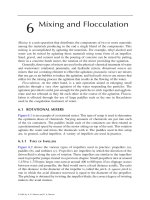
Figure 1.
PEGylation of graphene oxide. (a&b), photos of GO (a) and NGO-PEG (b) in different
solutions recorded after centrifugation at 10,000 g for 5 minutes. GO crashed out slightly in
PBS and completely in cell medium and serum (top panel). NGO-PEG was stable in all
solutions. (c&d) AFM images of GO (c) and NGO-PEG (d).
Liu et al. Page 4
J Am Chem Soc. Author manuscript; available in PMC 2009 August 20.
NIH-PA Author Manuscript NIH-PA Author Manuscript NIH-PA Author Manuscript
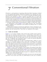
Figure 2.
SN38 loading on NGO-PEG. (a) schematic draw of SN38 loaded NGO-PEG. Inset: a photo of
NGO-PEG-SN38 water solution. (b) UV-VIS absorption spectra of NGO-PEG, NGO-PEG-
SN38, SN38 in methanol and difference spectrum of NGO-PEG and NGO-PEG-SN38. The
SN38 absorbance at 380 nm was used to determine the loading. (c) Fluorescence spectra of
SN38 and NGO-PEG-SN38 at [SN38]=1µM. Significant fluorescence quenching was
observed for SN38 adsorbed on NGO. (d) Retained SN38 on NGO-PEG over time incubated
in PBS and serum respectively. SN38 loaded on NGO-PEG was stable in PBS and released
slowly in serum. Error bars were based on triplet samples.
Liu et al. Page 5
J Am Chem Soc. Author manuscript; available in PMC 2009 August 20.
NIH-PA Author Manuscript NIH-PA Author Manuscript NIH-PA Author Manuscript
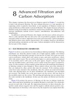
Figure 3.
In vitro cell toxicity assay. (a) Relative cell viability (versus untreated control) data of HCT-116
cells incubated with CPT-11, SN38 and NGO-PEG-SN38 at different concentrations for 72 h.
Free SN38 was dissolved in DMSO and diluted in PBS. Water soluble NGO-PEG-SN38
showed similar toxicity as SN38 in DMSO and far higher potency than CPT-11. (b) Relative
cell viability data of HCT-116 cells after incubation with NGO-PEG with (red) and without
(black) SN38 loading. Plain NGO-PEG exhibited no obvious toxicity even at very high
concentrations. Error bars were based on triplet samples.
Liu et al. Page 6
J Am Chem Soc. Author manuscript; available in PMC 2009 August 20.
NIH-PA Author Manuscript NIH-PA Author Manuscript NIH-PA Author Manuscript