Structural characterization and biochemical analysis of ID2, an inhibitor of DNA binding 9
Bạn đang xem bản rút gọn của tài liệu. Xem và tải ngay bản đầy đủ của tài liệu tại đây (5.08 MB, 43 trang )
!
64!
CHAPTER 5: RESULTS and DISCUSSION
(Biochemical Studies)
!
!
5.1 ID2 protein activity
To ensure that the purified ID2 protein was functionally active, competitive
electromobility shift assays (EMSAs) were performed to look for loss of binding of a
Group A bHLH transcription factor to its cy5-labeled DNA probe with an increasing
concentration of ID2.
E47, a Group A bHLH transcription factor and known binding partner of ID2 was
cloned, expressed and purified (Appendix 3) for use in these studies. E47 bound an
E-box element found in the DNA sequence of the MCK promoter containing the motif
(CANNTG). The sequence GGATCCCCCCAACACCTGCTGCCTGA was ordered as
cy5-labeled forward and reverse probes and annealed in a thermal cycler. Negative
controls were used to ensure that ID proteins did not bind DNA (Figure 22A) and a
mutant e-box element showed that E47 only bound to the wild-type e-box element
(Figure 22B).
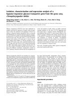
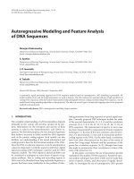
Figure 22: EMSA controls
(A) ID proteins did not bind e-box DNA, only E47, a bHLH transcription factor bound to DNA
(B) bHLH transcription factors (E47, Mash1, MYOD1) did not bind to mutant e-box DNA. The ID
proteins did not bind the mutant e-box probe.
!
!
65!
ID2 at different concentrations was incubated with E47 for 10 mins at room
temperature in EMSA binding buffer. Cy5-labeled DNA was added and allowed to
compete with ID2 for E47 binding for another 15 mins at room temperature to a final
reaction volume of 20 µl. The mixtures were loaded on a native gel and scanned for
cy5 intensity. Results showed that the ID2 N-HLH82-L protein used in the
crystallization experiments was successful in binding E47 and was therefore
functionally active (Figure 23).
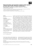
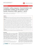
Figure 23: EMSA 6% native gel showing that increasing concentration of ID2 inhibited E47
binding to DNA. Lanes without ID2 (lanes 1 and 2) denoted by “-“. Number of “+” denoted
relative concentration of ID2 added. All lanes contained 2 μM E47. This showed that the purified
ID2 used for crystallization was active.
!
!
!
66!
5.2 ID heterodimer binding specificity and affinity
Although the ID2 and ID3 structures revealed a very similar fold to other classes
of bHLH-containing proteins such as E47, MYOD1, NeuroD1 and MAX, previous
studies showed that the ID proteins did not bind to any class of bHLH-containing
proteins. Instead, they preferentially bound to Group A bHLH-containing proteins.
Previous reports showed that ID1 and ID2 bound to E47 and MYOD1 (Group A).
Using EMSAs, it was found that ID1, ID2 and ID3 all bound to E47 (Figure 24). ID1
and ID2 but not ID3 bound weakly to MYOD1 (Figure 25). This confirmed the
reported observations by Langlands (Langlands, et al., 1997). The E47-MYOD1
heterodimer EMSA experiment (Figure 26) showed the same binding pattern as with
E47 and MYOD1 homodimers. This affirmed that IDs preferentially bound E47 and
likely inhibited MYOD1 indirectly by binding to MYOD1’s functional dimeric partner. It
was clear from the EMSA that MYOD1 preferentially bound E47 than itself and
confirmed MYOD1’s weak homodimerization capablities.
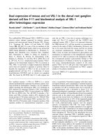
Figure 24: EMSA 6% native gel showing the different ID-HLH binding affinities to 0.05 µM human
E47. Residues for each human ID protein given in parentheses. “+” denoted presence of E47. All
lanes contained 200nM DNA. Concentrations of each ID protein provided in the table above the
gel. All ID proteins bound E47 to varying degrees.
!
!
!
!
67!
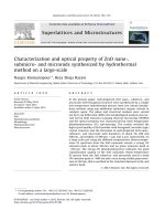
Figure 25: EMSA 6% native gel showing the different ID-HLH binding affinities to 0.2 µM human
MYOD1 (tagged with His-MBP). Residues for each human ID protein given in parentheses. “+”
denoted presence of MYOD1. All lanes contained 100nM DNA. Concentrations of each ID protein
provided in the table above the gel. ID1 and ID2 showed weak interactions with MYOD1 where a
large fraction seemed to form an intermediate rather than complete inhibition. ID3 did not bind
MYOD1.
!
!
!
!
!
Figure 26: EMSA 6% native gel showing the different ID-HLH binding affinities to 0.2 µM human
MYOD1 (tagged with His-MBP) heterodimerized with E47 (0.05µM). Residues for each human ID
protein given in parentheses. “+” denotes presence of MYOD1 and/or E47. All lanes contained
200nM DNA. Concentrations of each ID protein provided in the table above the gel. MYOD1 had
high propensity to bind E47. All IDs showed the same binding pattern as seen in Figures 24 and
25.
!
68!
5.3 ID helix-1 residues in binding specificity
Since IDs shared an average 80% identity across the HLH domain, differences
between ID2 and ID3 had to account for the differential binding to MYOD1.
Langlands had proposed that three residues in helix-1 were important for binding
specificity using a mammalian-2-hybrid system (Langlands, et al., 1997). In the ID2
structure, these three residues pointed away from the dimer interface (Figure 21).
The only residue that could potentially be affected was the lysine at position 47
because of its interaction with the positive ion in the loop. To find out if these
residues alone played a role in binding specificity, equivalent human positions from
the Langlands paper were mutated in ID2 as follows Y37D (Y in ID1), D41H (G in
ID1), K47R (K in ID1) as well as a double mutant Y37D-D41H. The expectation was
that binding to E47 would not change but binding to MYOD1 (Appendix 3) would be
affected. Using EMSAs and comparing binding of these mutants to E47 (Figure 27)
and MYOD1 (Figure 28), it was found that there were no significant differences and
they all bound like wild-type ID2. As with the previous EMSA for wild-type ID proteins,
the mutants showed similar binding patterns for the MYOD1-E47 heterodimer as with
MYOD1 and E47 homodimers (Figure 29). Hence, these residues were unlikely to
confer binding specificity of the different ID proteins as was hypothesized from the
ID2 and ID3 structures.
!
69!
Figure 27: EMSA 6% native gel showing ID2 helix-1 mutants binding affinities to 0.2 µM human
E47. “+” denotes presence of E47. All lanes contained 100nM DNA. Concentrations of each ID
protein provided in the table above the gel. All mutants bound to E47.
!
!
!
!
!
Figure 28: EMSA 6% native gel showing ID2 helix-1 mutants binding affinities to 0.2 µM human
MYOD1 (HisMBP tagged). “+” denotes presence of MYOD1. All lanes contained 100nM DNA.
Concentrations of each ID protein provided in the table above the gel. All ID2 helix-1 mutants
bound to MYOD1 weakly just like wild-type ID2.
!
!
70!
Figure 29: EMSA 6% native gel showing ID2 helix-1 mutants binding affinities to 0.2 µM human
MYOD1 (HisMBP tagged) heterodimerized with 0.2 µM E47. “+” denotes presence of MYOD1
and/or E47. All lanes contained 100nM DNA. Concentrations of each ID protein provided in the
table above the gel. IDs bound with similar affinities as with the E47 and MYOD1 homodimers.
!
71!
5.4 Exploring other differences in ID residues
Other residues where ID1 and 2 differed from ID3 were at ID2 positions Q55, N56,
K58, K61 and H67. Of these, Q55 and K61 had associated interactions in the
structure; Q55 to the ion and K61 as an intra-chain hydrogen bond to N40. The latter
bond was also found in the E47 homodimer at N353-K375 and was not lost in the
heterodimeric structure, E47-NeuroD1, at E47.N559-E47.K581. Mutations were
made to these residues to change their size as well as mirror the opposing ID protein.
ID2 mutations were Q55A, Q55R, K61A, K61Q and corresponding ID3 mutations
were R60A, R60Q, Q66A and Q66K. Of these mutations, ID2.K61A, ID3.Q66K and
ID3.Q66A did not produce soluble protein (Figure 17), suggesting a requirement for
this bond to form a stable monomer. ID2.K61Q had expression similar to wild type
and bound both E47 (Figure 30, top gel) and MYOD1 (Figure 30, bottom gel) that
was similar to wt-ID2.
Finally, a deletion at ID1.Q78 (ID2.Q55) caused partial binding loss to MYOD1
but not to E47 (Pesce, et al., 1993). It was found that ID2.Q55A and ID2.Q55R
showed partial binding loss to both MYOD1 (Figure 31, bottom gel) and E47 (Figure
31, top gel). We postulate that the alanine causes a loss of one of the interactions to
the positive ion and arginine would add a repelling force at that position. The reverse
mutation in ID3 was striking. ID3.R60A and ID3.R60Q had higher protein solubility
than wildtype, suggesting added stability. All previous experiments were done with
the solubility tag, His-MBP attached to ID3. However, these 2 mutants were soluble
without the tag and EMSAs were performed with these untagged ID3 mutants. It was
found that they bound better to E47 than wt-ID3, both with (Figure 32) and without
(Figure 33) the solubility tag. R60A made no difference to MYOD1 binding. But R60Q
showed partial binding to MYOD1.
!
72!
Pesce and Benezra had shown that the loop of ID1 was not as flexible and was
important for specificity (Pesce, et al., 1993). It is believed that this is also true for ID2.
These results show that an ionic interaction in the loop makes for a more rigid loop
that when disrupted, causes a conformational change that is not conducive to
heterodimerization. The hypothesis is that ID1 and 2 preferentially bind other bHLH-
containing proteins with a similar loop formation. Further, the arginine at position 60
in the loop of ID3 could have implications for its preference to different binding
partners than ID1 and ID2.
Figure 30: EMSA 6% native gels showing ID2 loop region mutants. wt = wild-type ID2, E47
concentration=100nM, DNA concentration=100nM, MYOD1 concentration=200nM.
Concentrations of ID2 are labeled. Top gel shows binding to E47, bottom gel to MYOD1. Apart
from the double mutant, the other ID2 mutants bound to E47 and MYOD1 as well as wild-type ID2.
!
73!
Figure 31: EMSA 6% native gel showing ID2 loop mutants. wt = wild-type ID2, E47
concentration=100nM, DNA concentration=100nM, MYOD1 concentration=200nM.
Concentrations of ID2 are labeled. Top gel shows binding to E47, bottom gel to MYOD1. Both
mutants showed partial binding loss compared to wild-type ID2.
!
74!
Figure 32: EMSA 6% native gels showing ID3 loop region mutants. wt = wild-type ID3 (His-MBP
tag), E47 concentration=100nM, DNA concentration=100nM, MYOD1 concentration=200nM.
Concentrations of ID3 are labeled. Top gel shows binding to E47, bottom gel to MYOD1. R60Q
and R60A were both tagged with His-MBP. R60Q appeared to bind better than wild-type ID3.
!
75!
Figure 33: EMSA 6% native gels showing ID3 loop region mutants. wt = wild-type ID3 (His-MBP
tag), E47 concentration=100nM, DNA concentration=100nM, MYOD1 concentration=200nM.
Concentrations of ID3 are labeled. Top gel shows binding to E47, bottom gel to MYOD1. R60Q
and R60A were both untagged. Tagged (Figure 32) or untagged, R60Q showed better binding
than wild-type ID3.
!
76!
5.5 MASH1 and the ID proteins
As mentioned in the introduction, MASH1 transcription factor played a role in
neurogenesis. It was hypothesized that IDs indirectly inhibited the function of MASH1
by sequestering E47, thus removing MASH1’s functional binding partner. However,
there had been conflicting reports on whether IDs bound MASH1 directly. An initial
report showed that all 4 ID proteins dimerized with E47 but not with human MASH1
(Jogi, et al., 2002). Later co-immunoprecipitation studies identified ID1 as a direct
binding partner of MASH1 (Obayashi, et al., 2009). As with the previous studies,
EMSAs were performed with MASH1 homodimer (Figure 34), MASH1-E47
heterodimer (Figure 34) and MASH1-MYOD1 heterodimer (Figure 35). None of them
showed ID1, ID2 or ID3 bound directly to MASH1. Based on all the EMSAs, it was
found that IDs bound strongly to E47, weakly to MYOD1 but not to MASH1.
Figure 34: EMSA 6% native gel showing ID proteins bound to MASH1 (left gel) and MASH1-E47
heterodimer (right gel). E47 concentration=50nM, MASH1 concentration = 0.5µM, DNA
concentration=100nM. Concentrations of ID2 are labeled on top of the gels. IDs did not bind
MASH1, only E47.
!
77!
Figure 35: EMSA 6% native gel showing ID proteins bound to MASH1-MYOD1 heterodimer.
MYOD1 concentration=0.2µM, MASH1 concentration = 0.5µM, DNA concentration=100nM.
Concentrations of ID2 are labeled on top of the gel. IDs bound weakly to MYOD1 but did not bind
to MASH1.
!
78!
CHAPTER 6: CONCLUSION and FUTURE DIRECTIONS
!
!
6.1 Conclusions
Constructs were created to enhance solubility and stability by choosing to omit
the C-terminal region of ID2 that contained the D-box motif that would signal the
protein for degradation via APC and Cdh1. However, even with the D-box region
removed and the addition of surrounding residues to stabilize the HLH domain, the
protein was insoluble without the help of a solubility tag like His6-GST. A cloning
artifact that ended up adding a short C-terminal polypeptide completely stabilized the
protein. The polypeptide was initially used only for the main HLH domain (residues
24-82). However, it was found that even adding the full N-terminus did not stabilize
ID2 so the polypeptide was added to terminate ID2 (residues 1-82) and it was then
found to be stable and soluble at room temperature.
With the short and long form of ID2, crystallization trials yielded crystals in
conditions of high salt. The crystals were optimized for growth and tested for
diffraction quality before being shipped to a synchrotron light source for data
collection. The short form of ID2 had very different looking crystals to the long form of
ID2 and ID2-Se-Met short form. It was noted that the long form and the Se-Met form
had similar looking crystals despite being grown in completely different mother
liquors.
The short form of ID2-Se-Met was solved as a SAD dataset using the Peak
energy. The structure had poor statistics in the highest resolution shells and was
used as a template for molecular replacement of the long form. The structure of the
long form of ID2 was solved at 2.1Å. The ID2 short form without the Se-Met could not
be solved.
Structural comparisons were made between the ID2 crystal structure and
published results about residues that were predicted to play an important role in
!
79!
dimerization. There were 4 main regions within the protein that were deemed crucial
for dimerization.
The first region was Helix-1. This region contained 3 residues that were
interchanged between ID1 and ID3. This residue exchange was proposed to confer
increased binding affinities of ID3 for MYOD1, which it did not otherwise bind
(Langlands, et al., 1997). The second region was a cysteine residue that was shown
to aid in homodimerization (Liu, et al., 2000). The third was the loop region where
mutations in certain residues abolished dimerization (Pesce, et al., 1993). And lastly,
a set of proposed bonds based on an ID3 homology model (Wibley, et al., 1996).
The helix-1 residues that were mentioned in the publication all pointed
outwards in the ID2 model. This was confirmed in the ID3 NMR model. The only
residue that could affect the dimer would be the lysine at position 47 in ID2, which
had a backbone oxygen that interacted with a loop ion believed to stabilize the
helices. To test these hypotheses, mutants at these positions were made in ID2 to
mirror both ID1 and ID3. As expected, none of the mutants changed the binding
affinities of ID2 to either E47 or MYOD1 in competitive EMSA studies. Even the
mutation at K47R did not change the binding much. So it was concluded that these
residues were not important for dimer formation.
The cysteine residue mentioned was proposed to create disulfide bonds to aid in
dimerization. But the ID2 model showed that this residue pointed away from the
dimer interface and also pointed outwards in the two monomers. The atomic distance
was too large for a disulfide bond. However, it could not be discounted that such a
bond could transiently bring two monomers in close proximity together to form the
homodimer.
The ID3 homolog model predicted 3 bond types, a hydrogen bond, a repelling
force and a salt bridge between 2 helices to form the homodimer. However, an
!
80!
inspection of both ID2 and ID3 showed that these bonds were unlikely due to the
sheer distances between the side chains.
Finally, the loop region, at the first turn after helix-1, contained density in Fo-
Fc maps that was too large to be water. A potassium ion was modeled in the density
based on the diameter of the density, the coordination of the surrounding residues
and the fact that the crystals were grown in a high concentration of potassium. The
ion was found in the loops of both monomers and was coordinated by four residues,
one from helix-1 and three from the loop with distances within 3Å. The closest
interaction was with I53 and the farthest was with Q55. A study of ID1’s equivalent
positions showed that mutations at these positions caused complete loss of
expression and a partial loss respectively (Pesce, et al., 1993). It is possible then that
ID1 shared this loop phenomenon especially since it was reported to have a rigid
loop. It is highly likely that the ion has a part to play in the loop’s rigidity; probably to
aid in a stable monomer by holding the 2 helices together. Looking at residues that
differed between ID2 and ID3 near the loop region, mutants were made to exchange
the ID2 for the ID3 residue and vice versa to see if there would be differences in
binding. At residue position 55 in ID2, mutation of Q to an R/A caused binding to be
slightly worse than wild-type. Conversely, mutation of the ID3 residue at the
equivalent position 61 from a K to and R or A conferred better binding than wild-type.
It was possible that since there was some interaction of this residue with the loop’s
ion, that the positively charged arginine repelled the positive ion enough to cause a
change that did not favour the ion’s placement to hold the loop rigidly in place. If
rigidity was required for sucessful heterodimer formation, it would explain the
differences in binding affinity.
Other interactions that are conserved between the ID2 crystal structure and
the ID3 NMR structure were the hydrogen bonds and the hydrophobic core. Wibley
!
81!
had noted that the hydrophobic core was all that was needed to hold the ID dimer
together but mutation studies to residues conferring interhelical hydrogen bonds such
as those of Y71 (chain A) and Q76 (chain B) completely abolished soluble protein
production and was required for proper folding of the monomer and hence, the
stability of the dimer.
In conclusion, the loop region was shown to be critical to dimer formation as well
as overall stability of the monomers. Apart from the loop, interhelical hydrogen bonds
also played a pivotal role in the protein’s stability.
!
6.2 Future Directions
A detailed study of the loop region of ID2 would be prudent. Loop swaps between
ID2 and the other IDs as well as with the binding partners to look for differences in
binding or perhaps even in vivo to allow transcription of a downstream gene.
Residue by residue mutations at each of the positions near the loop till the end of
the loop to study if the predicted residues affecting binding to the ion would either
abolish soluble protein production or binding to E47.
Only one hydrogen bond mutant was done so far. The others such as the Y43
that were conserved in ID as well as other bHLH-containing proteins should be tested.
Many proteins in the PDB are bound to divalent ions. Experiments such as using
crown ether to chelate the potassium ion, as well as EDTA and EGTA to chelate
divalent ions would be a way to check that the ion really was a potassium, especially
since the crystallization solution had such a high potassium salt content and could
have competed off the biological ion. In addition, the other ID proteins should also
have the same assay done to assess whether they also contained the loop ion and if
chelating the ion would result in misfolded protein.
!
82!
Crystal structures of the heterodimers of ID, such as ID2-E47 and/or ID3-E47
would provide a definitive answer as to the specific residues involved in these
interactions.
A high-resolution crystal structure of E47 or biochemical assays would be
worthwhile to test for the presence of a loop ion as predicted from the ID2-E47 model
comparisons. Mutations of residues predicted to hold the E47 loop in place could
also be tested.
Mutant forms of ID2 that caused misfolded proteins could be tested in vivo in cell
culture systems to look for phenotypic changes in cancer cells as a way to validate
the hypothesis that the ion interactions and the specific hydrogen bonds were
necessary for ID function.
!
83!
BIBLIOGRAPHY:
1. Adams, P. D., Grosse-Kunstleve, R. W., Hung, L. W., Ioerger, T. R., McCoy, A. J.,
Moriarty, N. W., Read, R. J., Sacchettini, J. C., Sauter, N. K. and Terwilliger, T. C.
(2002) "PHENIX: building new software for automated crystallographic structure
determination", Acta Crystallogr D Biol Crystallogr, 58, 1948-54
2. Alani, R. M., Hasskarl, J., Grace, M., Hernandez, M. C., Israel, M. A. and Munger,
K. (1999) "Immortalization of primary human keratinocytes by the helix-loop-helix
protein, Id-1", Proc Natl Acad Sci U S A, 96, 9637-41
3. Aronheim, A., Ohlsson, H., Park, C. W., Edlund, T. and Walker, M. D. (1991)
"Distribution and characterization of helix-loop-helix enhancer-binding proteins from
pancreatic beta cells and lymphocytes", Nucleic Acids Res, 19, 3893-9
4. Atchley, W. R. and Fitch, W. M. (1997) "A natural classification of the basic helix-
loop-helix class of transcription factors", Proceedings of the National Academy of
Sciences of the United States of America, 94, 5172-6
5. Ayer, D. E., Kretzner, L. and Eisenman, R. N. (1993) "Mad: a heterodimeric
partner for Max that antagonizes Myc transcriptional activity", Cell, 72, 211-22
6. Bain, G., Gruenwald, S. and Murre, C. (1993) "E2A and E2-2 are subunits of B-
cell-specific E2-box DNA-binding proteins", Mol Cell Biol, 13, 3522-9
7. Bain, G., Maandag, E. C., Izon, D. J., Amsen, D., Kruisbeek, A. M., Weintraub, B.
C., Krop, I., Schlissel, M. S., Feeney, A. J., van Roon, M. and et al. (1994) "E2A
proteins are required for proper B cell development and initiation of immunoglobulin
gene rearrangements", Cell, 79, 885-92
8. Benezra, R., Davis, R. L., Lassar, A., Tapscott, S., Thayer, M., Lockshon, D. and
Weintraub, H. (1990) "Id: a negative regulator of helix-loop-helix DNA binding
proteins. Control of terminal myogenic differentiation", Ann N Y Acad Sci, 599, 1-11
9. Benezra, R., Davis, R. L., Lockshon, D., Turner, D. L. and Weintraub, H. (1990)
"The protein Id: a negative regulator of helix-loop-helix DNA binding proteins", Cell,
61, 49-59
10. Berman, H. M., Westbrook, J., Feng, Z., Gilliland, G., Bhat, T. N., Weissig, H.,
Shindyalov, I. N. and Bourne, P. E. (2000) "The Protein Data Bank", Nucleic Acids
Res, 28, 235-42
!
84!
11. Beroukhim, R., Mermel, C. H., Porter, D., Wei, G., Raychaudhuri, S., Donovan, J.,
Barretina, J., Boehm, J. S., Dobson, J., Urashima, M., Mc Henry, K. T., Pinchback, R.
M., Ligon, A. H., Cho, Y. J., Haery, L., Greulich, H., Reich, M., Winckler, W.,
Lawrence, M. S., Weir, B. A., Tanaka, K. E., Chiang, D. Y., Bass, A. J., Loo, A.,
Hoffman, C., Prensner, J., Liefeld, T., Gao, Q., Yecies, D., Signoretti, S., Maher, E.,
Kaye, F. J., Sasaki, H., Tepper, J. E., Fletcher, J. A., Tabernero, J., Baselga, J., Tsao,
M. S., Demichelis, F., Rubin, M. A., Janne, P. A., Daly, M. J., Nucera, C., Levine, R.
L., Ebert, B. L., Gabriel, S., Rustgi, A. K., Antonescu, C. R., Ladanyi, M., Letai, A.,
Garraway, L. A., Loda, M., Beer, D. G., True, L. D., Okamoto, A., Pomeroy, S. L.,
Singer, S., Golub, T. R., Lander, E. S., Getz, G., Sellers, W. R. and Meyerson, M.
(2010) "The landscape of somatic copy-number alteration across human cancers",
Nature, 463, 899-905
12. Biggs, J., Murphy, E. V. and Israel, M. A. (1992) "A human Id-like helix-loop-helix
protein expressed during early development", Proc Natl Acad Sci U S A, 89, 1512-6
13. Blackwood, E. M. and Eisenman, R. N. (1991) "Max: a helix-loop-helix zipper
protein that forms a sequence-specific DNA-binding complex with Myc", Science, 251,
1211-7
14. Brunger, A. T., Adams, P. D., Clore, G. M., DeLano, W. L., Gros, P., Grosse-
Kunstleve, R. W., Jiang, J. S., Kuszewski, J., Nilges, M., Pannu, N. S., Read, R. J.,
Rice, L. M., Simonson, T. and Warren, G. L. (1998) "Crystallography & NMR system:
A new software suite for macromolecular structure determination", Acta Crystallogr D
Biol Crystallogr, 54, 905-21
15. Cai, L., Morrow, E. M. and Cepko, C. L. (2000) "Misexpression of basic helix-
loop-helix genes in the murine cerebral cortex affects cell fate choices and neuronal
survival", Development, 127, 3021-30
16. Campuzano, S. (2001) "Emc, a negative HLH regulator with multiple functions in
Drosophila development", Oncogene, 20, 8299-307
17. Card, P. B., Erbel, P. J. and Gardner, K. H. (2005) "Structural basis of ARNT
PAS-B dimerization: use of a common beta-sheet interface for hetero- and
homodimerization", J Mol Biol, 353, 664-77
18. Christy, B. A., Sanders, L. K., Lau, L. F., Copeland, N. G., Jenkins, N. A. and
Nathans, D. (1991) "An Id-related helix-loop-helix protein encoded by a growth factor-
inducible gene", Proc Natl Acad Sci U S A, 88, 1815-9
19. Church, G. M., Ephrussi, A., Gilbert, W. and Tonegawa, S. (1985) "Cell-type-
specific contacts to immunoglobulin enhancers in nuclei", Nature, 313, 798-801
!
85!
20. Colombo, N. and Cabrele, C. (2006) "Synthesis and conformational analysis of
Id2 protein fragments: impact of chain length and point mutations on the structural
HLH motif", J Pept Sci, 12, 550-8
21. Cooper, C. L., Brady, G., Bilia, F., Iscove, N. N. and Quesenberry, P. J. (1997)
"Expression of the Id family helix-loop-helix regulators during growth and
development in the hematopoietic system", Blood, 89, 3155-65
22. Crews, S. T. (1998) "Control of cell lineage-specific development and
transcription by bHLH-PAS proteins", Genes & development, 12, 607-20
23. Davis, R. L., Weintraub, H. and Lassar, A. B. (1987) "Expression of a single
transfected cDNA converts fibroblasts to myoblasts", Cell, 51, 987-1000
24. Davis, R. L., Weintraub, H. and Lassar, A. B. (1987) "Expression of a Single
transfected cDNA converts fibroblasts to myoblasts.", Cell, 51, 987-1000
25. Deed, R. W., Bianchi, S. M., Atherton, G. T., Johnston, D., Santibanez-Koref, M.,
Murphy, J. J. and Norton, J. D. (1993) "An immediate early human gene encodes an
Id-like helix-loop-helix protein and is regulated by protein kinase C activation in
diverse cell types", Oncogene, 8, 599-607
26. Deed, R. W., Hirose, T., Mitchell, E. L., Santibanez-Koref, M. F. and Norton, J. D.
(1994) "Structural organisation and chromosomal mapping of the human Id-3 gene",
Gene, 151, 309-14
27. DeLano, W. L. (2002) "The PyMOL Molecular Graphics System", DeLano
Scientific, San Carlos, CA, USA.
28. Depinho, R. A., Hatton, K., Ferrier, P., Zimmerman, K., Legouy, E., Tesfaye, A.,
Collum, R., Yancopoulos, G., Nisen, P. and Alt, F. (1986) "Myc family genes: a
dispersed multi-gene family", Annals of clinical research, 18, 284-9
29. Desprez, P. Y., Lin, C. Q., Thomasset, N., Sympson, C. J., Bissell, M. J. and
Campisi, J. (1998) "A novel pathway for mammary epithelial cell invasion induced by
the helix-loop-helix protein Id-1", Mol Cell Biol, 18, 4577-88
30. Di Tommaso, P., Moretti, S., Xenarios, I., Orobitg, M., Montanyola, A., Chang, J.
M., Taly, J. F. and Notredame, C. (2011) "T-Coffee: a web server for the multiple
sequence alignment of protein and RNA sequences using structural information and
homology extension", Nucleic Acids Res, 39, W13-7
!
86!
31. Eletsky, A., Wang, D., Kohan, E., Janjua, H., Acton, T. B., Xiao, R., Everett, J. K.,
Montelione, G. T. and Szyperski, T. (2011) "Solution NMR Structure of the Helix-loop-
Helix Domain of Human ID3 Protein, Northeast Structural Genomics Consortium
Target HR3111A", Protein Data Bank,
32. Ellenberger, T., Fass, D., Arnaud, M. and Harrison, S. C. (1994) "Crystal
structure of transcription factor E47: E-box recognition by a basic region helix-loop-
helix dimer", Genes Dev, 8, 970-80
33. Emsley, P. and Cowtan, K. (2004) "Coot: model-building tools for molecular
graphics", Acta Crystallogr D Biol Crystallogr, 60, 2126-32
34. Ephrussi, A., Church, G. M., Tonegawa, S. and Gilbert, W. (1985) "B lineage
specific interactions of an immunoglobulin enhancer with cellular factors in vivo",
Science, 227, 134-40
35. Evans, P. (2006) "Scaling and assessment of data quality", Acta Crystallogr D
Biol Crystallogr, 62, 72-82
36. Finn, R. D., Mistry, J., Tate, J., Coggill, P., Heger, A., Pollington, J. E., Gavin, O.
L., Gunasekaran, P., Ceric, G., Forslund, K., Holm, L., Sonnhammer, E. L., Eddy, S.
R. and Bateman, A. (2010) "The Pfam protein families database", Nucleic Acids Res,
38, D211-22
37. Flicek, P., Amode, M. R., Barrell, D., Beal, K., Brent, S., Carvalho-Silva, D.,
Clapham, P., Coates, G., Fairley, S., Fitzgerald, S., Gil, L., Gordon, L., Hendrix, M.,
Hourlier, T., Johnson, N., Kahari, A. K., Keefe, D., Keenan, S., Kinsella, R.,
Komorowska, M., Koscielny, G., Kulesha, E., Larsson, P., Longden, I., McLaren, W.,
Muffato, M., Overduin, B., Pignatelli, M., Pritchard, B., Riat, H. S., Ritchie, G. R.,
Ruffier, M., Schuster, M., Sobral, D., Tang, Y. A., Taylor, K., Trevanion, S.,
Vandrovcova, J., White, S., Wilson, M., Wilder, S. P., Aken, B. L., Birney, E.,
Cunningham, F., Dunham, I., Durbin, R., Fernandez-Suarez, X. M., Harrow, J.,
Herrero, J., Hubbard, T. J., Parker, A., Proctor, G., Spudich, G., Vogel, J., Yates, A.,
Zadissa, A. and Searle, S. M. (2012) "Ensembl 2012", Nucleic Acids Res, 40, D84-90
38. Fong, S., Debs, R. J. and Desprez, P. Y. (2004) "Id genes and proteins as
promising targets in cancer therapy", Trends Mol Med, 10, 387-92
39. Gabellini, C., Del Bufalo, D. and Zupi, G. (2006) "Involvement of RB gene family
in tumor angiogenesis", Oncogene, 25, 5326-32
40. Gray, M. J., Dallas, N. A., Van Buren, G., Xia, L., Yang, A. D., Somcio, R. J.,
Gaur, P., Mangala, L. S., Vivas-Mejia, P. E., Fan, F., Sanguino, A. M., Gallick, G. E.,
Lopez-Berestein, G., Sood, A. K. and Ellis, L. M. (2008) "Therapeutic targeting of Id2
!
87!
reduces growth of human colorectal carcinoma in the murine liver", Oncogene, 27,
7192-200
41. Guillemot, F. and Joyner, A. L. (1993) "Dynamic expression of the murine
Achaete-Scute homologue Mash-1 in the developing nervous system", Mech Dev, 42,
171-85
42. Guo, G., Huss, M., Tong, G. Q., Wang, C., Li Sun, L., Clarke, N. D. and Robson,
P. (2010) "Resolution of cell fate decisions revealed by single-cell gene expression
analysis from zygote to blastocyst", Dev Cell, 18, 675-85
43. Hara, E., Hall, M. and Peters, G. (1997) "Cdk2-dependent phosphorylation of Id2
modulates activity of E2A-related transcription factors", EMBO J, 16, 332-42
44. Hara, E., Uzman, J. A., Dimri, G. P., Nehlin, J. O., Testori, A. and Campisi, J.
(1996) "The helix-loop-helix protein Id-1 and a retinoblastoma protein binding mutant
of SV40 T antigen synergize to reactivate DNA synthesis in senescent human
fibroblasts", Dev Genet, 18, 161-72
45. Hara, E., Yamaguchi, T., Nojima, H., Ide, T., Campisi, J., Okayama, H. and Oda,
K. (1994) "Id-related genes encoding helix-loop-helix proteins are required for G1
progression and are repressed in senescent human fibroblasts", J Biol Chem, 269,
2139-45
46. Henthorn, P., Kiledjian, M. and Kadesch, T. (1990) "Two distinct transcription
factors that bind the immunoglobulin enhancer microE5/kappa 2 motif", Science, 247,
467-70
47. Hu, Y. C., Lam, K. Y., Law, S., Wong, J. and Srivastava, G. (2001) "Identification
of differentially expressed genes in esophageal squamous cell carcinoma (ESCC) by
cDNA expression array: overexpression of Fra-1, Neogenin, Id-1, and CDC25B
genes in ESCC", Clin Cancer Res, 7, 2213-21
48. Iavarone, A., Garg, P., Lasorella, A., Hsu, J. and Israel, M. A. (1994) "The helix-
loop-helix protein Id-2 enhances cell proliferation and binds to the retinoblastoma
protein", Genes Dev, 8, 1270-84
49. Israel, M. A., Hernandez, M. C., Florio, M., Andres-Barquin, P. J., Mantani, A.,
Carter, J. H. and Julin, C. M. (1999) "Id gene expression as a key mediator of tumor
cell biology", Cancer Res, 59, 1726s-1730s
50. Jacobs, Y., Vierra, C. and Nelson, C. (1993) "E2A expression, nuclear
localization, and in vivo formation of DNA- and non-DNA-binding species during B-
cell development", Mol Cell Biol, 13, 7321-33
!
88!
51. Jen, Y., Manova, K. and Benezra, R. (1996) "Expression patterns of Id1, Id2, and
Id3 are highly related but distinct from that of Id4 during mouse embryogenesis", Dev
Dyn, 207, 235-52
52. Jen, Y., Manova, K. and Benezra, R. (1997) "Each member of the Id gene family
exhibits a unique expression pattern in mouse gastrulation and neurogenesis", Dev
Dyn, 208, 92-106
53. Jen, Y., Weintraub, H. and Benezra, R. (1992) "Overexpression of Id protein
inhibits the muscle differentiation program: in vivo association of Id with E2A
proteins", Genes Dev, 6, 1466-79
54. Jogi, A., Persson, P., Grynfeld, A., Pahlman, S. and Axelson, H. (2002)
"Modulation of basic helix-loop-helix transcription complex formation by Id proteins
during neuronal differentiation", J Biol Chem, 277, 9118-26
55. Kajimoto, Y., Kawamori, R., Umayahara, Y., Watada, H., Iwama, N., Morishima,
T., Yamasaki, Y. and Kamada, T. (1994) "Identification of amino-acid polymorphism
within the leucine zipper motif of mouse transcription factor A1", Gene, 139, 247-9
56. Kebebew, E., Treseler, P. A., Duh, Q. Y. and Clark, O. H. (2000) "The helix-loop-
helix transcription factor, Id-1, is overexpressed in medullary thyroid cancer", Surgery,
128, 952-7
57. Kee, B. L. (2009) "E and ID proteins branch out", Nat Rev Immunol, 9, 175-84
58. Kim, D., Peng, X. C. and Sun, X. H. (1999) "Massive apoptosis of thymocytes in
T-cell-deficient Id1 transgenic mice", Mol Cell Biol, 19, 8240-53
59. Klambt, C., Knust, E., Tietze, K. and Campos-Ortega, J. A. (1989) "Closely
related transcripts encoded by the neurogenic gene complex enhancer of split of
Drosophila melanogaster", The EMBO journal, 8, 203-10
60. Kurooka, H. and Yokota, Y. (2005) "Nucleo-cytoplasmic shuttling of Id2, a
negative regulator of basic helix-loop-helix transcription factors", J Biol Chem, 280,
4313-20
61. Landschulz, W. H., Johnson, P. F. and McKnight, S. L. (1988) "The leucine
zipper: a hypothetical structure common to a new class of DNA binding proteins",
Science, 240, 1759-64