Endofin is a novel component in EGR EGFR oncogenic signaling
Bạn đang xem bản rút gọn của tài liệu. Xem và tải ngay bản đầy đủ của tài liệu tại đây (3.4 MB, 135 trang )
ENDOFIN IS A NOVEL COMPONENT IN EGF/EGFR
ONCOGENIC SIGNALING
TOY WEI YI
B.SC (LIFE SCIENCES) (HONS)
NUS
A THESIS SUBMITTED FOR THE DEGREE OF
DOCTOR OF PHILOSOPHY
DEPARTMENT OF PATHOLOGY
NATIONAL UNIVERSITY OF SINGAPORE
2010
ACKNOWLEDGEMENTS
I would like to express my gratitude and sincere thanks to my supervisor, Dr Lim
Yoon Pin for giving me the opportunity to pursue my PhD studies in his laboratory
and his kind patience and guidance throughout these years.
My thanks also extend to my co-supervisor, Associate Prof. Richie Soong and my
thesis advisory committee members, Associate Prof. Low Boon Chuan and Associate
Prof. Shen Han-ming for their suggestions and advice.
My heartfelt thanks to Dr Lim Shen Kiat and Dr Shirly Chong for their ready help and
invaluable advice whenever needed and Ms Lee Huiyin for all the help she has given
me throughout the years. I would also like to thank Dr Lim Shen Kiat and Emma May
Stanford for taking their precious time to proof-read this thesis.
I would like to thank all the past and present members of YPL lab for the wonderful
working experience, especially Dr Bobby, Dr Yang Yixuan, Dr Man Xiaohui, Ms
Choong Lee Yee, Mr Victor Tan, and Ms Emily Chen whom all have contributed to
the success of this thesis in one way or another. It has been a real pleasure working
with all of you throughout these years.
My deepest gratitude to my dearest friends, Ms Chang Jaw-shin, Ms Lim Simin and
Ms Peh Bee Keow for always being there to listen to my problems and give
encouragement. Thanks for being such wonderful friends and I’ll always treasure our
friendships.
Lastly, I would like to express my most sincere thanks to my family, especially my
parents for their constant moral support, my sister for her listening ears and my little
brother for his IT expertise and help in drawing the diagrams. Without them, I would
never been able to finish this.
Thank You.
Toy Weiyi
January 2010
I
TABLE OF CONTENTS
ACKNOWLEDGEMENTS I
TABLE OF CONTENTS II
LIST OF FIGURES VI
LIST OF TABLES VII
ABBREVIATIONS VIII
SUMMARY XII
Chapter 1 Introduction
1.1 Cellular communication and receptor tyrosine kinase
1
1.2 Receptor tyrosine kinase
2
1.3 Epidermal growth factor receptor
3
1.3.1 EGFR activation 6
1.3.2
EGFR signaling pathways 10
1.3.2.1 Ras/Raf/MEK/ERK pathway 10
1.3.2.2 PI3K/PDK1/Akt pathway 13
1.3.2.3 PLC-γ/DAG/IP
3
pathway 16
1.3.2.4 JAK/STAT pathway 18
1.3.2.5
Src kinases 19
1.4
EGFR endocytosis
20
1.4.1 Trafficking of EGFR in the absence of ligands 21
1.4.2 Ligand-induced endocytosis 21
II
1.4.2.1 Clathrin-independent endocytosis 22
1.4.2.2 Clathrin-dependent endocytosis 23
1.5 Post-endocytic trafficking of EGFR
27
1.5.1 Trafficking of EGFR through the endosomes 27
1.5.2 EGFR sorting at MVBs 28
1.6 Receptor recycling
32
1.7 Endosomal signaling
32
1.7.1 Continuous EGFR signaling from endosomes 33
1.7.2 Endosomal-specific signaling 35
1.8 FYVE domain-containing proteins
36
1.8.1 The FYVE zinc-finger domain 36
1.8.2 FYVE domain proteins and membrane trafficking 37
1.8.3 FYVE domain proteins and signal transduction 40
1.8.4 FYVE domain proteins that have enzymatic activity 41
1.8.5 FYVE domain proteins and cytoskeleton regulation 42
1.9
Endofin
43
1.10 Research objectives
47
Chapter 2 Materials and Methods
2.1 Chemicals and reagents
48
2.2 Antibodies
48
2.3 Plasmid constructs
49
2.4 Site-directed Mutagenesis
50
III
2.5 Cell culture
50
2.6 Transfection
51
2.7 Cell lysis
51
2.8 Subcellular fractionation
52
2.9 Immunoprecipitation
52
2.10 Immunoblotting
53
2.11 In vitro kinase assay
54
2.12 Immunofluorescence
54
2.13 Proliferation assay
56
Chapter 3 Results
3.1 Characterization of Endofin phosphorylation
57
3.1.1 Tyrosine phosphorylation of Endofin occurs upon TGF-α
stimulation and is dependent on EGFR activation
57
3.1.2 TGF-β does not induce tyrosine phosphorylation of
Endofin
60
3.1.3 Endofin phosphorylation correlates closely to EGFR
activity
61
3.1.4 Endofin is phosphorylated in the cytosol and clathrin-
dependent endocytosis is essential for Endofin
phosphorylation
64
3.1.5 EGF-induced PI3K activity and proper localization of
Endofin are necessary for its tyrosine phosphorylation
69
3.1.6 EGF-dependent co-localization of Endofin with EGFR
requires a functional FYVE domain
74
3.2 Determination of Endofin tyrosine phosphorylation site and
function
78
3.2.1 Tyrosine 515 is a major phosphorylation site of Endofin
78
IV
3.2.2 Phosphorylation at Y515 does not affect the localization of
Endofin and its co-localization with EGFR
84
3.2.3 Endofin’s localization and phosphorylation increased the
amplitude of EGF-induced MAPK pathway
87
3.2.4 Proliferation rates of Y515F and C753S expressing cells
were elevated
94
Chapter 4 Discussion
4.1 Endofin’s regulatory actions on EGFR signaling
96
4.2 Possible mechanisms utilized by Endofin in the regulation of
MAPK signaling
99
4.3 Future areas of research
101
References
103
Publications
122
V
LIST OF FIGURES
1.3 Schematic representation of EGFR structural domains
6
1.3.1 Schematic representation of phosphorylated tyrosine residues on
EGFR and its binding substrates
9
1.3.2.1 Ras/Raf/MEK/ERK pathway
12
1.3.2.2 PI3K/PDK1/Akt
15
1.3.2.3 PLC-γ/DAG/IP3 pathway
17
1.5 Schematic diagram of receptor endocytosis
31
1.9 Schematic representation of the various domains and interacting
partners of Endofin
46
3.1.1 Tyrosine phosphorylation of Endofin occurs upon TGF-α
stimulation and is dependent on EGFR activation
59
3.1.2 Endofin is not phosphorylated upon TGF-β stimulation
60
3.1.3 Endofin phosphorylation is dependent on EGFR activity
63
3.1.4A Phosphorylated Endofin was detected in non-nuclear intracellular
compartment
66
3.1.4B-C Endofin phosphorylation is dependent on EGFR endocytosis
67
3.1.4D-E Endofin phosphorylation requires EGFR clathrin-dependent
endocytosis
68
3.1.5A Endofin phosphorylation is dependent on PI3K activity
71
3.1.5B-C C753S Endofin mutant is unable to localize at the early endosomes
72
3.1.5D Proper localization of Endofin is essential for its phosphorylation
73
3.1.6A EGFR and Endofin co-localize at EEA1-positive endosomes
76
3.1.6B Co-localization of Endofin with EGFR requires its FYVE domain
77
3.2.1A-B Y515 of Endofin is a major phosphorylation site for EGFR
82
VI
3.2.1C-D Endofin is a direct substrate of EGFR
83
3.2.2A-B Endosomal localization of Endofin is not dependent on its Y515
phosphorylation
85
3.2.2C Co-localization of Endofin with EGFR does not require Y515
phosphorylation
86
3.2.3A Endofin expression and transfection efficiency in various cell lines
88
3.2.3B Endofin overexpression has no effect on EGFR modulation or
signaling
88
3.2.3C-D Mislocalization of Endofin amplified EGF-induced MAPK
signaling
90
3.2.3E-G Y515F mutant increased MAPK2 activity in Endofin knockdown
cells
93
3.2.4 Y515F and C753S expressing cells have increased proliferation
rates
95
4.1 Schematic diagram of Endofin’s role in EGFR signaling
98
LIST OF TABLES
3.2.1 Predicted tyrosine phosphorylation sites on Endofin
79
VII
ABBREVIATIONS
°C
degree Celsius
3'UTR
3' untranslated region
AA
arachidonic acid
Akt
AKR mouse T-cell lymphoma-derived oncogenic product
AP2
adaptor protein complex 2
APPL
adaptor protein, phosphotyrosine interaction, PH domain and leucine
zipper-containing ½
ARF1
ADP-ribosylation factor 1
ATP
adenosine triphosphate
bFGF
basic fibroblast growth factor
BMP
bone morphogenetic protein
BSA
bovine serum albumin
C
cysteine
Ca
2+
calcium
c-Cbl
c-Casitas B-lineage lymphoma
CCP
clathrin-coated pits
CDE
clathrin-dependent endocytosis
CIE
clathrin-independent endocytosis
CO
2
carbon dioxide
Cox II
cytochrome-c oxidase subunit II
CR
cysteine-rich domain
C-terminal
carboxyl (COOH)-terminal
DAG
1,2-diacylglycerol
DH
Dbl homology
DN
dominant-negative
DNA
deoxyribonucleic acid
DTT
dithiothreitol
E. coli
Escherichia coli
ECL
enhanced chemiluminescence
EDTA
ethylene-diamine tetra-acetic acid
EEA1
early endosomal antigen 1
EGF
epidermal growth factor
EGFR
epidermal growth factor receptor
EHD
epsin-homology domain
ERC
endocytic recycling compartment
ERK
extracellular signal-regulated kinase
ESCRT
endosomal sorting complex required for transport complexes
F
phenylalanine
FAB
F-actin filament-binding domain
FBS
fetal bovine serum
FYVE
Fab1p, YOTB, Vac1p EEA1
GDP
guanosine diphosphate
VIII
GEF
guanine-nucleotide exchange factor
GFP
green fluorescent protein
GM-CSF
granulocyte-macrophage colony-stimulating factor
GPCR
G-protein coupled receptors
Grb2
growth factor receptor-bound protein 2
GTP
guanosine triphosphate
GTPase
guanosine triphosphatase
HA
haemagglutinin
HGF
hepatocyte growth factor
HRP
horseradish peroxidase
Hrs
hepatocyte growth factor-regulated tyrosine kinase substrate
IGF-1
insulin growth factor 1
ILV
intraluminal vesicles
IP
immunoprecipitation
IP
3
inositol 1,3,5-trisphosphate
JAK
Janus kinase
JM
juxtamembrane region
JNK
c-Jun N-terminal kinase
kDa
kilo Dalton
LB
Luria Bertani
LBPA
lysobisphosphatic acid
L
leucine
MAPK
mitogen-activated protein kinase
MEK
mitogen activated extracellular signal regulated kinase
MEM
modified eagles medium
mg
milligram
μg
microgram
MgCl
2
magnesium chloride
mL
millilitre
μl
microlitre
mM
millimolar
μM
micromolar
MTM
myotubularin
MTS
3-(4,5-dimethylthiazol-2-yl)-5-(3-carboxymethoxyphenyl)-2-(4-
sulfophenyl)-2H-tetrazolium, inner salt
MVB
multivesicular bodies
Na
3
VO
4
sodium orthovanadate
NaCL
sodium chloride
NaF
sodium fluoride
ng
nanogram
NID
non-ionic denaturing
N-terminal
amino (NH
2
)-terminal
PA
phosphatidic acid
PBD
protein phosphatase catalytic subunit (PP1c) binding domain
IX
PBS
phosphate buffered saline
PBST
phosphate buffered saline with Tween 20
PDGF
platelet-derived growth factor
PDK-1
phosphoinositide-dependent kinase-1
PH
Pleckstrin homology
PI3,5P
2
phosphatidylinositol-3,5-bisphosphate
PI3K
phosphatidylinositol 3-kinase
PI3P
phosphatidylinositol 3-phosphate
PIP
2
phosphatidylinositol-4,5-bisphosphate
PIP
3
phosphatidylinositol-3,4,5-trisphosphate
PKC
protein kinase C
PLCγ
phospholipase Cγ
PNS
post nuclear supernatant
PTB
phosphotyrosine binding
PVDF
polyvinylidene difluoride
pY
phosphotyrosine
PY20H
phosphotyrosine antibody conjugated to horseradish peroxidase
Rab11
Ras-associated protein 11
Rab4
Ras-associated protein 4
Rab5
Ras-associated protein 5
Rab7
Ras-associated protein 7
Raf
Rapidly growing fibrosarcoma
rpm
revolutions per minute
RPMI
Roswell Park Memorial Institute
RTK
receptor tyrosine kinase
S
serine
SARA
Smad anchor for receptor activation
SBD
Smad binding domain
SDS-PAGE
sodium dodecyl sulphate-polyacrylamide gel electrophoresis
SH2
Src-homology 2
SH3
Src-homology 3
Shc
SH2 domain containing transforming protein C1
SOS
Son of Sevenless
STAM 1/2
signal transduction adaptor molecule 1/2
STAT
signal transducers and activators of transcription
T
threonine
TEMED
N,N,N',N'-tetramethyl-ethylene-diamine
TGF-α
transforming growth factor-α
TGF-β1
transforming growth factor β1
TSG101
tumor susceptibility gene 101
UIM
ubiquitin interacting motif
V
voltage
VHS
VPS-27, Hrs and STAM domain
WT
wild type
X
Y
tyrosine
XI
SUMMARY
Endofin is an endosomal protein that localizes to the early endosomes. It is
characterized by a zinc-finger domain, referred to as the FYVE domain. This domain
targets Endofin to the early endosomes by binding to the phosphatidylinositol 3-
phosphate within the endosomal membrane. Endofin functions as a regulator of
specific signaling pathways, such as BMP and TGF-β signaling, whereby it plays the
role of an adaptor. Furthermore, it has been identified as a novel tyrosine
phosphorylation target downstream of EGFR.
To date, there have only been a few functional studies published on Endofin
and consequently our understanding of Endofin’s functions is very limited, especially
with respect to EGFR signaling. In this study, an attempt was made to map the
signaling events associated with Endofin following activation of EGFR with EGF.
Tyrosine phosphorylation of Endofin was shown to be dependent on clathrin-
dependent endocytosis of EGFR and EGFR activity. Phosphatidylinositol 3-kinase
activity and FYVE domain-mediated localization of Endofin to early endosomes were
found to be necessary for the tyrosine phosphorylation of Endofin. Tyrosine 515 was
identified as a major phosphorylation site on Endofin however disruption of
phosphorylation at Y515 neither affects Endofin’s localization nor its co-localization
with EGFR at the endosomes. Instead, the abrogation of Y515 phosphorylation and
the mislocalization of Endofin were found to enhance the amplitude of the MAPK
cascade and increase cell proliferation, suggesting a possible role of Endofin in the
modulation of MAPK pathway. Collectively, this study has identified a novel
signaling cascade involving EGFR, PI3K, Endofin and MAPK in the EGFR signaling
network.
XII
Chapter 1
Introduction
1.1 Cellular communication
Cellular communication is a core aspect of a functional biological system.
From multicellular to unicellular organisms, communication between organs, tissues
and even single cells plays a decisive role in development and survival. This
communication not only determines vital cellular processes such as proliferation,
differentiation and apoptosis, it also allows cells to react appropriately to the ever-
changing surrounding environment (Pawson, 1995).
Cellular communication comprises of a complex system of signaling
networks, with each signaling pathway described as a “signal transduction” (King,
2010). A signal transduction begins with the extracellular signals, usually in the form
of endocrine, paracrine hormones, or signaling molecules, binding to specific proteins
on the cell membrane. The binding of these specific membrane proteins activates
signaling cascades that transverse across the cytosol and into the nucleus, ultimately
leading to changes in gene expression which will determine the correct biological
response. In addition, these signaling networks are controlled by finely-tuned positive
and negative feedback cascades and the balance between them determines the final
outcome (Freeman, 2000). Conversely when the balance between the two networks is
disrupted, it often results in uncontrollable proliferation or untimely cell death which
manifests in the form of diseases, such as cancer (Hanahan, 2000). Hence, it is crucial
for the cells to interpret these cellular signals with the utmost accuracy in order for
them to give the correct response. This ability of the cells to perceive and respond
correctly to the microenvironment therefore forms the basis of development, tissue
repair, immunity and normal tissue homeostasis. In view of the importance of cellular
1
signaling in cell biology, tremendous effort has been invested in trying to comprehend
the signaling networks involved, in both the normal and diseased state.
1.2 Receptor tyrosine kinase
Receptor tyrosine kinases (RTKs) are a class of transmembrane proteins that
play a pivotal role in cellular communication. They act as primary mediators in the
interpretation of extracellular mitogen activity and have a high-affinity for many
growth factors which allows them to regulate and coordinate cellular processes (Fantl
et al, 1993; Schlessinger and Ullrich, 1992). Binding of these external signals/ligands
to the RTKs at the plasma membrane activates the tyrosine kinases of the receptors
which in turn initiates signaling cascades within the cells. The signal is eventually
conveyed into the nucleus, leading to the transcription of specific genes. In this way,
RTKs integrate these external signals with the various internal signal transduction
pathways and activate gene transcription within the cells, allowing the cell to respond
to the extracellular stimuli (Kholodenko, 2006).
Currently, there are 58 receptor tyrosine kinase proteins identified out of the
90 unique tyrosine kinase genes present in the human genome (Robinson et al., 2000)
and they can be classified into 20 subfamilies based on their structural characteristics
(Grassot et al., 2003; Lemmon and Schlessinger, 2010). All RTKs have a general
structure consisting of an extracellular N-terminal region, a hydrophobic
transmembrane domain (25-38 amino acids) and an intracellular C-terminal region
(Grassot et al., 2006). The extracellular N-terminal region of the RTKs is made up of
various conserved elements such as the immunoglobulin (Ig)-like or epidermal growth
factor (EGF)-like domains, fibronectin type III repeats or cysteine-rich regions which
are characteristic of each RTK subfamily (Hubbard and Till, 2000). In addition, the
2
ligand-binding site which binds to the receptor’s specific ligand is also situated at the
extracellular N-terminal region. The C-terminal region displays the highest level of
conservation with the tyrosine kinase catalytic domain, which is responsible for
receptor autophosphorylation and the phosphorylation of RTK substrates (Yarden and
Ullrich, 1988).
1.3 Epidermal growth factor receptor
Epidermal growth factor receptor (EGFR), a typical RTK, lies at the head of a
complex signal transduction network that modulates numerous cellular processes.
EGFR was first discovered by Stanley Cohen in 1980 as a cell surface receptor for the
epidermal growth factor (EGF) he extracted from salivary gland extracts in 1962
(Cohen, 1962; Cohen et al., 1980). Since then, there have been extensive studies done
on EGFR, including the characterization of the protein and its functional roles in the
regulation of important cellular processes such as cell growth and differentiation
(Yarden and Sliwkowski, 2001). EGFR (also known as ErbB1) belongs to the ErbB
family of receptors and is classified under subclass I of the superfamily of RTKs.
Besides EGFR, there are 3 other ErbB family members, namely ErbB2, ErbB3 and
ErbB4, each playing different roles in development and differentiation (Britsch,
2007).
The importance of EGFR in regulating mammalian development has been
asserted through the use of genetically modified transgenic mice in which the
expression of both the receptor and its ligands has been manipulated. Depending on
the genetic background of the mice, EGFR knockout can be either embryonic or
perinatal lethal and generally carry abnormalities in multiple organs, including lung,
skin, brain, kidney, liver, central nervous system, placenta and gastrointestinal tract
3
(Miettinen et al., 1995; Sibilia and Wagner, 1995; Threadgill et al., 1995). EGFR
deficient mice have been shown to develop progressive neurodegeneration in the
frontal cortex, olfactory bulb and thalamus postnatal in a strain dependent manner
(Kornblum et al., 1998; Sibilia et al., 1998). In addition, mice with an EGFR kinase
domain mutation or expressing a dominant-negative EGFR exhibit impaired ductal
growth, indicating that EGFR is essential for promoting ductal growth in the
mammary glands (Fowler et al., 1995; Xie et al., 1997). Contrary to the lethal effects
of EGFR knockouts, mice that lack EGFR ligands display less severe phenotypes,
demonstrating the redundancy built into EGFR signaling. For instance, TGF-α null
mice are viable, fertile and display eye and hair follicles abnormalities (Luetteke et
al., 1993; Mann et al., 1993). Female mice with triple deficiency of EGF,
amphiregulin and TGF-α show reduced mammary gland ductal outgrowth during
puberty and display difficulties in nursing their young, indicative of possible
collaborative roles of the three ligands in mammopoiesis and lactogenesis (Luetteke et
al., 1999). In gastrointestinal development, mice lacking all three EGFR ligands
(EGF, amphiregulin and TGF-α) present with spontaneous duodenal lesions and
decreased cell proliferation in crypts leading to truncated ileal villi (Troyer et al.,
2001).
The wide variation of phenotypes arising from EGFR null mice demonstrate
the extensive role that EGFR plays in tissue development and maintenance. While
EGFR is one of the main regulators of cellular processes such as cell proliferation,
differentiation, and migration, aberrant signaling activity of this receptor has been
shown to play a key role in the development and growth of tumor cells. Numerous
studies on the involvement of EGFR in various human cancers, including breast,
gastric, ovarian, non-small cell lung cancer, head and neck cancer, prostate and many
4
others have been reported (Salomon et al., 1995). Dysregulation of EGFR as detected
in human cancers usually occur by several mechanisms, such as EGFR
overexpression, activating mutations, defective or limited receptor downregulation,
activation of the autocrine growth factor loop and deficiency of specific phosphatases
that deactivate EGFR (Bhargava et al., 2005; Lee et al., 2005; Zandi et al., 2007). In
addition, mutations of the EGFR gene that lead to the production of different receptor
forms have also been reported in certain cancers. One of the most common forms of
mutation is the mutation that results in the expression of a truncated EGFR in
glioblastoma, termed EGFRvIII (Collins, 1994). This truncated EGFR lacks
information from exons 2-7, thereby resulting in the constitutive ligand-independent
activation of the receptor tyrosine kinase (Chu et al., 1997).
The EGFR protein is made up of 1186 amino acids after cleavage at the N-
terminal of its 1210 amino acids polypeptide precursor. 20% of the receptor is N-
linked glycosylated and this is required for the translocation of the receptor to the cell
membrane and its ligand binding ability (Slieker et al., 1986). The structural features
of EGFR consist of a glycosylated, extracellular N-terminal region (621 amino acids),
a transmembrane segment (amino acids 622-644), followed by an intracellular
domain that contains a juxtamembrane region, a tyrosine kinase and a C-terminal
region which consists of several phosphorylation sites (Normanno et al., 2005)(Fig.
1.3). The extracellular N-terminal sequence of EGFR, also known as ectodomain,
contains 4 domains, L1, CR1, L2 and CR2. The ligand-binding pocket is located
between L1 and L2 domains and a dimerization arm is present between them. The
juxtamembrane region of EGFR has been shown to possess a few regulatory
functions. For instance, the dileucine motif, L679/680, in the juxtamembrane region
was shown to be involved in facilitating the transport of EGF-EGFR complexes into
5
the internal vesicles of multivesicular bodies (Kil and Carlin, 2000). Several proteins
were also found to bind to the juxtamembrane region of EGFR, namely eps8 and
calmodulin and these interactions prevented the phosphorylation of Thre654 by
protein kinase C (Castagnino et al., 1995; Li and Villalobo, 2002; Martin-Nieto and
Villalobo, 1998). The C-terminal region of EGFR contains several tyrosine residues,
which has been shown to modulate EGFR signaling upon phosphorylation and
serine/threonine residues whose phosphorylation are important for receptor down-
regulation and sequences required for endocytosis.
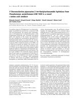
L1 CR1 L2 CR2 JM kinase CT
151 312 481 621 644 687 955 1186
Ligand binding domain
Fig. 1.3 Schematic representation of EGFR structural domains
The ligand binding pocket is located between L1 and L2 domain while the
transmembrane region is between CR2 and juxtamembrane region. The abbreviations
used: L and CR are for ligand binding and cysteine-rich domains, JM and CT refer to
juxtamembrane region and carboxyl-terminal respectively.
1.3.1 EGFR activation
To activate and initiate the downstream signaling cascades, EGFR has to first
bind to its ligand. Ligands that bind to EGFR are known as EGF-related peptide
growth factors (Yarden, 2001; Yarden and Sliwkowski, 2001). Their binding
specificities are conferred by an EGF-like domain (~ 60 amino acids) which is made
up of three disulfide-bonded intramolecular loops. In addition to the EGF-like
domain, they also possess other structural motifs, such as heparin-binding sites,
glycosylation sites and immunoglobulin-like domains. These growth factors are
6
synthesized as active transmembrane precursors, which can interact with the receptors
on neighboring cells, and are released by proteolysis as soluble growth factors. The
growth factors that bind specifically to EGFR include EGF, amphiregulin (AR) and
transforming growth factor-α (TGF-α). Betacellulin (BTC), heparin-binding EGF
(HB-EGF) and epiregulin (EPR) are able to bind to both EGFR and ErbB4, due to
their dual-specificity property (Jones et al., 1999).
Ligand binding results in the exposure of a dimerization arm that induces the
receptors to either homo- or heterodimerize with itself or other ErbB receptors. The
formation of various EGFR dimers is dependent on the binding ligand and the ErbB
receptors present on the cell surface. EGFR homodimers are better characterized
comparing to its heterodimers. The most common EGFR heterodimer formed is the
EGFR-ErbB2 heterodimer as ErbB2 is the preferred dimerization partner of all ErbB
receptors (Graus-Porta et al., 1997). Receptor dimerization not only amplifies signal
strength but it also leads to signal diversification as different receptor dimers can
result in different signaling outputs. For instance, EGF-activated EGFR-ErbB2
heterodimer was shown to be endocytosis-deficient in comparison to EGFR
homodimers (Haslekas et al., 2005; Wang et al., 1999). In addition, EGF activated
EGFR, but not EGFR-ErbB4 heterodimer, was able to recruit Grb2 (Olayioye et al.,
1998). Hence, the diversification of signals arises from the unique properties acquired
by the heterodimer as a whole and is not due to the signaling properties of the
individual receptors in the dimer.
EGFR dimerization induces the activation of its tyrosine kinase and
autophosphorylation of the receptors whereby several tyrosine residues in the C-
terminal region are phosphorylated. These phosphorylated residues in turn recruit a
group of protein substrates which bind to the phosphorylated residues mainly via their
7
PTB (phosphotyrosine binding) or SH2 (Src-homology 2) domains. The substrates
that bind to the phosphorylated tyrosine residues include adaptors like, Grb2, Shc and
Dok-R that bind to pY1068, pY1086 and pY1148, pY1173 and pY1086, pY1148
respectively, phospholipase PLC-γ which binds to pY1173 and pY992, phosphatase
like PTB-1B and SHP-1 are recruited by pY992, pY1148 and pY1173 respectively,
ubiquitin ligase c-Cbl that interacts with pY1045, transcription factor Stat5 binds to
pY978, pY998 and tyrosine kinase Abl binds to pY1086 (Jorissen et al., 2003;
Schulze et al., 2005)(Fig. 1.3.1). The presence of multiple binding sites of a substrate
on EGFR indicates that the differential substrate binding pattern, which depends on
the ligand, dimerization partner, substrate availability and competing substrates, can
occur and this contributes to the diverse signal outputs. Besides autophosphorylation
of residues, the C-terminal region of EGFR can also be phosphorylated by other
intracellular tyrosine kinases, such as the Src kinase and Jak2 kinase (Lombardo et al.,
1995; Yamauchi et al., 1997). Jak2 was proposed to phosphorylate Tyr1068 on EGFR
upon growth hormone stimulation, suggesting a potential cross-talk signaling pathway
between the cytokine and growth factor receptor. Reports have suggested that Tyr845,
Tyr891, Tyr920, Tyr1101 were phosphorylated by Src and src-mediated
phosphorylation of EGFR was shown to be involved in the regulation of the growth
factor receptor function and signal transduction (Biscardi et al., 1999; Stover et al.,
1995). The interaction of EGFR with its substrates greatly enhances its substrate
phosphorylation efficiency and assists in the formation of highly organized
multicomponent signaling complexes, therefore propagating the signal further
throughout the cell.
8
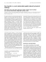
Fig. 1.3.1 Schematic representation of phosphorylated tyrosine residues on
EGFR and its binding substrates
Several substrates have more than one phosphotyrosine binding sites on the
intracellular portion of EGFR. This suggests that differential binding patterns of the
substrates can result in diverse signaling outputs from the receptor.
9
1.3.2 EGFR signaling pathways
Based on the diversity of protein complexes formed at EGFR, multiple
signaling pathways can be elicited from the activated EGFR. The major signaling
pathways activated by EGFR are the Ras/Raf/MEK/ERK, PI3K/PDK1/Akt, PLC-
γ/DAG/IP
3
and JAK/STAT pathways. All of these signaling pathways contribute to
the regulation of cellular processes such as cell proliferation, survival, adhesion and
migration (Yarden and Sliwkowski, 2001).
1.3.2.1 Ras/Raf/MEK/ERK pathway
The Ras/Raf/MEK/ERK pathway is one of the best characterized signaling
pathways emanating from EGFR (Fig. 1.3.2.1). This pathway begins with the adaptor
protein, Grb2, which binds constitutively to the proline-rich sequences of a guanine
nucleotide exchange factor (GEF), Son of Sevenless (SOS), in the cytosol under
normal circumstances, via its SH3 domain. However upon EGFR activation, Grb2 is
recruited to the C-terminus of EGFR where it can either bind directly to pY1068 and
pY1086 or indirectly, through EGFR bound tyrosine phosphorylated Shc, via its SH2
domain (Sasaoka et al., 1994). The recruitment of Grb2/SOS complex to the receptor
at the plasma membrane allows SOS to interact with the membrane-associated Ras, a
small guanosine triphosphatase (GTPase), thereby resulting in the exchange of the
Ras-bound GDP for GTP and the activation of Ras. The activated Ras in turn recruits
and activates one of its effector proteins, Raf, a serine/threonine kinase by displacing
it from an adaptor protein, namely 14-3-3 (Hallberg et al., 1994). The activation of the
Raf kinase triggers the activation/phosphorylation of a series of serine/threonine
kinases consecutively, namely the mitogen activated extracellular signal regulated
kinase (MEK) and the extracellular signal regulated kinase (ERK). Subsequently, the
10
activated ERK phosphorylates a wide range of substrates either in the cytosol or in the
nucleus. ERK nuclear substrates include transcription factors like Elk1, c-fos, c-myc,
c-jun, which are key regulators of proliferation, apoptosis, and differentiation
(Krishna and Narang, 2008; Yoon and Seger, 2006). On top of this, the activation of
ERK also acts as a negative feedback loop for this pathway. Reports have shown that
the phosphorylation of SOS by ERK caused the dissociation of SOS from Grb2 and
ERK phosphorylation of Raf inhibited Raf/Ras interaction, both resulting in the
attenuation of the Ras/Raf/MEK/ERK signaling pathway (Dougherty et al., 2005;
Langlois et al., 1995).
11
Fig. 1.3.2.1 Ras/Raf/MEK/ERK pathway
This pathway begins with Grb2, in association with SOS, that binds to activated
EGFR either directly or via Shc. SOS then activates Ras, promoting the exchange of
its GDP for GTP and this in turn leads to the consecutive activation of a series of
kinases, namely, Raf, MEK and ERK. The activated ERK translocates into the
nucleus and activates the transcription of genes through the phosphorylation of
transcription factors such as c-fos, c-myc and c-jun.
12