Purified herba leonuri and leonurine protect middle cerebral artery occluded rats from brain injury through antioxidative mechanism and mitochondrial protection 1
Bạn đang xem bản rút gọn của tài liệu. Xem và tải ngay bản đầy đủ của tài liệu tại đây (785.64 KB, 48 trang )
Chapter 2: Introduction
Department of Pharmacology,
YLL School of Medicine
11
Chapter 2
Introduction: Ischemic Stroke, CNS
mitochondria and Therapeutic Potential
of Traditional Chinese Medicine
Chapter 2: Introduction
Department of Pharmacology,
YLL School of Medicine
12
2.1 Pathophysiology of stroke
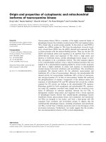
Acute stroke can be divided into 2 categories: hemorrhagic stroke and ischemic stroke.
Hemorrhagic stroke (Figure 2-1a) describes sudden rupture of the blood vessels within
the brain that causes the leakage of the blood into brain cavity, and therefore brain
damage results (Zemke et al, 2004). For ischemic stroke (Figure 2-1b), which accounts
for 80% of all stroke cases, the brain damage is caused by a reduction or complete
blockage of blood flow, resulting in the deficiency of glucose and oxygen supply to the
territory of the affected region (Zemke et al, 2004). Neurons are the most vulnerable cells
to hypoxia due to their strong dependence on the oxidative metabolism of glucose for
energy. Since ischemia is the major problem among all the stroke patients, many
researches have been targeting on the treatment of ischemia. This project focused also
ischemic stroke as a model to test on the therapeutic potential of studied drugs (see
below).
Figure 2-1: a) Ischemic stroke; and b) hemorrhagic stroke (arrow)
a) b)
Chapter 2: Introduction
Department of Pharmacology,
YLL School of Medicine
13
2.1.1 Ischemic stroke
As mentioned, ischemic stroke results from a transient or permanent reduction in cerebral
blood flow (CBF) by an embolus or a thrombus, leading to brain injury. A critical
reduction of CBF causes failure of cellular transport mechanisms and massive release of
potentially toxic neurotransmitters, subsequently the formation of free radicals,
inflammation, induction of immediate early genes and later, cell death by necrosis or
apoptosis (Barber, 2008). Therefore, the extent of brain injury is dependent on level of
CBF reduction, duration of ischemic insult, tissue temperature, blood glucose
concentration and many other physiological variables.
The two principle models of human stroke are global ischemia and focal ischemia, either
permanent or transient. Global ischemia occurs when CBF is reduced throughout most or
all parts of the brain as a result of cardiac arrest or other causes of collapse of system
circulation, and subsequently failure of brain perfusion. The tissue injury is dominated by
neurons, occurring especially in the most vulnerable region (CA
1
region of hippocampus)
of the brain first and then proceeding to the less vulnerable region such as thalamus or
caudate putamen (Miller, 1999; Canese et al, 1997). Neuronal death in global ischemia is
always detected in the hippocampus, striatum, neocortex with most susceptible
population lies within CA1 and CA4 area of hippocampus and layer 2 and 5 of the
cerebral cortex (Taoufik and Probert, 2008).
Chapter 2: Introduction
Department of Pharmacology,
YLL School of Medicine
14
As for focal ischemia, it is represented by a reduction of blood flow to a specific brain
region, such as the occlusion of middle cerebral artery. Although occlusion of vessel
occurs during focal ischemia, there is rarely complete blockade of CBF to the area
supplied by the occluded vessel because plethora of collateral vessels provides some flow
to the area (Horst and Korf, 1997). During focal ischemia, there will be a core infarct area
which does not receive sufficient perfusion to sustain any of the neurons, glia, or even the
vasculature. The volume of core infarct is correlated to the severity of neurological deficit.
On multi-tracer O Positron emission tomography (PET), ischemic core exhibits very low
CBF, cerebral blood volume (CBV), and metabolic rates of oxygen and glucose (Marchal
et al, 1999). In this region, a CBF of <10ml/100g of brain tissue per minute severely
impair the cellular function by depleting the energy metabolites and causing the failure of
the cell membrane to maintain ion homeostasis. This manifests as massive efflux of
potassium and reciprocal influx of sodium, calcium and water. In addition, the anaerobic
respiration is initiated due to the impairment of mitochondrial oxidative phosphorylation.
Anaerobic respiration leads to the production lactic acid which could cause acidosis
toxicity to the cell. The cells in this area are destined to have irreversible damage and
undergo necrosis, which means out of therapeutic rescue (Marchal et al, 1999).
In addition, within the necrotic core, the vasculature may also be severely damaged,
exposing the risk of undergoing haemorrhagic transformation. This is especially
important for the case of extensive infarction and with the use of thrombolytics for the
stroke treatment, as it might worsen the clinical condition. Therefore, reduction the risk
of haemorrhagic transformation is one of the therapeutic goals as for instance, new
Chapter 2: Introduction
Department of Pharmacology,
YLL School of Medicine
15
thrombolytic agents that do not interfere with endothelial function or induce matrix
metalloproteinase dysregulation (Moustafa and Barron, 2008).
Surrounding this core will be hypoperfused tissue, namely ischemic penumbra that
receives collateral flow that sufficient to prevent cells from undergoing necrosis. At
reduced CBF of about 20ml/100g of brain tissue per minute, the cerebral metabolic rate
of oxygen (CMRO
2
) starts to fall, thereby impairing the normal blood flow
autoregulatory mechanisms, and the neuronal electroencephalographic activity ceases.
Therefore, this threshold represents the threshold for loss of neuronal electrical function
(Wise et al, 1983). Cells in this region are functionally silent but remain metabolically
active and maintain a very low level of adenosine triphosphate (ATP). Tissue within this
area is potentially salvageable. Reports (Touzani et al, 1997; Heiss et al, 1998) showed
that large volumes of tissue with penumbral level of CBF escape necrosis if arterial
recanalization is achieved in time. However, with prolonged ischemic insult, the cells in
penumbra is continuously bombarded by waste products from the dead cells in ischemic
core, the cell recovery will therefore decrease over time and cells in this area undergo
delayed (hours or days) cell death (Zemke et al, 2004).
Furthermore, as if ischemia persists, large slow voltage shifts occur at the borders of the
core infarct and propagate as spreading depolarization waves that compromise the
survival of surrounding tissue (Selman et al, 2004), together with spreading of the
inflammation and excitotoxicity in the ischemic core, tissue in ischemic penumbra will
gradually transform into the core. The course of events varies from patients to patients,
Chapter 2: Introduction
Department of Pharmacology,
YLL School of Medicine
16
most exhibit substantial volumes of penumbra for many hours, with exceptionally days
after stroke onset (Moustafa and Baron, 2008). Using multi-tracer PET, substantial
volumes of cortical penumbra have been reported to decline over time, that being present
in over 50% of the patients studied within 9 hours after stroke onset, and in about one-
third of the patients studied between 5 to 18 hours (Mousfara and Baron, 2008). Thus, it
emphasizes the urgency of acute stroke management. The reduction or prevention of the
cell death in the ischemic penumbra so to prevent the growth of the ischemic core lesion
within the temporal therapeutic window is the main target of pharmacological
intervention studies.
2.1.2 Cell death in stroke
2.1.2.1 Ischemic cascade
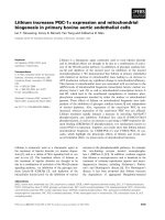
At cellular level, ischemic cascade is resulted from a severe prolonged ischemic insult
(Figure 2-1). It begins with progressive derangements in energy and substrate metabolism
(Horst and Korf, 1997). Energy deficiency leads to interruption of ATP dependent
process, such as sodium/potassium ATPase (Na
+
/K
+
ATPase) which can subsequently
causes the disruption of ion homeostasis as most of the ATP generated from
mitochondrial oxidative phosphorylation is used for the stabilization of transmembrane
ion concentration gradients of sodium, potassium and calcium which are important for
neuronal impulse conduction and synaptic function. Therefore, onset of ischemia results
in the movement of ions down their electrochemical gradients such that intracellular
Chapter 2: Introduction
Department of Pharmacology,
YLL School of Medicine
17
calcium and sodium concentration and extracellular potassium concentration increase
dramatically within one or two minutes after ischemia (Horst and Korf, 1997).
Increase in extracellular potassium triggers depolarization and reversal of direction of
action of the amino acid (such as glutamate) transporters. Under these conditions, Ca
2+
enters the cells via voltage-dependent channel and a massive release of excitatory amino
acid such as glutamate out of the cells, resulting in excitotoxicity. This initiates a positive
feedback loop where excessive glutamate activates AMPA, kainate and N-methyl-D-
aspartate (NMDA) receptor to consume more ATP and promote further release of
glutamate. Ionotropic NMDA receptor potentiates the efflux of K
+
and influx of Na
+
together with water, leading to the cell edema. Together with the Ca
2+
entry via voltage
dependent channel, ionotropic NMDA receptor also promotes excessive Ca
2+
influx,
leading to intracellular Ca
2+
overload. A range of downstream nuclear and cytoplasmic
lethal metabolic derangement will be resulted by Ca
2+
overload. These include the
activation of phospholipases and proteases that could degrade membrane and proteins
that are essential for cellular integrity. Ca
2+
overloaded mitochondria will be severely
impaired and hence the inhibition of ATP production (Nakka et al, 2008). Augmented
intracellular Ca
2+
further promotes the release of glutamate and thus propagates the
excitotoxicity. Increase in Ca
2+
level causes also the increase in free radicals production
which will be discussed later.
As mentioned, oxygen deficiency during ischemia results in anaerobic respiration due to
the inability of mitochondria to perform oxidative phosphorylation. However, energy
Chapter 2: Introduction
Department of Pharmacology,
YLL School of Medicine
18
obtained from anaerobic respiration is not enough to compensate the energy needed for
neurons since brain has limited amount of glycogen stores. In addition, anaerobic
respiration leads to accumulation of lactic acid which causes a local rise of lactate
production and a fall in pH, leading to intra- and extra-cellular acidosis, reflecting a
marked imbalance between energy use and production (Barber, 2008). Low oxygen level
will also cause free radical generation by incomplete oxidative phosphorylation. Free
radicals are known to react with and damage whole range of organelles and plasma
membrane (Zemke et al, 2004). In conclusion, mechanisms that contribute to the
neuronal cell death predominantly occur via 3 major mediators: unregulated intracellular
increase of Ca
2
, tissue acidosis, nitric oxide (NO·) and free-radical production (Barber,
2008).
Figure 2-2: A diagram illustrates the ischemic cascade (Adapted from Crack and Taylor,
2005)
Chapter 2: Introduction
Department of Pharmacology,
YLL School of Medicine
19
2.1.2.2 Apoptosis
In the past, neuronal cell death after cerebral ischemia was considered to be exclusively
necrotic. Research over the past decade has revealed that a portion of cells in ischemic
penumbra or periinfarct zone undergo programmed cell death (PCD), namely apoptosis,
via caspase dependent or caspase independent pathways, after stroke. Thus they are
potentially recoverable after the onset of stroke via pharmacological intervention of PCD
(Brad et al, 2009).
Necrosis is commonly resulted from the accumulation of deleterious changes that disrupt
vital cell viability. Necrosis is irreversible massive cell death characterized by shrunken
cells with darkened nuclei, swelling of cytoplasms and organelles and loss of membrane
integrity, resulting in cell lysis and release of the cellular content that in turn lead to local
inflammation to surrounding tissue (Taoufik and Probert, 2008). In contrast to necrosis,
apoptosis is orderly process of energy dependent programmed cell death characterized by
morphological features as cell shrinkage, membrane blebbing, chromatin condensation,
and DNA fragmentation (Nakka et al, 2008). Cell undergoing apoptosis will be
recognized and removed in an organized way (phagocytosis) to avoid inflammation and
minimize the damage and disruption of neighboring cells (Taylor et al, 2008). A more
unique morphological characteristic of neuron undergoing apoptotsis is the neurite
fragment (dendrites and axons) that occurs early during the cell death process (Taoufik
and Probert, 2008). Mixed morphologies of apoptosis and necrosis observed during
Chapter 2: Introduction
Department of Pharmacology,
YLL School of Medicine
20
ischemic insult could be result from the initiation of apoptosis that are then overtaken by
the molecular event associated with necrosis (Roy and Sapolosky, 1999).
Evidences of involvement of apoptosis in stroke comes from a small number of studies
showing that neuronal apoptosis is involved in human stroke (Guglielmo et al, 1998;
Love S et al, 1998), as well as a large body of support from animal studies where
apoptotic markers are co-localized at the ischemic affected regions (Taoufik and Probert,
2008). Apoptosis was showed to contribute to ischemic damage by TUNEL staining,
which could detect the DNA fragmentation of cell death. Through TUNEL staining,
apoptosis was found scattered throughout the ischemic territory with more apparent at the
perifocal tissue (Sims and Anderson, 2002).
Generally, apoptosis can be executed via two pathways: Extrinsic pathway and intrinsic
pathway (Figure 2-3). Extrinsic pathway initiates apoptosis through the engagement of
plasma membrane death receptors, therefore also referred as “death receptor pathway”
(Ashe and Berry, 2003). Death receptors belong to the tumor necrosis factor receptor
(TNFR) family. They transmit the apoptotic signal through binding of death ligand. Fas is
one of the best characterized family members. Its preferred ligand is (Fas ligand) FasL
(Ashe and Berry, 2003). There were reports on Fas/FasL system that it is also involved in
neuronal apoptosis following traumatic brain injury and cerebral ischemia (Beer et al,
2000; Martin-Villalba et al, 1999; Rosenbaum et al, 2000). Trimerization of Fas followed
by ligation of FasL promotes the recruitment of the cytosolic adaptor protein Fas-
associated death domain protein (FADD) through complementary death domain (DD).
Chapter 2: Introduction
Department of Pharmacology,
YLL School of Medicine
21
FADD contains also death effector domain (DED) which is responsible to bind with
complementary DED in procaspase 8 and 10. This complex (FasL, Fas, FADD,
procaspase 8 or 10) is referred as death-inducing signaling complex (DISC). DISC close
positions the DED containing initiator caspase (procaspase 8) and therefore cause the
activation of initiator caspase by their autolytic cleavage (Figure 2-3). Activation of these
initiator caspases results in the execution of the apoptotic programme by cleavage of
downstream targets (Ashe and Berry, 2003).
Generally, there are two types of Fas-mediated apoptosis. Type 1 requires the activation
of caspase 8 that is closely followed by the activation of caspase 3. Apoptosis in Type 1
cells cannot be rescued by inhibitor of Bcl-2 family which plays a central role in intrinsic
pathway of apoptosis. Type II has limited activation of caspase 8. Caspase 8 in type II
cells involves cleavage of the BH3-only protein, Bcl-2 interacting domain (BID), to
release truncated BID (tBID). BID is a proapoptotic cytosolic member of Bcl-2 family
that translocates to mitochondria when cell receives death signal (Sugawara et al, 2004)
which is crucial for the release of cytochrome c and Smac/DIABLO from mitochondria.
Activation of Bid by caspase 8 results in an amplification loop by means of extrinsic
apoptotic pathway recruits an intrinsic apoptotic pathway (Ashe and Berry, 2003).
Regardless the type, caspase 8 is the apical caspase in DR signaling and its activity is
detected after permanent middle cerebral artery occlusion (MCAO) (Taoufik and Probert,
2008).
Chapter 2: Introduction
Department of Pharmacology,
YLL School of Medicine
22
The onset of stroke causes the cytotoxic intracellular accumulation of Ca
2+
which triggers
the activation of intrinsic pathway of apoptosis (Dirnagl et al, 1999). Activation of
calpain by increased Ca
2+
or stimulation of caspase-8 via extrinsic pathway results in the
activation of BID to its truncated active form tBID (Brad et al, 2009). Recent studies
have shown the involvement of BID in cerebral ischemia that Plesnila and colleagues
(2002) found that deletion of BID gene in mice reduced ischemic infarct size. tBID
causes the conformational changes of other proapoptotic proteins situated on
mitochondria, such as BAX and Bcl-xS so to execute the apoptotic signaling (Figure 2-3).
These proapoptotic proteins can also heteromerize with antiapoptotic members of bcl-2
family situated on outer mitochondrial membrane, such as Bcl-xL, Bcl-2, so to counteract
their antiapoptotic function (Saito et al, 2003). In addition, studies showed that BAX can
form channel across the mitochondrial membrane that are large enough to allow the
passage of cytochrome c (Kirkland et al, 2002).
After the disruption of mitochondria or the opening of mitochondrial permeability
transition pore (MPTP), mitochondrial proapoptotic protein such as cytochrome c,
Smac/DIABLO, serine protease HtrA2/Omi will be released into the cytoplasm. Once
released, these proteins will be involved in caspase-dependent apoptotic pathway.
Cytochrome c, a water soluble mitochondrial protein that is an essential component of
mitochondrial respiratory chain, forms apoptosome by binding to Apaf-1, ATP and pro-
caspase 9. Caspase 9 will then be activated and subsequently activate caspase 3 as a
executor of apoptosis (Brad et al, 2009). Caspase 3 has been documented to be involved
in cerebral ischemia (Asahi et al, 1997) and it cleaves many substrates such as poly
Chapter 2: Introduction
Department of Pharmacology,
YLL School of Medicine
23
(ADP-ribose) polymerase (PARP). For Smac/DIABLO, it lifts the inhibition of caspase 9
and caspase 3 via neutralizing the caspase-inhibitory properties of the IAP (inhibitor of
apoptosis) family of proteins, particularly XIAP thereby allowing apoptosis to occur
(Christophe and Nicholas, 2006).
Both intrinsic and extrinsic pathways of apoptosis lead to activation of caspase 3.
Caspase 3 is the executioner caspase in the cascade. Caspase 3 activation was observed in
neurons 24 hours after MCAO. Administration of caspase 3 inhibitor reduces the infarct
size after focal ischemia. Caspase 3 inhibition also protected mice from transient MCAO
(Taoufik and Probert, 2008).
Increasing evidences showed the significance of caspase independent apoptotic pathway
is involved in ischemic stroke (Elmore, 2007). A group of proteins will be released out
from the mitochondria during apoptosis such as apoptosis inducing factor (AIF),
endonuclease G and Bcl-2/adenovirus E1B 19kDa-interacting protein (BNIP3). Studies
have demonstrated that the involvement of AIF and endonuclease G in cerebral ischemia
that both AIF and endonuclease G translocate from mitochondria to nucleus after cerebral
ischemia (Culmsee et al, 2005; Lee et al, 2005). In particular, AIF causes large scale of
DNA fragmentation and peripheral condensation of peripheral nuclear chromatin, which
is distinct from the global chromatin condensation and oligonucleosomal DNA
fragmentation of caspase-dependent death (Cho and Toledo, 2008).
Chapter 2: Introduction
Department of Pharmacology,
YLL School of Medicine
24
With the sudden increase of intracellular Ca
2+
after excitotoxicty insult, calpains,
cytoplasmic calcium sensitive cysteine proteases have been implicated in the
pathogenesis of ischemic stroke (Lau and Tymianski, 2010). Previous report showed a
modest neuroprotection in hippocampal cell cultures from NMDA insults by calpain
inhibitors Faddis et al, 1997). Calpain proteolytic activity is necessary for the cleavage
and release of AIF from mitochondria (Polster et al, 2005). Neuronal cultures subjected
to oxygen-glucose deprivation and calpain inhibitor treatment was shown to be prevented
from undergoing neuronal death due to the inhibition of AIF translocation into nucleus
(Cao et al, 2007)
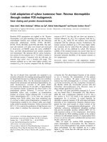
Figure 2-3: A schematic diagram of apoptosis. There are considerable cross talks between
intrinsic and extrinsic pathway of apoptosis which could ultimately lead to cell death.
(Adapted from Nakka et al, 2008)
Chapter 2: Introduction
Department of Pharmacology,
YLL School of Medicine
25
In addition to the necrosis and apoptosis, it was suggested that a third type of cell death,
autophagy might be involved in the stroke pathology. Autophagy is a fusion process
which enables cells to dispose cytoplasms or organelles by fusion of vesicles containing
these cellular compartments with lysosomes (Taoufik and Probert, 2008). However, a
more detailed understanding is needed for this type of cell death.
2.1.3 Oxidative stress of stroke
Under physiological condition, reactive oxygen species (ROS) including superoxide
anion (O
2
˙¯), and nitric oxide (NO˙) is produced at low level and plays a role in cellular
signaling such as regulation of blood flow and neurotransmission. Intracellular sources of
ROS include xanthine oxidase, mitochondrial electron transport chain, arachidonic acid
and NADPH oxidase (Brad et al, 2009). Most importantly, ROS production is controlled
by endogenous antioxidants for instances superoxide dismutase (SOD) to dismutate O
2
˙¯,
glutathione peroxidase and catalase to detoxify H
2
O
2
.
Increased levels of ROS are the major cause of tissue injury after cerebral ischemia
(Figure 2-4), in which there are overproduction of ROS, inactivation of antioxidant
enzymes, consumption of antioxidants such that endogenous antioxidant defense
mechanisms are failed to protect neurons from oxidative damage (Brad et al, 2009).
Oxidative stress is the state of imbalance between the two opposing antagonistic forces,
ROS and antioxidant, in which the effects of former predominate over the compensating
action of latter (Fernández-Checa et al, 1997).
Chapter 2: Introduction
Department of Pharmacology,
YLL School of Medicine
26
NO˙ and O
2
˙¯ are two major free radicals responsible in oxidative stress. These two free
radicals react with each other to produce powerful oxidant peroxynitrite (ONOO¯). Other
ROS includes hydrogen peroxide (H
2
O
2
) and hydroxyl radical (OH˙). As reported by Zhu
et al, (2004), there are multiple possible mechanisms of free radical production. In
addition to the basal level of O
2
˙¯ generation by mitochondria, disruption of the
mitochondria electron transport chain can result in autoxidation of flavoprotein and
ubisemiquinone to form O
2
˙¯. Ischemia induced excessive release of glutamate results in
increased intracellular Ca
2+
which will in turn activates Ca
2+
dependent nitric oxide
synthase (NOS) and NO˙ production. Metabolism of phospholipase A
2
and subsequent
release of arachidonic acid, prostaglandins, leukotrienes, thromboxanes, and platelet
activating factor will be activated during ischemic cascade and produce free radicals as
intermediates.
In healthy tissue, xanthine oxidase exits as NAD reducing dehydrogenase (Lindsay et al,
1991). However, under ischemic condition, Ca
2+
stimulated proteases irreversibly convert
xanthine dehydrogenase to free-radical producing xanthine oxidase. ATP hydrolysis
during ischemic condition causes the accumulation of hypoxanthine. Xanthine oxidase
catalyzes the oxidation of hypoxanthine to xanthine which can be further oxidized by
xanthine oxidase to produce uric acid, O
2
˙¯ and H
2
O
2
(Parks and Granger, 1986; Warner
et al, 2004). It was shown that xanthine oxidase was increased significantly from 8% to
44% after 30 mins of global ischemia (Kinuta et al, 1989). Allopurinol, a competitive
Chapter 2: Introduction
Department of Pharmacology,
YLL School of Medicine
27
inhibitor of xanthine oxidase, provides protection against ischemic injury in intestine,
heart, kidney and brain (Parks and Granger, 1986; Isik et al, 2005)
In addition, NADPH oxidase is believed to be another major source of O
2
˙¯ during
cerebral ischemia (Jackman et al, 2009; Abramov et al, 2007). NADPH oxidase is
expressed in neurons, microglia and astrocytes constitutively (Bedard and Krause, 2007).
According to Abramov and co-workers (2007), during ischemic condition, the ROS
production in neurons is initiated from mitochondria, followed by the secondary phase of
ROS generation associated by xanthine oxidase. The third phase of ROS generation is
associated with NADPH oxidase.
In response to inflammatory response, leukocytes will generate large amounts of O
2
˙¯
and H
2
O
2
. In the extracellular compartment, autoxidation of catecholamines is another
pathway for free radical production. Endothelial cells also produce free radicals such as
NO˙ which is a major component of endothelial-derived relaxing factor (Zhu et al, 2004).
Free radicals damage the membrane lipids, peroxidize the docosahexaenoic acid, a
precursor of neuroprotective docosanoids proteins, cleave DNA during the hydroxylation
of guanine, and methylate the cytosine. Free radicals block the mitochondrial respiration
and facilitate the formation of mitochondrial transition pore permeability (MPTP),
resulting in the initiation of apoptosis. Free radicals also activate various cell signaling
pathway and transcription factors such as nuclear factor-kappa B (NFkB) which regulates
the cell death and survival (Nakka et al, 2008). While more intense oxidative stresses can
Chapter 2: Introduction
Department of Pharmacology,
YLL School of Medicine
28
cause cell death, a moderate oxidative stress is a potent promoter for the apoptosis
pathway. The mechanism by which oxidative stress promotes the apoptosis is far from
understood. It has been reviewed that oxidative stress and redox state of neurons are
implicated in the signaling pathway that involves phosphatidylinositol 3-kinase/Akt and
downstream signaling, which is important for the cell survival (Yamamoto and Takahara,
2009). The possible mechanisms include increased expression of p53, a redox sensitive
transcriptional activator of several proapoptotic genes and activation of mitochondrial
permeability transition pore (MPTP) to release of cytochrome c from mitochondrial
(Fiskum et al, 2004).
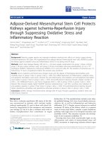
Figure 2-4: A flow chart showing the involvement of ROS in multiple ischemic cascades.
(adapted from Nakka et al, 2008)
Chapter 2: Introduction
Department of Pharmacology,
YLL School of Medicine
29
Compared to other organs, brain is particularly vulnerable to oxidative stress due to the
reason: (i) Brain depends almost exclusively on oxidative phosphorylation for energy
production. The neurons utilize 20% of the oxygen consumed by the body but constitute
only 2% of the body weight, indicating the need of brain for high oxygen consumption
and the potential generation of ROS during oxidative phosphorylation in brain; (ii) A
high content of iron has been reported in some areas of brains, which can catalyze the
formation of ROS; (iii) The brain is rich in poly-unsaturated fatty acids, the targets of
ROS attack; (iv) Brain contains relatively low antioxidant defense mechanisms such as
SOD, catalase, glutathione peroxidase; and (v) Loss of neurons cannot generally be
compensated by regenerating new neurons (Ralf D, 2000).
Several lines of evidence indicate that oxidative stress is a primary mediator of
neurologic injury during cerebral ischemia. Most of the evidences that ROS participates
in neuronal ischemic injury comes from the use of antioxidants and free radical
scavengers that prevent the infarct expansion and restore the neurological deficit function
after ischemia (Braughler and Hall, 1989; Tagami et al, 1999). Cerebral protection was
observed with mice with overproduction of free radical scavenging enzymes (Weisbrot-
Leftkowitz et al, 1998). Furthermore, the extent of delayed neuronal death correlates well
with prelethal markers of oxidative molecular alterations (Fiskum et al, 2004).
Neuroprotection was observed in vivo when the animals subjected to stroke insult were
treated with antioxidant or inhibitors of free-radicals producing enzymes. Studies on
genetic animal models demonstrated that neuroprotection could be observed where genes
Chapter 2: Introduction
Department of Pharmacology,
YLL School of Medicine
30
encoding for free radical producing enzymes are knocked out or genes encoding for
antioxidant enzymes are over-expressed (Fiskum et al, 2004). Therefore, it is believed
that pharmacological modification of oxidative damage is one of the most promising
avenues for stroke therapy.
2.1.4 Rodent ischemic stroke models
Currently, there are two types of animal models of cerebral ischemia used in brain
ischemia studies: global ischemia and focal ischemia (Zemke et al, 2004).
Global ischemia affects the entire brain, which results most commonly from cardiac
arrest or other causes of collapse of system circulation, and subsequently failure of brain
perfusion. The tissue injury is dominated by neurons, occurring especially in the most
vulnerable region of the brain first and then proceeding to the least vulnerable region
(Miller, 1999). Global ischemia can be imitated by the occlusion of both carotid arteries.
Two rodent models of global ischemia are routinely used: the 4-vessel occlusion (4-VO)
transient severe forebrain ischemia model (Pulsinelli and Brierley, 1979) and the 2-VO
plus hypotension model (Smith et al, 1984). 4-VO is caused by the permanent
coagulation of the vertebral arteries and temporary ligation of two common carotid
arteries while 2-VO is caused by the ligation of the two common carotid arteries with the
reduction of blood pressure (Taoufik and Probert, 2008). Both of them have been used for
the examination of selective hippocampal CA
1
and neuronal death. These models create a
transient oligemia in the hippocampus, cortex and striatum during ischemia. The
Chapter 2: Introduction
Department of Pharmacology,
YLL School of Medicine
31
occlusion is followed by a complete restoration of energy by blood reperfusion.
Therefore, the ischemic insult is brief but severe. Hypoxia from both cases is termed
incomplete with residual 1-4% of blood flow. However, complete hypoxia from global
ischemia can be achieved by cardiac arrest or ligation of all arteries streaming from the
heart. In summary, global ischemia involves a short but very intense insults that results in
the drastic reduction of ATP and delayed type of cell death to a portion of specific
neuronal population, making this model is relatively simplified and less informative to
stroke in humans (Taoufik and Probert, 2008).
In contrast, focal ischemia causes the damage only to a portion of brain. The size and part
of affected area depends on which vessel is occluded (Zemke et al, 2004). In addition,
collateral flow contributes another major difference between global and focal ischemia
(Horst and Korf, 1997). Regions of the brain with most severely impaired blood flow will
be rapidly and irreversibly injured. This region is commonly termed as ischemic core.
Surrounding the ischemic core is hypoperfused region where cells receive moderate
blood flow, referred as ischemic penumbra. Cells within ischemic penumbra are
functionally impaired but metabolically silent. Most of the cells in ischemic penumbra
undergo delayed cell death and therefore it is potentially salvageable (Brouns and De
Deyn, 2009). Therefore, focal ischemia involves much more complicated ischemic
cascade events as compared to global ischemia, and represents the closest model to stroke
in human; therefore it is most widely used model in stroke study. In rodent models, focal
ischemia can be mimicked by the occlusion of one of the major blood vessels that supply
the brain, such as common carotid artery and the middle cerebral artery, permanent or
Chapter 2: Introduction
Department of Pharmacology,
YLL School of Medicine
32
transient followed by reperfusion. There are a few models of focal ischemia, for
examples, intraluminal suture (Hata et al, 2000), a more distal extravascular clip (Buchan
et al, 1992) and clot embolic model (Kaplan et al, 1991). Intraluminal suture induces
severe ischemia in striatum but mild ischemia to cortex while extravascular clip induces
more severe cortical ischemia. Clot embolic models (Kaplan et al, 1991) have the
disadvantage of controlling the accurate timing for reperfusion.
In order to resemble a massive and potentially fatal ischemic stroke in humans,
permenant focal ischemic stroke by left middle cerebral artery occlusion (MCAO) was
chosen as a model for studies in this research. Most experiment and clinical research have
focused much on MCAO as the infarct formed by MCAO is similar to the brain damage
of ischemic stroke in humans (Miller, 1999). MCA can be occluded close to its branching
from internal carotid so that caudate putamen, most neocortical regions, the
somatosensory and entorhinal cortex will be affected, and at the distal part where the
flow to the basal ganglia will not be blocked and the damage spans the parietal cortex
(Taoufik and Probert, 2008). In the case of left middle cerebral artery, it supplies the
blood to left cortical areas and also corpus striatum. Damage of these areas results in
impairment of motor, speech and swallowing functions.
Early metabolic responses following MCAO has been well characterized by Folbergrová
et al (1992, 1995), that little changes was observed in between 15 minutes and 2 hours
after ischemia. Impaired glucose delivery in the core infarct causes the decreased to
glucose to the affected territory to 10-26% of non-ischemic values, glycogen was
Chapter 2: Introduction
Department of Pharmacology,
YLL School of Medicine
33
essentially depleted to 5-12%. During ischemia, major losses of ATP (18-32%) and
phosphocreatine (16-28%) are resulted. Lactate was greatly increased to 5-14 times in
severely ischemic core regions. For ischemic penumbra, ATP and phosphocreatine are
moderately decreased, 53% and 70% of non-ischemic region, respectively. However, the
lactate accumulation was substantial.
Interruption of blood flow by MCAO to the supplied basal ganglia, white matter and
cortex causes a gradient of hypoperfusion to emerge, rather than a complete homogenous
ischemia of the entire MCA territory (Figure 2-5). The striato-capsular and
opercular/insular regions are often the earliest to exhibit irreversible damage.
Subsequently, as the penumbra is recruited into the core, the latter progressively expands
to other areas, including the cortical mantle. The maximum extent of the core will
become the final infarct volume (Moustafa and Baron, 2008).
Figure 2-5: The spatial pattern of cerebral blood flow (CBF) in MCAO. The figure
illustrate the CBF reduction following middle cerebral artery (MCA) occlusion in the
baboon brain, demonstrating a gradient from ischemic core (red) through to penumbra
and oligaemia (blue) to normally perfused cortex (grey). Values indicate approximate
CBF in ml100g
-1
min
-1
. (adapted from Moustafa and Baron, 2008)
Chapter 2: Introduction
Department of Pharmacology,
YLL School of Medicine
34
In this study, permanent left MCAO was induced by transcranial approach in rats. Rats
are currently the best species to perform MCAO, because it is relatively inexpensive, its
cerebrovascular anatomy and physiology resemble that of higher species, and physiologic
parameters can be easily monitored. The transcranial approach requires a careful removal
of a section of the skull and the underlying dura in order to occlude the middle cerebral
artery. Tamura et al (1981) developed a subtemporal approach of proximal MCAO at the
point near the origin of the lateral striate arteries, which produced infarction of both
cortex and the caudate putamen. The original technique, however, was very invasive and
the rats survived only for a few hours. Subsequent modifications including preserving the
zygoma and the masseter muscle improved the postoperative survival and eventually the
subtemporal approach becomes a standard technique of permanent focal ischemia in rats
(Duverger et al, 1988; Nakayama et al, 1988; Menzies et al 1992).
Chapter 2: Introduction
Department of Pharmacology,
YLL School of Medicine
35
2.2 CNS Mitochondria
2.2.1 Protective physiological roles of CNS mitochondria
The principle role of mitochondria is producing the high energy phosphate bond ATP for
cellular function. Mitochondria consume nearly 85% to 90% of a cell’s oxygen to support
oxidative phosphorylation for ATP production. Thus mitochondria are called the
“powerhouse of the cell”, that produce 70%-80% of ATP (Szeto, 2006; Willis, 1992).
The mitochondrial respiratory chain (electron transport chain, ETC) located within the
inner mitochondrial membrane is a highly regulated set of reactions carried out by four
electron transporting complexes (complex I-IV) and one H
+
-translocating ATP synthetic
complex (Complex V) with the role of producing ATP. The metabolites produced by
glycolysis are incorporated into tricarboxylic cycle which produces NADH and succinate
and provides the substrate for ETC: NADH is the substrate for complex I (NADH
ubiquinone reductase) while succinate is the substrate for complex II (succinate
dehydrogenase). The oxidation-reduction reactions along the ETC results in the flow of
electron from complex I and complex II to complex III (ubiquinol-cytochrome C
oxidoreductase) via uniquinol. The electrons will then be carried to complex IV
(cytochrome c oxidase) via cytochrome c. At complex IV, oxygen will be reduced by the
electrons to produce water, which is the final product of ETC. Energy resulting from the
glycolysis is converted to proton motive force, as together with these oxidation-reduction
reactions, electrochemical proton gradient is generated by means of proton pumps from
matrix out to the cytosolic site of inner mitochondrial membrane, through complex I,