PERICYTES ARE MORE THAN MSCS a COMPARISON OF THREE CELL POPULATIONS
Bạn đang xem bản rút gọn của tài liệu. Xem và tải ngay bản đầy đủ của tài liệu tại đây (2.74 MB, 80 trang )
PERICYTES ARE MORE THAN MSCS:
A COMPARISON OF THREE CELL POPULATIONS
WANG YINGTING
NATIONAL UNIVERSITY OF SINGAPORE
2012
1
PERICYTES ARE MORE THAN MSCS:
A FUNCTIONAL COMPARISON OF THREE CELL
POPULATIONS
WANG YINGTING
B.Eng (Hons). NUS
A THESIS SUBMITTED
FOR THE DEGREE OF MASTER OF ENGINEERING
DEPARTMENT OF BIOENGINEERING
NATIONAL UNIVERSITY OF SINGAPORE
2012
2
Declaration
I hereby declare that this thesis is my original work and it has been written by me
in its entirety. I have duly acknowledged all the sources of information which have
been used in the thesis.
This thesis has also not been submitted for any degree in any university
previously.
_______________________________
Wang Yingting
27 May 2012
3
Acknowledgement
I am particularly grateful to my supervisor, Prof Michael Raghunath, for his kind
support and advice throughout the project.
I would like to extend my gratitude to Ms Anna Blocki, who has mentored me during
the past year. This study would not have been possible without her patient teaching
and her insightful advice. Her passion for research has been an inspiration.
Last but not the least, I would like to thank my seniors and labmates in Tissue
Modulation Laboratory, who have not only saved a first year graduate student from
many crises and disasters in lab, but more importantly, have also made this journey
enjoyable.
4
Table of Contents
Declaration .............................................................................................................. 3
Acknowledgement .................................................................................................. 4
Summary ................................................................................................................. 6
List of tables ............................................................................................................ 8
List of figures .......................................................................................................... 8
List of symbols and abbreviations .......................................................................... 8
1. Background: MSCs and pericytes —interweaving identities ......................... 9
2. Hypothesis and Objective ............................................................................. 34
3. Methods: ....................................................................................................... 36
4. Results .......................................................................................................... 43
Summary of marker expression profile of Pl-Prc, MSC, and fibroblast ........... 43
4.1.
Pericytes displayed a typical MSC antigen expression profile ............. 44
4.2.
Pericytes demonstrated multipotent differentiation potential ............... 48
4.3.
Pl-Prc expressed pericyte-related markers that MSCs lacked .............. 51
4.4.
Only pericytes maintained EC-formed network in MatrigelTM
angiogenic assay ...................................................................................................... 53
4.4.1. Only EC was able to develop networks on MatrigelTM alone. .............. 54
4.4.2. Pl-Prc, MSCs, and fibroblasts co-localized with EC-formed network . 55
4.4.3. Pl-Prc maintained the EC networks over time ...................................... 56
5. Discussion .................................................................................................... 61
5.1.
The expression of MSC marker profile is not sufficient for
distinguishing Pl-Prc, MSCs, and fibroblasts. Differentiation assay shows that PlPrc possess multi-potent differentiation potential as MSCs do............................... 61
5.2.
NG2, desmin and Tie2 may serve as pericyte-specific markers ........... 65
5.3.
EC-network maintenance, not co-localization, is characteristic of
pericytes 67
Bibliography ......................................................................................................... 71
5
Summary
Pericytes are cells located inside the basement membrane of blood vessels. They play
an essential role in angiogenesis as well as in vessel maintenance and stabilization.
Recently it has been found that pericytes from various tissues demonstrated features
of mesenchymal stem cells (MSCs). It has thus been proposed that some pericytes
may be MSCs residing in a perivascular niche and serving as a progenitor reserve for
tissue regeneration in response to injury by differentiation into other lineages. In this
study, we hypothesized that apart from possessing MSC-like characteristics, pericytes
further possess angiogenic functions that conventional MSC cannot substitute for. To
verify if commercially purchased placenta pericytes are truly MSC-like, the
expression of pericytes, MSCs, and fibroblasts (negative control) of the MSC antigen
profile was compared. It was found that the marker expressions profile of all three
cell types all fulfilled the marker panel required of MSCs. Interestingly, CD146, the
surface marker which is used to isolate pericytes from various tissues, was expressed
by all three cell types. To conclude, a conventional MSC marker profile is not
sufficient to identify MSC. Therefore we further investigated the differentiation
potential of the three cell types and found that only pericytes and MSCs were capable
of adipogenesis and osteogenesis, indicating that pericytes as MSC are multipotent.
Once we were able to show that pericytes behave like MSC, we posed the question if
pericytes are more than just MSC. The three cell types were therefore compared for
pericytic features. It was found that pericytes expressed NG2, desmin and Tie2,
which are pericytic markers linked to important functions in angiogenesis that MSCs
and fibroblasts do not share. As CD146 is not selective for the pericytes we propose a
6
new set of potential markers, which will have to be verified in the isolation of
pericytes. The in vitro pro-angiogenic ability of pericytes, MSCs, and fibroblasts were
also investigated using a MatrigelTM assay, and it was observed that pericytes, MSCs
and fibroblasts all co-localized with endothelial cell networks. However, MSCs and
fibroblasts contracted the network in a cell-ratio dependent manner. These findings
suggested that pericytes are truly MSC-like cells, with additional role in angiogenesis
distinct from that of MSCs.
In conclusion, the traditionally employed in vitro method to identify pericytes by the
co-localization of cells with tubular network on MatrigelTM is inconclusive and not
sufficient. In order to distinguish pericytes from other cells in the tube formation
assay pericyte and non-pericyte standards have to be considered and the contraction
of the network over time observed.
7
List of tables
Table 1. List of antibodies used for immunocytochemistry and flow cytometry .......................................... 36
Table 2. Cell types used and their respective media and detachment kit ...................................................... 37
Table 3. Expression profile of Pl-Prc, MSC, and fibroblast .......................................................................... 43
List of figures
Figure 1. EC-mural cell interaction ....................................................................................................... 17
Figure 2. EC form capillary-like networks when cultured on MatrigelTM ............................................. 33
Figure 3. Microscopic photos of cells in culture. .................................................................................. 37
Figure 4: Pl-Prc, MSC, and FB expressed MSC markers...................................................................... 45
Figure 5. Pl-Prc, MSCs, and FB lacked endothelial markers and hematopoietic markers expression.. 46
Figure 6. None of Pl-Prc, MSCsand FB expressed the histocompatibility antigen HLA-DR, monocyte
related marker CD11b, and the B cell markers CD11b and CD19. ....................................................... 47
Figure 7. Osteoblast and adipocyte induction of Pl-Prc, MSCs and fibroblasts (FB). .......................... 48
Figure 8. chondrocyte induction of Pl-Prc, MSC, and fibroblast. ......................................................... 50
Figure 9: Pl-Prc, MSCs, and fibroblasts all expressed pericytic markers α-SMA and PDGFR-β.. ....... 52
Figure 10. NG2 expression is weak in all three cell types. .................................................................... 52
Figure 11: Pl-Prc showed the strongest expression of desmin. ............................................................. 53
Figure 12. Pl-Prc showed positive staining for TIE2, ........................................................................... 53
Figure 13. Only EC formed networks when cultured alone on MatrigelTM. .......................................... 54
Figure 14. Pericyte co-localize with EC-formed networks on MatrigelTM in vitro................................ 55
Figure 15. Pl-Prc, MSCs, and FB all co-localized with EC formed network on MatrigelTM................ 56
Figure 16. Pl-Prc/ MSC/ FB co-culture with EC on MatrigelTM 4 hours after seeding. . ..................... 57
Figure 17. Pl-Prc/ MSC/ FB co-culture with EC on MatrigelTM 8 hours after seeding ......................... 58
Figure 18. Pl-Prc/ MSC/ FB co-culture with EC on Matrigel TM 12 hours after seeding ...................... 59
Figure 19. Pl-Prc / MSC/ FB co-culture with EC on MatrigelTM 24 hours after seeding. ..................... 60
List of symbols and abbreviations
BSA: bovine serum albumin
DMEM: Dulbecco's modified Eagle medium
EC: endothelial cells
FB: fibroblasts
FBS: fetal bovine serum
FC: Flow Cytometry
HBSS: Hanks' balanced salt solution
HUVEC: human umbilical vein endothelial cells
ICC: Immunocytochemistry
MSCs: mesenchymal stem cells
PBS: phosphate buffered saline buffer
Pl-Prc: placenta pericytes
p/s: antibiotic-penicillin/streptomycin
SMC: smooth muscle cells
8
1.
Background: MSCs and pericytes —interweaving
identities
Mesenchymal stem/stromal cells (MSCs) have been under the spotlight of stem cell
therapy because of its multi-lineage differentiation capacity (reviewed by Ankrum, et
al., 2010), immunosuppressive effect (Nauta, et al., 2007), and increasingly
importantly, its ability to secret trophic factors that induce tissue regeneration
(reviewed by Ankrum, et al., 2010). According to the US Public Clinical Trials
Database (U. S. National Institutes of Health, 2012), there is nearly 300 clinical trials
exploiting MSCs for their therapeutic values. Most of the current clinical trials target
diabetics, ischemia, myocardial infarction, inflammation, and immune diseases. The
trial outcomes, on the other hand, are encouraging but not yet satisfactory. Implanted
or infused MSCs often have low efficacy in vivo. It is reasoned that the improvement
of MSC therapy is hindered by the limited understanding of MSC cell fate in vivo
(reviewed by Ankrum, et al., 2010). The consensus on MSC identification is solely
based on its marker expression and differentiation potential under in vitro conditions
(Augello, et al., 2010; Dominici, et al., 2006). Although MSC in vitro characteristics
are intensively researched upon, their in vivo counterpart still remains to be found
(reviewed by Corselli, et al., 2012).
A few discoveries in recent years provide hints on the in vivo niche of MSCs. The
first piece of evidence comes from the successful isolation of MSC from a wide
spectrum of tissues. Conventionally extracted from bone marrow, MSCs have now
been isolated from virtually all postnatal connective tissues, such as the adipose
tissue, dental pulp, and so on (reviewed by Bianco, et al., 2008; da Silva Meirelles, et
9
al., 2006). These studies suggest that the in vivo source of MSC must be widely
distributed across different tissues and organs.
Following this line of thought, several research groups have come up with the
hypothesis that the in vivo MSC reservoir is most likely to be associated with the
blood vessels, which is present in all tissues in the body. More specifically, they
propose that MSCs in situ are perivascular. To prove this theory, perivascular cells
have been isolated and purified by flow cytometric cell sorting. The sorted cells were
shown to display a MSC marker profile, and to demonstrate adipogenic (Crisan, et al.,
2008; Corselli, et al., 2012; Zannettino, et al., 2008), osteogenic (Sacchetti, et al.,
2007; Crisan, et al., 2008; Corselli, et al., 2012; Zannettino, et al., 2008),
chondrogenic (Corselli, et al., 2012; Zannettino, et al., 2008), and even myogenic
potentials (Crisan, et al., 2008; Dellavalle, et al., 2007). Therefore, perivascular cells
are shown to be bona fide MSCs. Some even go so far as to pose the question that if
all MSCs are pericytes (Caplan, 2008).
Under such circumstances, pericytes, one of the perivascular cells and are found
around small blood vessels (Gaengel, et al., 2009), have attracted great research
interest. Until recently, pericytes have been a cell type that is not well studied and
understood. They have been shown to play an essential role in the maturation and
stabilization of blood vessels (Armulik, et al., 2005). The recent evidences on their
additional function as MSC-like progenitor cells (reviewed by Crisan, et al., in press)
put them under new attention as candidates for cell therapy and regenerative
medicine. These cells, not only multipotent but also have pro-angiogenesis properties,
may become a promising alternative for MSC in stem cell therapy. Also, the study on
10
the relationship between pericytes and MSCs may shine light on the obscure in vivo
identity of MSC.
However, the identification of pericytes is no easier problem. Different from MSCs,
pericytes are traditionally identified not by their in vitro characteristics, but by their in
vivo location. Pericytes are defined as cells located within the basement membrane of
endothelial cells. This is until now the ultimate standard for pericyte identification,
which is unfortunately impractical and sometimes impossible to verify for in vitro
cultures. Besides the definition, pericyte identification is further complicated by its
heterogeneity. Pericytes are widely distributed around virtually all small blood
vessels in the body, and their maker expression depends on their tissue of origin as
well as degree of maturation of the associated blood vessels (reviewed by Bergers, et
al., 2005). To date, there is no marker or combination of markers that is available for
identification of pericytes from all tissues reviewed by (Armulik, et al., 2011). A
vigorous study that claims to have isolated pericytes by a set of markers would often
verify the in vivo location of the cells in their tissue of origin.
Most of the recent studies on pericyte-MSC relationship concentrate on flow
cytometric sorting isolated pericytes, and their in vivo or in vitro characterization for
MSC-specific features (Péault, et al., 2007; Crisan, et al., 2008; Covas, et al., 2008;
Castrechini, et al., 2010; Corselli, et al., 2012). Side by side comparison of MSCs and
pericytes are rare. For example, few papers have been published on comparing MSCs
and pericytes from the same bone marrow source (reviewed by Bouacida, et al.,
2012). However, such comparative assays are essential for finding out the differences
and similarities of the two cell populations.
11
This study thus proposes an unbiased comparison between a typical pericyte
population (pericytes from human placenta isolated by CD146 expression, Promocell)
and a typical MSC population (MSCs isolated from human bone marrow by plastic
adherence, Lonza) for MSC as well as pericyte related characteristics. In this way,
this study aims to generate novel insights on several elusive aspects of the MSCpericyte relationship:
The first motivation of the study is to address the unanswered question: are MSCs
really pericytes? Although pericytes have been shown to possess the major
characteristics of MSC (Crisan, et al., 2008; Dellavalle, et al., 2007; Shi, et al., 2003;
Zannettino, et al., 2008; Díaz-Flores, et al., 2009), the reverse question is rarely
posed. Do MSCs possess the typical pericyte features? Pericytes have been shown to
interact with endothelial cells through a number of pathways, and to play a specific
role in angiogenesis and blood vessel maintenance (Bergers, 2008; Bergers, et al.,
2005; Hirschi, et al., 1996). These functions are rarely associated with MSCs, and
would need to be verified before being able to conclude if MSCs are truly pericytes.
That is why this study chose to test both pericytes and MSCs not only for MSC
related characteristics, but also pericyte and angiogenesis related features.
The second motivation of the study is to seek a way to identify pericyte in vitro. By
screening both pericytes and MSCs for a spectrum of marker and functional assays,
we expect to establish a set of in vitro assays that is sensitive enough to distinguish
pericytes from other mesenchymal lineages, for example MSCs, if there is any
differences between the two. Many who claim that they have identified pericytes rely
on one or a few markers, while to this day there is no pericyte specific/ pan-pericyte
12
marker available (reviewed by Armulik, et al., 2011). It is to be verified if these
“pericytes”, isolated from various tissues using different sets of markers, refer indeed
to the same population. The ultimate test still requires verifying the in vivo
perivascular location of the cells. It would be of great interest to have a set of
standardized assays that enables identification of pericytes in vitro. Such assays
would also need to be able to identify functional pericytes, i.e. cells that maintains
their pro-angiogenic properties and the ability to interact with endothelial cells. This
would provide a platform to differentiate pericytes from other cell populations in
vitro. Moreover, it would also allow for standardization of pericytes for research
purposes as well as for clinical application.
Besides providing a tool for facilitating future research, a third motivation of the
study is to obtain insights of the in vivo characteristics of pericytes and MSCs.
Although the in vivo function and properties of MSCs and pericytes are beyond the
scope of this study, some clues may be obtained from their in vitro characteristics and
behaviors.
1.1
Mesenchymal Stem Cells (MSCs)
Before moving on to compare the different cell types, it is important to review the
current definition and methods of identification and characterization for each of them.
The cell population that is called mesenchymal stem cells today was first described
by Friedenstein (1968), who found a non-hematopoietic progenitor population in the
bone marrow that is capable of forming single clones in culture (colony-forming
units-fibroblastic or CFU-Fs) and is capable to undergo osteogenesis in vitro
13
(Friedenstein, et al., 1970).The term “mesenchymal stem cells”, or MSCs, are later
made popular by Pittenger et al. (1999), who showed that these plastic adherent,
colony-forming cells isolated from bone marrow were able to differentiate into
osteoblasts, adipocytes, and chondrocytes in vitro when induced by a cocktail of
small molecules. They further suggested that this particular cell population may be
the reservoir for adult connective tissue regeneration. Nowadays the sources of MSCs
have been expanded beyond bone marrow. MSCs have been isolated from virtually
all types of postnatal tissues, such as adipose tissue, dental pulp, and so on (reviewed
by Bianco, et al., 2008; da Silva Meirelles, et al., 2006). The in vivo location of MSCs
still remains to be confirmed, which is difficult due to the lack of a MSC-specific
marker set (reviewed by Bianco, 2011).
One of the currently most accepted definition of MSCs is proposed by the
International Society for Cellular Therapy (ISCT) (Dominici, et al., 2006), who
suggested three minimal conditions for a cell population to be called MSCs. Firstly,
the cells have to be plastic adherent, Secondly, they should be positive for surface
antigens CD105, CD73, CD90, and at the same time be negative for CD45, CD34,
CD14 or CD11b, CD79α or CD19, and HLA-DR. Lastly, they should be able to
differentiate in vitro into three mesenchymal lineages, namely osteoblasts, adipocytes,
and chondrocytes, under standard differentiation conditions.
Recent years have seen a shift of interest in the clinical application of MSCs. MSCs
were initially regarded as the earliest progenitor cells in mesenchymal lineage
(Caplan, 1994). The earlier studies focused on their ability to self-renew and to
differentiate into multiple mesenchymal lineages, and tried to explore their
14
therapeutic potential for tissue repair or even for gene therapy (Bonab, et al., 2006).
Mesenchymal stem cells from bone marrow have already been used for clinical
applications (Gerson, 1999). It has been since observed that MSC implantation
resulted somehow in reduced inflammation, fibrosis and apoptosis, even when there
is a lack of effective MSC differentiation in situ (reviewed by Ankrum, et al., 2010;
Bianco, 2011). Systematically infused auto- or allogeneic MSCs were able to home to
damaged tissues and to establish a conductive microenvironment for tissue
regeneration. It has thus been suggested that other factors than differentiation and
proliferation must be contributing to the therapeutic effect of MSC in clinical trials.
However, the actual mechanism of the effect of MSCs in vivo is still unclear.
(Ankrum, et al., 2010; Caplan, 2007; Bianco, 2011)
Although numerous clinical trials are ongoing to exploit the therapeutic effect of
MSCs, few have proved to be significantly effective. It has been suggested that the
current bottleneck of MSC cell therapy is the lack of understanding of their in vivo
cell fate (Ankrum, et al., 2010). The dilemma is that the definition and
characterization of MSCs have depended exclusively on in vitro cultures, leaving the
in situ identity and behavior of these cells elusive (Bianco, 2011).
Even the nomenclature of MSC is now being challenged. The use of “stem cells” is
considered not vigorous. MSCs only have limited renewing ability in vitro.
Furthermore, proliferation and differentiation in culture do not necessarily mean selfrenewal and multi-potency in vivo (Bianco, 2011). The word “mesenchymal” is also
often debated, since muscle and bone are derived from different progenitors during
the early embryonic development (Bianco, 2011; Nombela-Arrieta, et al., 2011)
15
Therefore, the search of the in vivo counterpart of MSC is an important ongoing
research topic both for elucidating on the identity of MSCs as well as for improving
the clinical outcome of MSC-based therapy. Pericytes, with numerous features shared
with MSCs, may promise to provide valuable clues on the subject.
1.2
Pericytes
The discovery of pericytes is attributed to the French scientist Charles Rouget in
1873. They carried thus the name "Rouget cells". The term "pericytes" was first
coined by Zimmermann in 1923, referring to their close association with endothelial
cells (Armulik, et al., 2011; Hirschi, et al., 1996). The definition of pericytes has since
depended heavily on the in vivo location of the cells relative the endothelial cells.
Pericytes are originally defined as extensively branched cells located in non-muscular
microvessels, capillaries and postcapillary venules (Díaz-Flores, et al., 2009). The
currently accepted and most vigorous definition of pericytes is cells that are located
within the basement membrane of blood vessels, which come from the electron
microscopy observation of pericytes in situ (reviewed by Sims, 1986).
In the vasculature system, pericyte is one of the two categories of mural cells that are
found around blood vessels (Figure 1). In specific, pericytes are found around small
blood vessels. They wrap the selves around the inner single-layer vessel lumen
formed by endothelial cells (EC). Pericytes are in physical contact with EC and have
intimate interactions with the EC-formed vessels (McDonald, 2008). The other type
of mural cells, smooth muscle cells, is found around large blood vessels. They form
multiple layers (tunica media) around the endothelial cells-formed vessels (tunica
16
intia). They are further enveloped by the tunica adventitia, which consists of
fibroblasts and connective tissue (Corselli, et al., 2010).
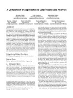
Figure 1. EC-mural cell interaction (adapted from (Gaengel, et al., 2009)). Blood vessels consist of
two cell types: endothelial cells (EC, in yellow) which form the internal lumen, and mural cells (in
green) which wrap around the EC-formed vessels. Under the class of mural cells, there is a subcategory of cells named pericytes (at lower left corner of the diagram) that are embedded within the
basement membrane of blood vessels in close association with EC. The interaction and exchange of
signal molecules between pericytes and EC are essential for the stabilization and maturation of small
blood vessels. For example, the PDGF-B/PDGFR-β pathway and the Ang1/Tie2 pathway (represented
by a and b, respectively).
The prominent feature of pericytes is that they sit in the basement membrane of the
blood vessels. They are in close contact with the endothelial cell through various
mechanisms such as gap junction or peg-socket contact (Armulik, et al., 2011;
Hirschi, et al., 1996).
1.2.1
Pericyte distribution in tissues
17
Pericytes are widely distributed in the body. Pericytes are found in almost all tissue
types in blood microvasculature, but not in normal lymphatic system (Armulik, et al.,
2011). The most prominent feature is their close association with endothelial cell
vessels. Pericytes are located more frequently around microvasculature such as
capillaries and small venules, as well as pre-capillary arterioles (Sims, 1986).
Pericytes are often found at the junction points of capillaries or of small vessels and
capillaries, where they stretch themselves along the length of blood vessels across
several branches (Armulik, et al., 2011; Bergers, 2008). The EC-pericyte ratio around
blood vessels is tissue specific. It can vary from 1:1 in retina tissues and down to
100:1 in human skeletal muscle, for example (reviewed by Díaz-Flores, et al. (2009)).
Besides the variation in the EC-pericyte ratio, pericyte distribution in tissue also
varies in the form in which pericytes wrap themselves around EC. They can come in
the form of single, discontinuous cells to a mono-cell layer around EC-formed vessels
(Gerhardt, et al., 2003; Hirschi, et al., 1996)
Pericytes are found also at sprouting blood vessels. EC recruit pericytes during
angiogenesis by secreting platelet-derived growth factor (PDGF), which promote the
proliferation and migration of pericytes (Armulik, et al., 2005).
Depletion of
pericytes through inhibition of platelet-derived growth factor receptor β (PDGFR-β)
in vivo leads to leaky and dilated vessels in mice as a results of lack of mural cells
around the blood vessels (Hellström, et al., 2001).
So far, pericytes have been isolated from a wide spectrum of human tissues, such as
skeletal muscle, myocardium, placenta, pancreas, skin, brain, and bone marrow
(Crisan, et al., 2008), Zannettino and colleagues (2008) have isolated multipotent
18
pericyte-like cells from human adult adipose tissues by the markers STRO-1, CD146
or 3G5. However, it is worth noting that the isolated “pericytes” have a different
marker profile compared to Crisan’s group, and common pericyte markers, like
desmin, NG2, PDGFR-β, has not been tested. The markers used for isolation are not
restricted to small vessels, and the expression of STRO-1 was not exclusively
perivascular, based on the immunofluorescence staining of frozen sections. Moreover,
only a small portion of the isolated cells possessed multipotency. The group of Paolo
Bianco (Dellavalle, et al., 2007) isolated ALP+ CD56- cells from human adult muscle
that exhibited a typical pericyte marker profile (annexin V, alkaline phosphatase,
desmin, smooth muscle actin, vimentin and PDGFR-β), though they have weak
expression for CD90, CD105 and CD146. It demonstrates that pericytes isolated
using different markers may have different marker profiles, while those isolated with
CD146 resemble most that of MSCs.
1.2.2
Pericyte origin
Pericytes can develop from a variety of tissues (Lamagna, et al., 2006). For example,
brain pericytes are shown to originate from neurocrest (Bergwerff, et al., 1998). It has
also been proposed that VEGFR2+ angioblasts can differentiate into EC or pericytes
under different stimuli (Yamashita, et al., 2000). There are also research groups who
suggested that pericytes originate from myofibroblasts (Díaz-Flores, et al., 2009). It
has equally been shown that bone marrow derived cells, when systematically infused
into mice, can home to perivascular locations, infiltrate with microvasculature, and
express pericytic markers, indicating that some pericytes may also come from the
bone marrow (Ozerdem, et al., 2005; Rajantie, et al., 2004)
19
Finally, MSCs have as well been proposed as pericyte precursors. It has been shown
that when co-cultured with endothelial cells, MSCs (10T1/2, ATCC) are able to
differentiate into pericyte-like phenotype. They expressed NG2 and αSMA, stabilized
EC formed networks on matrigel, and homed to perivascular locations when
implanted into mice developing vessels (Darland, et al., 2001; Hirschi, et al., 1998).
1.2.3
Increasing interest in pericyte research arising from newly discovered
pericyte functions: an implication for their therapeutic potential
The research on pericyte function is still ongoing and recent years have seen rapid
advances in understanding of the role of pericytes in microvascular system.
Nevertheless, three main pericyte functions have been pointed out. The first function
of pericyte is the maintenance of blood vessels through secreting growth factors that
are indispensable for EC survival (Gaengel, et al., 2009; Gerhardt, et al., 2003). Three
well-known ligand/receptor pairs in EC-pericyte interaction are VEGF/VEGFR,
PDGF-B/PDGFR-βand Ang1/Tie2. Pericytes are able to produce vascular endothelial
growth factor (VEGF) which binds to the VEGF receptors in EC. VEGF is essential
for EC survival and regulates EC immigration (Darland, et al., 2003; Senger, et al.,
1996; Franco, et al., 2011). PDGF-B is important for mural cell recruitment towards
EC-formed vessels (Hellström, et al., 1999). Inhibition of PDGF-B impaired EC’s
ability to recruit mesenchymal cells to EC vessels on MatrigelTM in vitro (Hirschi, et
al., 1998). Pericytes also secret Ang1, the main agonistic ligand for Tie2 receptor on
EC (Gaengel, et al., 2009). Ang1/Tie2 pathway is shown to be essential for blood
vessel maturation and stabilization. Mouse with Ang1 or Tie2 depletion died from
cardiovascular failure as embryos (Suri, et al., 1996).
20
A second function of pericytes is to provide mechanical support and to control blood
circulation through providing mechanical forces. Pericytes express a number of
contractile proteins, for instance α-SMA, desmin and tropomyosin have been
identified in pericytes in vivo or in vitro (Bergers, et al., 2005). Some research groups
proposed that the pericytes are able to constrain blood vessels to contribute to the
regulation of blood flow in small vessels (Rucker, et al., 2000; Bergers, et al., 2005).
However, there is some controversy on if pericytes really act to provide contractile
force to blood vessels, because there is a lack of direct evidence. Observation of
pericyte contraction in vivo is a difficult issue, due to the lack of specific pericyte
markers (reviewed by Armulik et al.) (2011).
Besides these two traditional functions, there is an increasingly popular theory that
pericyte further processes the ability to serve as a reservoir of progenitor cells in
different tissues (Augello, et al., 2010). As mentioned earlier, recent studies have
reported that perivascular cells express MSC markers and possess multi-lineage
differentiation potential (Crisan, et al., 2008; Covas, et al., 2008; da Silva Meirelles,
et al., 2006; Shi, et al., 2003). As early as in 1988, it has been found that alkaline
phosphates positive cells in the bone marrow are able to differentiate into adipocytes
(Bianco, et al., 1988). More recently, pericytes derived from various tissues have
been demonstrated to possess myogenic capacities (Crisan, et al., 2008). It has been
further suggested that pericytes exhibit stem cell features and may even be
mesenchymal stem cells (MSCs). It has been proposed that pericyte-like populations
reside in a perivascular niche and may serve as local stem cell reservoirs (Crisan, et
al., 2008; Zannettino, et al., 2008; da Silva Meirelles, et al., 2006; Shi, et al., 2003). It
21
is found that perivascular cells, isolated from adipose tissues by pericyte related
markers STRO-1, CD146 or 3G5, expressed also stromal cell related markers (CD44,
CD90, CD105, CD106, CD146, CD166, STRO-1, and alkaline phosphatase). These
cells equally demonstrated the potential to differentiate into cells from different
lineages (Zannettino, et al., 2008). This suggests that pericytes, besides their
angiogenic properties, may also serve as a local stem cell source that response quickly
to damaged tissues or growth signals in their proximity.
The group led by Bruno Péault in Pittsburgh published the ground-breaking article in
Cell Stem Cell in July 2008 (Crisan, et al.), where they identified NG2, CD146,
PDGFR-β as exclusive markers for cells at perivascular location. They thus isolated
“pericytes” from different adult and fetal tissues by sorting for CD146+ CD34CD45- CD56- population. They found that this cell population has the potential to
differentiate into myogenic, osteogenic, adipogenic, and chondrogenic lineages,
maintains the expression of pericytic markers NG2, CD146, and αSMA, as well as
typical MSC antigens. They equally demonstrated by immunohistochemistry that
MSC marker expressing cells were found in perivascular locations, and that they coexpressed CD146.
1.3
in vitro identification methods for MSCs
The international consensus for defining MSCs is by their three features: plastic
adherence, marker expression, and multipotency (Dominici, et al., 2006).MSCs in
culture are characterized by their plastic-adherent well-spread morphology (Pittenger,
et al., 1999; Dominici, et al., 2006). Furthermore, there is a set of markers that are
22
generally agreed upon to be expressed by MSCs. MSCs are expected to express CD90
(Thy-1), CD105 (Endoglin), CD73, CD13 (APN) (Jiang, et al., 2002). At the same
time, MSCs normally do not express CD11b (monocyte marker), CD45 (leukocyte
marker), CD34 (hematopoietic stem cell marker), CD117 (c-kit, hematopoietic
progenitor cell marker), CD19 (B cell marker), HLA-DR (antigen presenting cell
marker), glycophorin-A, and CD31 (EC marker) (Kolf, et al., 2007; Dominici, et al.,
2006).
1.3.1. Three MSC hallmark antigens CD90, CD105, and CD73
CD90, CD105, and CD73 are the three MSC markers that are part of the minimal
criteria for defining MSC proposed by the International Society for Cellular Therapy
(ISCT) (Dominici, et al., 2006). This publication has been intensively cited as a
standard of MSC identification in vitro.
CD90, also named Thy-1, is an important surface glycoprotein that regulates cell-cell
interactions (Rege, et al., 2006). MSCs are shown to express CD90 in culture
(Pittenger, et al., 1999). It is expressed in fibroblasts, brain cells, thymocytes, T cells,
myoblasts, epidermal cells and keratinocytes (Pont, 1987; Haeryfar, et al., 2004) . It is
also found in activated endothelial cells, smooth muscle cells, and a restricted
population of hematopoietic cells (Craig, et al., 1993; Haeryfar, et al., 2004). In
fibroblasts, CD90 is found to affect cell proliferation, collagen production, and
migration (reviewed by Rege, et al., 2006).
CD105 (endoglin), is a dimeric protein that form part of the transforming growth
factor-beta receptor complex (Yamashita, et al., 1994). CD105 is strongly expressed
23
in vascular EC and plays a role in angiogenesis. It is also expressed in stromal cells
and fibroblasts, as reviewed by Fonsatti (2001).
CD73 (ecto-5'-nucleotidase (S'-NT)) is an ecto-enzyme commonly found on the cell
membrane which catalyzes the dephosphorylation of monophosphates (Resta, et al.,
1998). It is found to be expressed in mesenchymal stem cells as well as in
lymphocytes (Barry, et al., 2001).
1.3.2. MSCs frequently express CD29, CD13, CD166, and CD146
Integrins are the major surface adhesion receptors. They consist of αβ heterodimers
(Hynes, 1992). CD29 is the integrin β1 subunit, which are the receptors for collagen
(α1β1, α2β1, α10β1, α11β1), laminin (α3β1, α6β1, α7β1), and RGD (α5β1, αVβ1,
α8β1), a tripeptide present in fibronectin and vitronectin (Hynes, 2002).Most of them
are expressed in endothelial cells (Francis, et al., 2002). Integrins β1 are equally
found in the center nervous system and are important for cerebral angiognenesis,
especially α5β1 (Li, et al., 2012). All four integrin β1 isoforms are expressed in
MSCs, with β1A showing the highest expression. (Ip, et al., 2007). As reviewed by
(Francis, et al., 2002), β1 integrins or CD29 have been shown to play an essential part
in vascular development. Angiogenesis is haulted after inhibition of α1β1 and α2β1
(Senger, et al., 1996).
CD13 is a membrane bound ectopeptidase named aminopeptidase N (APN) which
contribute to the degradation of certain proteins and peptides. Besides its enzyme
activity, it is also involved in other cell activities, especially in the migration,
differentiation, and angiogenesis of malignant tumor cells (Wickström, et al., 2011).
24
The expression of CD13 is found in a wide range of cell types including epithelial,
endothelial, and fibroblast-like cells. It is also strongly expressed in stem cells. It is
used as a differentiation marker for granulocytes and monocytes, as reviewed by
Bauvois, et al., (2006)
CD166, also named as activated leukocyte cell adhesion molecule (ALCAM), is a cell
surface immunoglobulin. As its name suggests, CD166 is important for cell adhesion.
It is expressed on hematopoietic progenitor cells, and endothelial cells, as reviewed
by Ohneda, et al., (2001).
CD146 or S-endo 1 is a membrane glycoprotein that is located at the cell-cell contact
point, and is possibly involved in cell-cell adhesion and cell-matrix interaction.
CD146 is one of the markers that interest us the most, because it is often used for
pericyte identification for research or commercial applications. It is reported to be
expressed in EC, smooth muscle tissues, cerebellum, hair follicles of normal tissues,
as well as melanomas and some other malignant tissues (Shih, et al., 1994). Recent
discoveries have shown that CD146 is found in cells that co-express pericyte markers
such as α-SMA and 3G5 (Shi, et al., 2003). Zimmerlin and colleagues and also shown
that CD146+/CD31- cells identifies pericytes in tissue verified by histology
(Zimmerlin, et al., 2009). CD146 has routinely been used as a marker for pericyte
sorting from heterogeneous populations (Péault, et al., 2007; Crisan, et al., 2008;
Covas, et al., 2008; PromoCell).
1.3.3. MSCs are supposed to be negative for EC markers CD144, and VEGFR2,
and hematopoietic markers for CD45, CD34, and CD117
25