Design and experimentation of surgical devices for voice restoration
Bạn đang xem bản rút gọn của tài liệu. Xem và tải ngay bản đầy đủ của tài liệu tại đây (3.14 MB, 86 trang )
DESIGN AND EXPERIMENTATION OF SURGICAL DEVICES
FOR VOICE RESTORATION
CHNG CHIN BOON
(B.Eng. (Hons.). NUS)
A THESIS SUBMITTED
FOR THE DEGREE OF MASTER OF ENGINEERING
DEPARTMENT OF MECHANICAL ENGINEERING
NATIONAL UNIVERSITY OF SINGAPORE
2011
Acknowledgement
Graduating from a purely mechanical background, the work I’ve done for this thesis
has been an eye opening experience. I would like to firstly express my gratitude to my main
supervisor, Dr. Chui Chee Kong for his kind patience and support throughout my candidature
which would not have been possible without his valuable guidance and encouragement. I
would also like to thank my co-supervisor, Senior Consultant Dr. David Lau Pang Cheng from
the Department of Otolaryngology, Singapore General Hospital, whose insightful feedback
and ideas provided not only the springboard for the projects, but often lead to the
conceptualization of most of the solutions presented.
I would also like to thank the all staff in Control and Mechantronics Laboratory 1 –
Mr Sakthiyavan s/o Kuppusamy, Mr Yee Choon Seng, Ms Ooi-Toh Chew Hoey, Ms Hamidah
Bte Jasman, Ms Tshin Oi Meng, whose friendly technical support and assistance provided a
pleasant and enjoyable environment to work in throughout my candidature.
Lastly, I would like to thank all fellow students and researchers in Dr Chui’s research
group for their advice, particularly Mr Yang Liang Jing and Mr Wen Rong, whose dedication
to their research has been a great source of inspiration.
The two projects report in this thesis is a collaboration between Singapore General
Hospital and National University of Singapore was supported in part by the National Medical
Research
Council
(Singapore)
under
Grant
NMRC/EDG/0006/2007
and
NMRC/EDG/0043/2008.
I
List of publications
Journal
C.B. Chng, D.P. Lau, J.Q. Choo, C.K. Chui, Bio-absorbable Micro-clip for Laryngeal
Microsurgery
Design
and
Evaluation.
Acta
Biomaterialia,
2012
( />D.P. Lau, C.B. Chng, J.Q. Choo, N. Teo, Bunte RM, C.K. Chui, Development of a micro-clip for
laryngeal microsurgery: Initial animal studies. The Laryngoscope. 2012; (Accepted).
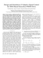
J.Q. Choo, D.P. Lau, C.K. Chui, T. Yang, C.B. Chng, S.H. Teoh. Design of a mechanical larynx
with agarose as a soft tissue substitute for vocal fold applications. J Biomech Eng. 2010
Jun;132(6):065001.
Conference
C.B. Chng, C.K. Chui, D.P. Lau. A novel handheld device for tracheo-esophageal puncture and
prosthesis insertion. 2011 IEEE/SICE International Symposium on System Integration (SII
2011), 20-22 Dec 2011; 2011; Piscataway, NJ, USA: IEEE.
L. Yang, C. B. Chng, C.-K. Chui, D.P Lau. Model-based Design Analysis for Programmable
Remote Center of Motion in Minimally Invasive Surgery. 4th IEEE international Conference
on Robotics, Automation and Mechatronics 2010, Singapore, 2010.
II
Table of Contents
Acknowledgement ...................................................................................................................... I
List of publications ..................................................................................................................... II
Table of Contents ...................................................................................................................... III
Summary ................................................................................................................................... VI
List of figures ........................................................................................................................... VIII
List of tables .............................................................................................................................. XI
1.
Introduction ....................................................................................................................... 1
1.1. Background ..................................................................................................................... 1
1.1.1. Voice microsurgery .................................................................................................. 1
1.1.2. Voice restoration after total laryngectomy ............................................................. 3
1.2. Motivation and objective ................................................................................................ 5
1.3. Research scope and organization of thesis..................................................................... 6
2.
Literature review................................................................................................................ 7
2.1. Bio-absorbable clips for vocal fold wound healing ......................................................... 7
2.1.1. Vocal fold morphology ............................................................................................. 7
2.1.2. Laryngeal microsurgery............................................................................................ 8
2.1.3. Current methods for laryngeal wound healing........................................................ 9
2.1.4. Surgical clips ........................................................................................................... 12
III
2.1.5. Magnesium as the core material ........................................................................... 13
2.2. Device for TEP and prosthesis insertion ....................................................................... 14
2.2.1. TEP creation ........................................................................................................... 14
2.2.2. Transnasal esophagoscopy (TNE) guided TEP ........................................................ 15
3.
Bio-absorbable micro-clips for vocal fold wound closure ............................................... 18
3.1. Design requirements and considerations ..................................................................... 18
3.2. Selected design and implementation ........................................................................... 20
3.3. Experiments and results ............................................................................................... 22
3.3.1. In-vitro study .......................................................................................................... 22
3.3.2. Ex-vivo study .......................................................................................................... 24
3.3.3. In-vivo study ........................................................................................................... 26
3.4. Discussion...................................................................................................................... 32
3.4.1. Bioabsorption and biocompatibility of magnesium for the microclip ................... 32
3.4.2. Recommendation for future work ......................................................................... 34
4.
Device for TEP and voice prosthesis insertion ................................................................. 36
4.1. Design requirements and proposed solution ............................................................... 36
4.2. Designs .......................................................................................................................... 37
4.2.1. Initial design ........................................................................................................... 37
4.2.2. Handheld device .................................................................................................... 40
IV
4.2.3. Manipulator system ............................................................................................... 45
4.3. Experiments and results ............................................................................................... 50
4.3.1. Manual device ........................................................................................................ 50
4.3.2. Handheld device .................................................................................................... 52
4.4. Discussion...................................................................................................................... 53
4.4.1. Recommendations and future work ...................................................................... 53
5.
Conclusion ........................................................................................................................ 57
6.
References ....................................................................................................................... 58
7.
Appendix I –Technical drawings....................................................................................... 63
7.1. Micro-clip ...................................................................................................................... 63
7.2. Manual TEP device ........................................................................................................ 64
7.3. Handheld TEP device..................................................................................................... 69
7.4. Calculation of the end effector position for the robotic manipulator .......................... 73
V
Summary
Voice production is an ability that allows us to verbally communicate our thoughts
and ideas. The total loss of such a crucial skill has enormous psychosocial and economic
consequences on patients’ lives. Two common procedures in head and neck surgery for the
restoration of voice are voice microsurgery and implantation of a voice prosthesis for
patients after total laryngectomy. This thesis thus presents details of the development and
experimental evaluation of devices to aid respectively in the two surgical procedures – 1)
bio-absorbable micro-clips to aid in vocal fold wound closure and 2) a tracheoesophageal
puncture and voice prosthesis insertion device for immediate voice restoration.
For voice microsurgery, surgical removal of benign lesions often results in an incision
in the vocal folds. In order to reduce scarring, the edges of the wound need to be
approximated in order for healing by primary intention. Current techniques for vocal fold
wound include the usage of micro-sutures or fibrin glue. While current methods have been
found to provide good treatment results, each are limited by different constraints which
increase procedural difficulty and time. Hence, based on combining the ease and efficiency
of using fibrin glue with the precision and security of micro-sutures, a novel bio-absorbable
micro-clip made from magnesium is specifically designed to reduce technical complexity in
achieving apposition of epithelial flaps, possibly providing comparable wound holding and
healing properties to current methods. The micro-clips were tested in an in-vivo survival
study with a pig model for their ease of application, bio-absorption and bio-compatibility.
Experimental results revealed a lack of significant inflammation and achievable bioabsorption rates.
VI
For the implantation of a voice prosthesis for patients after total laryngectomy, a
tracheoesophageal puncture (TEP) is used to create a fistula in which a voice prosthesis can
be inserted. Traditionally, this procedure is performed in an operating theatre under general
anaesthesia. However, the recent trend has been towards in-office unsedated techniques
which leverage the visual advantages transnasal esophagoscopy (TNE) provides. Even so,
due to difficulties in appropriately sizing a voice prosthesis at the time of TEP, immediate
voice restoration for patients is seldom achieved. A device consisting of two concentric
metal cannulae is therefore developed to insert the voice prosthesis (VP) accurately and
immediately at the time of initial puncture. The device and its various embodiments were
also tested in-vivo to evaluate their feasibility. Experimental results showed that with the
devices, appropriately sized voice prosthesis could be easily inserted at the time of TEP,
enabling immediate voice restoration.
The devices described in this thesis have the potential to improve current methods
of voice restoration. Both procedural time and recovery time can be reduced, cumulating in
cost savings for patients.
VII
List of figures
Figure 1 – Common tools used in laryngeal microsurgery. (Left) Laryngoscopes with a Fibre
optic light carrier. (Right) Microlaryngeal forceps and suction tubing. ..................................... 2
Figure 2 - Before and after Laryngectomy. Images courtesy of InHeath Technologies [8]
which supplies Blom-Singer voice prostheses. .......................................................................... 3
Figure 3 – Tracheoesophageal Voice Prosthesis [8] .................................................................. 4
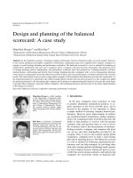
Figure 4 - Vocal fold morphology (Coronal view). Picture adapted from [30]. ......................... 7
Figure 5 – Microflap technique in practice, (Left) after removal of benign lesion and (Right)
redraping of microflap. .............................................................................................................. 9
Figure 6 – From Baxter [48] (Left) TISSEEL fibrin glue and applicator. (Right) FIBRINOTHERM
heating and stirring device. ..................................................................................................... 11
Figure 7 - Mini-Trach II – Seldinger Portex Minitracheotomy Kit [64] ..................................... 16
Figure 8 - Initial shapes of clips. ............................................................................................... 19
Figure 9 - Loaded Micro-clips in a typical microlaryngeal forcep ............................................ 20
Figure 10 – CAD drawing of clip (Isometric view) .................................................................... 21
Figure 11 - Micro-clip design. (Left) Before application. (Right) After application. ................. 22
Figure 12 - The weight gain of specimens (%wt) and pH of m-PBS plotted as functions of
corrosion time. ......................................................................................................................... 23
Figure 13 - Mounted trachea on ex-vivo setup ....................................................................... 25
Figure 14 – Ex-vivo experiment with excised larynx. (Left) Applied micro-clips within larynx.
(Right) Tensioning of micro-clips exhibiting their security. ..................................................... 26
Figure 15 - In-vivo setup .......................................................................................................... 28
Figure 16 – Microscopic view of the micro-clips after application in-vivo. ............................. 30
VIII
Figure 17 – Histological results showing inflammatory reaction to the micro-clips. (Left) Unpolished magnesium micro -clips. (Right) PCL coated micro-clips. ......................................... 31
Figure 18 – Applicator design. ................................................................................................. 34
Figure 19 – Non-indwelling voice prostheses from (Left) Provox NID [83] (Right) Blom-Singer.
................................................................................................................................................. 38
Figure 20 – Initial design. (Left) CAD drawing of 1) the measurement cannula and 2) its
adaptor cannula. (Right) Fabricated prototype used to test solution concept. ...................... 39
Figure 21 – The finalized design. From left to right, the modified measurement cannula,
handle, loader and plunger. ..................................................................................................... 40
Figure 22 – 3D CAD drawing of the handheld device. ............................................................. 41
Figure 23 – (a) Loaded puncture tool before puncture. (b) Loaded puncture tool after the
puncture stroke. (c) Measurement cannula after retraction stroke. Main body is concealed
for better view. ........................................................................................................................ 43
Figure 24– Loaded insertion tool with measurement cannula (a) just before voice prosthesis
insertion. (b) after the insertion stroke. .................................................................................. 44
Figure 25 – Fabricated main body of the handheld device with the measurement cannula
and puncture tool. (Left) After the puncture stroke. (Right) After the insertion stroke. ........ 45
Figure 26 - 3D CAD drawing of manipulator system with handheld device mounted. .......... 46
Figure 27 - Frame assignment for the manipulator with an alternate end effector for gripping
a trocar. .................................................................................................................................... 46
Figure 28 – Fabricated manipulator system for handheld device ........................................... 48
Figure 29 – User interface of manipulator system. (Left) Teleoperation mode and (Right)
predefined mode. .................................................................................................................... 48
Figure 30 – Simulated environment. (Left) Matlab simulation of the configuration of the
manipulator. (Right) Sub-manipulator mounted with an earlier version of the handheld
device ....................................................................................................................................... 49
IX
Figure 31 – In-vivo experiment. (Left) Simulated tracheostome. (Right) TNE system. ........... 50
Figure 32 - Experimental evaluation of the manual device. (A) Palpitation of anterior tracheal
wall. (B) Initial puncture with needle. (C) Guide wire insertion. (D) Removal of needle. (E)
Insertion of dilator with manual device. (F) Insertion of prosthesis after remove of dilator. (G)
Removal of measurement cannula. (H) External view of the surgical site. ............................. 51
Figure 33– Handheld device used to Inserted the voice prosthesis within the fistula. (Left)
Measurement cannula within fistula. (Right) Inserted duckbill voice prosthesis.................... 52
Figure 34 – Schematic diagram of the handheld device. ......................................................... 54
Figure 35– In-vivo force data of 14G needle insertion through posterior tracheal wall and
anterior esophageal wall. ........................................................................................................ 55
X
List of tables
Table 1 – Summary of In-vivo Experimental Results................................................................ 29
Table 2 ̶ DH parameters for the manipulator ......................................................................... 47
XI
1. Introduction
This chapter introduces the relevant background information, states the motivation
and defines the objectives of the projects in this thesis.
1.1. Background
Voice production is an essential skill that enables humans to communicate
information and convey our emotions. Generation of the sound waves required for speech
starts in the larynx, where the vocal folds vibrate due to fluctuations in pressure. However,
the larynx is a fragile organ susceptible to various disorders which can result in the reduction
or loss of voice capabilities. In moderate cases like the growth of benign vocal fold lesions,
voice microsurgery is commonly used for treatment. In more severe cases like total
laryngectomy, voice restoration methods are required instead.
1.1.1. Voice microsurgery
One of the most common causes of voice disorders are benign vocal fold lesions
such as nodules, polyps and cysts. While these lesions are non-cancerous, they may result in
impaired vocal fold closure and vibration, and reduction of voice quality. Treatment is
divided into two main categories based on the surgical instruments used – either laser
surgery or cold surgery. In laser surgery, a CO2 laser is used to ablate tissue and for
coagulation of the target region [1]. Together with a micro-manipulator for precise cutting,
the reduced blood loss during laser surgery enables a relatively clear view of the surgical
field. Although studies have found no significant difference in surgical outcomes between
laser and cold surgery [2-4], risk of thermal damage to surrounding tissues is still dependent
on familiarity with the equipment and surgical technique. This coupled with the increased
1
cost of equipment, maintenance, additional personnel and their training [1], has maintained
the relevance of traditional “cold” voice microsurgery techniques. Cold surgery allows for
tactile feedback and is better utilized in techniques like the micro-flap excision of benign
vocal fold lesions [4].
Figure 1 – Common tools used in laryngeal microsurgery. (Left) Laryngoscopes with a Fibre optic light carrier.
(Right) Microlaryngeal forceps and suction tubing.
Laryngeal microsurgery involves operating on the vocal folds under general
anaesthesia [5], where access to the vocal folds typically utilizes suspension laryngoscopy [6].
A rigid laryngoscope (Figure 1) inserted via the patient’s oral cavity provides a direct view of
the vocal folds. The laryngoscope is suspended over the patient’s chest to free up the
surgeon’s hands for operating. A binocular operating microscope is used to provide
magnification. Due to the prohibitive space constraints of laryngoscopes, microlaryngeal
instruments are thin and long to access the lesion while maximizing vision of the surgical
field (Figure 1). A significant level of dexterity is needed to handle the microlaryngeal tools,
especially considering the fragile structure of the vocal fold. Epithelial micro-flaps may be
created and elevated with micro-laryngeal instruments during surgery and these micro-flaps
require re-approximation on completion of the procedure. Current techniques for reapproximation are the focus of this thesis and will be discussed in the Chapter 2.
2
1.1.2. Voice restoration after total laryngectomy
In the medical field of otolaryngology or ENT (Ear, nose and throat), one of the most
commonly diagnosed malignancies in Singapore is laryngeal cancer [7], often the result of
heavy smoking and alcohol abuse. Total laryngectomy (TL) is often used as surgical
treatment for locally advanced laryngeal and pharyngeal cancer, in which the larynx is
detached from the trachea and excised. A tracheostome is subsequently constructed with
the remaining trachea for breathing (Figure 2).
Figure 2 - Before and after Laryngectomy. Images courtesy of InHeath Technologies [8] which supplies BlomSinger voice prostheses.
There are a number of options available for voice rehabilitation, ranging from
oesophageal speech, electrolaryngeal speech and prosthetic valve speech. Prosthetic voice
restoration provides the closest approximation to normal laryngeal voice and is considered
to be the gold standard of choice [9-11]. Prosthetic voice restoration involves the
implantation of voice prosthesis in a surgically created fistula between the posterior tracheal
3
wall and the anterior esophageal wall (Figure 3). This fistula is created by means of a
tracheoesophageal puncture (TEP) which can be carried out either as a primary procedure at
the time of TL or as a secondary procedure in a subsequent surgery [12-15]. In difficult
circumstances like if primary surgery is extensive and requires free flap or gastric pull-up
reconstruction, after heavy neck irradiation, or when alternative voicing techniques
including primary TEP have failed [16], secondary TEP has been found to prevent potential
complications such as cervical cellulitis, mediastinitis and salivary leakage [17-22] which may
adversely affect healing of the TL site.
Figure 3 – Tracheoesophageal Voice Prosthesis [8]
Traditionally, secondary TEP is performed in an operating theatre with rigid
esophagoscopy and under general anesthesia. Advancements in endoscopy have allowed
the development of transnasal esophagoscopy (TNE) and together with various secondary
TEP techniques, there is an increasing trend towards unsedated in-office TEP [18, 23-27]. Inoffice TEP avoids risks of general anesthesia and those associated with rigid esophagoscopy
4
such as esophageal perforation, and oral injury [28]. It enables cannulation of the esophagus
in patients with limited neck extension or stenosis of the neopharynx [29], allows rapid
recovery and decreases the need for patient monitoring. Also, performing the procedure in
an outpatient, office-based setting reduces patient cost. A stent in the form of a nasogastric
tube or catheter is often left in the fistula for one or two weeks while it matures. After which,
the patient returns to have a voice prosthesis sized and inserted, restoring his ability to
speak.
1.2. Motivation and objective
Our ability to express and communicate our thoughts and ideas is one that many of
us cannot afford to lose. The disabling psychosocial and economic consequence of losing
one’s voice is an ordeal for many patients and it is of great interest to reduce or mitigate
such issues/problems. This thesis is split into two main studies - voice microsurgery and
voice restoration.
For voice microsurgery, the main objective is to develop a small clip to be applied to
close incisions on the vocal fold, with at least comparable wound holding and healing
properties to current methods. Based on combining the ease and efficiency of using fibrin
glue with the precision and security of micro-sutures by specifically designing the micro-clip
and application technique to reduce technical complexity in achieving apposition of
epithelial flaps, it is hoped that such surgical micro-clips have the potential to reduce
procedure time and vocal fold scar, cumulating in better surgical outcomes and cost savings
for patients.
5
For voice restoration, an improvement in the current voice restoration method of
voice prosthesis insertion is required. By developing a device to insert the voice prosthesis
immediately at the time of initial puncture and leveraging on the visual advantages TNE
provides, it is hoped that such a device allows immediate voice restoration for patients,
reducing recovery time and thus translating to lower costs.
1.3. Research scope and organization of thesis
This study focuses on the design and development of two separate surgical devices
to aid the restoration of voice in patients in both voice microsurgery and total laryngectomy.
This thesis is organized as follows: Chapter One introduces the background of the research
topic as well as the motivation and objectives of the project. Chapter Two reviews the
current state of the art and related topics. Chapter Three presents the development of the
bio-absorbable micro-clips for would closure and their evaluation in ex-vivo and in-vivo
experiments. Chapter Four presents the development of various versions of the device for
TEP and voice prosthesis insertion and their evaluation in-vivo. Finally the contributions of
this work are concluded in Chapter Five.
6
2. Literature review
A range of relevant topics was reviewed to better understand and appreciate the
advantages and limitations in the current state of the art.
2.1. Bio-absorbable clips for vocal fold wound healing
2.1.1. Vocal fold morphology
Current voice microsurgery techniques are based on Hirano’s discovery of the
layered structure of the vocal folds [5, 30, 31]. Based on his microscopic work, the vocal fold
was found to be composed of three well defined layers - the epithelium, lamina propria and
vocalis muscle. The lamina propria was further subdivided into 3 layers, the superficial layer
of the lamina propria (SLLP), intermediate layer and deep layer. Figure 4 illustrates the
morphology of an adult vocal fold.
Epithelium
Cover
Superficial Layer
Intermediate Layer
Lamina
Propria
Deep Layer
Vocalis
Muscle
Interface
Body
Figure 4 - Vocal fold morphology (Coronal view). Picture adapted from [30].
In the SLLP, elastin and collagen fibres are loosely arranged within a matrix, whereas
dense elastin fibres make up most of the intermediate layer. Collagen is densely packed in
7
the deep layer, providing most of the support for the lamina propria [30]. Hirano also
proposed a cover-body concept, providing an explanation for the vibratory characteristics of
the vocal fold. Based on his theory, the cover (consisting of stratified squamous epithelium
and the underlying SLLP) is attached to the body (consisting of the vocalis and
thyroarytenoid muscles) by an elastic interface or ligament (composed of the intermediate
and deep layers of the lamina propria), with an increasing stiffness from superficial to deep.
This allows the cover to oscillate independently due to its elastic characteristics, resulting in
the mucosal wave seen on stroboscopy and most of the vibratory dynamics required for
good voice production and phonation [31].
The vocal fold is a layered structure, and the depth from the epithelial surface to
vocal ligament layer is approximately 1 mm [32, 33]. Surgical dissection is usually limited to
the surface layers including the epithelium and superficial lamina propria.
2.1.2. Laryngeal microsurgery
Early treatments for benign vocal fold lesions consisted of stripping (deepithelialization) of the entire vocal fold [34]. The healing process after this method of
treatment often results in significant vocal fold scar formation which causes a change in the
stiffness and viscoelastic layered structure of the lamina propria. This inhibits normal
vibration of the vocal fold and can cause significant dysphonia and possible glottic
incompetence. However with the discovery by Hirano of the layered structure of the vocal
foldand its implications on healing, treatment is now focused on preserving as much of the
normal vocal fold structure as possible [35-38]. Avoiding injury to the deeper structures is
important during voice microsurgery to minimize vocal fold scarring and persistent postoperative hoarseness.
8
Figure 5 – Microflap technique in practice, (Left) after removal of benign lesion and (Right) redraping of
microflap.
The microflap technique has been accepted as the standard approach for cold
surgical removal of benign vocal fold lesions [37, 39, 40], achieving the main principles of
vocal fold surgery by minimal tissue excision and minimal trauma to SLLP and epithelium.
This technique typically involves the initial creation of an epithelial incision beside the lesion.
Blunt dissection is used to elevate the microflap while taking care to minimize trauma to the
deeper layers of the lamina propria. Only pathologic tissue is excised and the microflap is
then reapproximated [34] as seen in Figure 5.
2.1.3. Current methods for laryngeal wound healing
Typically following excision of the lesion, the microflap is redraped to promote
primary healing. If there is loss of epithelium or dislodgement of the microflap, then healing
can occur by secondary intention. In this case, granulation tissue formation and epithelial
migration occur and there is correspondingly more scar tissue formation [37, 41]. Voice rest
is usually prescribed after surgery [42], but even with a totally compliant patient, apposition
of epithelial flaps edges can be difficult to maintain. More advanced techniques for
apposition of epithelial flaps during laryngeal microsurgery include using micro-sutures to
re-approximate the edges [35, 41, 43] and the application of fibrin glue to seal the flaps,
improve wound closure and minimize scar tissue formation [5, 44-47].
9
2.1.3.1. Micro-sutures
The use of micro-sutures in vocal fold wound closure was proposed by Woo et al. in
1995, hypothesizing that micro-sutures would allow precise positioning of wound edges and
maintenance of the approximation [41]. This would reduce exposure of the wound site and
permit primary healing to occur. They carried out the procedure in 18 patients, finding
improved voice results after surgery.
As there was no control group and basis for
comparison in Woo et al.’s study, Fleming et al. attempted to compare the amount of scar
formation with and without micro-sutures in a canine model [35]. A small sample group of 4
dogs were used, with bilateral micro-flaps defects created in each dog. 6-0 fast absorbing
gut sutures were used to close the microflap on only one side, leaving the contra-lateral side
unclosed. The amount of scar was evaluated between 39 and 49 days post surgery. Unsutured vocal folds were found to have at around 75% larger scar formation than sutured
vocal folds, concurring with Woo et al.’s hypothesis that the use of microsutures improves
postoperative wound healing.
However, suturing of tissue is still challenging due to restrictions imposed by the
laryngoscope. These restrictions include limitation of instrument movement to 4 degrees of
freedom, reduced force feedback and loss of stereopsis. High level of skill is required during
suturing and surgeons must exercise care not to grasp the deeper structures of the vocal
folds. Fleming et al. [35] also identified the length of time required for suture placement as
the main disadvantage of this technique, suggesting that practice and familiarization with
the technique using larger sutures before actual surgery could help mitigate the learning
curve.
10
Tsuji et al. recently proposed an improvement to the microsuture technique [43] by
pre-tying a small length of 4-0 non-absorbable nylon suture to the free end of a 7-0
absorbable suture. The nylon acted as an anchor at the epithelial surface, preventing the
thread from escaping and removing the need for an assistant surgeon to maintain tension
on the free end of the suture. This improved the ease of performing the technique. Their
new technique was tested on human cadaveric larynges for a total of 10 sutures and they
reported a placement time of 5 to 7 minutes per suture.
2.1.3.2. Fibrin glue
Despite good wound healing results demonstrated by micro-sutures, many surgeons
prefer using adhesives to hold down epithelial flaps to achieve wound closure. Tissue
adhesives such as cyanoacrylates and fibrin glue have been used [44] and are easier to apply
than sutures. Tisseel from Baxter is one such fibrin glue that is current used. Figure 6 shows
the current tools from Baxter used to prepare and apply fibrin glue to vocal fold wounds.
Figure 6 – From Baxter [48] (Left) TISSEEL fibrin glue and applicator. (Right) FIBRINOTHERM heating and
stirring device.
The components of TISSEEL from Baxter needs to be firstly, diluted based on
required concentration and heated before loading each into its allocated syringes for
11
application. Its applicator consists of a Y-tubing which combines both components and
expels it through a blunted hypodermic needle onto the application site.
A potential limitation of tissue adhesives includes increased scar tissue formation if
glue accumulates between the epithelial edges preventing proper approximation, or by
adhering the epithelium to the underlying connective tissue without proper reformation of
the intervening layered structure. Depending on the concentration used, the lack of tensile
strength of the adhesive is another possible concern. Fibrin glue can take several minutes for
initiation of curing and several hours to develop its full strength. Especially during the curing
phase, it may not possess sufficient tensile strength to withstand rupture of its bond [41].
Conversely, rapid curing can also restricts the surgeon from re-apposing malpositioned flaps,
resulting in even more scarring. Lastly, as the vocal folds vibrate at high frequencies during
speech, constant shearing against the adhesive glue causes wear and the resultant debris
may impede the vibratory properties of the vocal fold. Healing by secondary intention and a
broader scar may also occur as a result.
2.1.4. Surgical clips
While surgical clips or staples have been used in various areas of the body, they have
not been described previously for use in vocal folds. Nevertheless, as an alternative to time
consuming suturing, surgical staples provide a rapid solution to closure of long incisions [49].
A number of materials have also been studied in the design of surgical clips. Stainless steel
clips and newer materials such as titanium and tantalum have been used in areas where
surgical dissection is difficult, such as ligating the cystic duct and artery in laparoscopic
cholecystectomy[50]. However, major limitations of these materials include significant
foreign body reaction, poor holding power and significant interference with roentgenologic
12
studies like computerized tomography (CT) and magnetic resonance imaging (MRI), making
them unfavourable for application[51-54]. The introduction of ligating clips manufactured
from novel polymers such as polydioxanone in laparoscopic cholecystectomy helped to
address these limitations. These clips are completely absorbed in the process of ester bond
hydrolysis over a period of 180 days and the by-products are excreted by urine. Moreover,
these clips produce minimal tissue reactivity with good adhesion, and are radiolucent [54].
However, earlier studies with such clips proved them to be unsuitable for our requirements,
as they could not provide adequate structural strength due to the minute size of the microclips.
2.1.5. Magnesium as the core material
There have been a number of reviews on the potential and viability of magnesium as
a biomaterial [52, 55, 56]. Most of these studies, which focus on the use of magnesium in
orthopaedic implants and bio-absorbable vascular stents, concentrate on improving its
mechanical properties by alloying with various elements. Zhang et al. [57] reported
significant improvement of both biocompatibility and mechanical properties using Zn as an
additional alloying element to Mg-Si. Gu et al. [58] reported good biocompatibility of
magnesium with various alloying elements, recommending Al and Y for stents and Al, Ca, Zn,
Sn, Si and Mn for orthopaedic implants. Drynda et al. [59] developed and evaluated fluoride
coated Mg-Ca alloys for cardiovascular stents, reporting good biocompatibility and better
degradation behaviour.
However, as pure magnesium corrodes too quickly in the low pH environment of
physiological systems, much effort has also been placed into developing alloys or coatings to
limit its degradation behaviour [55]. Rosalbino et al. [53] reported improved corrosion
13