Professional guide to signs symptoms, 6th edition
Bạn đang xem bản rút gọn của tài liệu. Xem và tải ngay bản đầy đủ của tài liệu tại đây (11.44 MB, 822 trang )
LWBK395-FM_pi-x.qxd
11/30/09
4:02 PM
Page i
PROFESSIONAL GUIDE TO
SIGNS & SYMPTOMS
SIXTH EDITION
LWBK395-FM_pi-x.qxd
11/30/09
4:02 PM
Page ii
LWBK395-FM_pi-x.qxd
11/30/09
4:02 PM
Page iii
PROFESSIONAL GUIDE TO
SIGNS & SYMPTOMS
SIXTH EDITION
LWBK395-FM_pi-x.qxd
11/30/09
4:02 PM
STAFF
Executive Publisher
Judith A. Schilling McCann, RN, MSN
Clinical Director
Joan M. Robinson, RN, MSN
Clinical Project Manager
Jennifer Meyering, RN, BSN, MS, CCRN
Art Director
Elaine Kasmer
Product Manager
Rosanne Hallowell
Marketing Manager
Kimberly Schonberger
Copy Editor
Amy Furman
Vendor Manager
Beth Martz
Composition Services
Aptara, Inc.
Manufacturing Manager
Beth J. Welsh
Page iv
The clinical treatments described and recommended in
this publication are based on research and consultation
with nursing, medical, and legal authorities. To the best
of our knowledge, these procedures reflect currently accepted practice. Nevertheless, they can’t be considered
absolute and universal recommendations. For individual
applications, all recommendations must be considered
in light of the patient’s clinical condition and, before administration of new or infrequently used drugs, in light
of the latest package-insert information. The authors
and publisher disclaim any responsibility for any adverse effects resulting from the suggested procedures,
from any undetected errors, or from the reader’s misunderstanding of the text.
© 2011 by Lippincott Williams & Wilkins. All rights reserved. This book is protected by copyright. No part of it
may be reproduced, stored in a retrieval system, or
transmitted, in any form or by any means—electronic,
mechanical, photocopy, recording, or otherwise—without prior written permission of the publisher, except for
brief quotations embodied in critical articles and reviews and testing and evaluation materials provided by
publisher to instructors whose schools have adopted its
accompanying textbook. For information, write Lippincott Williams & Wilkins, 323 Norristown Road, Suite
200, Ambler, PA 19002-2756.
Printed in China
PGSS6E—010310
Library of Congress
Cataloging-in-Publication Data
Professional guide to signs & symptoms. — 6th ed.
p. ; cm.
Includes bibliographical references and index.
ISBN 978-1-60831-098-2 (alk. paper)
1. Symptoms—Handbooks, manuals, etc. I.
Lippincott Williams & Wilkins. II. Title:
Professional guide to signs and symptoms.
[DNLM: 1. Nursing Assessment—methods—
Handbooks. 2. Signs and Symptoms—Handbooks.
WY 49 P964 2011]
RC69.P77 2011
616Ј.047—dc22
2009033038
LWBK395-FM_pi-x.qxd
11/30/09
4:02 PM
Page v
TABLE OF
CONTENTS
Contributors and consultants
Foreword
SIGNS & SYMPTOMS (A–Z)
APPENDICES
Selected signs & symptoms
Potential agents of bioterrorism
Adverse effects associated with herbs
Obtaining a health history
Guide to laboratory test results
Selected references
Index
vi
viii
1
724
744
746
750
752
756
757
v
LWBK395-FM_pi-x.qxd
11/30/09
4:02 PM
Page vi
CONTRIBUTORS
AND CONSULTANTS
Diane Dixon Abercrombie,
MA, MMSc,
PhD Candidate, PA-C
Assistant Professor and Academic Coordinator
Department of Physician Assistant Studies
University of South Alabama
Mobile, Alabama
Marylee Bressie, RN, MSN, CCRN,
CCNS, CEN
Instructor
Spring Hill College Division of Nursing
Mobile, Alabama
Julie Carman,
RN, MS
Instructor
University of Arkansas
Fort Smith, Arkansas
RN, PhD, CNS-BC, ONC,
CCRN, CCNS, CNRN, CEN, CFRN
Clinical Nurse
Oregon Health & Science University
Portland, Oregon
RNC, PhD, WHNP-BC
Senior Associate
Coastline Writing Consultants
Assistant Professor (Retired)
University of North Carolina—Wilmington
School of Nursing
Wilmington, North Carolina
Julia Anne Isen,
RN, MS, FNP-C
Assistant Clinical Professor
University of California
San Francisco, California
Internal Medicine
Uniformed Services University of the Health
Sciences
Bethesda, Maryland
vi
PhD, ACNP/ACNS, BC
APN/Program Manager, Rapid Response Team
Central Arkansas Veterans Healthcare System
Little Rock, Arkansas
Cynthia Miculan,
RN, MSN, ONC, CE-BC
Clinical Manager
The University Hospital
Cincinnati, Ohio
Steven Noakes,
MPAS, PA-C
Division Officer, Acute Care Clinic
Marine Corps Recruit Depot
San Diego, California
Allen Phelps,
Laura M. Criddle,
Shelton M. Hisley,
Anna Lee Jarrett,
MPAS, PA-C
Physician Assistant
Naval Medical Center
San Diego, California
Rexann G. Pickering,
RN, BSN, MS, MSN,
PhD, CIM, CIP
Administrator, Human Protection–Research
Methodist Healthcare
Memphis, Tennessee
Roseanne Hanlon Rafter,
RN, MSN,
GCNS, BC
Director of Nursing, Professional Practice
Chestnut Hill Hospital
Philadelphia, Pennsylvania
Sundaram V. Ramanan,
MD, FRCP
Professor of Medicine
St. Francis Hospital—University of Connecticut
Hartford, Connecticut
LWBK395-FM_pi-x.qxd
11/30/09
4:02 PM
Page vii
C O N T R I B U T O R S A N D C O N S U LT A N T S
Richard R. Roach,
MD, FACP
Assistant Professor of Internal Medicine
Michigan State University
Kalamazoo Center for Medical Studies
Kalamazoo, Michigan
Ora V. Robinson,
RN, PhD
Assistant Professor
California State University
San Bernardino, California
Phillip Todd Smith,
MHS, PA-C
Assistant Professor
Department of Physician Assistant Studies
University of South Alabama
Mobile, Alabama
Allison J. Terry,
RN, MSN, PhD
Director, Center for Nursing
Alabama Board of Nursing
Montgomery, Alabama
Daniel T. Vetrosky,
PhD, PA-C
Assistant Professor
University of South Alabama
Mobile, Alabama
Gail A. Viergutz,
MS, ANP-C
Nurse Practitioner, Emergency Department and
Urgent Care
Ministry Corporation
St. Michael’s Hospital
Stevens Point, Wisconsin
vii
LWBK395-FM_pi-x.qxd
11/30/09
4:02 PM
Page viii
FOREWORD
With continuing advances in medical technology, laboratory studies, and diagnostic testing,
clinical diagnosis and physical examination
skills are in danger of becoming a lost art. I
have seen too many students and novice practitioners become overly dependent on frequently
imperfect, unreliable, and expensive tests to diagnose the cause of their patients’ illnesses. The
sixth edition of Professional Guide to Signs &
Symptoms will help ensure that this doesn’t happen. This fully reviewed and updated edition
provides a comprehensive yet easy-tounderstand compilation of many important
signs and symptoms seen in clinical practice,
and can help guide initial interventions and the
appropriate use of laboratory and diagnostic
studies.
The scope and organization of this sixth edition make it a valuable reference for students,
nurses, and practitioners at all levels of training
and expertise. More than 500 clinical signs and
symptoms are arranged alphabetically and discussed in the body of the text. The new full-color
format is appealing and enables quick and easy
retrieval of relevant information. Easy-to-read
tables, charts, and illustrations make difficultto-grasp physiologic and clinical concepts understandable. Potentially obscure pathologic
signs are clearly explained and should become
more readily apparent to the astute clinical observer. New sections examining troublesome infectious diseases (methicillin-resistant Staphylococcus aureus, vancomycin-resistant
enterococci, and vancomycin-resistant S.
aureus) and popcorn lung disease (diacetyl exposure) are included.
viii
Each sign and symptom is reviewed in a concise and standard format. Every entry begins
with a brief review of the sign or symptom and
is followed, where applicable, by a focused discussion of possible emergency interventions.
Relevant history and physical findings are then
reviewed and possible medical causes are discussed. Special considerations for caregivers
provide practical advice, and pointers for pediatric and elderly populations should be particularly helpful for those who care for patients at
either end of the age spectrum. Detailed differential diagnosis matrixes and flowcharts interspersed throughout the text aid patient assessment and diagnosis, while patient counseling
sections provide helpful recommendations for
patients and families once the diagnosis is established.
An additional 250 less frequently encountered selected signs and symptoms are briefly
reviewed in the first appendix. Updated sections
on the signs and symptoms of bioterrorism
agents and the adverse effects of herbal remedies are particularly timely. The guide to obtaining a patient history provides helpful tips for
conducting a medical interview, collecting primary clinical data, and performing a thorough
review of systems. The index is crossreferenced and thorough, and the inside-thecover listing of common signs and symptoms in
both English and Spanish make this sixth edition a valuable reference for students, nurses,
and practitioners living or traveling abroad.
I believe anyone who provides clinical care to
patients and who is interested in the focused
and appropriate use of medical technology,
LWBK395-FM_pi-x.qxd
11/30/09
4:02 PM
Page ix
F O R E WO R D
diagnostic testing, and initial interventions will
find this comprehensive text extremely valuable.
The standardized format with its easy-to-read
tables, charts, and illustrations make this sixth
edition an indispensable tool for the inquisitive
student, nurse, or clinical practitioner.
Charles W. Mackett III,
MD, FAAFP
Associate Professor and Executive
Vice Chairman
Department of Family Medicine
University of Pittsburgh (Pa.) Medical Center
ix
LWBK395-FM_pi-x.qxd
11/30/09
4:02 PM
Page x
LWBK395-A_p1-75.qxd
11/30/09
3:09 PM
Page 1
A
Abdominal distention
Abdominal distention refers to increased abdominal girth—the result of increased intraabdominal pressure forcing the abdominal wall
outward. Distention may be mild or severe, depending on the amount of pressure. It may be
localized or diffuse and may occur gradually or
suddenly. Acute abdominal distention may signal life-threatening peritonitis or acute bowel
obstruction.
Abdominal distention may result from fat, flatus, a fetus (pregnancy or intra-abdominal mass
[ectopic pregnancy]), or fluid. Fluid and gas are
normally present in the GI tract but not in the
peritoneal cavity. However, if fluid and gas are
unable to pass freely through the GI tract, abdominal distention occurs. In the peritoneal
cavity, distention may reflect acute bleeding, accumulation of ascitic fluid, or air from perforation of an abdominal organ.
Abdominal distention doesn’t always signal
pathology. For example, in anxious patients or
those with digestive distress, localized distention in the left upper quadrant can result from
aerophagia—the unconscious swallowing of air.
Generalized distention can result from ingestion
of fruits or vegetables with large quantities of
unabsorbable carbohydrates, such as legumes,
or from abnormal food fermentation by microbes. Don’t forget to rule out pregnancy in all
females with abdominal distention.
EMERGENCY INTERVENTIONS If the patient displays abdominal distention, quickly
check for signs of hypovolemia, such as pallor,
diaphoresis, hypotension, rapid and thready pulse,
rapid and shallow breathing, decreased urine output, poor capillary refill, and altered mentation.
Ask the patient if he’s experiencing severe abdominal pain or difficulty breathing. Find out about any
recent accidents, and observe the patient for signs
of trauma and peritoneal bleeding, such as
Cullen’s sign or Turner’s sign. Then auscultate all
abdominal quadrants, noting rapid and highpitched, diminished, or absent bowel sounds. (If
you don’t hear bowel sounds immediately, listen
for at least 5 minutes.) Gently palpate the abdomen for rigidity. Remember that deep or extensive palpation may increase pain.
If you detect abdominal distention and rigidity
along with abnormal bowel sounds, and the patient complains of pain, begin emergency interventions. Place the patient in the supine position, administer oxygen, and insert an I.V.
catheter for fluid replacement. Prepare to insert
a nasogastric tube to relieve acute intraluminal
distention. Reassure the patient and prepare him
for surgery.
HISTORY AND PHYSICAL
EXAMINATION
If the patient’s abdominal distention isn’t acute,
ask about its onset and duration and associated
signs. A patient with localized distention may
report a sensation of pressure, fullness, or tenderness in the affected area. A patient with generalized distention may report a bloated feeling,
a pounding heartbeat, and difficulty breathing
deeply or breathing when lying flat. The patient
may also feel unable to bend at his waist. Be
1
LWBK395-A_p1-75.qxd
2
11/30/09
3:09 PM
Page 2
ABDOMINAL DISTENTION
sure to ask about abdominal pain, fever,
nausea, vomiting, anorexia, altered bowel
habits, and weight gain or loss.
Obtain a medical history, noting GI or biliary
disorders that may cause peritonitis or ascites,
such as cirrhosis, hepatitis, or inflammatory
bowel disease. (See Detecting ascites.) Also note
chronic constipation. Has the patient recently
had abdominal surgery, which can lead to abdominal distention? Ask about recent accidents, even minor ones, like falling off a stepladder.
Perform a complete physical examination.
Don’t restrict the examination to the abdomen
because you could miss important clues to the
cause of abdominal distention. Next, stand at
the foot of the bed and observe the recumbent
patient for abdominal asymmetry to determine
if distention is localized or generalized. Then
assess abdominal contour by stooping at his
side. Inspect for tense, glistening skin and
bulging flanks, which may indicate ascites. Observe the umbilicus. An everted umbilicus may
indicate ascites or an umbilical hernia. An inverted umbilicus may indicate distention from
gas; it’s also common in obese individuals. Inspect the abdomen for signs of an inguinal or
femoral hernia and for incisions that may point
to adhesions; both may lead to intestinal obstruction. Then auscultate for bowel sounds,
abdominal friction rubs (indicating peritoneal
inflammation), and bruits (indicating an
aneurysm). Listen for a succussion splash—a
splashing sound normally heard in the stomach
when the patient moves or when palpation disturbs the viscera. An abnormally loud splash
indicates fluid accumulation, suggesting gastric
dilation or obstruction.
Next, percuss and palpate the abdomen to
determine if distention results from air, fluid, or
both. A tympanic note in the left lower quadrant
suggests an air-filled descending or sigmoid
colon. A tympanic note throughout a generally
distended abdomen suggests an air-filled peritoneal cavity. A dull percussion note throughout
a generally distended abdomen suggests a fluidfilled peritoneal cavity. Shifting of dullness laterally when the patient is in the decubitus position also indicates a fluid-filled abdominal
cavity. A pelvic or intra-abdominal mass causes
local dullness upon percussion and should be
palpable. Obesity causes a large abdomen with
generalized rather then localized dullness and
without shifting dullness, prominent tympany,
or palpable bowel or other masses.
Palpate the abdomen for tenderness, noting
whether it’s localized or generalized. Watch for
peritoneal signs and symptoms, such as
rebound tenderness, guarding, rigidity,
McBurney’s point, obturator sign, and psoas
sign. Female patients should undergo a pelvic
examination; males, a genital examination. All
patients who report abdominal pain should undergo a digital rectal examination with fecal
occult blood testing. Finally, measure abdominal girth for a baseline value. Mark the flanks
with a felt-tipped pen as a reference point for
subsequent measurements. (See Abdominal distention: Causes and associated findings, pages 4
and 5.)
MEDICAL CAUSES
◆ Abdominal cancer. Generalized abdominal
distention may occur when the cancer—most
commonly ovarian, hepatic, or pancreatic
cancer—produces ascites (usually in a patient
with a known tumor). It’s an indication of advanced disease. Shifting dullness and a fluid
wave accompany distention. Associated signs
and symptoms may include severe abdominal
pain, an abdominal mass, anorexia, jaundice, GI
hemorrhage (hematemesis or melena), dyspepsia, and weight loss that progresses to muscle
weakness and atrophy.
◆ Abdominal trauma. When brisk internal
bleeding accompanies trauma, abdominal distention may be acute and dramatic. Associated
signs and symptoms of this life-threatening disorder include abdominal rigidity with guarding,
decreased or absent bowel sounds, vomiting,
tenderness, and abdominal bruising. The patient may feel pain over the trauma site, or over
the scapula if abdominal bleeding irritates the
phrenic nerve. Signs of hypovolemic shock
(such as hypotension and rapid, thready pulse)
appear with significant blood loss.
◆ Bladder distention. Various disorders cause
bladder distention, which in turn causes lower
abdominal distention. Slight dullness on percussion above the symphysis indicates mild bladder
distention. A palpable, smooth, rounded, fluctuant suprapubic mass suggests severe distention;
a fluctuant mass extending to the umbilicus indicates extremely severe distention. Urinary
dribbling, frequency, or urgency may occur with
urinary obstruction. Suprapubic discomfort is
also common.
◆ Cirrhosis. In cirrhosis, ascites causes generalized distention and is confirmed by a fluid
wave, shifting dullness, and a puddle sign.
LWBK395-A_p1-75.qxd
11/30/09
3:09 PM
Page 3
ABDOMINAL DISTENTION
3
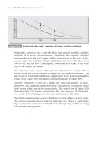
Detecting ascites
To differentiate ascites from other causes of abdominal distention, check for shifting dullness and
fluid wave, as described here.
Shifting dullness
Step 1. With the patient in a supine position, percuss from the umbilicus outward
to the flank, as shown. Draw a line on
the patient’s skin to mark the change
from tympany to dullness.
Step 2. Turn the patient onto his side.
(Note that this position causes ascitic fluid to shift.) Percuss again and mark the
change from tympany to dullness. Any
difference between these lines can indicate ascites.
Fluid wave
Have another person press deeply into
the patient’s midline to prevent vibration
from traveling along the abdominal wall.
Place one of your palms on one of the
patient’s flanks, as shown. Strike the opposite flank with your other hand. If you
feel the blow in the opposite palm, ascitic fluid is present.
Umbilical eversion and caput medusae (dilated
veins around the umbilicus) are common. The
patient may report a feeling of fullness or
weight gain. Associated findings include vague
abdominal pain, fever, anorexia, nausea, vomiting, constipation or diarrhea, bleeding tendencies, severe pruritus, palmar erythema, spider
angiomas, leg edema, and possibly splenomegaly.
Hematemesis, encephalopathy, gynecomastia,
or testicular atrophy may also occur. Jaundice
is usually a late sign. Hepatomegaly occurs initially, but the liver may not be palpable in advanced disease.
◆ Gastric dilation (acute). Left-upperquadrant distention is characteristic in acute
gastric dilation, but the presentation varies. The
patient usually complains of epigastric fullness
or pain and nausea with or without vomiting.
Physical examination reveals tympany, gastric
tenderness, and a succussion splash. Initially,
peristalsis may be visible. Later, hypoactive or
absent bowel sounds confirm ileus. The patient
may be pale and diaphoretic and may exhibit
tachycardia or bradycardia.
◆ Heart failure. Generalized abdominal distention due to ascites typically accompanies
(Text continues on page 6.)
LWBK395-A_p1-75.qxd
3:09 PM
Page 4
ABDOMINAL DISTENTION
SIGNS & SYMPTOMS
Abdominal distention: Causes and associated findings
Abdominal cancer
Abdominal trauma
Bladder distention
Cirrhosis
Gastric dilation
(acute)
•
• •
• •
•
•
•
•
•
•
•
• • •
•
•
Heart failure
Irritable bowel
syndrome
Large-bowel
obstruction
Mesenteric artery
occlusion (acute)
Peritonitis
Small-bowel
obstruction
Toxic megacolon
(acute)
•
•
•
• • • •
• •
•
syndrome
Paralytic ileus
• •
•
Nephrotic
Ovarian cysts
Edema
Diarrhea
Constipation
hypoactive
Bowel sounds,
hyperactive
Bowel sounds,
absent
Bowel sounds,
Anorexia
Abdominal rigidity
Common
causes
Abdominal pain
Major associated signs and symptoms
Abdominal mass
4
11/30/09
•
• •
•
• •
•
•
•
•
•
•
•
•
•
•
•
•
•
•
• •
•
•
•
•
•
•
•
•
•
•
•
•
•
•
•
•
•
• •
•
•
•
•
•
•
•
•
•
Weight change
Vomiting
Urinary frequency
Tachypnea
Tachycardia
splash
Succussion
tenderness
Rebound
3:09 PM
Oliguria
Nausea
distention
11/30/09
Jugular vein
Jaundice
Hypotension
Hepatomegaly
Fever
LWBK395-A_p1-75.qxd
Page 5
ABDOMINAL DISTENTION
s
•
•
•
• •
•
5
LWBK395-A_p1-75.qxd
6
11/30/09
3:09 PM
Page 6
ABDOMINAL DISTENTION
severe cardiovascular impairment and is confirmed by shifting dullness and a fluid wave.
Signs and symptoms of heart failure are numerous and depend on the disease stage and degree of cardiovascular impairment. Hallmarks
include peripheral edema, jugular vein distention, dyspnea, and tachycardia. Common
associated signs and symptoms include hepatomegaly (which may cause right-upper-quadrant
pain), nausea, vomiting, productive cough,
crackles, cool extremities, cyanotic nail beds,
nocturia, exercise intolerance, nocturnal
wheezing, diastolic hypertension, and cardiomegaly.
◆ Irritable bowel syndrome (IBS). IBS may
produce intermittent, localized distention—the
result of periodic intestinal spasms. Lower abdominal pain or cramping typically accompanies these spasms. The pain is usually relieved
by defecation or by passage of intestinal gas
and is aggravated by stress. Other possible
signs and symptoms include diarrhea that may
alternate with constipation or normal bowel
function; nausea; dyspepsia; straining and urgency at defecation; feeling of incomplete evacuation; and small, mucus-streaked stools.
◆ Large-bowel obstruction. Dramatic abdominal distention is characteristic in large-bowel
obstruction, a life-threatening disorder; in fact,
loops of the large bowel may become visible on
the abdomen. Constipation precedes distention
and may be the only symptom for days. Associated findings include tympany, high-pitched
bowel sounds, and sudden onset of colicky lower abdominal pain that becomes persistent. Fecal vomiting and diminished peristaltic waves
and bowel sounds are late signs.
◆ Mesenteric artery occlusion (acute). In
mesenteric artery occlusion—a life-threatening
disorder—abdominal distention usually occurs
several hours after the sudden onset of severe,
colicky periumbilical pain accompanied by rapid
(even forceful) bowel evacuation. The pain later
becomes constant and diffuse. Related signs
and symptoms include severe abdominal tenderness with guarding and rigidity, absent bowel sounds and, occasionally, a bruit in the right
iliac fossa. The patient may also experience
vomiting, anorexia, diarrhea, or constipation.
Late signs include fever, tachycardia, tachypnea, hypotension, and cool, clammy skin. Abdominal distention or GI bleeding may be the
only clue if pain is absent.
◆ Nephrotic syndrome. Nephrotic syndrome
may produce massive edema, causing general-
ized abdominal distention with a fluid wave and
shifting dullness. It may also produce elevated
blood pressure, hematuria or oliguria, fatigue,
anorexia, depression, pallor, periorbital edema,
scrotal swelling, and skin striae.
◆ Ovarian cysts. Typically, large ovarian cysts
produce lower abdominal distention accompanied by umbilical eversion. Because they’re thin
walled and fluid filled, these cysts produce a fluid wave and shifting dullness—signs that mimic
ascites. Lower abdominal pain and a palpable
mass may be present.
◆ Paralytic ileus. Paralytic ileus, which produces generalized distention with a tympanic
percussion note, is accompanied by absent or
hypoactive bowel sounds and, occasionally,
mild abdominal pain and vomiting. The patient
may be severely constipated or may pass flatus
and small, liquid stools.
◆ Peritonitis. In peritonitis—a life-threatening
disorder—abdominal distention may be localized or generalized, depending on the extent of
peritonitis. Fluid accumulates first within the
peritoneal cavity and then within the bowel lumen, causing a fluid wave and shifting dullness.
Typically, distention is accompanied by rebound
tenderness, abdominal rigidity, and sudden and
severe abdominal pain that worsens with movement.
The skin over the patient’s abdomen may appear taut. Associated signs and symptoms usually include hypoactive or absent bowel sounds,
fever, chills, hyperalgesia, nausea, and vomiting. Signs of shock, such as tachycardia and hypotension, appear with significant fluid loss into
the abdomen.
◆ Small-bowel obstruction. Abdominal distention, which is characteristic in small-bowel
obstruction—a life-threatening disorder—is
most pronounced during late obstruction, especially in the distal small bowel. Auscultation reveals hypoactive or hyperactive bowel sounds,
whereas percussion produces a tympanic note.
Accompanying signs and symptoms include
colicky periumbilical pain, constipation, nausea, and vomiting; the higher the obstruction,
the earlier and more severe the vomiting. Rebound tenderness reflects intestinal strangulation with ischemia. Associated signs and symptoms include drowsiness, malaise, and signs of
dehydration. Signs of hypovolemic shock appear with progressive dehydration and plasma
loss.
◆ Toxic megacolon (acute). Toxic megacolon
is a life-threatening complication of infectious
LWBK395-A_p1-75.qxd
11/30/09
3:09 PM
Page 7
ABDOMINAL MASS
or ulcerative colitis that produces dramatic abdominal distention. The distention usually develops gradually and is accompanied by a tympanic percussion note, diminished or absent
bowel sounds, and mild rebound tenderness.
The patient also experiences abdominal pain
and tenderness, fever, tachycardia, and dehydration.
7
GERIATRIC POINTERS
As people age, fat tends to accumulate in the
lower abdomen and near the hips, even when
body weight is stable. This accumulation,
together with weakening abdominal muscles,
commonly produces a potbelly, which some
elderly patients interpret as fluid collection or
evidence of disease.
SPECIAL CONSIDERATIONS
PATIENT COUNSELING
Position the patient comfortably, using pillows
for support. Place him on his left side to help flatus escape or, if he has ascites, elevate the head
of the bed to ease his breathing. Administer
drugs to relieve pain, and offer emotional support.
Prepare the patient for diagnostic tests, such
as abdominal X-rays, endoscopy, laparoscopy,
ultrasonography, computed tomography scan,
or possibly paracentesis.
If the patient’s anxiety triggers air swallowing or
deep breathing that causes discomfort, advise
him to take slow breaths. If the patient has an
obstruction or ascites, explain food and fluid restrictions. Stress good oral hygiene to prevent
dry mouth.
PEDIATRIC POINTERS
Because a young child’s abdomen is normally
rounded, distention may be difficult to observe. However, a child’s abdominal wall is
less well developed than an adult’s, so palpation is easier. When percussing the abdomen,
remember that children normally swallow air
when eating and crying, resulting in louderthan-normal tympany. Minimal tympany with
abdominal distention may result from fluid accumulation or solid masses. To check for abdominal fluid, test for shifting dullness instead
of for a fluid wave. (In a child, air swallowing
and incomplete abdominal muscle development make the fluid wave difficult to
interpret.)
Some children won’t cooperate with a physical examination. Try to gain the child’s confidence, and consider allowing him to remain in
the parent’s or caregiver’s lap. You can gather
clues by observing the child while he’s coughing, walking, or even climbing on office furniture. Remove all the child’s clothing to avoid
missing any diagnostic clues. Also, perform a
gentle rectal examination.
In neonates, ascites usually results from GI or
urinary perforation; in older children, from heart
failure, cirrhosis, or nephrosis. Besides ascites,
congenital malformations of the GI tract (such
as intussusception and volvulus) may cause abdominal distention. A hernia may cause distention if it produces an intestinal obstruction. In
addition, overeating and constipation can cause
distention.
Abdominal mass
Commonly detected on routine physical examination, an abdominal mass is a localized
swelling in one abdominal quadrant. Typically,
this sign develops insidiously and may
represent an enlarged organ, a neoplasm,
an abscess, a vascular defect, or a fecal
mass.
Distinguishing an abdominal mass from a
normal structure requires skillful palpation. At
times, palpation must be repeated with the patient in a different position or performed by a
second examiner to verify initial findings. A palpable abdominal mass is an important clinical
sign and usually represents a serious—and perhaps life-threatening—disorder.
EMERGENCY INTERVENTIONS If the
patient has a pulsating midabdominal
mass and severe abdominal or back pain, suspect an aortic aneurysm. Quickly take his vital
signs. Because the patient may require emergency surgery, withhold food or fluids until the
patient is examined. Prepare to administer oxygen and to start an I.V. infusion for fluid and
blood replacement. Obtain routine preoperative
tests, and prepare the patient for angiography.
Frequently monitor blood pressure, pulse rate,
respirations, and urine output.
Be alert for signs of shock, such as tachycardia,
hypotension, and cool, clammy skin, which may
indicate significant blood loss.
HISTORY AND PHYSICAL
EXAMINATION
If the patient’s abdominal mass doesn’t suggest
an aortic aneurysm, take a detailed history. Ask
LWBK395-A_p1-75.qxd
8
11/30/09
3:09 PM
Page 8
ABDOMINAL MASS
the patient if the mass is painful. If so, ask if the
pain is constant or if it occurs only on palpation.
Is it localized or generalized? Determine if the
patient was already aware of the mass. If he
was, find out if he noticed any change in its size
or location.
Next, review the patient’s medical history,
paying special attention to GI disorders. Ask the
patient about GI symptoms, such as constipation, diarrhea, rectal bleeding, abnormally
colored stools, and vomiting. Has the patient
noticed a change in appetite? If the patient is female, ask whether her menstrual cycles are regular and when the 1st day of her last menstrual
period was.
Perform a complete physical examination.
Next, auscultate for bowel sounds in each quadrant. Listen for bruits or friction rubs, and check
for enlarged veins. Lightly palpate and then
deeply palpate the abdomen, assessing any
painful or suspicious areas last. Note the patient’s position when you locate the mass. Some
masses can be detected only with the patient in
a supine position; others require a side-lying
position.
Estimate the size of the mass in centimeters.
Determine its shape. Is it round or sausage
shaped? Describe its contour as smooth, rough,
sharply defined, nodular, or irregular. Determine
the consistency of the mass. Is it doughy, soft,
solid, or hard? Also, percuss the mass. A dull
sound indicates a fluid-filled mass; a tympanic
sound, an air-filled mass.
Next, determine if the mass moves with your
hand or in response to respiration. Is the mass
free-floating or attached to intra-abdominal
structures? To determine whether the mass is
located in the abdominal wall or the abdominal
cavity, ask the patient to lift his head and
shoulders off the examination table, thereby
contracting his abdominal muscles. While these
muscles are contracted, try to palpate the mass.
If you can, the mass is in the abdominal wall; if
you can’t, the mass is within the abdominal
cavity. (See Abdominal masses: Locations and
causes.)
After the abdominal examination is complete,
perform pelvic, genital, and rectal examinations.
MEDICAL CAUSES
◆ Abdominal aortic aneurysm. An abdominal
aortic aneurysm may persist for years, producing only a pulsating periumbilical mass with a
systolic bruit over the aorta. However, it may
become life-threatening if the aneurysm expands and its walls weaken. In such cases, the
patient initially reports constant upper abdominal pain or, less often, low back or dull abdominal pain. If the aneurysm ruptures, he’ll report
severe abdominal and back pain. And after rupture, the aneurysm no longer pulsates.
Associated signs and symptoms of rupture
include mottled skin below the waist, absent
femoral and pedal pulses, lower blood pressure
in the legs than in the arms, mild to moderate
tenderness with guarding, and abdominal
rigidity. Signs of shock—such as tachycardia
and cool, clammy skin—appear with significant
blood loss.
◆ Bladder distention. A smooth, rounded,
fluctuant suprapubic mass is characteristic. In
extreme distention, the mass may extend to the
umbilicus. Severe suprapubic pain and urinary
frequency and urgency may also occur.
◆ Cholecystitis. Deep palpation below the
liver border may reveal a smooth, firm,
sausage-shaped mass. However, in acute inflammation, the gallbladder is usually too tender to be palpated. Cholecystitis can cause severe right-upper-quadrant pain that may
radiate to the right shoulder, chest, or back;
abdominal rigidity and tenderness; fever; pallor; diaphoresis; anorexia; nausea; and vomiting. Recurrent attacks usually occur 1 to 6
hours after meals. Murphy’s sign (inspiratory
arrest elicited when the examiner palpates the
right upper quadrant as the patient takes a
deep breath) is common.
◆ Cholelithiasis. A stone-filled gallbladder
usually produces a painless right-upperquadrant mass that’s smooth and sausageshaped. However, passage of a stone through
the bile or cystic duct may cause severe rightupper-quadrant pain that radiates to the epigastrium, back, or shoulder blades. Accompanying
signs and symptoms include anorexia, nausea,
vomiting, chills, diaphoresis, restlessness, and
low-grade fever. Jaundice may occur with obstruction of the common bile duct. The patient
may also experience intolerance of fatty foods
and frequent indigestion.
◆ Colon cancer. A right-lower-quadrant mass
may occur in cancer of the right colon, which
may also cause occult bleeding with anemia
and abdominal aching, pressure, or dull cramps.
Associated findings include weakness, fatigue,
exertional dyspnea, vertigo, and signs and
symptoms of intestinal obstruction, such as obstipation and vomiting.
LWBK395-A_p1-75.qxd
11/30/09
3:09 PM
Page 9
ABDOMINAL MASS
Abdominal masses: Locations and causes
The location of an abdominal mass provides an important clue to the causative disorder. Below
you’ll find the disorders responsible for abdominal masses in each of the four abdominal quadrants.
Right upper quadrant
Left upper quadrant
◆ Aortic aneurysm (epigastric area)
◆ Aortic aneurysm (epigastric area)
◆ Cholecystitis or cholelithiasis
◆ Gastric carcinoma (epigastric area)
◆ Gallbladder, gastric, or hepatic carcinoma
◆ Hydronephrosis
◆ Hepatomegaly
◆ Pancreatic abscess (epigastric area)
◆ Hydronephrosis
◆ Pancreatic pseudocysts (epigastric area)
◆ Pancreatic abscess or pseudocysts
◆ Renal cell carcinoma
◆ Renal cell carcinoma
◆ Splenomegaly
Right lower quadrant
Left lower quadrant
◆ Bladder distention (suprapubic area)
◆ Bladder distention (suprapubic area)
◆ Colon cancer
◆ Colon cancer
◆ Crohn’s disease
◆ Diverticulitis
◆ Ovarian cyst (suprapubic area)
◆ Ovarian cyst (suprapubic area)
◆ Uterine leiomyomas (suprapubic area)
◆ Uterine leiomyomas (suprapubic area)
◆ Volvulus
9
LWBK395-A_p1-75.qxd
10
11/30/09
3:09 PM
Page 10
ABDOMINAL MASS
Occasionally, cancer of the left colon also
causes a palpable mass. Usually though, it produces rectal bleeding, intermittent abdominal
fullness or cramping, and rectal pressure. The
patient may also report fremitus and pelvic discomfort. Later, he develops obstipation, diarrhea, or pencil-shaped, grossly bloody, or
mucus-streaked stools. Typically, defecation
relieves pain.
◆ Crohn’s disease. In Crohn’s disease, tender,
sausage-shaped masses are usually palpable in
the right lower quadrant and, at times, in the
left lower quadrant. Attacks of colicky rightlower-quadrant pain and diarrhea are common.
Associated signs and symptoms include fever,
anorexia, weight loss, hyperactive bowel
sounds, nausea, abdominal tenderness with
guarding, and perirectal, skin, or vaginal
fistulas.
◆ Diverticulitis. Most common in the sigmoid colon, diverticulitis may produce a leftlower-quadrant mass that’s usually tender,
firm, and fixed. It also produces intermittent
abdominal pain that’s relieved by defecation
or passage of flatus. Other findings may include alternating constipation and diarrhea,
nausea, low-grade fever, and a distended and
tympanic abdomen.
◆ Gallbladder cancer. Gallbladder cancer
may produce a moderately tender, irregular
mass in the right upper quadrant. Accompanying it is chronic, progressively severe epigastric
or right-upper-quadrant pain that may radiate
to the right shoulder. Associated signs and
symptoms include nausea, vomiting, anorexia,
weight loss, jaundice, and possibly
hepatosplenomegaly.
◆ Gastric cancer. Advanced gastric cancer
may produce an epigastric mass. Early findings
include chronic dyspepsia and epigastric discomfort, whereas late findings include weight
loss, a feeling of fullness after eating, fatigue,
and occasionally coffee-ground vomitus or melena.
◆ Hepatic cancer. Hepatic cancer produces a
tender, nodular mass in the right upper quadrant or right epigastric area accompanied by severe pain that’s aggravated by jolting. Other
effects include weight loss, weakness, anorexia,
nausea, fever, dependent edema, and occasionally jaundice and ascites. A large tumor can also
cause a bruit or hum.
◆ Hepatomegaly. Hepatomegaly produces a
firm, blunt, irregular mass in the epigastric region or below the right costal margin.
Associated signs and symptoms vary with the
causative disorder but commonly include ascites, right-upper-quadrant pain and tenderness, anorexia, nausea, vomiting, leg edema,
jaundice, palmar erythema, spider angiomas,
gynecomastia, testicular atrophy, and possibly
splenomegaly.
◆ Hydronephrosis. By enlarging one or both
kidneys, hydronephrosis produces a smooth,
boggy mass in one or both flanks. Other findings
vary with the degree of hydronephrosis. The patient may have severe colicky renal pain or dull
flank pain that radiates to the groin, vulva, or
testes. Hematuria, pyuria, dysuria, alternating
oliguria and polyuria, nocturia, accelerated hypertension, nausea, and vomiting may also
occur.
◆ Ovarian cyst. A large ovarian cyst may produce a smooth, rounded, fluctuant mass, resembling a distended bladder, in the suprapubic region. Large or multiple cysts may also cause
mild pelvic discomfort, low back pain, menstrual irregularities, and hirsutism. A twisted or ruptured cyst may cause abdominal tenderness,
distention, and rigidity.
◆ Pancreatic abscess. Occasionally, pancreatic
abscess may produce a palpable epigastric mass
accompanied by epigastric pain and tenderness.
The patient’s temperature usually rises abruptly
but may climb steadily. Nausea, vomiting, diarrhea, tachycardia, and hypotension may also
occur.
◆ Pancreatic pseudocysts. After pancreatitis,
pseudocysts may form on the pancreas, causing
a palpable nodular mass in the epigastric area.
Other findings include nausea, vomiting, diarrhea, abdominal pain and tenderness, lowgrade fever, and tachycardia.
◆ Renal cell carcinoma. Usually occurring in
only one kidney, renal cell carcinoma produces
a smooth, firm, nontender mass near the affected kidney. Accompanying it are dull, constant
abdominal or flank pain and hematuria. Other
signs and symptoms include elevated blood
pressure, fever, and urine retention. Weight
loss, nausea, vomiting, and leg edema occur in
late stages.
◆ Splenomegaly. Lymphomas, leukemias, hemolytic anemias, and inflammatory diseases
are among the many disorders that may cause
splenomegaly. Typically, the smooth edge of
the enlarged spleen is palpable in the left upper quadrant. Associated signs and symptoms
vary with the causative disorder but often
include a feeling of abdominal fullness,
LWBK395-A_p1-75.qxd
11/30/09
3:09 PM
Page 11
A B D O M I N A L PA I N
left-upper-quadrant abdominal pain and tenderness, splenic friction rub, splenic bruits, and
low-grade fever.
◆ Uterine leiomyomas (fibroids). If large
enough, these common, benign uterine tumors
produce a round, multinodular mass in the
suprapubic region. The patient’s chief complaint
is usually menorrhagia; she may also experience a feeling of heaviness in the abdomen, and
pressure on surrounding organs may cause
back pain, constipation, and urinary frequency
or urgency. Edema and varicosities of the lower
extremities may develop. Rapid fibroid growth
in perimenopausal or postmenopausal women
needs further evaluation.
SPECIAL CONSIDERATIONS
Discovery of an abdominal mass commonly
causes anxiety. Offer emotional support to the
patient and his family as they await the diagnosis. Position the patient comfortably, and
administer drugs for pain or anxiety as
needed.
If an abdominal mass causes bowel obstruction, watch for indications of peritonitis—
abdominal pain and rebound tenderness—and
for signs of shock, such as tachycardia and hypotension.
PEDIATRIC POINTERS
Detecting an abdominal mass in an infant can
be quite a challenge. However, these tips will
make palpation easier for you: Allow an infant
to suck on his bottle or pacifier to prevent crying, which causes abdominal rigidity and interferes with palpation. Avoid tickling him because
laughter also causes abdominal rigidity. Also,
reduce his apprehension by distracting him with
cheerful conversation. Rest your hand on his
abdomen for a few moments before palpation.
If he remains sensitive, place his hand under
yours as you palpate. Consider allowing the
child to remain on the parent’s or caregiver’s
lap. A gentle rectal examination should also be
performed.
In neonates, most abdominal masses result
from renal disorders, such as polycystic kidney
disease or congenital hydronephrosis. In older
infants and children, abdominal masses usually
are caused by enlarged organs, such as the liver
and spleen.
Other common causes include Wilms’ tumor,
neuroblastoma, intussusception, volvulus,
Hirschsprung’s disease (congenital megacolon),
pyloric stenosis, and abdominal abscess.
11
GERIATRIC POINTERS
Ultrasonography should be used to evaluate a
prominent midepigastric mass in thin elderly
patients.
PATIENT COUNSELING
Carefully explain diagnostic tests, which may
include blood and urine studies, abdominal Xrays, barium enema, computed tomography
scan, ultrasonography, radioisotope scan, and
gastroscopy or sigmoidoscopy. A pelvic or rectal
examination is usually indicated.
Abdominal pain
Abdominal pain usually results from a GI disorder, but it can also be caused by a reproductive,
genitourinary (GU), musculoskeletal, or vascular
disorder; drug use; or ingestion of toxins. At
times, such pain signals life-threatening complications.
Abdominal pain arises from the abdominopelvic viscera, the parietal peritoneum,
or the capsules of the liver, kidney, or spleen. It
may be acute or chronic and diffuse or localized. Visceral pain develops slowly into a deep,
dull, aching pain that’s poorly localized in the
epigastric, periumbilical, or lower midabdominal (hypogastric) region. In contrast, somatic
(parietal, peritoneal) pain produces a sharp,
more intense, and well-localized discomfort
that rapidly follows the insult. Movement or
coughing aggravates this pain. (See Abdominal
pain: Types and locations, page 12.)
Pain may also be referred to the abdomen
from another site with the same or similar nerve
supply. This sharp, well-localized, referred pain is
felt in skin or deeper tissues and may coexist
with skin hyperesthesia and muscle hyperalgesia.
Mechanisms that produce abdominal pain include stretching or tension of the gut wall, traction on the peritoneum or mesentery, vigorous
intestinal contraction, inflammation, ischemia,
and sensory nerve irritation.
EMERGENCY INTERVENTIONS If the
patient is experiencing sudden and severe
abdominal pain, quickly take his vital signs and
palpate pulses below the waist. Be alert for signs
of hypovolemic shock, such as tachycardia and
hypotension. Obtain I.V. access.
Emergency surgery may be required if the patient also has mottled skin below the waist and a
pulsating epigastric mass or rebound tenderness
and rigidity.
LWBK395-A_p1-75.qxd
12
11/30/09
3:09 PM
Page 12
A B D O M I N A L PA I N
Abdominal pain: Types and locations
Affected organ
Visceral pain
Parietal pain
Referred pain
Stomach
Middle epigastrium
Middle epigastrium and
left upper quadrant
Shoulders
Small intestine
Periumbilical area
Over affected site
Midback (rare)
Appendix
Periumbilical area
Right lower quadrant
Right lower quadrant
Proximal colon
Periumbilical area and
right flank for ascending
colon
Over affected site
Right lower quadrant
and back (rare)
Distal colon
Hypogastrium and left
flank for descending
colon
Over affected site
Left lower quadrant and
back (rare)
Gallbladder
Middle epigastrium
Right upper quadrant
Right subscapular area
Ureters
Costovertebral angle
Over affected site
Groin; scrotum in men,
labia in women (rare)
Pancreas
Middle epigastrium and
left upper quadrant
Middle epigastrium and
left upper quadrant
Back and left shoulder
Ovaries, fallopian Hypogastrium and groin Over affected site
tubes, and uterus
HISTORY AND PHYSICAL
EXAMINATION
If the patient has no life-threatening signs or
symptoms, take his history. Ask him if he has
had this type of pain before. Have him describe
the pain—for example, is it dull, sharp, stabbing, or burning? Ask if anything relieves the
pain or makes it worse. Ask the patient if the
pain is constant or intermittent and when
the pain began. Constant, steady abdominal
pain suggests organ perforation, ischemia, or
inflammation or blood in the peritoneal cavity.
Intermittent, cramping abdominal pain suggests the patient may have an obstruction of a
hollow organ.
If pain is intermittent, find out the duration of
a typical episode. In addition, ask the patient
where the pain is located and if it radiates to
other areas.
Find out if movement, coughing, exertion,
vomiting, eating, elimination, or walking worsens or relieves the pain. The patient may report
abdominal pain as indigestion or gas pain, so
have him describe it in detail.
Inner thighs
Ask the patient about substance abuse and
any history of vascular, GI, GU, or reproductive
disorders. Ask the female patient the date of her
last menses and if she has had changes in her
menstrual pattern or dyspareunia.
Also ask about appetite changes and the onset
and frequency of nausea or vomiting. Find out
about increased flatulence, constipation, diarrhea,
and changes in stool consistency. When was his
last bowel movement? Ask about urinary frequency, urgency, or pain. Is the urine cloudy or pink?
Perform a physical examination. Take the patient’s vital signs, and assess skin turgor and
mucous membranes. Inspect his abdomen for
distention or visible peristaltic waves and, if indicated, measure his abdominal girth.
Auscultate for bowel sounds and characterize
their motility. Percuss all quadrants, noting the
percussion sounds. Palpate the entire abdomen
for masses, rigidity, and tenderness. Check for
costovertebral angle (CVA) tenderness, abdominal tenderness with guarding, and rebound tenderness. (See Abdominal pain: Causes and associated findings, pages 14 to 19.)
LWBK395-A_p1-75.qxd
11/30/09
3:09 PM
Page 13
A B D O M I N A L PA I N
MEDICAL CAUSES
◆ Abdominal aortic aneurysm (dissecting).
Initially, abdominal aortic aneurysm—a lifethreatening disorder—may produce dull lower
abdominal, lower back, or severe chest pain. In
most cases, however, it produces constant upper abdominal pain, which may worsen when
the patient lies down and may abate when he
leans forward or sits up. Palpation may reveal
an epigastric mass that pulsates before rupture
but not after it.
Other findings may include mottled skin below the waist, absent femoral and pedal pulses,
blood pressure that’s lower in the legs than in
the arms, mild to moderate abdominal tenderness with guarding, and abdominal rigidity.
Signs of shock, such as tachycardia and tachypnea, may appear.
◆ Abdominal cancer. Abdominal pain usually
occurs late in abdominal cancer. It may be accompanied by anorexia, weight loss, weakness,
depression, an abdominal mass, and abdominal
distention.
◆ Abdominal trauma. Generalized or localized abdominal pain occurs with ecchymoses
on the abdomen; abdominal tenderness; vomiting; and, with hemorrhage into the peritoneal
cavity, abdominal rigidity. Bowel sounds are decreased or absent. The patient may have signs
of hypovolemic shock, such as hypotension and
a rapid, thready pulse.
◆ Adrenal crisis. Severe abdominal pain appears early along with nausea, vomiting, dehydration, profound weakness, anorexia, and
fever. Later signs are progressive loss of consciousness, hypotension, tachycardia, oliguria,
cool and clammy skin, and increased motor activity, which may progress to delirium or
seizures.
◆ Anthrax, GI. Anthrax is an acute infectious
disease that’s caused by the gram-positive,
spore-forming bacterium Bacillus anthracis. Although the disease most commonly occurs in
wild and domestic grazing animals, such as cattle, sheep, and goats, the spores can live in the
soil for many years. The disease can occur in
humans exposed to infected animals, tissue
from infected animals, or biological agents.
Most natural cases occur in agricultural regions
worldwide. Anthrax may occur in cutaneous, inhaled, or GI forms.
GI anthrax is caused by eating contaminated
meat from an infected animal. Initial signs and
symptoms include anorexia, nausea, vomiting,
13
and fever. Late signs and symptoms include abdominal pain, severe bloody diarrhea, and hematemesis.
◆ Appendicitis. Appendicitis is a lifethreatening disorder in which pain initially
occurs in the epigastric or umbilical region.
Anorexia, nausea, and vomiting may occur
after the onset of pain. Pain localizes at
McBurney’s point in the right lower quadrant
and is accompanied by abdominal rigidity,
increasing tenderness (especially over
McBurney’s point), rebound tenderness, and retractive respirations. Later signs and symptoms
include malaise, constipation (or diarrhea),
low-grade fever, and tachycardia.
◆ Cholecystitis. Severe pain in the right upper quadrant may arise suddenly or increase
gradually over several hours, usually after
meals. It may radiate to the right shoulder,
chest, or back. Accompanying the pain are
anorexia, nausea, vomiting, fever, abdominal
rigidity and tenderness, pallor, and diaphoresis. Murphy’s sign (inspiratory arrest elicited
when the examiner palpates the right upper
quadrant as the patient takes a deep breath) is
common.
◆ Cholelithiasis. Patients may suffer sudden,
severe, and paroxysmal pain in the right upper
quadrant lasting several minutes to several
hours. The pain may radiate to the epigastrium,
back, or shoulder blades. The pain is accompanied by anorexia, nausea, vomiting (sometimes
bilious), diaphoresis, restlessness, and abdominal tenderness with guarding over the gallbladder or biliary duct. The patient may also experience fatty food intolerance and frequent
indigestion.
◆ Cirrhosis. Dull abdominal aching occurs early and is usually accompanied by anorexia, indigestion, nausea, vomiting, and constipation or
diarrhea. Subsequent right-upper-quadrant pain
worsens when the patient sits up or leans forward. Associated signs include fever, ascites,
leg edema, weight gain, hepatomegaly, jaundice, severe pruritus, bleeding tendencies, palmar erythema, and spider angiomas. Gynecomastia and testicular atrophy may also be
present.
◆ Crohn’s disease. An acute attack causes severe cramping pain in the lower abdomen, typically preceded by weeks or months of milder
cramping pain. Crohn’s disease may also cause
diarrhea, hyperactive bowel sounds, dehydration, weight loss, fever, abdominal tenderness
(Text continues on page 18.)