BAI GIANG HE TUAN HOAN (circulation)
Bạn đang xem bản rút gọn của tài liệu. Xem và tải ngay bản đầy đủ của tài liệu tại đây (3.53 MB, 75 trang )
circulatory system
Color angiogram of a healthy heart
C.L. Standfield.2011. Principles of Human Physiology, 4th edition.
Circulation system
1.
2.
3.
4.
5.
6.
7.
8.
9.
Structure of the heart and heart muscle to function as a pump generating
driving force for the bulk flow of the blood
autorhythmicity of the heart
Heart cycle and its phases
Cardiac out and factors affect cardiac output
Vasculature : arteries, arterioles, veins and their function
Arterioles and the distribution of blood flow to organs
Mean arterial pressure (MAP) as the driving force for the blood flow
Bulk flow of fluid across capillary walls
Role of the lymphatic circulation
Specific terms
• Cardio– Cardiology
– Cardiologist
– Electrocardiogram (ECG)
• Vasculature
– Vascular disease
– Cardiovasculature
– Cardiovascular system
Functions of Cardiovascular System
1. Carrying of oxygen and carbon dioxide
2. Carrying of fuel, hormones etc
3. Immunity
4. Heat Transfer
Components of a circulatory system
• Circulatory fluid: blood, lymph
• Pump (heart): generates the driving force for the bulk flow of
blood
• Vessels
• Valves
– Keep the blood flowing in one direction
Circulatory systems in human
• Cardiovascular system (Hệ tuần hoàn máu)
• Lymphatic system (Hệ tuần hoàn bạch huyết)
Cardiovascular system
Blood flow in the circulation is bulk flow
• Q= ΔP/R
•Contraction of the heart generates the driving
force for the flow
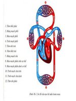
•Path of blood flow: right atrium->right ventricle- >
pulmonary arteries->pulmonary veins->left atrium>right trium->aorta-> systemic veins->venae cavae>right atrium
•The valves in the heart help the blood flow in one
direction
•Blood pressure values:
Pulmonary Circulation = 24/8 mm Hg (Systolic/Diastolic)
Systemic Circulation = 120/80 mm Hg(Systolic/Diastolic)
Huyết áp ở các loại mạch trong tuần hoàn phổi và
tuần hoàn hệ thống
Location of the heart in the thoracic cavity
Xương ức
40o
5
8-12 cm
6
Structure of the heart
Tĩnh mạch chủ trên
Cung động mạch chủ
Động mạch phổi
Van động mạch phổi
Tâm nhĩ phải
Tâm nhĩ trái
Van bán nguyệt
Van 2 lá
Van 3 lá
Tâm thất phải
Vách nhĩ thất
Tâm thất trái
Mỏm tim
Động mạch chủ dưới
Tĩnh mạch chủ dưới
•4 chambers, 2 halves (right and left), each half contains:
•Atrium :receives blood coming back to the heart from the vasculature
•Ventricle: receives blood from atrium, generates force to push the blood
through the vasculature
•valvesLeft ventricle- Aorta, right ventricle-pulmonary artery
Brief Overview of Electrical Activity of the Heart
There are three main types of heart cells:
•Heart muscle cells‐ These are the contractile cells of the heart.
•Conducting cells‐ Modified muscle cells that rapidly conduct
electrical charge.
•Pacemaker Cells‐ Located in Sinoatrial Node (SAN) these cells
spontaneously electrically discharge and set the pace or rhythm
of the heart rate.
In order for heart muscle cells to mechanically contract or generate forc
they must first be electrically excited.
Myocardium/ cardiac muscle
• properties of both skeletal muscle
and smooth muscle
• Striation appearance
• Cardiac muscle cells are
interconnected by intercalated
discs -> action potential is
transmitted rapidly
The autorhythmicity of the heart
•
•
The heart can generate signals triggering its own contraction on a periodic basic –
the heart has autorhymthmicity because it has a conduction system
(contractile activity of cardiac muscle is said to be myogenic)
Conduction system of the heart contains autorhythmic cells:
– Pacemaker cells can generate action potential and establish the heart rhymth:
• Sinoatrial node (SA node)
• Atrioventricukar node
– Conduction fibers transmit action potential through the heart in highly coordinated manner
- Bundle of His
- Purkinje fibers
Hạch xoang
Hạch nhĩ thất
Bó His
Sợi Purkinje
Spread of action potential between cardiac
muscle cells
Electric current/action potential (ions) can be passed from one cells to
other rapidly through gap junctions (intercalated disks)
The spread of action potentials through the heart
Stanius experiment
1
2
Xoang nhĩ
3
4
5
Electrocardiogram-ECG (Điện tim)
-ECG is a record of the overall spread of
electrical current through the heart as a
function of time during the cardiac cycle
-ECG reflects patterns of AP firing in entire
population of heart muscle cells
- recorded by electrodes placed on the
skin
-Willem Einthoven ‘s techmique
-Einthoven’s triangle: right arm, left arm,
left leg:
- Limb electrodes: Lead I, II, III
-Chest electrodes: V1-V6
/>
a basic ECG
− P wave: atrial depolarization
- QRS: ventricular depolarization
-T: ventricular repolarization
- QT: time for contraction of
ventricles
-PQ interval: time of conduction
through AV node
-TQ: time for relaxation of
ventricles
- RR: time between heart beats
- rate of record: 25mm/sec;
1mV/cm
C.L. Standfield.2011. Principles of Human Physiology, 4th edition.
Abnormal ECG
Cardiac cycle
(Chu kì hoạt động của tim/chu chuyển tim)
Cardiac cycle
• Cardiac cycle contains all the events
associated with the flow of blood through the
heart during a single complete heart beat
Duration of a cardiac cycle
• 0,8 second (human)
• 2 stages: - systole (tim co/tâm thu);
- diastole (tim giãn/ tâm trương)
Atria contraction: 0.1 sec.
ventricle contraction: 0.3sec
relaxation
of 8 seconds of a cardiac cycle, atria have
7 sec. and ventricles have 5 sec. of
relaxation !
4 phases of a cardiac cycle
The main variables and events during a cardiac cycle:
• atrial, ventricular, and aortic pressures
• ventricular volume
• valves opening , closure and heart sounds