NUÔI CẤY MÔ VÀ TẾ BÀO THỰC VẬT
Bạn đang xem bản rút gọn của tài liệu. Xem và tải ngay bản đầy đủ của tài liệu tại đây (1.69 MB, 24 trang )
NỘI DUNG
PROTOCORM-LIKE BODY INDUCTION AND SUBSEQUENT
PLANT REGENERATION FROM ROOT TIP CULTURES OF
DORITAENOPSIS
Introduction
Doritaenopsis is a hybrid between Doritis and Phalaenopsis.
Doritaenopsis
Introduction
Several micropropagation methods such as:
Shoot tips
Leaf segments
Nodal
Flower stalk cuttings
Introduction
•
Orchid roots have a well developed metabolic
capacity, their ability to form buds is relatively
limited under natural conditions.
•
In this study, we attempted in vitro root tip
culture in Doritaenopsis.
Materials and methods
Plant materials :
Roots of 3-month old in vitro plantlets were used as the source of
explants. the latter of which were obtained from in vitro culture of flower
stalk sections.
Root tip culture
Root tips (< 0.5 cm) were dissected from plantlets and were cultured
on half strength Murashige and Skoog (MS) medium supplemented with
different concentration TDZ, BA or zeatin, 20% coconut water, 10 mg/l
adenine sulfate, and solidified with 2.3g/l Gelrite.
The pH of medium was adjusted to 5.5, and all media were autoclaved
for 20 min at 115o C. Zeatin was added by filter sterilization to the
autoclaved medium.
Cultures were incubated at 25oC under cool-white florescent lamps at
an intensity of 30 mmol/m2 per s photosyn-thetic photon flux (PPF) 16
h/day.
Materials and methods
Plantlet regeneration and transplantation of
plantlets
Culture in a modified Hyponex medium supplemented with 30
g/l potato homogenate, 2g/l pepton, 0,5 g/l activated charcoal, 20
g/l sucrose, and 5,5 g/l plant agar.
The pH was adjusted to 5.5 before autoclaving. The cultures
were maintained at 25o C and a 16-h light:8-h dark photoperiod
The plantlets were subcultured every 8 weeks to fresh medium.
Plantlets (5-6cm length) were transplanted to pots and grown in
the green- house under conditions of high humidity (60/70%) at a
day/night temperature 25/15o C
photoperiod.
and 16-h light:8-h dark
Results
The root tip explants cultured on
a half Murashige and Skoog
medium
(MS)
supplemented
with
medium
different
cytokinins become swollen after 4
weeks of culture
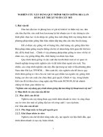
Fig. 1. Induction of PLBs and plant regeneration from
root tips of Doritaenopsis. (A) PLB development on root
tip after 4 weeks of culture on 1/2 MS medium with 2.3
mM TDZ. PLBs developed after 5 (B) and 6 (C) weeks of
culture. (D) Developing PLBs on modified Hyponex
medium. (E) PLBs with expanding sheath leaves. (F) A
rooted plantlet.
On a half MS medium supplemented with 2.3 mM TDZ 71.1% of root tips
survived and 47.2% of them developed 2 to 6 PLBs.
Fig. 2. Survival rate and frequency of PLB developing root tips of
Doritaenopsis on MS medium supplemented with different concentrations of
BA, TDZ, or zeatin.
Observations made on serial
longitudinal and cross sections of
root tips revealed the occurrence
of two differential developmental
pathways
during
PLB
regeneration. :
Root meristems were involved
in the direct development of
PLBs. And callus mediated PLB
induction from cortical cells
Fig. 3. Histological observations during PLB
regeneration from root tips of Doritaenopsis
Plantlets grew further after
another two subcultures.
The regenerated plantlets about
3 - 4 cm in height with a pair of
leaves and three to four roots were
then potted to sphagnum moss and
acclimatized in greenhouse. These
plants grew well and developed
into normal plants after 12 weeks
of transplantation.
Fig. 4. (A) Actively growing plantlets on Hyponex
medium 8 weeks after culture. (B) Acclimated plants
growing in greenhouse.
Discussion
The study of orchid root morphogenesis has not received as much attention as
the development of aerial plant organs.
Root tips of orchids species have been considered generally recalcitrant to form
PLB or callus in vitro, such is the case, for example, with Epidendrum,
Oncidium, and Cattleya plants
Among the three cytokinins used in this study, TDZ was efficient in induction
of PLBs
Discussion
Although extensive research has been carried out on PLB induction from in vitro
cultures in orchids, there are few structural details known on the development of
PLBs or their cellular origin.
Using root tip culture plants can be regenerated on a large scale, and the mother
plant can also be conserved since using root tips as the explant does not destroy
mother plants.
The number of roots available through the year, and ease of using them as
explants makes roots an ideal explant for in vitro propagation of Doritaenopsis.
KẾT QUẢ THỰC HÀNH THU ĐƯỢC
NUÔI CẤY
MÔ TẾ BÀO
THỰC VẬT
1. MỘT SỐ KỸ THUẬT CƠ BẢN
Môi trường sau khi hấp khử trùng nấu để nguội
thấy không đông hoặc ở dạng sệt
Nguyên nhân :
Đo pH chưa chính xác
Hóa chất có vấn đề ( xuất hiện cặn, kết tủa
dưới bình MS
Do quá trình thao tác nhiễm tạp chất
Đo lượng thành phần các chất trong môi
trường không chuẩn xác
2. NUÔI CẤY ĐỈNH SINH TRƯỞNG - KỸ THUẬT TÁCH ĐỈNH
SINH TRƯỞNG & PHÔI
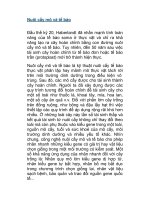
TÁCH ĐỈNH SINH TRƯỞNG
Từ mẫu chồi ban đầu sau
khi phân tách trên kính hiển
vi thu được khối đỉnh sinh
trường màu xanh có kích
thước nhỏ 1-3 mm
Hình 2.1: Mẫu lan và mẫu đỉnh sinh trưởng lan
Sau khi tách lớp vỏ bên ngoài, thu được phôi lúa màu xanh
đen (phần mầm)
Hình 2.2 :cấu tạo hạt lúa và mẫu phôi của hạt lúa
3. CẢM ỨNG CALLUS
-
Trường hợp 1:
Mẫu cấy cảm ứng callus theo
mong muốn do thực hiện chính
xác từ khâu chuẩn bị nguyên
liệu đến khâu xử lí mẫu và cấy
mẫu. Các vết thương của mẫu
tiếp xúc đồng nhất với môi
trường.
Hình 3.1: Mẫu lá cảm ứng callus
3. CẢM ỨNG CALLUS
-
Trường hợp 2:
Mẫu cấy phát sinh chồi. (đoạn thân bên trái).
Do cắt trúng mẫu thân có những mắc chồi nên
khi cấy vào bình, mắc chồi tiếp xúc với môi
trường phát triển thành chồi.
-
Trường hợp 3:
Mẫu cấy không có hiện tượng gì. (Hình 3.2 : 3
2
3
Hình 3.2 : mẫu thân phát sinh
chồi (trường hợp 2) và mẫu không
Do khi cấy ta đặt mẫu thân vào môi trường không có hiện tượng gì (trường hợp 3).
đồng đều, các vết thương trên thân không tiếp xúc
đoạn thân bên phải).
được với môi trường
3. CẢM ỨNG CALLUS
Từ các kết quả trên ta thấy mẫu lá có khả năng cảm ứng callus
tốt hơn mẫu thân. Do lợi thế về bề mặt lá mỏng và lớn có thể tạo
được nhiều vết thương hơn và diện tích tiếp xúc bề mặt với môi
trường lớn nên các vết thương tiếp xúc với môi trường sẽ tối ưu
hơn.
4. NUÔI CẤY HUYỀN PHÙ TẾ BÀO
•
Dưới tác dụng của NAA và KIN cho thấy
tế bào callus có sự tăng sinh về sinh khối.
•
Việc nuôi cấy trong môi trường lỏng lắc
đã tạo điều kiện cho sự tăng sinh cuả tế
bào
•
Việc chọn nguồn callus cứng và rời rạc
đảm bảo cho tế bào callus không bị ảnh
hưởng của quá trình lắc và tế bào không bị
dính vào nhau cản trở việc tiếp xúc với
A
B
chất dinh dưỡng
Hình 4.1 Tế bào callus sau khi cấy(A) và tế bào tăng sinh sau 14 ngày(B)
4. NUÔI CẤY HUYỀN PHÙ TẾ BÀO
Bảng 4.1 Số liệu khối lượng tể bào sau khi lọc (m ướt) và
khối lượng tế bào sau khi đi sấy ( m khô)
Giấy
M ướt
M khô
I
0.79
0.55
0.062
II
0.78
0.58
0.064
III
0.79
0.6
0.069
THE END