Vai trò của siêu âm tim 3d
Bạn đang xem bản rút gọn của tài liệu. Xem và tải ngay bản đầy đủ của tài liệu tại đây (9.45 MB, 46 trang )
3D ECHOCARDIOGRAPHY
IN DIAGNOSIS OF
CARDIOVASCULAR DISEASE
Nguyen Tuan Vu MD PhD
MEDIC HCMC
PNT-U
MAINZ University
Introduction
3D Echocardiography was introduced after the
year 2000.
Fully sampled matrix array transducers .
Live 3D, 3D Zoom and Full Volume modes.
Used to select and guide the most appropriate
intervention during BMV, MitralClip, TAVI…
3D Echocardiography applied in diagnosis of
cardiovascular disease at MEDIC HCMC since
January 2012.
Patients & Methods
-Pts with 3D TTE and 3D TEE at Medic HCM
since January 2012
-Instrument:
Phillip ie 33
3D TTE Probe 2 MHz and 3D TEE 5MHz Probe
-Techniques: 2D X plane, Live 3D, 3D Zoom,
Full Volume
- Case series report study
- Compare the diagnosis before and after
intervention or operation.
3 D Echocardiography
Fully sampled MatrixArray Transducer
3000 Piezoelectric
Elements
TTE 2-4MHz
TEE 5-7MHz
Data Acquisition
Live 3 D : real time 3 D
mode, narrow sector
Full volume: gated
acquisition
Zoom 3D: focused wide
sector
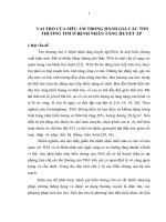
Results
44%
20%
MR, chordea rupture
Bicuspid Aorta
Coronary Fistula
Mechanical Valve
Endocarditis
Myxoma
Aortic Dissection
Philips IE 33
Mitral Stenosis
Advantages of 3D Echocardiography
Visualize the entire MV and associated structures
More reliable Planimetry of the mitral orifice by using
MPR.
Detection of complicated commissural fusion.
The 3 D dataset may be oriented to view the MV from
the surgeon view.
Leaflet mobility particularly well analyzed in the 3D Zoom
mode.
More sensible in detecting LA and LAA Thrombi.
Monitor and guide the catheter during BMV.
Mitral Regurgitation
Anatomical detail of MV
Clip 1
Echocardiographic Anatomy
Mitral leaflets
TEE and surgeon view
Normal MV fr LA view
MS: 3D TTE
LAX View
SAX View
MS: 3D TTE
Apical 4 C View
Live 3 D with color
MS, LAA Thrombus
Mitral Stenosis 3D Zoom
Clip 3 D Zoom
LA Thrombus
I crop LA Thrombus
LAA Thrombus
Normal LAA
LAA Thrombus
Guide the catheter during BMV
Fluoroscopy
Live 3D Echocardiography
Mitral Regurgitation
Mitral Regurgitation
Mitral Regurgitation
3D Zoom
3D Live with color
Mitral Regurgitation
Mitral Regurgitation: 2 Jets
Graduation of MR: PISA
Method
Mitral Regurgitation
3D ECHOCARDIOGRAPHY
Mechanism of MR.
Graduation: Vena Contracta, MR Volume (
PISA 2D, 3D ).
Detection of multiple Jets.
Location of lesion: segment I,II,III
Monitoring MitralClip procedure
Criteria for MitralClip : functional MR,
segment II, PML <10mm .
Guiding septal puncture and
MitralClip