Evaluation of invivo wound healing and anti inflammatory activity of 80% methanol crude extracts of the leaves and fruits of b antidysentrica j f mill (simaroubaceae) in mice
Bạn đang xem bản rút gọn của tài liệu. Xem và tải ngay bản đầy đủ của tài liệu tại đây (1.73 MB, 68 trang )
ADDIS ABABA UNIVERSITY
SCHOOL OF GRADUATE STUDIES
Evaluation of invivo wound healing and anti-inflammatory activity of
80% methanol crude extracts of the leaves and fruits of Brucea
antidysentrica J .F. Mill (Simaroubaceae) in mice
By:
Zenaw Tessema (B.pharm)
December, 2016
Addis Ababa, Ethiopia
Evaluation of invivo wound healing and anti-inflammatory activity of
80% methanol crude extracts of the leaves and fruits of B.
antidysentrica J. F. Mill (Simaroubaceae) in mice
By: Zenaw Tessema (B.pharm)
A Thesis submitted to the Department of Pharmacology, School of
Medicine, College of Health Sciences in partial fulfilment of the
requirements for the Degree of Master of Science in Pharmacology.
Under the supervision of:
Prof. Eyasu Makonnen, PhD, Department of Pharmacology, School of
medicine, Addis Ababa University, Ethiopia and
Asfaw Debella, PhD, Directorate of Traditional and Modern Medicine
Research, Ethiopian Public Health Institute, Addis Ababa, Ethiopia
December, 2016
Addis Ababa, Ethiopia
I
Addis Ababa University
School of Graduate Studies
This is to certify that the thesis prepared by Zenaw Tessema, entitled: Evaluation
of in vivo wound healing and anti-inflammatory activity of 80% methanol crude
extracts of the leaves
and
fruits of
Brucea
antidysentrica
J.F.Mill
(Simaroubaceae) in mice and submitted in partial fulfillment of the requirements
for the Degree of Master of Science in Pharmacology complies with the
regulations of the university and meets the accepted standards with respect to
originality and quality.
Signed by the Examining Committee:
Prof. Yalemtsehay Mekonnen
External Examiner
Prof. Tefera Abula
Internal Examiner
_______
Signature
_______
Date
_______
Signature
_______
Date
Prof. Eyasu Makonnen (PhD)
Advisor
_________
Signature
__________
Date
Asfaw Debella (PhD)
Co- advisor
_________
Signature
________
Date
_________________________________________________
Chair of Department or Graduate program coordinator
II
DECLARATION
I, the undersigned, declare that this thesis is my original work and has not been presented for a
degree in any other university.
Name: Zenaw Tessema Wolie
Signature: ____________________
Place and date of submission: Addis Ababa, Ethiopia, December, 2016
III
ABSTRACT
Evaluation of invivo wound healing and anti-inflammatory activity of 80%
methanol crude extracts of the leaves and fruits of Brucea antidysentrica J .F.
Mill (Simaroubaceae) in mice.
Zenaw Tessema
Addis Ababa University, 2016
Introduction: Brucea antidysentrica locally known as “Abalo” is traditionally used to treat
conditions like scabies and external parasites, dysentery, gonorrhea, eczema, cancer, malaria,
and trypanosomosis among others. The fruits and leaves of B. antidysentrica are also claimed
to promote wound healing and anti-inflammatory activities. However, there is no scientific
confirmation that substantiates the traditional claims.
Objective: to evaluate the wound healing and anti-inflammatory activities of both fruits and
leaves extracts of B. antidysentrica in mice model.
Materials and methods: Mice were used for wound healing and anti-inflammatory studies,
while rats were used for skin irritation test. For studying healing activity, 80% methanolic
extracts of the leaves and fruits were formulated in strength of 2% and 4% and 1% and 2% as
ointment base respectively for topical applications of excision and incision wound models. The
negative controls were treated with simple ointment while positive controls with nitrofurazone
(0.2%) skin ointment. Extract solutions of the leaves and fruits in 2% Tween 80 at a dose of
100 mg/kg, 200 mg/kg and 400 mg/Kg body weight were used for anti-inflammatory activity
tests orally against the inflammation produced by carrageenan injection. Negative controls for
anti-inflammatory test were treated with 2% Tween80 and the positive controls with
Indomethacin 10mg/kg. Parameters, including rate of wound contraction, period of complete
epithelialization, skin breaking strength and edema inhibition were evaluated.
Results: On the last day of treatment, 80% methanol fruits and leaves extracts showed a
significant wound healing activity in strengths of 2% compared with negative control as
evidenced by an increase in % wound contraction (p < 0.01) and a decrease in epithelization
period (p<0.05). The 4% MLE also showed the highest % wound contraction (P<0.001) and the
IV
shortest epithelialization period than the rest of the extracts (P<0.01). One percent MFE was
found to increase the % wound contraction significantly on the last day of treatment (P<0.01)
and its effect on the epithelialization period was insignificant. In the incision wound model,
both 2% and 4% extract ointments of the leaves and only the 2% MFE resulted in a significant
increase in tensile strength (p < 0.01) compared with negative control. The same extracts also
revealed a significant anti-inflammatory effect compared with negative control particularly 3 to
4 h after extract administration as shown by a decrease in edema expressed as % reduction of
edema. All doses of the leaves extract exhibited a higher effect on the 3rd (P<0.05) and the 4thh
(P<0.001) compared to the negative control. Similar effect was also found for the 200mg/kg
and 400mg/kg doses of the fruits extract, while its 100mg/kg dose reduced the edema
significantly on the 4th h (P<0.001).
Conclusion: The 80% methanol extracts of the fruits and leaves of B. antidysentrica supports
the traditional claims for healing of wounds as evidenced by an increase in wound contraction
rate and tensile strength, decrease in epithelization period and anti-inflammatory activity.
Key words: wound healing, anti-inflammatory, excision, incision, carrageenan induced paw
edema, Brucea antidysentrica.
V
ACKNOWLEDGMENTS
First and above all, I praise the almighty God and his mother St. Virgin Marry for providing me
this opportunity and granting me the capability to accomplish this task successfully.
I want to express my deep thanks to my esteemed advisor Prof. Eyasu Makonnen (PhD) and
Co-advisor Asfaw Debella (PhD) for the trust, the motivation, the enthusiasm, offering
valuable advice and unreserved support during the whole period of this thesis work. Besides
my advisors, I would like to thank Mr. Bekesho Geleta for his patience, encouragement and
insightful comments during my laboratory work.
My sincere thanks also goes to Mr. Hailemeskel Meshesha and Ms. Fantu Assefa for their
unfailing help during the laboratory activities and Ms. Yewubdar Haile for her dutiful and
uninterrupted concern of the experimental animals.
It is also my interest to show gratitude to my families particularly to my beloved wife and
friends for never ending support throughout my life. I additionally wish to give my appreciation
to staffs of Traditional and Modern Medicine Research Directorate (TMMRD) at the Ethiopian
Public Health Institute (EPHI) for their kind welcome and consent to use premises and facilities
at Pharmacology laboratory.
Last but not least, I would like to thank Addis Ababa University for financial support for this
work and Debre Markos University for sponsoring my postgraduate education.
VI
TABLE OF CONTENTS
ABSTRACT ............................................................................................................................................................. IV
ACKNOWLEDGMENTS ........................................................................................................................................ VI
TABLE OF CONTENTS......................................................................................................................................... VII
LIST OF TABLES .....................................................................................................................................................IX
LIST OF FIGURES ....................................................................................................................................................X
LIST OF ABBREVATIONS AND ACRONYMS ....................................................................................................XI
1.
2.
3.
INTRODUCTION .............................................................................................................................................1
1.1.
Wound and basic principle of its formation................................................................... 1
1.2.
Wound healing processes............................................................................................... 3
1.2.1.
Hemostasis phases .................................................................................................. 4
1.2.2.
Inflammatory Phase ................................................................................................ 4
1.2.3.
Proliferation/granulation/ contraction Phase .......................................................... 5
1.2.4.
Remodeling /Maturation Phase .............................................................................. 6
1.3.
Factors that can interfere with healing ........................................................................... 7
1.4.
Management of wounds ................................................................................................. 7
1.5.
Plant medicines traditionally used in would healing ..................................................... 8
1.6.
Overview of the experimental plant............................................................................... 9
1.7.
Statement of the problem ............................................................................................. 12
OBJECTIVES ..................................................................................................................... 13
2.1.
General objective ......................................................................................................... 13
2.2.
Specific objectives ....................................................................................................... 13
MATERIALS AND METHODS ........................................................................................ 14
3.1.
Drugs and chemicals .................................................................................................... 14
3.2.
Instruments and Apparatus .......................................................................................... 14
3.3.
Collection of plant materials ........................................................................................ 15
3.4.
Experimental animals .................................................................................................. 16
3.5.
Ethical approval ........................................................................................................... 16
3.6.
Preparation of the crude extracts of B. antidysentrica leaves and fruits...................... 16
3.7.
Ointment formulation .................................................................................................. 17
VII
3.8.
Grouping and dosing of experimental animals ............................................................ 18
3.9.
Wound healing studies ................................................................................................. 18
3.9.1.
Excision wound model ......................................................................................... 19
3.9.2.
Incision wound model .......................................................................................... 19
3.9.3.
Anti-inflammatory activities ................................................................................ 21
3.10. Phytochemical screening ............................................................................................. 21
3.11. Acute toxicity studies................................................................................................... 23
3.11.1. Acute oral toxicity study ..................................................................................... 23
3.11.2. Skin irritation test ................................................................................................. 24
3.12. Statistical analysis ........................................................................................................ 26
4.
RESULTS ........................................................................................................................... 27
4.1.
Yields of extraction ...................................................................................................... 27
4.2.
Wound healing Effect of the extracts .......................................................................... 27
4.2.1.
Excision wound model ......................................................................................... 27
4.2.2.
Incision wound model .......................................................................................... 30
4.3.
Anti-inflammatory effect of the extracts ..................................................................... 31
4.4.
Phytochemical screening ............................................................................................. 33
4.5.
Acute toxicity studies................................................................................................... 33
4.5.1.
Acute oral toxicity study ...................................................................................... 33
4.5.2.
Skin irritation study .............................................................................................. 34
5.
DISCUSSION ..................................................................................................................... 36
6.
CONCLUSION ................................................................................................................... 42
7.
RECOMMENDATIONS .................................................................................................... 43
8.
REFFERENCES ................................................................................................................. 44
9.
APPENDIXES .................................................................................................................... 54
9.1.
Photos of plant material collection from Debre Markos, E/Gojjam Zone, Amhara
Region.......................................................................................................................... 54
9.2.
Photos showing the drying of plant materials at Pharmacology Department laboratory,
SoM, CHS, AAU ......................................................................................................... 54
9.3.
Some of the instruments used during the experiment .................................................. 55
9.4.
Photos showing some of the procedure during experiment ......................................... 55
VIII
LIST OF TABLES
Table1. The four phases of wound healing process…………………………………………… 6
Table 2. Formula used for preparation of simple and medicated ointment……………
…….17
Table 3: Classification of erythema and oedema scores used to determine the primary irritation
index……………………………………………………………………....................25
Table4: Categories of irritation response in rats……………………………………………….26
Table 5: Effect of topical application of methanol extracts of Brucea antidysentrica leaves and
fruits on percentage wound contraction and epithelization time of an excision wound
in mice……………………………………………………………………………….29
Table 6: Effect of topical application of 80 % methanol extracts of Brucea antidysentrica
leaves and fruits on breaking strength of an incision wound on day 10 of wound
creation………………………………………………………………………............30
Table 7: Anti-inflammatory effect of 80 % methanol extracts of leaves and fruits of B.
antidysentrica on carrageenan-induced paw edema following oral administration
…………………………………………………………….........................................32
Table8. Results of phytochemical screening of 80% methanol extracts of leaves and fruits of
B. antidysentrica in mice…………………………………………………………....33
Table 9: Score of irritation and edema after application of ointments containing extracts of
B.antidysentrica with their respective bases………………………………………...35
IX
LIST OF FIGURES
Fig.1. Leaves and Fruits of B.antidysentrica J. F. Mill…………………………...……………11
Fig.2.Map of B.antidysentrica plant collection area…………………………………………...15
Fig.3 Incised mice and continuous water flow method for determination of tensile strength
……………….…………………………………………………………………………20
Fig.4. Excision wound immediately after wounding and a healing progresses on excision
wound…………………………………………………………………………………..28
Fig.5. Animals tested for the skin irritation with the respective indicated medicated
formulations ……………………..…………………………………………………......35
X
LIST OF ABBREVATIONS AND ACRONYMS
AAU
Addis Ababa University
AFRO
Africa Regional Office
ANOVA
Analysis of Variance
BP
British Pharmacopeia
CHS
College of Health Science
COX
Cyclooxygenase
DNA
Deoxyribonucleic Acid
ECM
Extra Cellular Matrix
EGF
Epidermal Growth Factor
EP
Epithelialization Period
EPHI
Ethiopian Public Health Institute
FGF
Fibroblast Growth Factor
HIV/AIDS
Human Immune Virus/Acquired Immune Deficiency virus
IL-1
Interleukin –One.
ILAR
Institute for Laboratory Animal Research
IP
Intrapertonial
IV
Intravenous
LD50
Medial Lethal Dose
MF
Master Formula
MFE
Methanol Fruits Extract
XI
MLE
OECD
Methanol Leaves Extract
Organization for Economic Cooperation and Development
PDGF
Platelet-Derived Growth Factor
PGE
prostaglandin E
PII
Primary Irritation Index
PMN
PolyMorphonuclear Neutrophils
RF
Reduced Formula
RNA
Ribonucleic Acid
RPM
Revolution Per Minute
SEC
Scientific Ethical Committee
SEM
Standard Error of the Mean
SoM
School of Medicine
SPI
Scoring of Primary Irritation
SPSS
Statistical Package for the Social Sciences'
TMMRD
Traditional and Modern Medicine Research Directorate
TGF-b
Transfer Growth Factor b
TNF-a
Tumor Necrosis Factor a
TS
Tensile Strength
UK
United Kingdom
USD
United States Dollar
USP
United States Pharmacopeia
WHO
World Health Organization
WHS
Wound Healing Society
XII
1. INTRODUCTION
1.1.
Wound and basic principle of its formation
The skin is the largest organ of the body that acts as a barrier against external agents. The loss
of skin tissue integrity can cause lesions or illnesses that bring disability or even death (Panda
et al., 2011; Asghar et al., 2015).
Wound which is inescapable event of life (Majumdar, 2005) is a clinical problem as old as
mankind and may be defined in different ways. But the most acceptable one is “a loss or
breaking of cellular and anatomical or functional continuity of living tissues˶ (Raina et al.,2008;
Kumar et al., 2013; Mulisa et al., 2015). According to the Wound Healing Society (WHS),
wounds are physical injuries that result in an opening, breaking or interrupting of tissue
integrity that cause disturbance in the normal skin anatomy and function (Murthy et al., 2013;
Hussain et al., 2014; Subalakshmi et al., 2014; Ositadimma et al., 2015) which in turn have a
significant impact on public health and expenditure of health care resources (Fikru et al., 2012).
Physical, chemical, thermal, microbial, or immunological insults to the tissue are among the
factors mentioned in wound production (Majumdar, 2005; Thakur et al., 2011; Hussain et al.,
2014).
Based on different classification criteria such as etiology, location, type of injury or presenting
symptoms, wound depth and tissue loss or clinical appearance (Udaya et al .,2010; Sabale et
al.,2012); there are different types of wounds, including injuries, cuts and bites, diabetic,
gastric and duodenal ulcers. These wounds can be broadly classified as acute or chronic
depending on physiology or the time it takes to heal. Without complications, most wounds are
acute wounds and tend to heal within few weeks. Chronic wounds in contrast, require
prolonged time to heal, do not heal, or recur frequently. These wounds tend to occur when the
normal wound healing process has been compromised due to microbial infection, metabolic
disturbances, or an underlying disease (Agyepong et al., 2015).
1
Based on the underlying cause of wound creation; it can also be categorized as open and closed
wounds (Alam et al., 2011). In open wounds, the blood escapes the body and bleeding is
clearly visible. It can be further classified as incised wound, laceration or tear wound, abrasions
or superficial wounds, puncture wounds, penetration wounds and gunshot wounds. On the other
hand in the case of closed wounds, blood escapes the circulatory system but remains in the
body and includes contusion or bruises, heamatomas or blood tumor, crush injury etc (Alam et
al., 2011; Shrimanker et al., 2013).
Acute wounds are tissue injuries that normally proceed through an orderly and timely
reparative process that result in sustained restoration of anatomic and functional integrity. They
are usually caused by cuts or surgical incisions and complete the wound healing process within
the expected time frame (Diegelmann and Evans, 2004).
Chronic wounds, rarely seen in healthy individuals and usually associated with diseases like
diabetes and obesity, are defined as wounds, which have failed to progress through an orderly
and timely reparative process of healing and therefore enter a state of pathologic inflammation.
As a result, the healing process is delayed, incomplete, and does not proceed in a coordinated
manner, subsequently resulting in poor anatomic and functional integrity over a period of 3
months (Menke et al., 2007;Trostrup et al., 2013).To mention some from this category; foot
ulcers and pressure ulcers are complications of diabetes and spinal cord injuries, respectively.
All wound types have the potential to become chronic and, as such, chronic wounds are
traditionally divided etiologically. Identifying and treating the underlying aetiology of a chronic
wound such as venous insufficiency, arterial perfusion, diabetes, or unrelieved pressure as well
as systemic factors such as nutritional status, immunosuppression, and infection that may
contribute to poor wound healing are key to successful wound treatment (Werdin et al.,2009).
Chronic wounds result in significant functional impairment, reduction in quality of life, and
large financial costs for patients and the health care system. Yet the epidemiological profile of
chronic wounds hasn‟t been well established (Graves and Zheng, 2014). Current estimates
indicate about 6 million people suffer from chronic wounds worldwide (Agyepong et al., 2015)
which is responsible for loss of USD 25 billion for its clinical management representing an
incredible burden in public health expenditure. Also in developed countries, the population
2
experiencing chronic wound during their lifetime is estimated to be 1-2% posing a public health
problem. Loss of 2-4% of the total health care expenses for the clinical management of chronic
wounds in Scandinavian countries is a proof of the reality (Sen et al., 2009).
1.2.Wound healing processes
The wound healing process, particularly in skin, has been well characterized histologically in
studies extending back more than 100 years (Shawi and Martin, 2009). The term “wound
healing” embraces all types of wounds, burns, and ulcerations. Complete wound healing
includes restoration of function hardly ever achieved in those disfigured by wounds, especially
when one includes the appearance of the skin or absence of an appendage (WHO,
2010).Wound healing is a complex and dynamic interplay between various cell types, the
extracellular matrix (ECM), cytokines, and growth factors (Pakyari et al.,2012). It is a
normal biological process that is initiated by trauma and often terminated by scar formation
which reveals that healing is essentially a survival mechanism and represents an attempt to
maintain normal anatomical structure and function (Fikru et al., 2012; Kumar et al., 2013;
Mohsenikia et al., 2015). It comprises a series of coordinated and overlapping processes that
have been characterized over many years (Ansell et al., 2012). The processes involved include
hemostasis, inflammation, fibroblast activation and migration, re-epithelization, proliferation of
endothelial cells, and remodeling (Fikru et al., 2012; Hussain et al., 2014; Mohsenikia et al.,
2015). Wound healing remains a challenging clinical problem, and requires appropriate and
efficient management. Much has been focused on wound care with an emphasis on new
therapeutic approaches and the development of technologies for acute and chronic wound
management (Velnar et al., 2009).
The process of wound repair differs a little from one kind of tissue to another and is
independent of the form of injury. Even though the different steps in the wound healing process
occur in a continuous, integrated manner (Majumdar, 2005), it is convenient to classify the
physiological process involving through four temporarily and spatially overlapping phases:
hemostasis, inflammation, proliferation, and remodeling phases (Ud-Din and Bayat, 2014;
Frykberg and Banks., 2015) and for proper healing to occur these phases need to be well
controlled (Gould et al, 2008
3
1.2.1. Hemostasis phases
As soon as injury occurred, an important step of initiation and continuation of the healing
process called hemostasis appears. It is characterized by vasoconstriction, platelet
degranulation and aggregation, and fibrin deposition leading to formation of a clot and bleeding
cessation (Pakyari et al., 2012). Platelets being the primary subset of cells that enter to the
injured site release various types of growth factors such as platelet-derived growth factor
(PDGF), ,epidermal growth factor (EGF), and fibroblast growth factor (FGF) and inflammatory
cytokines like tumor necrosis factor alpha (TNF-a), transforming growth factor beta (TGF-b),
all together encourage the inflammatory phase and some of them function as chemo-attractants
(Frykberg and Banks., 2015).Immediately after the production of these initiation factors,
epithelial cells travel under the newly formed granulation tissue being activated by several
cytokines and growth factors; specifically, interleukin (IL)-1a appears to be expressed within
the epidermis and released upon the dermal injury, which in turn stimulates various genes
including adhesion molecules, chemokines, cytokines, proteolytic enzymes, and matrix proteins
in different types of skin cells (Pakyari et al.,2012,Bodnar, 2014).
1.2.2. Inflammatory Phase
The inflammatory response following tissue injury and lasts from day 0 to 5 plays crucial roles
both in normal and pathological healing (Koh and DiPietro, 2013). During this phase of wound
healing macrophages, epithelial cells, and lymphocytes secret to much amount of
proangiogenic molecules (growth factors and cytokines) (Bodnar, 2014).The response from
inflammatory phase is initiated at the moment of injury. Shape and architecture of tissues are
disrupted owing to surgical or traumatic wounds and cause hemorrhage. In the beginning,
blood fills the wound and exposure of this blood to collagen in the wound leads to platelet
deregulation and activation of a plasma protein (coagulation factor XII also known as Hageman
factor). As a result a number of biological amplification systems including the complement
kinin and clotting cascades and plasmin generation are followed. This condition serves to
amplify the original injury signal and lead not only to clot formation, which unites the wound
edges, but also to the accumulation of a number of mitogens and chemo-attractants at the site of
wound. Production of both kinins and prostaglandins leads to vasodilatation and increased
4
small vessel permeability in the region of the wound leading to edema in the area of the injury.
Within 6 hours, circulating immune cells start to appear in the wound. Polymorphonuclear
neutrophils (PMN) are the first blood leukocytes to enter the wound sites. Their main functions
appear to be phagocytes of the bacteria, which have been introduced into the wound during
injury. In the absence of infection, Polymorphonuclear neutrophils (PMNs) have a relatively
short life span in the wound and their numbers decrease rapidly after the third day. The next
cellular, immune component enter to the wound is macrophages. These macrophages have a
much longer life span than the Polymorphonuclear neutrophils (PMN) and persist in the wound
until healing is complete (Kumar et al., 2013).
Like neutrophils, macrophages phagocyte and digest organisms responsible for pathological
process and secrete collagenase and elastases which break down the affected tissues and release
cytokinins. Macrophages release different types of biologically active substances; in addition
growth factors and other substances are also released which are essential for the initiation and
progression of granulation formation (Majumdar, 2005).
1.2.3. Proliferation/granulation/ contraction Phase
This phase lasts approximately from days 3-14 and in the absence of significant infection or
contamination, the inflammatory phase is short, and after the wound has been successfully
cleared of devitalized and unwanted material, it gives away to the proliferative phase of
healing. Granulation tissue consists of a combination of cellular elements, including fibroblasts
and inflammatory cells. Fibroblasts which are the primary synthetic element in the repair
process and are responsible for production of the majority of structural proteins first appear in
significant numbers in the wound on the third day post-injury and achieve peak numbers on the
seventh day. This rapid expansion in the fibroblast population at the wound site occurs via a
combination of proliferation and migration (Majumdar, 2005) and the migration of fibroblasts
to the wound site is assisted by contraction of extra cellular matrix (ECM) and the formation of
granular tissue (Ayuk,2012). Then the fibroblasts produce large quantities of collagen which
forms the main constituent of the extracellular wound matrix, and are ultimately responsible for
imparting tensile strength to the scar which finally leads to restoration of an epithelial integrity
at the wound surface. This phase comprised of events such as angiogenesis, fibroblasia and
5
granulation tissue formation, collagen deposition, epithelialization and contraction that overlap
each other (Guo and Dipietro, 2010;Ayuk, 2012).
1.2.4. Remodeling /Maturation Phase
The final stage of wound healing process which starts from day 7 and involves remodeling,
realignment and well organization of the collagen tissue to produce greater tensile strength, cell
and capillary density reduction and a balance between synthesis and degradation can take up to
2 years and results in the development of normal epithelium and maturation of the scar tissue.
Eventually they will regain a structure similar to that seen in unwounded tissue. The main cells
involved in this process are the fibroblasts (Orsted et al., 2004; Sinno and Prakash, 2013). The
four phases of wound healing process are shown in Table1.
Table1. The four phases of wound healing process
Phase of healing
Time post injury
Cells involved in the phases
Function or activity
Homeostasis
immediately
Platelets
Clotting
inflammation
Day 0-5
Neutrophils or macrophages
Phagocytosis
Proliferation(granula
Day 3-14
Macrophages
Fill defect
Lymphocytes
Re –establish
Neurocytes
Skin function closures
tion or contraction)
Fibroblasts
Keratinocytes
Remodeling
(maturation)
Day7-2 yrs
Fibrocytes
Develop tensile
strength
6
1.3.
Factors that can interfere with healing
“The germ is nothing. It is the terrain in which it is found that is everything.” Stated by Louis
Pasteur. This is similar with wounds! Factors that affect wound healing must be addressed in a
holistic fashion as stated above, at the terrain in which the wound is found. The individual with
a wound has a wide terrain, from the local wound environment to the environment in which he
or she lives, and that terrain may determine the healing ability. In other words, wounds do not
exist in isolation from the patient as a whole (Orsted et al., 2004). Multiple factors can lead to
impaired wound healing and these factors can be categorized into local and systemic. Local
factors are those that directly influence the characteristics of the wound itself, while systemic
factors are the overall health or disease state of the individual that affect his or her ability to
heal (Guo and DiPietro, 2010). Local factors affect the features of the wound and they are
mainly oxygenation, infection, presence of a foreign body and venous insufficiency while the
systemic factors are provoked by the physiological state of an individual which may impair
wound healing. Some of these factors include age and gender, temperature, chemicals, sex
hormones, stress, moisture, nutritional status, diabetes, HIV/AIDS, cancer heredity healing
disorders and obesity. Alcoholism, smoking, and certain medications such as steroids and
chemotherapy also affect the wound healing processes (Thomas, 2011; Ayuk, 2012).
1.4.
Management of wounds
The correct approach of treating wounds should effectively assist the healing process, and can
have an important impact on the final clinical outcome. Physiological, endocrine and nutritional
support at a clinical level significantly influence repair and, without which, wound healing
often fails completely. Assessment of the wound and the patient starting with a diagnosis of the
wound‟s aetiology and continues with optimizing the patient‟s medical condition, particularly
blood flow to the wound area is considered to be the first stage in wound management. The
wound needs to be debrided and dressed correctly. The next important stage in wound
management is the lavage of micro organisms, dead tissues and foreign bodies which decrease
tissue bacterial count using bacitracin or normal saline solution. Currently novel techniques
such as topical growth factor application and incisional priming with PDGF or IL-1 can
optimize both the cellular and molecular environment, thus decreasing healing time by
7
modifying inflammation and accelerating the proliferative phase. Electrical field stimulation
may also optimize the remodeling phase by promoting more efficient fibroblast recruitment and
collagen deposition (Velnar et al., 2009).
1.5.
Plant medicines traditionally used in would healing
According to World health organization(WHO) traditional medicine is defined as health
practices, approaches, knowledge and beliefs incorporating plant, animal and mineral based
medicines, spiritual therapies, manual techniques and exercises applied to treat, diagnose and
prevent illnesses or maintain wellbeing(Lulekal et al.,2008).
Traditional people around the world possess unique knowledge of plant resources on which
they depend for food and medicine (Bekele and Reddy, 2015). Trends in the use of traditional
and complementary medicine are on the increase in many developed and developing countries
(Limenih et al., 2015).
As estimated by the World Health organization, 80% of the populations of Asia, Africa and
Latin America use traditional medicine to meet their primary health care needs (WHO-AFRO,
2010).
In Ethiopia, it has been estimated that traditional remedies are the most important and
sometimes the only source of therapeutics for nearly 80% of the population of which 95% of
traditional medicinal preparations are of plant origin (Getaneh and Girma, 2014) due to the
cultural acceptability, relatively low cost and limited access to modern health facilities
(Kassaye et al., 2006).
There are many plants which are traditionally used for wound healing in Ethiopia, These
include Achyranthes aspera (Fikru et al., 2012),Rumexa byssinicus (Mulisa et al., 2015),
Brucea antidysentrica, Datura stramonium, Croton macrostachyus, Acokanthera schimperi
(Taye et al,2011), Rhusvulgaris ficuscaricus, Acacia abysinica, Vernonia amygdalina Del
(Gebeyehu et al.,2014), Commelin abengalensis L, Solanum incanum, Ximenia americana
(Teklehaymanot,2009), Acalypha volkensii Pax, Amorphophallus gallaensi (Gidey et
al.,2009),Clematis hirsute Guill.&Perr (Gidey et al.,2007), Bersama abyssinica, Cynodon
8
dacytylon, (Abera,2003), Cordial africana, Coffee arabica (Regassa,2013) and many others
are being used in the treatment of wounds and other diseases in the traditional health care
system of the country.
The
study done
on
the
in
vivo
wound
healing
activity of
methanol
extract
from Achyranthes aspera L. leaves showed significant wound healing activity compared to
group of rats treated with simple ointment (Fikru et al., 2012). In addition, it was also reported
that Wound treated with 5 % and 10 % (w/w) hydroalcoholic extract ointment from rhizomes
of Rumexa byssinicus J. exhibited significant wound healing activity in both excision and
incision models (Mulisa et al., 2015). Since the in vivo wound healing activity of Brucea
antidysentrica is not reported, this study will focus on evaluation of its wound healing and antiinflammatory activity using incision & excision wound models and carrageenan induced hind
paw edema model.
1.6.
Overview of the experimental plant
The Simaroubaceae family includes 32 genera and more than 170 species of trees and brushes
of pantropical distribution. It is characterized by its content of bitter substances, mostly
responsible for its pharmaceutical properties. The family is characterized by the presence of
quassinoids, secondary metabolites responsible for a wide spectrum of biological activities such
as antitumor, antimalarial, antiviral, feeding deterrent, amebicide, antiparasitic and herbicidal
(Alves et al., 2014) and antimicrobial and antioxidant (Viswanad,2011) .
Brucea (a family of Simaroubaceae) is widely distributed genus occurring in tropical Africa
and tropical Asia. It is very bitter monoecius or dioecius shrub or small tree which is grouped
into ten species. But the most common acceptable species are the following: B.javanica,
B.mollis, B.antidysentrica, and B.quineensis (Roberts, 1994).
The study on the Methanolic-chloroform and methanolic-aqueous root extracts of Brucea
mollis showed significant in vitro antiplasmodial activity which was also supported by their
promising in vivo activity, respectively (Sharma et al., 2013).
9
The fruit of Brucea javanica is currently recorded in the Pharmacopoeia of the People's
Republic of China (2010 edition) for treatment of fever, malaria and amebic dysentery (Liang
et al., 2015).
Brucea antidysenterica J. F. Mill belongs to a genus Brucea and a family of Simaroubaceae is
commonly known as Waginos (Geeze) (Limenih et al., 2015), Abalo (Amharic), Meleta
(Tigrigna), Hadawi (Somaligna), Atanico (Sidamigna) (Getahun, 1976), and it is among the
commonly used traditional medicinal plants. The plant Brucea’: is named after James Bruce
(1730-1794), a Scottish man who travelled to Ethiopia in the years 1768-1773 and took seeds
of this plant to Europe. ‟Antidysenterica‟: derived from the Greek 'anti' = 'against', and
'dusenteria' = 'bad bowels'; so, active against e.g. dysentery (Jansen, 1981).
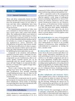
It is an ever green shrub or tree up to seven meters high (fig.1). The plant grows at moderate
elevations, usually to 2,500 metres and exceptionally to 3,700 metres in the moisture tropics of
Africa (Getahun, 1976). The plant is mainly distributed in Ethiopia, Sudan, Tanzania,
Cameroon, Nigeria, Angola, Malawi and Zambia (Jansen, 1981).
Brucea antidysenterica J. F. Mill has a number of therapeutic applications like treating scabies
and external parasites (Bekele and Reddy, 2015), as an antidysentric agent (Limenih et al.,
2015; Teklehaymanot, 2009), wound healing effect (Getahun, 1976;Taye et al,2011;
Regassa,2013;Getaneh and Girma, 2014; d‟Avigdor et al.,2014;), treatment of Gonorrhoea
(Lulekal et al.,2008), eczema and hookworm (Gebeyehu et al.,2014), as anticancer and antimalaria (Abera,2003) and treatment of Trypanosomosis (Tamiru et al., 2013) among others.
10
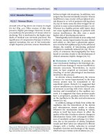
Fig, 1. Leaves (A) and Fruits (B) of B.antidysentrica J. F. Mill
(Source: Photograph taken from floral site (Debre Markos) during collection in
January, 2016)
The methanolic extract in vitro wound healing activity from Brucea antidysenterica showed
35% growth inhibition on wound causing bacteria like S. aureus, S. pyogens, E. coli and
P.aeuruginosa (Taye et al., 2011). Since its wound healing and anti-inflammatory activity is
not investigated; the traditional claim enforces to evaluate its in vivo activity.
11
1.7.
Statement of the problem
Wound is one of the most common diseases often having severe complications in relation to
health and posing high costs for therapy. In order to establish integrity of the damaged tissue;
series of events must be progressed orderly in well controlled manner that unless otherwise
cause physical disability even lead to death (Paulan, 2013; Taye et al, 2011; Gautama et al.,
2011).
Wounds are also significant causes of morbidity and mortality worldwide. Studies show that for
every million wound patients, at least 10,000 die from microbial infections (Wong et al., 2015).
Currently available methods of wound management including debridement, irrigation,
antibiotics, tissue grafts and proteolytic enzymes are found to be associated with major
drawbacks such as invasiveness and being expensive (Werdin et al., 2009). Emergence of
resistant strains along with lack, high cost and retarded rate of newly generated antibiotics
increase wound related mortality and morbidity (Akinsulire et al., 2007).
Hence it is paramount important to urgently intensify research to emerge new, cheap and
effective wound healing agents. Now a day, scientists and researchers turn their attention to the
medicinal plants as a noble source in the development of wound healing agents. In line with
this, there is a need for conducting investigation towards medicinal plant claimed to be
effective in the management of wound and inflammation.
12