Acupuncture in manual therapy 12 transcutaneous electrical nerve stimulators for pain management
Bạn đang xem bản rút gọn của tài liệu. Xem và tải ngay bản đầy đủ của tài liệu tại đây (2.76 MB, 19 trang )
Transcutaneous electrical nerve
stimulators for pain management
12
Professor Mark Johnson
CHAPTER CONTENTS
Introduction . . . . . . . . . . . . . . . . . . . . . . . . . . 205
Definition and techniques . . . . . . . . . . . . . . . 206
Conventional TENS . . . . . . . . . . . . . . . . . . . . . . 206
Acupuncture-like TENS (AL-TENS) . . . . . . . . . . . 208
Intense TENS . . . . . . . . . . . . . . . . . . . . . . . . . . . 208
Contraindications . . . . . . . . . . . . . . . . . . . . . . . . 208
Precautions . . . . . . . . . . . . . . . . . . . . . . . . . . . . 209
Clinical technique . . . . . . . . . . . . . . . . . . . . . 210
Indications . . . . . . . . . . . . . . . . . . . . . . . . . . . . . 210
Timing and dosage . . . . . . . . . . . . . . . . . . . . . . . 210
Electrode location . . . . . . . . . . . . . . . . . . . . . . . . 210
TENS on acupuncture points . . . . . . . . . . . . . . . 210
Electrical characteristics of TENS . . . . . . . . . . . . 211
Research evidence . . . . . . . . . . . . . . . . . . . . 211
Mechanism of action . . . . . . . . . . . . . . . . . . . . . 211
Clinical effectiveness . . . . . . . . . . . . . . . . . . . 212
References . . . . . . . . . . . . . . . . . . . . . . . . . . 220
Introduction
Transcutaneous electrical nerve stimulation (TENS)
is a peripheral stimulation technique that is noninvasive, allowing patients the ability to selfadminister treatment. The purpose of TENS is to
deliver pulsed electrical currents across the intact
surface of the skin to activate underlying nerves
© 2010
2009 Elsevier Ltd.
DOI: 10.1016/B978-0-443-06782-2.00012-8
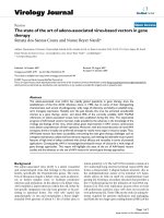
and reduce pain (Fig. 12.1). Effective treatment is
facilitated when administered to produce a strong
non-painful electrical paraesthesia. The effects are
usually rapid in onset and offset, allowing treatment administration throughout the day. TENS is
inexpensive and can be purchased without prescription in the UK. However, a practitioner who has
been trained in the principles and practice of TENS
should supervise patient’s use in the first instance
and provide a point of contact to troubleshoot any
problems.
Electrotherapy became popular in the eighteenth and nineteenth centuries following the
invention of electrostatic generators. However,
increasing use of pharmacological treatments in the
twentieth century meant that electrotherapy disappeared from mainstream medicine until the mid1960s. Interest in electrotherapy for pain relief
increased with the publication of Melzack and
Wall’s Pain Mechanisms: A New Theory (Melzack &
Wall 1965). They suggested that large diameter
non-noxious transmitting peripheral afferents
could be stimulated using electrical stimuli, reducing onward transmission of noxious information
arising from tissue damage. In 1967 Wall & Sweet
reported that electrical stimulation of peripheral
nerves reduced pain in eight chronic pain patients
(Wall & Sweet 1967). Pain relief was also demonstrated in patients during electrical stimulation of
dorsal columns (Shealy et al 1967) and the periaqueductal grey of the midbrain, forming part of the
descending pain inhibitory pathways (Richardson &
Akil 1977). Originally, TENS was used to predict
c h apte r 1 2
Transcutaneous electrical nerve stimulators for pain management
Figure 12.1 l Transcutaneous electrical nerve stimulation (TENS)
the success of dorsal column stimulation implants
until it was realized that it could be used as a successful modality on its own (Long 1973; Shealy
1972).
Definition and techniques
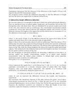
Healthcare professionals use the term TENS to
refer to currents administered using a ‘standard
TENS device’ (Fig. 12.2). Differences in the design
between manufacturers tend to be cosmetic with
limited effect on physiological and clinical outcome.
Some manufacturers have designed TENS devices
that markedly differ from a standard device. These
TENS-like devices include interferential therapy,
microcurrent therapy, and transcutaneous electrical
acupoint stimulation. A critical review of TENSlike devices can be found in Johnson (2001a, b).
A standard TENS device should be used for pain
in the first instance and will be the focus of this
chapter.
The purpose of TENS is to stimulate nerve fibres
and to generate nerve impulses that elicit pain modulation. Different techniques are used to stimulate
different populations of nerve fibres (Table 12.1).
The main techniques are:
Conventional TENS: low-intensity, highfrequency currents, to elicit segmental
analgesia;
l
206
Acupuncture-like TENS: high-intensity, lowfrequency currents, to elicit extrasegmental
analgesia; and
Intense TENS: high-intensity high-frequency
currents, to elicit peripheral nerve blockade, and
segmental and extrasegmental analgesia.
l
l
Conventional TENS is used for most patients in
the first instance.
Conventional TENS
The International Association for the Study of Pain
(IASP) defines conventional TENS as high frequency (50–100 Hz), low intensity (paraesthesia, not
painful), small pulse width (50–200 s) (Charlton
2005). Conventional TENS is used to activate lowthreshold, large diameter myelinated afferent fibres
(A) normally transmitting information related to
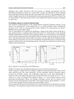
non-painful touch and pressure (Fig. 12.3). This
inhibits onward transmission of nociceptive information at synapses in the central nervous system (see
Mechanism of Action). Patients are instructed to
increase TENS pulse amplitude until a strong, comfortable, non-painful paraesthesia is experienced
beneath the electrodes, indicating large diameter
myelinated afferent fibre activity. A painful TENS
paraesthesia beneath the electrodes is not appropriate. Theoretically, high-frequency (10–200 pulses
per second (pps)) currents are optimal because they
generate a large afferent barrage leading to greater
Professor Mark Johnson
c h apte r 1 2
Figure 12.2 l A standard TENS device
Table 12.1 Types of TENS
Physiological
intention
TENS
parameters
Patient
experience
Electrode
location
Analgesic
profile
Regimen
Conventional
TENS
To stimulate large
diameter non-noxious
afferents (A) to
produce segmental
analgesia
Low intensity
(amplitude),
high frequency
(10–200 pps)
Strong, nonpainful TENS
paraesthesia
with minimal
muscle activity
Dermatomes
Site of pain
Usually rapid
onset and
offset
Use TENS
whenever in
pain
AL-TENS
To stimulate small
diameter cutaneous
and motor afferents
(A) to produce
extrasegmental
analgesia
High intensity
(amplitude), low
frequency (1–5
bursts of 100
pps)
Strong
comfortable
muscle
twitching
Myotomes
Site of pain
Muscles
Motor nerves
Acupuncture
points
May be delayed
onset and
offset
Use TENS
for 20–30
minutes at a
time
Intense
TENS
To stimulate
small diameter
cutaneous afferents
(A) to produce
counterirritation
High amplitude
(uncomfortable/
noxious), high
frequency
(50–200 pps)
Uncomfortable
(painful)
electrical
paraesthesia
Dermatomes
Site of pain
Nerves
proximal to
pain
Rapid onset
and delayed
offset
Short periods
only 5–15
minutes at a
time
207
Transcutaneous electrical nerve stimulators for pain management
c h apte r 1 2
TENS
electrodes
TENS
Skin
TENS
Paraesthesia
'Touch' afferent (A-beta)
Blockade of incoming
nociceptive input
within spinal cord
Nociceptive afferent (A-delta fibre)
Nociceptive afferent (C-fibre)
PNS
CNS
Figure 12.3 The physiological intention of conventional TENS
Arrows indicate direction of TENS-induced nerve impulses; PNS peripheral nervous system; CNS central nervous
system.
l
inhibition of nociceptive transmission. Pulse durations between 50 and 200 s allow optimal precision in achieving the desired intensity when titrating
pulse amplitude.
Acupuncture-like TENS (AL-TENS)
AL-TENS was developed to harness the mechanisms of action of TENS and acupuncture by activating segmental and extrasegmental mechanisms
(descending pain inhibitory pathways) (Eriksson &
Sjölund 1976). IASP define AL-TENS as a form
of hyperstimulation achieved using currents that
are low frequency (2–4 Hz), higher intensity (to
tolerance threshold), and longer pulse width
(100–400 s) (Charlton 2005). Intermittent trains
or bursts (2–4 Hz) of high-frequency pulses (100–
200 pps) are often used in clinical practice to reduce
discomfort experienced using high-intensity single
pulses. The intention of AL-TENS is to stimulate
small diameter, higher threshold afferents (A) using
high-intensity, low-frequency TENS. Research suggests that small muscle afferents produce greatest
analgesia so some practitioners administer AL-TENS
to generate non-painful muscle twitches which
indirectly generates impulses in small diameter
muscle afferents (Fig. 12.4). Electrodes are positioned at the site of pain, over myotomes, muscles,
acupuncture points, and trigger points. AL-TENS is
used to treat patients who are resistant to conventional TENS and patients are advised to administer
it less frequently than conventional TENS, e.g. 20
208
minutes, 3 times a day (Eriksson & Sjölund 1976).
AL-TENS can also be used for muscle and visceral
pain arising from deep-seated structures, radiating
neuropathic pain, and in situations where prolonged
analgesia is required (Johnson 1998).
Intense TENS
Intense TENS is a counterirritant and is delivered
for short periods of time over nerve bundles close
to the site of pain. High-frequency (up to 200 pps),
high-intensity currents that are painful but tolerable are used. The intention of intense TENS is to
stimulate small diameter, higher threshold cutaneous afferents (A) to block transmission of nociceptive information in peripheral nerves (Fig. 12.5).
Intense TENS activates diffuse noxious inhibitory controls (Le Bars et al 1979), and can be used
for minor procedures such as wound dressing and
suture removal.
Contraindications
Manufacturers list cardiac pacemakers, epilepsy, and
pregnancy as contraindications because it may be
difficult to exclude TENS as a potential cause from a
medico-legal perspective. The Chartered Society for
Physiotherapy (CSP) suggest that TENS can be used
in pregnancy and in epilepsy providing electrodes are
placed well away from the abdomen, sacrum, and
neck respectively (i.e. local contraindication) (CSP
Professor Mark Johnson
TENS
electrodes
Skin
Muscle
twitch
c h apte r 1 2
TENS
Motor efferent (A-alpha)
Activation of
descending pain
inhibitory pathways
Cutaneous afferent (A-delta fibre)
Muscle afferent (A-delta fibre)
Blockade of incoming
nociceptive input
within spinal cord
Nociceptive afferent (C-fibre)
PNS
CNS
Figure 12.4 l The physiological intention of acupuncture-like TENS.
Arrows indicate direction of TENS-induced nerve impulses; PNS peripheral nervous system; CNS central nervous
system.
TENS
electrodes
TENS
Skin
‘Touch’ afferent (A-beta)
Noxious
stimulus
TENS
Paraesthesia
Blockade of incoming
nociceptive information
in peripheral nerves
Nociceptive afferent
(A-delta fibre)
Figure 12.5 l Intense TENS
Arrows indicate direction of TENS-induced nerve impulses and direction of nerve impulses arising from damaged
tissue
2006). The CSP also lists bleeding tissue as a contraindication and suggests that TENS should not
be delivered over active epiphysis or over an active,
treatable tumour.
Precautions
TENS should not be administered over the anterior neck, eyes, and testes or through the chest
using anterior and posterior positions. TENS may
interfere with foetal and cardiac monitoring equipment and should not be administered close to
transdermal drug delivery systems. There is no
known evidence that adverse events occur when
TENS is used with metal implants, stents, percutaneous central catheters, or drainage systems. It
should not be used while driving and should only be
given internally using devices designed for that purpose (e.g. incontinence or dental analgesia). TENS
devices with timers that automatically switch off
are useful to aid sleep and may be used by children
with success (Lander & Fowler-Kerry 1993; Merkel
et al 1999).
Serious adverse events from TENS occur but are
extremely rare (Mann 1996; Rosted 2001). It has
209
c h apte r 1 2
Transcutaneous electrical nerve stimulators for pain management
been known to exacerbate pain and occasionally
causes nausea and light-headedness, but retains an
excellent safety and toxicity profile. No major drug
interactions occur; therefore it can be combined
with analgesics to reduce dosage and drug-related
side effects. It has been claimed that caffeine may
inhibit TENS effects (Marchand et al 1995).
Table 12.2 Clinical Indications
Pain
Chronic pain
Postoperative pain
Osteoarthritis, rheumatoid
arthritis, low back pain
Labour pain
Neuropathic pain including
amputee pain, postherpetic
and trigeminal neuralgias,
post-stroke pain, complex
regional pain syndrome
Dysmenorrhoea
Localized muscle pain
including muscle tension,
myofascial pain, postexercise soreness
Angina pectoris
Nociceptive pain including
inflammatory pains and
chronic wound pain
Orofacial pain
Cancer-related pain
Physical trauma including
fractured ribs and minor
medical procedures
Acute pain
Clinical technique
Indications
TENS is potentially useful for any type of pain
including that of nociceptive, neuropathic, and
musculoskeletal origins (Table 12.2). Clinical experience suggests it provides long-term benefit for
low back pain (LBP), osteoarthritis (OA), localized
muscle pain, and neuropathic pains of peripheral
origin such as postherpetic and trigeminal neuralgias, amputee pain, entrapment neuropathies, and
radiculopathies (Barlas & Lundeberg 2006). TENS
may also benefit metastatic bone disease, nerve
compression by a neoplasm, and post-mastectomy
and post-thoracotomy pains (Berkovitch & Waller
2005).
Timing and dosage
TENS is ideal when treatment needs to be dynamic
as effects are usually rapid in onset and offset, and
are maximal during stimulation. Electrodes are left
in situ and TENS may be administered intermittently throughout the day on an as-needed basis.
Patients can leave TENS switched on for long periods of time and should increase intensity for breakthrough or incident pain. It should be administered
before pain becomes moderate or severe but skin
hygiene is essential as minor skin irritation under
electrodes may occur.
Electrode location
TENS should be delivered over healthy sensate
skin; therefore skin sensitivity testing should be
undertaken at the site of electrode placement.
Electrodes are positioned at dermatomes related to
the site of pain for conventional TENS. As TENS
activates nerve fibres directly beneath the electrodes the primary site for electrodes is around the
210
site of pain (Fig. 12.6), or positioned paravertebrally
at the appropriate spinal segment or on contralateral dermatomes. If it is not possible to site electrodes close to the pain because of hypersensitivity
or skin damage (e.g. open wound, eczema), then
electrodes should be positioned on nerves proximal
to the pain. TENS may aggravate pain if electrodes
are placed on skin with tactile allodynia.
TENS on acupuncture points
The use of TENS to supplement acupuncture
analgesia over specific points, such as trigger and
acupuncture points, is done sparingly within clinical application. A common misconception is that
AL-TENS must be delivered at acupuncture points,
which is not the case, but it may be effective.
A review of research on TENS and acupuncture
points concluded that it may be useful when given
over acupuncture points but there were few studies
that compared TENS at acupuncture points versus
TENS at the site of pain (Walsh 1996).
Transcutaneous electrical acupoint stimulators
(TEAS) are watch-like devices worn on the underside
of the wrist over the Pericardium 6 (P6) acupuncture
point (Fig. 12.6). Good quality randomized controlled trials (RCTs) have found that TEAS reduced
Professor Mark Johnson
c h apte r 1 2
Figure 12.6 l Common sites for positioning electrodes during TENS
postoperative and chemotherapy-induced nausea and
vomiting (Coloma et al 2002; Zarate et al 2001).
between treatments whilst maintaining a strong but
comfortable intensity.
Electrical characteristics of TENS
Research evidence
The key determinant of TENS outcome is titration
of the pulse amplitude to activate different nerve
fibres (Table 12.1). For conventional TENS the user
should titrate pulse amplitude to produce a strong,
comfortable, non-painful paraesthesia beneath
the electrodes. Practitioners should be cautious of
claims about the best pulse frequencies, durations,
and patterns for different pain conditions. A systematic review of studies investigating the effects
of different pulse frequencies on experimental pain
in healthy humans concluded that research to find
optimal TENS settings for different conditions is
confusing (Chen et al 2008) suggesting that the
parameters may influence subjective comfort of
paraesthesia rather than having clinically meaningful
effects on TENS outcome (Johnson et al 1991a, b).
For this reason, pulse frequency, pattern, and duration are selected by trial and error according to ‘personal comfort’ for the pain at that time. Patients are
encouraged to experiment with settings within and
Mechanism of action
TENS causes antridromic activation of peripheral
nerves so that impulses travelling away from the
central nervous system will collide and extinguish
afferent impulses arising from peripheral receptors.
This may lead to peripheral blockade of impulses
arising from tissue damage (Fig. 12.5).
Animal studies show that conventional TENS
inhibits central transmission of nociceptive information in the spinal cord when applied to somatic
receptive fields (Garrison & Foreman 1994, 1996;
Leem et al 1995). The inhibitory neurotransmitter
gamma-amino butyric acid (GABA) appears to be
critical for conventional TENS effects (Duggan &
Foong 1985; Maeda et al 2007). It has also been
shown to reduce inflammation-induced sensitization
of dorsal horn neurons in anaesthetized rats (Ma &
Sluka 2001).
211
c h apte r 1 2
Transcutaneous electrical nerve stimulators for pain management
Higher intensities, e.g. AL-TENS, act via
extrasegmental mechanisms and activate structures
on the descending pain inhibitory pathways (e.g.
periaqueductal grey and ventromedial medulla) and
inhibit structures on descending pain facilitatory
pathways (Ainsworth et al 2006; Chung et al 1984a,
b). Higher intensities cause long-term depression of
central nociceptor cells for up to 2 hours post stimulation (Sandkühler et al 1997, 2000). Activation of
deep tissue peripheral afferents appears to produce
largest effects (Duranti et al 1988; Radhakrishnan
& Sluka 2005). Brief, intense, painful TENS probably elicits counterirritant mechanisms via diffuse
noxious inhibitory controls (Le Bars et al 1979).
Recent research has shown low-frequency TENS
to involve mu opioid receptors and high-frequency
TENS to involve delta opioid receptors (Kalra
et al 2001; Sluka et al 1999, 2000). Cholinergic,
adrenergic, and serotinergic systems also seem to
be involved (King et al 2005; Radhakrishnan et al
2003; Sluka & Chandran 2002).
Clinical effectiveness
There are over 500 RCTs cited in PubMed (10
September 2009) but many have methodological
shortcomings due to inappropriate technique and/or
under dosing. Systematic reviews of clinical research
for acute pain have been inconclusive for a mix of
acute pain conditions (Walsh et al 2009), positive
for primary dysmenorrhoea (Proctor et al 2003) and
negative for labour pain (Carroll et al 1997; Dowswell
et al 2009) and postoperative pain (Carroll et al 1996).
However, a systematic review of 21 RCTs on TENS
for postoperative pain revealed shortcomings in RCTs
that may have contributed to negative findings (Bjordal
et al 2003). The meta-analysis demonstrated TENS
reduced analgesic consumption during postoperative
care, provided it was administered using a strong, subnoxious electrical stimulation at the site of pain.
Systematic reviews for chronic pain are often inconclusive (Nnoaham and Kumbang 2008; Khadilkar et al
2005) although authors are often positive about TENS
effects. It may be of benefit for, knee OA (Osiri et al
2000; Bjordal et al 2008), rheumatoid arthritis of the
hand (Brosseau et al 2003), post-stroke shoulder pain
(Price & Pandyan 2000), whiplash, mechanical neck
disorders (Kroeling et al 2005), and chronic recurrent headache (Bronfort et al 2004). a meta-analysis
of 38 studies on TENS and peripheral electrical nerve
stimulation (PENS) for chronic musculoskeletal pain
reported significant decreases in pain at rest and on
movement (Johnson & Martinson 2007). There is
insufficient evidence to judge the effects of TENS for
cancer pain (Robb et al 2009)
Case Study 1
Anonymous
Introduction
Complex regional pain syndrome type 1 (CRPS 1) was
previously classified as reflex sympathetic dystrophy
(RSD) (Evans 1946) and refers to a functional disorder
of the spinal cord that involves the dorsal and ventral
horns, and the intermediolateral columns, to varying
degrees so as to produce sensory, motor, and autonomic
abnormalities (Loeser 2005; Wilson et al 2005a). Type I
CRPS is distinguished from type II solely by the presence
or absence of a clinically detectable injury or nerve
involvement. The condition is a form of neuropathic pain,
but not all neuropathic pain are caused by CRPS and
not all neuropathies lead to presentations of this type
(Loeser 2005). The symptoms of CRPS 1 may be caused
by an injury or by spontaneous events, manifesting via
pain and sensory changes disproportionate in intensity,
distribution, and duration to the underlying pathology
(Dunn 2000). Additional dysfunctional features may
involve motor changes, autonomic changes, trophic
changes, and psychological dysfunction.
CRPS 1 is now regarded as a systemic condition
involving the entire neuroaxis with manifestations of
inflammatory changes at the central and peripheral nerve
levels. It is a syndrome that represents a spectrum of
changes involving a myriad of multiple systems including
neurogenic both peripheral (PNS) and central nervous
systems (CNS); endocrine; vascular; musculoskeletal;
and biopsychosocial (Wilson et al 2005b). The condition
appears to have a cyclical presentation, with recurrences
of symptoms after dormant periods ranging from 6
months to 2 years; recurrent episodes are reported as
occurring in 10 to 30% of patients diagnosed with the
condition (Dunn 2000).
Current evidence is far from conclusive and a wide
variety of causative mechanisms have been described
(van Griensven 2005), with generalized sensory
and motor changes not explained by the peripheral
innervation (Rommel et al 1999) and even altered brain
responses (Juottonen et al 2002). There appears to be no
evidence of CRPS as a psychogenic condition, merely
(Continued )
212
Professor Mark Johnson
c h apte r 1 2
Case Study 1 (Continued)
anxiety and stress linked to the physical presentation
alongside sympathetic dysfunction (Covington 1996).
With this in mind, many treatment approaches have
been tried, but there is almost no reliable evidence of
genuine efficacy (Bengtson 1997). Early treatment, pain
modulation, and functional rehabilitation are essential,
together with a respectful approach to a highly sensitized
CNS and PNS; each treatment must be judged on
individual merits for each patient. The emphasis must
lie with the functional restoration or improvement of the
affected area. If untreated, CRPS 1 will progress through
acute, subacute (dystrophic), and finally, atrophic
phases. Each stage results in progressively greater
dysfunction and disability, with a diminishing chance of
successful resolution (Keller et al 1996).
The IASP renamed both types with their present
nomenclature in 1995. The IASP has agreed on four
diagnostic criteria for CRPS 1, the last three of which
must be present to confirm the diagnosis:
l The presence of an initiating noxious event or a cause
for immobilization;
l Continuing pain, allodynia, or hyperalgesia, which is
disproportionate to any inciting event;
l Evidence of oedema, changes in skin blood flow, or
abnormal sudomotor activity in the region of pain;
and
l The exclusion of other pathology that would
otherwise account for the degree of pain and
dysfunction.
With such a myriad of complex and debilitating
symptoms it is not surprising that physiotherapy provides
the mainstay of treatment of CRPS 1. If left unrecognized
and therefore untreated, atrophy, contracture, and
irreversible disablement can lead to despondency,
depression, and, in rare cases, amputation. The treatment
of CRPS still engenders much controversy because by
its very nature no single treatment produces predictable
results in every patient. Each treatment programme must
be individually tailored to the specific symptoms and the
personality of the patient. It is precisely because pain
in these patients is so pronounced and intractable that
gentle handling is essential.
Subject’s history
The subject was a male, aged 49 years, who sustained
a complex fracture to his left distal radius after falling
downstairs. X-rays detected a fracture of the left wrist,
and 2 days later he had an open surgical reduction with
internal fixation and bone grafting of the fractured ulna;
postoperatively he was placed in a plaster cast in which
he remained for 6 weeks. The subject presented 1 week
after the plaster was removed, having returned to work
as a project manager in the construction industry, but he
was experiencing problems with all aspects of daily living
and work.
The subject described his pain as sharp, deep, and
burning, affecting most of his wrist and hand, particularly
over the operation scars and in the interphalangeal (IP)
joints of his fingers over the radial aspect. The visual
analogue scale (VAS) was reported as 80.5/100 on
any activities involving the use of his hand. Changes
in temperature aggravated his pain, especially cold
weather. The subject reported no sleep disturbance,
although his wrist and fingers were stiff and painful in the
morning.
Objective examination
The following objectives signs were demonstrated:
l Swelling and oedema of the hand.
l Trophic skin changes which was dry and flaky.
l Active wrist movements were greatly limited by pain
and stiffness, particularly extension was only 10°.
flexion to 30°; and supination was so minimal it was
too difficult to measure accurately.
l Extension at the interphalangeal joint (IPJ) and
metacarpophalangeal joints (MPJ) were full, but
flexion was severely restricted, measured at 70 mm
from the palm.
l There were sensory changes to light touch to which
he was hypersensitive, particularly on his fingertips;
and
l Passive accessory movements were not examined
because of severe pain.
From the subjective history and objective examination
it was concluded that the patient’s problems were:
l Pain, severe and debilitating in nature;
l Oedema;
l Decreased range of movement (ROM);
l Altered sensation; and
l Decreased function.
Treatment
Initial treatment consisted of:
l An explanation of CRPS 1;
l A full explanation of the need for exercise,
desensitization, and pacing; and
l Restoration of full functional independence.
The subject was instructed into the use of contrast
baths and self-massage; desensitization of the skin with
different textures; and gentle active wrist and finger
exercises. During the next four treatments, with increased
handling and some gentle accessory glides to the wrist
and IPJ, he reported a definite improvement in pain levels
and light functional use; the subject felt generally more
comfortable, but ROM demonstrated little improvement.
The patient returned to see the consultant who
confirmed the diagnosis of CRPS 1 and also brought up
the possibility that, having viewed recent X-rays, perhaps
(Continued )
213
c h apte r 1 2
Transcutaneous electrical nerve stimulators for pain management
Case Study 1 (Continued)
some of the internal fixating metalwork could be acting
to block wrist extension.
A change in treatment was indicated as progress
had plateaued and more active pain inhibitory
mechanisms were required to facilitate restoration
of function. As wrist hypersensitivity remained the
overwhelming problem, acupuncture was considered too
invasive into an already sensitized sympathetic nervous
system (SNS); the skin texture and circulatory quality of
the limb were not sufficiently robust to tolerate needling
into the area.
TENS using AL-TENS at 4 Hz was administered to
Large Intestine 4 (LI4) bilaterally, LI10, and LI11
on the left arm. This treatment was administered in
the clinic and the subject asked to use it at home for
two periods of 30 minutes, twice daily whilst all the
normal physiotherapy rehabilitation activities were
continued.
At treatment three further use of conventional TENS
current was applied to the extra Baxie acupuncture
points between the second and third, third and fourth,
and fourth and fifth metacarpal heads found proximal
to the folds between the fingers (Hecker et al 2001).
Again, the patient was instructed to use this as a daily
home treatment whilst passive, active, and accessory
joint mobility was undertaken during the physiotherapy
intervention.
Outcome
After the first TENS treatment the subject complained
of aching and soreness in his hand which was different
in nature from his presenting pain and eased the
following day; the VAS was now 40/100, increasing
to a 70/100 after mobilizations and stretches but settling
after treatment. Active ROM had also improved: wrist
extension was now 25°; supination was 70°, but difficult
to maintain. The hand appearance has been the most
dramatic improvement, with resolution of oedema over
the dorsum of the hand and wrist; there was no longer
a general shiny appearance to the hand or increased
sweating, and the hypersensitivity in the fingertips had
resolved. There is unfortunately the appearance of
fixed flexion contractures in the distal IPJ of the little
and ring fingers; these digits remain very stiff and
lacked full ROM. Functionally there has been great
improvement and the subject has returned to driving,
although this involved changing gear, which remained
awkward.
Clinical reasoning
It is clear from both the subjective and objective findings
of the initial and subsequent examination that this patient
demonstrated CRPS 1 according to the recognized
signs and symptoms described in the literature (Janig
et al 1991; Koman et al 1999; Mitchell et al 1864).
The subject demonstrated classic hyperaesthesia,
allodynia, and vasomotor and labile sudomotor
changes.
Research into the effect of TENS on the nervous
system is well recognized (Johnson et al 1991b; King
et al 2005) and the analgesic effect produced by the
secretion of endorphins, enkephalins, dynorphin,
serotonin, and adrenaline as a result of TENS will
enhance descending inhibitory control (Johnson 1998).
After the first two treatments, the treatment was
extended to include acupuncture points as the hand
sensitivity had reduced and the subject was now
able to tolerate enhanced exercise and practitioner
handling of the affected limb. The non-meridian, extra,
Baxie points were used in between the metacarpal
heads of the index, middle, and ring fingers in the
contralateral limb, chosen for their action of alleviating
pain, stiffness, and swelling in the hand (Hecker 2008).
The He-Sea points, Pericardium 3 (PC3), Lung 5 (LU5),
and LI11 were used on the affected side to increase the
circulation and Qi flow to the hand and forearm. The
extra point Yintang was added to help with relaxation
and induce sleep.
Reflective practice
One limitation of this single case study is the use of
other physiotherapy modalities alongside that of TENS;
mobilizations, exercises, and gentle massage, along with
an extensive home exercise programme were all used
concurrently. The improvement in the symptoms and
objective measurements cannot be solely attributed to
the application of one modality.
The choice of acupuncture points appeared appro
priate for the condition but perhaps bilateral application
of LI4, into the affected tissue may have added to the
sensitization but it appeared to be well tolerated by
the subject. It would have been interesting to have the
opportunity to continue with a progression of active
acupuncture treatments for the stiffness in the ring and
little fingers, but unfortunately time constraints prevented
this progression from taking place.
Conclusion
CRPS 1 is a multifactorial condition that requires clear
diagnosis and an individually tailored treatment plan.
No two cases will respond in the same way; this
case study demonstrated the successful integration
of TENS and acupuncture into a complex management
programme, as a means of facilitating greater pain
modulation, empowering the subject in a home
management programme, and providing a costeffective means of managing a very complex, longterm condition.
(Continued )
214
Professor Mark Johnson
c h apte r 1 2
Case Study 2
Matthew Walmsley
Introduction
This case study presents a 78-year-old male with acute
on chronic cervical (Cx) and associated right arm pain.
After an episode of chronic pain in 1996, he underwent
a Cx laminectomy at the levels of C4 to C7 inclusively
and following his operation the pain resolved. He
subsequently received no physiotherapeutic followup. During 2008, he experienced an acute onset of Cx
pain following a rotation of his Cx spine whilst sitting.
Pain was initially centralized in his Cx spine, then
peripheralized, developing clawing and weakness in his
right arm and hand following an ulnar nerve distribution.
During initial assessment this patient had severe
functional difficulties. He presented with a pain-evoked
Cx block into right rotation and side flexion, limiting his
movement to approximately 50 and 30%, respectively,
compared to the opposite side. He had associated
ulna nerve pain with affected C7 to T1 myotomes and
dermatomes on his right. Manual therapy commenced
with exercise and taping and after three sessions of
physiotherapy he reported some level of satisfaction in
terms of pain resolution; however he still had moderate
pain and some functional limitations.
Following initial assessment, the priority was to
reduce pain, then unload the nerve and gain increased
movement at his Cx spine. Treatment included
education, taping, electroacupuncture (EA), and
progressive Cx stabilization exercises. After 8 sessions
of the above treatment over a period of 2 months, the
patient reported an 85% improvement in pain and a 75%
improvement in functional capacity. Moreover, clawing
of his right hand was completely eradicated and he was
able to complete all functional rehabilitation.
During the next five physiotherapy treatments
acupuncture was used to reduce pain further and help
stimulate nerve growth and effectiveness of C7 to T1
myotomes. Following these sessions the patient’s
strength in his right hand became similar to his left and
functional tasks were now manageable.
Subjective and objective examinations
The locations of symptoms, with frequency and intensity,
are summarized on the body chart in Fig. 12.7.
The objective assessment is summarized in Table 12.3.
Clinical reasoning and underlying mechanisms
Considering this patient’s previous surgery and the
aggravating factors it is likely that he has had a degree
of ulnar nerve damage. Therefore, the most likely pain
presentation is mechanism with a peripheral neuropathic
component, together with some nociceptive pain owing
to local tissue trauma. Neuropathic pain (NP) is initiated
by nervous system damage or dysfunction. It is often
difficult to manage due to a complex history with diverse
causes and it is often difficult to identify a specific cause
of NP; symptoms can include perceived temperature
changes, weakness, radiating pain, pins and needles,
numbness, and changes in skin condition (Colvin et al
2000; NICE 2008). Axons within the ulnar nerve may
have been damaged; therefore early intervention is
imperative in order to create the best environment
for axonal healing to help resolve and prevent further
problems (Colvin et al 2000).
Since the onset of pain, the subject had become
increasingly frustrated and was struggling to sleep. He
had commenced on a low dose of Amitriptylin to help
decrease pain, improve his low mood, and improve sleep
quality (Gilron 2006). Sleep is an important aspect of
self-healing, since during sleep hormones are released
that boost the immune system and promote selfhealing (Moldofsky 1995). However, the physiological
functions of sleep are partly unknown (Kryger et al 1994;
Parmeggiani 1994). Lack of sleep may lead to lower pain
threshold, centrally sensitising this subject to the neural
injury (Moldofsky et al 1975). As he had experienced
insomnia for the past 4 weeks, his pain threshold would
have been significantly reduced, increasing his NP and
further reducing his mood and ability to cope. Taking this
in to account, reducing this subject’s NP and insomnia
would help resolve his problems.
Treatment selection
During the first two sessions of physiotherapy attention
was paid to offloading the ulnar nerve, together with
positions of comfort for the Cx to decrease the subject’s
acute pain (Wheeless 2009). By the third session,
acupuncture was considered for reduction of insomnia
and pain and facilitate to improvement in function. In this
case, it was hypothesized that damage to the neural tis
sue had taken place in the ulnar nerve, resulting in a short
onset of afferent impulses, termed injury discharge which
has been linked to the onset of NP (Kryger et al 1994).
Many studies have been completed using
acupuncture for the treatment of NP, with varied results
and many conclude that traditional acupuncture,
using meridian points, is much more beneficial when
treating nociceptive pain rather than neuropathic pain
(Bradnam 2003; Budh et al 2006). This is thought to
be due to a difference in neuropeptides needed during
pain modulation (Han 2003). However, many studies
have found EA to be an effective analgesic and a
good treatment for NP, without any observed negative
side effects (Stener-Victorin et al 1999). EA has been
demonstrated to activate inhibitory systems within the
spinal cord, which results in segmental inhibition of the
sympathetic outflow (Sato et al 1997) and pain pathways,
as predicted by the gate control theory (Melzack & Wall
1965). In this instance the C7 to T1 segments could be
(Continued )
215
c h apte r 1 2
Transcutaneous electrical nerve stimulators for pain management
Case Study 2 (Continued)
A
B
B
Constant
Deep
Ache
Deep
I/M shooting pain
Followed by constant ache
Ags
C x R rotation - instant
C x R side flexion - instant
Reading > 10 mins
Sleeping
Ags
Using R arm to lift
> 5 kg - instant
C x R rotation I/M
C x R side flexion I/M
Eases
Laying supine > 20 mins
Heat
Anti inflammatory gel
Eases
Hand in pocket
Heat
Rest
A
B
SQ’s
No 5D’s
No pins and needles
Numbness over C7/T1
No headaches
10 mins of stiffness on walking
Wakes patient 3–4 times a night
Worst time evenings
Body chart showing the areas of pain;
SQ’s
I/M
Ags
Eases
Special questions
Intermittent
Aggravating factors
Easing factors
Figure 12.7 l Symptom location.
utilized by relevant, adjacent acupuncture points in order
to decrease localized pain, whilst other points may be
utilized to give the patient systemic relief.
Stener-Victorin (2003) used a combination of highand low-frequency (80 and 2 Hz, respectively) EA, and
found it lowered pain experienced by 24%, compared
to the control, using acupuncture points Governor
Vessel 20 (GV20) and Stomach 29 (ST29) at 80 Hz; Triple
Energizer 5 (TE5) and LI4 at 2 Hz; and ST36 with manual
stimulation. This identical study design was carried
out (Taguchi 2007) with a variation on point selection;
however, they found no statistical difference between
the two groups. These two studies identified 11 and 8%
reductions in anaesthetic requirement when using EA at
auricular points, respectively (Taguchi 2007). In contrast,
Morioka et al (2002) and Stener-Victorin et al (2003)
stimulated three acupoints ST36, GB34, and Bladder 60
(BL60), failing to reduce anaesthetic need.
Nedstrand et al (2005), using acupuncture in an
attempt to reduce hormonal symptoms in women,
found a decreased generalized pain threshold by using
EA. The points used were BL15, BL23, BL32, Heart 7
(H7), Spleen 6 (SP6) and SP9, LIV3, PC6, and GV20.
The choice of acupuncture points demonstrated no
significant decrease in pain scales that had been found
in previous studies during treatment of dysmenorrhoea.
Within all studies reviewed, there was no consistency of
points used; there was, however, a general consensus
about the use and the amount of stimulation to use for
NP relief. High frequency (100 Hz) was seen to be better
than low frequency (2 Hz) at reducing pain (Han et al
1999; Liang et al 2002; Morioka et al 2002).
Recent studies showed that EA in specific
frequencies applied to certain points could facilitate the
release of neuropeptides, eliciting profound physiological
effects, activating self-healing mechanisms (Han 2004).
(Continued )
216
Professor Mark Johnson
c h apte r 1 2
Case Study 2 (Continued)
Table 12.3 Objective assessment baseline measurements
Observation
Right trapezius lengthened
No muscle bulk loss
Protracted Cx and rounded shoulders
Kyphotic at Tx spine
Palpation
Tenderness over whole Cx spine, worse over R facets between C3 and C7
AROM
Right side flexion 1⁄3
Right rotation 1⁄2
ROM blocked by pain
No end-feel gained.
Neural function
No absence of triceps reflex
No absence of coracobrachialis and or biceps reflex
Diminished RC7 to T1 dermatome and myotome sensation
Functional tests
Instant pain on picking up anything heavier than 5 kg with R hand
Muscle tests
Unable to assess Cx spine due to pain
All GHJ muscles at R and L full power
R hand myotomal weakness in C7 to T1
Special tests
Repeated flexion and extension of Cx spine increased pain
Combined movements of Cx spine into R rotation, R side flexion and extension increased
both A and B pain
Upper limb tension test (ULTT) 3 positive on R
Investigations
Nil since X-ray following laminectomy 1997
No MRI
Medications
Anti-inflammatory gel, atenolol, ramipril, and lansoprazole
Hobbies
Before injury; walking, looking after grandchildren, and reading
Right shoulder full ROM
Pain on all movements
No active movement of middle, ring, and small
fingers on right
Notes: ULTT, upper limb tension test; Tx, thoracic spine; Cx, cervical spine; R, Right; L, Left.
At different frequencies, different neuropeptides are
released; these are most commonly dynorphin and
enkephalin (Han 2003). Using EA at 2 Hz accelerates
the release of enkephalin, whilst that of 100 Hz
increases the release of dynorphin (Han 2003). However,
a combination of the two frequencies produces a
simultaneous release of both, resulting in a maximal
therapeutic effect (Han 2004). This result was in direct
contrast with the hypothesis summarized by Verge et al
(1991) that central neuropeptides can be released
only by high-frequency stimulation. It is therefore
hypothesized that a combination of 2- and 100-Hz EA,
applied in unison, will result in two sites of stimulation,
which become merged and are perceived as 102 Hz,
almost indistinguishable from 100 Hz. As a result, only
dynorphin will be released (Han 2004).
In addition to decreasing pain, EA was found to
improve physical activity, sense of well being, and quality
of sleep, whilst reducing the need for medication (Hamza
2000). Hamza (2000) found that using frequencies of 15
and 30 Hz, repeated every 3 seconds, and using 0 Hz for
the sham treatments, respectively, the EA group reported
needing significantly less medication than the sham
group, which remained the same. Although this study
had some good findings, the acupuncture points used
were not disclosed.
There is also some evidence that EA can be beneficial
in treating insomnia (Hamza et al 2000; Spence et al
2004). Spence et al (2004) found that 10 sessions of
acupuncture could produce significant improvement
in sleep quality; however, this study failed to mention
the points used. With decreased sleep, an increase in
nociceptive substances such as substance P, bradykinin,
histamine, and prostaglandins would be released; this
would lead to greater central sensitization and reduce
the subject’s peripheral pain threshold, leading to a
further reduction in deep sleep (Ishimaru et al
1995; Kitade et al 1979; Taguchi 2007).
(Continued )
217
c h apte r 1 2
Transcutaneous electrical nerve stimulators for pain management
Case Study 2 (Continued)
Outcome measurements and results
Outcome measures were active Cx right rotation and
side flexion measured with a cervical goniometer.
Subjective information including pain and uninterrupted
sleep were measured with the VAS scale and patient
records, respectively. Table 12.4 gives an overview of
the points used and Table 12.5 summarizes the outcome
measures recorded in all physiotherapy sessions that
included acupuncture treatment. Following this treatment
the patient reported decrease in both pain and improved
sleep.
Limitations
Undoubtedly, there are some limitations; the subject
is undergoing a natural healing process, and therefore
it is difficult to ascertain how much EA had improved
Table 12.4 Acupuncture point rationale
Session
Aim
Points used
De Qi
Rationale
Time/frequency
B
1
Familiarize patient to
acupuncture and gain
general well being and
improved sleep
LI4 LIV3
Extra point
Yintang
Four gaits used for
general anaesthesia.
Ying tang for sleep.
20 mins
De Qi gained again @ 10
mins
2
Encourage neural
regeneration and
decrease pain. Plus
improve sleep
LI4 LIV3B
GB10B
BL10B
BL11B
EA 80 Hz
Segmental approach for
anaesthesia (BL11)
HFEA to stimulate opioid
release.
30 mins
80 Hz pulsed @ 2-s intervals.
De Qi gained 10 mins at
manual points
3
Encourage neural
regeneration and
decrease pain. Plus
improve sleep
LI4 LIV3B
GB10B
BL10B
BL11B
EA 80Hz
HJJ 80Hz
GV14
Expand on segmental
anaesthesia
(HJJ and GV) using HFEA
and LFEA to stimulate
dorsal horn effect
30 mins
80 Hz and 2 Hz separately
pulsed @ 2-s intervals. De
Qi gained
10 mins @ manual points
4
Encourage neural
regeneration and
decrease pain. Plus
improve sleep
LI4 LIV3B
GB10 B
BL10B
BL11B
EA 80Hz
HJJ 100Hz
GV14
LI11R
LI15R
As above plus adding
points on the LI meridian
as it passes over the
affected myotome.
30 mins
100 and 2 Hz separately
pulsed @ 2-s intervals. De
Qi gained
10 mins @ manual points
5
Encourage neural
regeneration and
decrease pain. Plus
improve sleep
LI4 LIV3B
GB10B
BL10B
BL11B
EA 80Hz
HJJ 100Hz
GV14
LI11R
LI15R
As above.
30 mins
100 and 2 Hz separately
pulsed @ 2-s intervals. De
Qi gained
10 mins @ manual points
Notes: B, Bilateral; R, Right; L, Left; GB, Gall Bladder; BL, Bladder; LIV, Liver; GV, Governor Vessel; LI, Large Intestine; HJJ, Huatuojiaji points;
EA, Electroacupuncture; HFEA, high-frequency, electroacupuncture; LFEA, low-frequency, electroacupuncture.
(Continued )
218
Professor Mark Johnson
c h apte r 1 2
Case Study 2 (Continued)
Table 12.5 Outcome measurements
Day
Power/grip
strength
Pain VAS
Cx ROM
C7/T1 myotomal function
Oxford Scale
Sleep
1
0.3 kg
80/100
R rotation 50% R
side flexion 30%
Full active elbow extension, nil finger
abduction and or wrist flexion
5.6
8
0.5 kg
71/100
R rotation 50% R
side flexion 50%
Full active elbow extension, 0/5 finger
abduction and 2/5 wrist flexion
6.7
22
1 kg
71/100
R rotation 60% R
side flexion 65%
Full active elbow extension, 3/5 finger
abduction and 3/5 wrist flexion
6.5
29
3 kg
50/100
R rotation 60% R
side flexion 70%
Full active elbow extension, 3/5 finger
abduction and 3/5 wrist flexion
7
36
5 kg
14/100
R rotation 80% R
side flexion 85%
Full active elbow extension, 4/5 finger
abduction and 5/5 wrist flexion
7.2
Notes: Power/grip, tested with a grip dynamometer; ROM, compared to L with a Cervical Goniometer; Sleep, average hours per night.
symptoms. Secondly, the measure of the amount of
sleep was very subjective and did not address quality
of sleep; a more specific questionnaire could have
been used to determine well being, tiredness, energy,
and mood (Hamza 2000). Finally, the acupuncture
protocol used in this study was not previously validated,
as no study has fully concluded specific points and or
frequencies of EA to use in the treatment of NP.
Discussion
This case study attempted to analyse the use of EA
and physiotherapeutic interventions on NP. Although
acupuncture is not commonly recognized for treating
such conditions, it was considered in this case, as it
was coupled with other interventions to help treat the
subject’s pain, insomnia, and reduced motor function.
During the first 3 sessions of physiotherapy the
patient made very limited improvement and EA was
considered in conjunction with the exercises regime.
Following 5 treatments of EA, outcome measurements all
improved significantly. Pain levels reduced from 92/100
to14/100 (VAS), Cx ROM in right side flexion improved
from 30 to 80%, and the average amount of sleep
improved from 5.6 to 7.2 hours per night.
According to traditional Chinese medicine, the ‘four
gates’, LI4 and LIV3 (Liang et al 2002) combined with
a segmental approach at C7 to T1, exhibit a powerful
analgesic effect (Han 2003) whilst the extra point Ying
Tang and EA in general can improve sleep (Hamza 2000).
Many theories can be considered to explain the
positive outcomes regarding pain relief. Manual
acupuncture given to healthy volunteers, at acupuncture
points LI4 and LIV3 has been shown to deactivate areas
in the brain that regulate pain modulation (Yan et al
2005). Acupuncture has been shown to be much more
effective when used with low-frequency EA, stimulating
the dorsal horn and giving longer lasting relief (Mo
et al 1996; Han 2003 Hamza 2000). This effect is
further enhanced when alternated with high-frequency
EA at segmental levels, in order to offer an overall
global analgesia (Hamza 2000; Morioka et al 2002;
Han 2003).
Two studies demonstrated the improvement in sleep
with the use of EA (Hamza 2000; Nedstrand et al 2005).
Although the results of both of these studies appeared
conclusive, different acupuncture points were used
and no relationships were formed with biochemical
changes at cellular level. Many authors consider this
effect to be psychological and may even be due to
acupuncture intervention facilitating increased time to
rest whilst the treatment is taking place (Renckens 2002;
Spiller 2007).
Considering the above, it appears that specific
molecular and chemical factors account for
acupuncture-induced pain modulation. However, it
is impossible to discount the power of suggestion
associated with expectancy and belief for pain reduction
(Pariente et al 2005). In some patient interactions this
could play a significant role, as human pain modulating
areas have been found to be activated in both
conditions, starting a chemical process that enabled the
release of neuropeptides crucial for the relief of pain (Han
2003, 2004). Therefore, it is impossible to be definitive
concerning the specific and non-specific factors in
facilitating decrease in the subject’s pain, increase in
motor function, and improvement in sleep.
(Continued )
219
c h apte r 1 2
Transcutaneous electrical nerve stimulators for pain management
Case Study 2 (Continued)
Conclusion
In conclusion, integration of manual therapy and EA
for this subject demonstrated good results. Initially
the advice and exercises approach helped to increase
and normalize movement, gain increased stability,
and desensitize the CNS. Later, EA was effective
in producing systemic and segmental analgesia,
decreasing right arm pain, and improving neural growth,
function, and strength. Furthermore, average hours of
sleep increased with the use of EA; however, further
studies are needed to determine the exact effect of EA
on the neuronal structures.
References
Ainsworth, L., Budelier, K.,
Clinesmith, M., et al., 2006.
Transcutaneous electrical nerve
stimulation (TENS) reduces chronic
hyperalgesia induced by muscle
inflammation. Pain 120, 182–187.
Barlas, P., Lundeberg, T., 2006.
Transcutaneous electrical nerve
stimulation and acupuncture.
In: McMahon, S., Koltzenburg,
M. (Eds.), Melzack and Wall’s
Textbook of Pain. Elsevier Churchill
Livingstone, Philadelphia, pp.
583–590.
Bengtson, K., 1997. Physical modalities
for complex regional pain syndrome.
Hand Clin. 13 (3), 453–454.
Berkovitch, M., Waller, A., et al., 2005.
Treating pain with transcutaneous
electrical nerve stimulation (TENS).
In: Doyle, D., Hanks, G., Cherny,
N.I. (Eds.), Oxford Textbook
of Palliative Medicine. Oxford
University Press, Oxford, pp.
405–410.
Bjordal, J.M., Johnson, M.I.,
Ljunggreen, A.E., 2003.
Transcutaneous electrical nerve
stimulation (TENS) can reduce
postoperative analgesic consumption
A meta-analysis with assessment of
optimal treatment parameters for
postoperative pain. European Journal
of Pain 7, 181–188.
Bjordal, J.M., Johnson, M.I., LopesMartins, R.A., Bogen, B., Chow, R.,
Ljunggren, A.E., 2007. Short-term
efficacy of physical interventions
in osteoarthritic knee pain. A
systematic review and meta-analysis
of randomised placebocontrolled
trials. BMC Musculoskelet Disord
8, 51.
Bradnam, L., 2003. A proposed clinical
reasoning model for Western
acupuncture. NZ J. Physiother. 31,
40–45.
Bronfort, G., Nilsson, N., Haas, M.,
et al., 2004. Non-invasive physical
220
treatments for chronic/recurrent
headache. Cochrane Database Syst.
Rev., CD001878.
Brosseau, L., Judd, M.G., Marchand, S.,
et al., 2003. Transcutaneous
electrical nerve stimulation (TENS)
for the treatment of rheumatoid
arthritis in the hand. Cochrane
Database Syst. Rev., CD004377.
Budh, C., Kowalski, J., Lundeberg, T.,
2006. A comprehensive pain
management programme comprising
education, cognitive and behavioural
interventions for neuropathic pain
following spinal cord injury. J.
Rehabil. Med. 38 (3), 172–180.
Carroll, D., Tramer, M., McQuay, H.,
et al., 1996. Randomization is
important in studies with pain
outcomes: systematic review of
transcutaneous electrical nerve
stimulation in acute postoperative
pain. Br. J. Anaesth. 77, 798–803.
Carroll, D., Moore, A., Tramer, M.,
et al., 1997. Transcutaneous
electrical nerve stimulation does
not relieve in labour pain: updated
systematic review. Contemp. Rev.
Obstet. Gynaecol. (September),
195–205.
Charlton, J., 2005. Core Curriculum for
Professional Education in Pain, third
ed. IASP Press, Seattle pp 93–96.
Chartered Society of Physiotherapy
(CSP), 2006. Guidance for the
clinical use of electrophysical agents.
Chen, C.C., Tabasam, G., Johnson, M.I.,
2008. Does the pulse frequency
of transcutaneous electrical nerve
stimulation (TENS) influence
hypoalgesia? A systematic review
of studies using experimental pain
and healthy human participants.
Physiotherapy 94 (1), 11–20.
Chung, J.M., Fang, Z.R., Hori, Y., et
al., 1984a. Prolonged inhibition of
primate spinothalamic tract cells by
peripheral nerve stimulation. Pain
19, 259–275.
Chung, J.M., Lee, K.H., Hori, Y., et al.,
1984b. Factors influencing peripheral
nerve stimulation produced
inhibition of primate spinothalamic
tract cells. Pain 19, 277–293.
Coloma, M., White, P.F., Ogunnaike,
B.O., et al., 2002. Comparison of
acustimulation and ondansetron
for the treatment of established
postoperative nausea and vomiting.
Anesthesiology 97, 1387–1392.
Colvin, L.A., 2000. Practical
management of pain. Br. J. Anaesth.
101, 119–127.
Covington, E., 1996. Psychological
issues in reflex sympathetic
dystrophy. In: Stanton-Hicks, M.,
Janig, W. (Eds.), Reflex Sympathetic
Dystrophy. IASP, Seattle.
Dowswell, T., Bedwell, C., Lavender, T.,
Neilson, J.P., 2009. Transcutaneous
electrical nerve stimulation
(TENS) for pain relief in labour.
Cochrane Database Syst Rev (2)
CD007214.
Duggan, A.W., Foong, F.W., 1985.
Bicuculline and spinal inhibition
produced by dorsal column
stimulation in the cat. Pain 22,
249–259.
Dunn, D., 2000. Chronic regional pain
syndrome, type 1: part 1. AORN J.
72 (3), 422–449.
Duranti, R., Pantaleo, T., Bellini, F.,
1988. Increase in muscular
pain threshold following low
frequency-high intensity peripheral
conditioning stimulation in humans.
Brain Res. 452, 66–72.
Eriksson, M., Sjölund, B.H., 1976.
Acupuncture-like electroanalgesia
in TNS resistant chronic pain.
In: Zotterman, Y. (Ed.), Sensory
Functions of the Skin. Pergamon
Press, Oxford/New York, pp.
575–581.
Evans, J., 1946. Reflex sympathetic
dystrophy. Surg. Clin. North Am.
26, 780–790.
Professor Mark Johnson
Garrison, D.W., Foreman, R.D., 1994.
Decreased activity of spontaneous
and noxiously evoked dorsal horn
cells during transcutaneous electrical
nerve stimulation (TENS). Pain 58,
309–315.
Garrison, D.W., Foreman, R.D., 1996.
Effects of transcutaneous electrical
nerve stimulation (TENS) on
spontaneous and noxiously evoked
dorsal horn cell activity in cats with
transected spinal cords. Neurosci.
Lett. 216, 125–128.
Gilron, I., Watson, C.P.N., Cahill, C.M.,
et al., 2006. Neuropathic pain: a
practical guide for the clinician. Can.
Med. Assoc. J. 175, 265–275.
Hamza, M., White, P., Craig, W., et
al., 2000. Percutaneous electrical
nerve stimulation. A novel analgesic
therapy for diabetic neuropathic
pain. Diabetes Care 23, 365–370.
Han, S.J., 2003. Acupuncture:
neuropeptide release produced by
electrical stimulation of different
frequencies. Trends Neurosci. 26
(1), 145–149.
Han, S.J., 2004. Acupuncture and
endorphins. Neurosci. Lett. 361,
258–261.
Han, Z., Jiang, Y.H., Wan, Y., et al.,
1999. Endorphin-1 mediates 2 Hz
but not 100Hz electroacupuncture
analgesia in the rat. Neurosci. Lett.
274, 75–78.
Hecker, H.U., Steveling, A., Peuker, E.,
et al., 2001. Color Atlas of
Acupuncture. Thieme Publishing,
Stuttgart.
Hecker, H.U., Steveling, A., Peuker, E.,
et al., 2008. Color Atlas of
Acupuncture Body Parts, Ear Points,
Trigger Points. Thieme Publishing,
Stuttgart.
Ishimaru, K., Kawakita, K., Sakita, M.,
1995. Analgesic effects induced
by TENS and electroacupuncture
with different types of stimulating
electrodes on deep tissues in human
subjects. Pain 63, 181–187.
Janig, W., Blumberg, H., Boas, R.A.,
et al., 1991. Reflex sympathetic
dystrophy syndrome. Consensus
statement and general
recommendations for diagnosis and
clinical research. Pain Res. Clin.
Manag. 4, 372–375.
Johnson, M.I., 1998. The analgesic
effects and clinical use of
acupuncture-like TENS (AL-TENS).
Phys. Ther. Rev. 3, 73–93.
Johnson, M.I., 2001a. Transcutaneous
electrical nerve stimulation (TENS)
and TENS-like devices. Do they
provide pain relief? Pain Rev. 8,
121–128.
Johnson, M.I., 2001b. A critical
review of the analgesic effects of
TENS-like devices. Phy. Ther. Rev.
6, 153–173.
Johnson, M., Martinson, M.,
2007. Efficacy of electrical
nerve stimulation for chronic
musculoskeletal pain: a meta-analysis
of randomized controlled trials. Pain
130, 157–165.
Johnson, M.I., Ashton, C.H.,
Thompson, J.W., 1991a. The
consistency of pulse frequencies and
pulse patterns of transcutaneous
electrical nerve stimulation (TENS)
used by chronic pain patients. Pain
44, 231–234.
Johnson, M.I., Ashton, C.H.,
Thompson, J.W., 1991b. An indepth study of long-term users
of transcutaneous electrical nerve
stimulation (TENS) Implications
for clinical use of TENS. Pain 44,
221–229.
Juottonen, K., Gockel, M., Silén, T.,
et al., 2002. Altered central
sensorimotor processing in patients
with complex regional pain
syndrome. Pain 98 (3), 315–323.
Kalra, A., Urban, M.O., Sluka, K.A.,
2001. Blockade of opioid receptors
in rostral ventral medulla prevents
antihyperalgesia produced by
transcutaneous electrical nerve
stimulation (TENS). J. Pharmacol.
Exp. Ther. 298, 257–263.
Keller, T., Goldstein, L., Chappell, T.,
1996. Gamekeepers thumb variant
complicated by reflex sympathetic
dystrophy. J. Trauma 40, 660–662.
Khadilkar, A., Milne, S., Brosseau, L.,
et al., 2005. Transcutaneous
electrical nerve stimulation (TENS)
for chronic low-back pain. Cochrane
Database Syst. Rev., CD003008.
King, E.W., Audette, K., Athman, G.A.,
et al., 2005. Transcutaneous
electrical nerve stimulation activates
peripherally located alpha-2A
adrenergic receptors. Pain 115,
364–373.
Kitade, T., Hyodo, M., 1979. The
effects of stimulation of ear
acupuncture points on the body’s
pain threshold. Am. J. Chin. Med. 7,
241–252.
Koman, L.A., Poehling, G.G.,
Smith, T.L., 1999. Complex regional
pain syndrome: RSD and causalgia.
In: Greens Operative Hand Surgery,
4th ed. Churchill Livingstone,
Philadelphia.
Kroeling, P., Gross, A.R., Goldsmith,
C.H., 2005. A Cochrane review of
electrotherapy for mechanical neck
disorders. Spine 30, 641–648.
c h apte r 1 2
Kryger, M.H., Roth, T., Carskadon, M.,
1994. Circadian rhythms in humans:
an overview. In: Kryger, M.H.,
Roth, T., Dement, W.C. (Eds.),
Principles and Practice of
Sleep Medicine. WB Saunders,
Philadelphia, pp. 301–308.
Lander, J., Fowler-Kerry, S., 1993.
TENS for children’s procedural pain.
Pain 52, 209–216.
Le Bars, D., Dickenson, A.H.,
Besson, J.M., 1979. Diffuse noxious
inhibitory controls (DNIC). Effects
on dorsal horn convergent neurones
in the rat. Pain 6, 283–304.
Leem, J., Park, E., Paik, K., 1995.
Electrophysiological evidence
for the antinociceptive effect
of transcutaneous electrical
stimulation on mechanically
evoked responsiveness of dorsal
horn neurons in neuropathic rats.
Neurosci. Lett. 192, 197–200.
Liang, X.B., Liu, X.Y., Li, F.Q., et al.,
2002. Long-term high-frequency
electro-acupuncture stimulation
prevents neuronal degeneration
and up-regulates BDNF mRNA
in the substantia nigra and ventral
tegmental area following medial
forebrain bundle axotomy. Mol.
Brain Res. 108, 51–59.
Loeser, J., 2005. Introduction to
complex regional pain syndrome.
In: Wilson, M., Stanton-Hicks, M.,
Harden, R. (Eds.), CRPS: Current
Diagnosis and Therapy. IASP,
Seattle, pp. 3A–7A.
Long, D.M., 1973. Electrical stimulation
for relief of pain from chronic nerve
injury. J. Neurosurg. 39, 718–722.
Ma, Y.T., Sluka, K.A., 2001.
Reduction in inflammation-induced
sensitization of dorsal horn neurons
by transcutaneous electrical nerve
stimulation in anesthetized rats. Exp.
Brain Res. 137, 94–102.
Maeda, Y., Lisi, T.L., Vance, C.G.,
et al., 2007. Release of GABA and
activation of GABA (A) in the spinal
cord mediates the effects of TENS
in rats. Brain Res. 1136, 43–50.
Mann, C., 1996. Respiratory
compromise: a rare complication
of transcutaneous electrical nerve
stimulation for angina pectoris.
Journal of Accident and Emergency
Medicine 13, 68.
Marchand, S., Li, J., Charest, J., 1995.
Effects of caffeine on analgesia from
transcutaneous electrical nerve
stimulation. N. Engl. J. Med. 333,
325–326.
Melzack, R., Wall, P., 1965. Pain
mechanisms: a new theory. Science
150, 971–979.
221
c h apte r 1 2
Transcutaneous electrical nerve stimulators for pain management
Merkel, S.I., Gutstein, H.B.,
Malviya, S., 1999. Use of
transcutaneous electrical nerve
stimulation in a young child with
pain from open perineal lesions.
J. Pain. Symptom Manage. 18,
376–381.
Mitchell, S.W., Morhouse, G.R.,
Keen, W.W., 1864. Gunshot Wounds
and Other Injuries of Nerves.
Lippincott, Philadelphia.
Mo, X., Chen, D., Ji, C., et al., 1996.
Effect of electroacupuncture and
transcutaneous electric nerve
stimulation on experimental diabetes
and its neuropathy. Chen Tzu Yen
Chiu 3, 55–59.
Moldofsky, H., 1995. Sleep,
neuroimmune and neuroendonocrine
functions in fibromyalgia and
chronic fatigue syndrome. Adv.
Neuroimmunol. 5, 39–56.
Moldofsky, H., Scarisbrick, P.,
England, R., et al., 1975.
Musculoskeletal symptoms and nonREM sleep disturbance in patients
with ‘fibrositis syndrome’ and
healthy controls. Psychosom. Med.
37, 341–351.
Morioka, N., Akça, O., Doufas, A.G.,
et al., 2002. Electro-acupuncture
at the Zusanli, Kanglingquan,
and Kunlun points does not
reduce anaesthetic requirement.
Anaesthesiol. Analogue 95, 98–102.
National Institute of Clinical Excellence
(NICE), 2008. Spinal cord
stimulation for chronic pain of
neuropathic or ischaemic origin.
Nedstrand, E., Wijma, K., Wyon, Y.,
et al., 2005. Vasomotor symptoms
decrease in women with breast
cancer randomised to treatment
with applied relaxation or electroacupuncture: a preliminary study.
Climacteric 8 (3), 243–250.
Nnoaham, K.E., Kumbang, J., 2008.
Transcutaneous electrical nerve
stimulation (TENS) for chronic pain.
Cochrane Database Syst Rev (3)
CD003222.
Osiri, M., Welch, V., Brosseau, L.,
et al., 2000. Transcutaneous
electrical nerve stimulation for knee
osteoarthritis. Cochrane Database
Syst. Rev., CD002823.
Pariente, J., White, P., Frackowiak, R.S.,
et al., 2005. Expectancy and belief
modulate the neuronal substrates
of pain treated by acupuncture.
Neuroimage 25, 1161–1167.
Parmeggiani, P.L., 1994. The autonomic
nervous system in sleep. In: Kryger,
M.H., Roth, T., Dement, W.C.
(Eds.), Principles And Practice of
Sleep Medicine. WB Saunders,
Philadelphia, pp. 194–203.
222
Price, C.I., Pandyan, A.D., 2000.
Electrical stimulation for preventing
and treating post-stroke shoulder
pain. Cochrane Database Syst. Rev.,
CD001698.
Proctor, M.L., Smith, C.A., Farquhar,
C.M., et al., 2003. Transcutaneous
electrical nerve stimulation
and acupuncture for primary
dysmenorrhoea (Cochrane Review).
Cochrane Database Syst. Rev.,
CD002123.
Radhakrishnan, R., Sluka, K.A.,
2005. Deep tissue afferents
but not cutaneous afferents
mediate transcutaneous electrical
nerve stimulation-Induced
antihyperalgesia. J. Pain 6, 673–680.
Radhakrishnan, R., King, E.W.,
Dickman, J.K., et al., 2003. Spinal
5-HT (2) and 5-HT (3) receptors
mediate low but not high frequency
TENS-induced antihyperalgesia in
rats. Pain 105, 205–213.
Renckens, C.N., 2002. Alternative
treatments in reproductive medicine:
much ado about nothing: the fact
that millions of people do not master
arithmetic does not prove that two
times two is anything else than four.
Hum. Reprod. 17, 528–533.
Richardson, D.E., Akil, H., 1977. Longterm results of periventricular gray
self-stimulation. Neurosurgery 1,
199–202.
Robb, K., Oxberry, S.G., Bennett, M.
I., Johnson, M.I., Simpson, K.H.,
Searle, R.D., 2009. A Cochrane
systematic review of transcutaneous
electrical nerve stimulation for
cancer pain. J Pain Sympton Manage
37 (4), 746–753.
Rommel, O., Gehling, M.,
Dertwinkel, R., et al., 1999.
Hemisensory impairment in
patients with complex regional pain
syndrome. Pain 80 (1–2), 95–101.
Rosted, P., 2001. Repetitive epileptic
fits–a possible adverse effect after
transcutaneous electrical nerve
stimulation (TENS) in a post-stroke
patient. Acupunct. Med. 19, 46–49.
Sandkühler, J., 2000. Long-lasting
analgesia following TENS and
acupuncture: Spinal mechanisms
beyond gate control. In: Devor, M.,
Rowbotham, M.C., WiesenfeldHallin, Z. (Eds.), Progress in Pain
Research and Management. IASP
Press, Seattle, pp. 359–369.
Sandkühler, J., Chen, J.G., Cheng, G.,
et al., 1997. Low-frequency
stimulation of afferent Adelta-fibers
induces long-term depression at
primary afferent synapses with
substantia gelatinosa neurons in the
rat. J. Neurosci. 17, 6483–6491.
Sato, A., Sato, Y., Schmidt, R.F., 1997.
The Impact of Somatosensory Input
on Autonomic Functions. SpringerVerlag, Heidelberg.
Shealy, C.N., 1972. Transcutaneous
electroanalgesia. Surg. Forum 23,
419–421.
Shealy, C.N., Mortimer, J.T., Reswick,
J.B., 1967. Electrical inhibition of
pain by stimulation of the dorsal
columns: preliminary clinical report.
Anesth. Analg. 46, 489–491.
Sluka, K.A., Chandran, P., 2002.
Enhanced reduction in hyperalgesia
by combined administration of
clonidine and TENS. Pain 100,
183–190.
Sluka, K.A., Deacon, M., Stibal, A.,
et al., 1999. Spinal blockade of opioid
receptors prevents the analgesia
produced by TENS in arthritic rats. J.
Pharmacol. Exp. Ther. 289, 840–846.
Sluka, K.A., Judge, M.A., McColley,
M.M., et al., 2000. Low frequency
TENS is less effective than high
frequency TENS at reducing
inflammation-induced hyperalgesia
in morphine-tolerant rats. Eur. J.
Pain 4, 185–193.
Spence, D.W., Kayumov, L., Chen, A.,
et al., 2004. Acupuncture increases
nocturnal melatonin secretion and
reduces insomnia and anxiety: a
preliminary report. J. Neuropsy
chiatry Clin. Neurosci. 16, 19–28.
Spiller, J., 2007. Acupuncture,
Ketamine and Piriformis Syndrome –
A case report from palliative care.
Acupunct. Med. 25, 109–112.
Stener-Victorin, E., Waldenstrom, U.,
Nilsson, L., et al., 1999. A
prospective randomised study of
electro-acupuncture versus alfentanil
as anaesthesia during oocyte
aspiration in in-vitro fertilization.
Hum. Reprod. 14, 2480–2484.
Stener-Victorin, E., Waldenstrom, U.,
Wikland, M., et al., 2003. Electroacupuncture as a preoperative
analgesic method and its effects
on implantation rate and
neuropeptide Y concentrations in
follicular fluid. Hum. Reprod. 18
(7), 1454–1460.
Taguchi, R., 2007. Acupuncture anaes
thesia and analgesia for acute pain in
Japan. Ann. Oncol. 5, 153–158.
van Griensven, H., 2005. Pain in
Practice: Theory and Treatment
Strategies for Manual Therapists.
Butterworth Heinemann, Oxford.
Verge, V.M., Richardson, P.M., Hockfelt, T.,
1991. Differential influence of nerve
growth factor on neuropeptide
expression in vivo: a novel role in
peptide suppression in adult sensory
neurons. J. Neurosci. 15, 2081–2096.
Professor Mark Johnson
Wall, P.D., Sweet, W.H., 1967.
Temporary abolition of pain in man.
Science 155, 108–109.
Walsh, D.M., 1996. Transcutaneous
electrical nerve stimulation and
acupuncture points. Complement.
Ther. Med. 4, 133–137.
Walsh, D., Howe, T., Johnson, M.I.,
Sluka, K.A., 2009. Transcutaneous
electrical nerve stimulation for
acute pain. Cochrane Database of
Systematic Reviews 138 (2), 1–72.
Wheeless, C., 2009. Textbook of
Orthopaedics. Data Trace Internet
Publishing.
Wilson, P., Stanton-Hicks, M.,
Harden, R. (Eds.), 2005a. CRPS:
Current Diagnosis and Therapy.
IASP Press, Seattle.
Wilson, P., Stanton-Hicks, M.,
Harden, R., 2005b. Progress in Pain
Research and Management. IASP
Press, Seattle.
c h apte r 1 2
Yan, B., Li, K., Xu, J., et al., 2005.
Acupoint-specific fMRI patterns in
human brain. Neurosci. Lett. 383,
236–240.
Zarate, E., Mingus, M., White, P.F.,
et al., 2001. The use of
transcutaneous acupoint electrical
stimulation for preventing nausea
and vomiting after laparoscopic
surgery. Anesth. Analg. 92, 629–635.
223