100 case da liễu. Chẩn đoán và điều trị
Bạn đang xem bản rút gọn của tài liệu. Xem và tải ngay bản đầy đủ của tài liệu tại đây (5.11 MB, 245 trang )
100 CASES
in Dermatology
This page intentionally left blank
100 CASES
in Dermatology
Rachael Morris-Jones PhD PCME FRCP
Consultant Dermatologist & Honorary Senior Lecturer, King’s College
Hospital, London, UK
Ann-Marie Powell
Consultant Dermatologist, Department of Dermatology, St Thomas’ Hospital,
London, UK
Emma Benton MB ChB MRCP
Post-CCT Clinical Research Fellow, St John’s Institute of Dermatology,
Guy’s and St Thomas’ NHS Trust, London, UK
100 Cases Series Editor:
Professor P John Rees MD FRCP
Dean of Medical Undergraduate Education, King’s College London School
of Medicine at Guy’s, King’s and St Thomas’ Hospitals, London, UK
First published in Great Britain in 2011 by
Hodder Arnold, an imprint of Hodder Education, a division of Hachette UK
338 Euston Road, London NW1 3BH
© 2011 Rachael Morris-Jones, Ann-Marie Powell and Emma Benton
All rights reserved. Apart from any use permitted under UK copyright law, this publication may
only be reproduced, stored or transmitted, in any form, or by any means with prior permission in
writing of the publishers or in the case of reprographic production in accordance with the terms
of licences issued by the Copyright Licensing Agency. In the United Kingdom such licences are
issued by the Copyright Licensing Agency: Saffron House, 6–10 Kirby Street, London EC1N 8TS
Hachette UK’s policy is to use papers that are natural, renewable and recyclable products and
made from wood grown in sustainable forests. The logging and manufacturing processes are
expected to conform to the environmental regulations of the country of origin.
Whilst the advice and information in this book are believed to be true and accurate at the date
of going to press, neither the author[s] nor the publisher can accept any legal responsibility
or liability for any errors or omissions that may be made. In particular, (but without limiting
the generality of the preceding disclaimer) every effort has been made to check drug dosages;
however it is still possible that errors have been missed. Furthermore, dosage schedules are
constantly being revised and new side-effects recognized. For these reasons the reader is
strongly urged to consult the drug companies’ printed instructions, and their websites, before
administering any of the drugs recommended in this book.
British Library Cataloguing in Publication Data
A catalogue record for this book is available from the British Library
Library of Congress Cataloging-in-Publication Data
A catalog record for this book is available from the Library of Congress
ISBN-13
978-1-444-11793-6
1 2 3 4 5 6 7 8 9 10
Commissioning Editor:
Project Editor:
Production Controller:
Cover Design:
Indexer:
Joanna Koster
Stephen Clausard
Jonathan Williams
Amina Dudhia
Laurence Errington
Typeset in 10/12 Rotis Serif by MPS Limited, a Macmillan Company, Chennai, India
Printed and bound in India
What do you think about this book? Or any other Hodder Arnold title?
Please visit our website at www.hoddereducation.com
CONTENTS
Preface
Acknowledgements
Glossary
1: An itchy, slow-growing infant
2: An agitated atopic child
3: An acute monomorphic eruption in a systemically unwell atopic child
4: A recurrent, unsightly facial eruption in a stressed but well young adult
5: Blistered hands and feet in an athletic man
6: Chronic erythematous pruritic eruption on the lower legs
7: An itchy localized eruption
8: An eczematous eruption complicating venous ulcers
9: A transient pruritic eruption exacerbated by heat
10: A toddler with brown patches which urticate
11: Acute soft tissue swelling associated with systemic symptoms
12: Chronic scaly plaques on the knees
13: Widespread scaly eruption appears after a sore throat
14: A patient presents acutely unwell with all his skin red and hot
15: An itchy eruption appearing on the chest and arms after sun exposure
16: Acute-onset linear blistering on the legs
17: Chronic blistering eruption on the dorsal hands
18: Sun-induced skin pain, redness and scarring in a child
19: Sudden-onset widespread rash
20: Recurrent annular erythematous lesions reactivating
at identical skin sites
21: Painful lip lesion associated with a localized
blistering rash and sore mouth
22: Painful eroded mucous membranes and skin lesions
23: Acute-onset extensive blistering and skin necrosis with
mucous membrane involvement
24: Fever, epilepsy and a widespread skin eruption
with marked facial oedema
25: Acute-onset multiple pustules on a background of erythematous skin
26: Acute non-blanching cutaneous eruption associated with a sore throat
27: An itchy papular eruption on the ankles
28: A generalized itchy blistering eruption in an elderly woman
29: Sudden onset of erosions, blisters and fragile skin
following gradually worsening mouth ulcers
30: An itchy, vesicular extensor eruption associated with malabsorption
31: An itchy blistering eruption recurring in a second pregnancy
32: Extremely itchy stretch marks in the third trimester
33: Asymptomatic sclerotic white plaques on the trunk
34: Insidious onset of tightening of the skin over the limbs
ix
x
xi
1
3
5
7
9
11
13
16
19
23
25
27
29
31
33
35
37
39
41
43
45
47
49
51
53
55
57
59
63
65
67
69
71
73
100 Cases in Dermatology
35: Acute facial rash, fever and joint pains in a young woman
36: Annular erythematous rash of sudden onset
37: Hair loss, scarring rash and photosensitivity
38: An erythematous rash and muscle weakness
39: Widespread maculopapular eruption on the trunk and
face with flu-like symptoms
40: Slow asymptomatic depigmentation of the skin
41: A young adult with high blood pressure,
irregular pigmentation and skin lumps
42: An overweight teenager with thickened skin around her neck
43: A dramatic and painful ulcer in a young patient
with no evidence of infection
44: Slow-onset asymptomatic lesions on the shins of a diabetic patient
45: Slowly progressive swelling and discolouration over the shins
46: Asymptomatic annular lesions on the limbs
47: An asymptomatic papular and annular eruption
48: Ulcer over the gaiter area on a background of aching legs
49: Slow-onset, unilateral, painless leg swelling
50: An infirm elderly man with arterial disease and an ulcerated heel
51: Non-healing foot ulcer in a diabetic patient
52: A regressing vascular lesion in a pre-school child
53: A livid red birthmark on a newborn child
54: Slow development of a scaly plaque on a finger
55: A slow-growing ulcerated non-healing nodule on the face
56: Multiple basal cell carcinomas in a young patient
57: An ulcerating lesion on the scalp, enlarging over 4 months
58: A rapidly growing lesion on the dorsum of the hand
59: A longstanding flesh-coloured nodule on the face
60: Multiple, slightly atypical looking naevi on the trunk
61: An enlarging pigmented macule on the face of an elderly man
62: A unilateral rash around the nipple
63: A changing pigmented lesion on the leg
64: A pigmented nodule on the back
65: Longstanding erythematous scaly patches
66: A slow-growing plum-coloured skin nodule
67: Papular and pustular eruption on the face with scarring
68: A red face with papules and pustules
69: Sudden-onset facial crusting and blistering in a child
70: An erythematous painful face
71: A hot, swollen leg
72: Painful areas of superficially eroded skin in the flexures of a child
73: Asymptomatic erythematous scaly patches on the palms and soles
74: Acute-onset blister on the lip with facial swelling and pain
75: A localized, painful, blistering eruption
76: Multiple flesh-coloured papules on the face
77: Multiple hyperkeratotic papules and nodules on the fingers
78: Sudden-onset maculopapular rash with conjunctivitis and malaise
79: Crops of blisters becoming widespread in a child with
gastrointestinal upset
80: Multiple cutaneous boils appearing over 12 months
81: Chronic, sore, macerated skin in the finger webs
vi
75
79
81
83
85
87
89
93
95
97
99
101
103
105
107
111
113
117
120
123
125
127
129
131
133
137
139
141
143
145
147
149
151
153
155
157
159
161
163
165
167
169
171
173
175
177
179
Contents
82: Asymptomatic purple skin lesions appearing on the limbs and trunk
83: Widespread itchy eruption preventing sleep
84: Painless erythematous lesion on the nose grows over four months
85: Scaling of the scalp with occipital lymphadenopathy in a child
86: A pruritic annular rash and family involvement
87: Progressive scaling of the palms and dystrophy of the fingernails
88: Patchy asymptomatic hair loss over the scalp
89: Frontal hair loss in a woman
90: Excessive facial hair in a young woman
91: Multiple skin lesions develop in a renal transplant recipient
92: Stiffness of the skin developing after bone marrow transplantation
93: Streaky skin changes in a toddler and a maternal history
of miscarriage
94: A young adult with seizures and markedly photo-damaged skin
95: A young man seeking genetic counselling advice regarding
his dry skin condition
96: Recurrent blisters on the extremities associated with
minor pressure/friction
97: An increasing number of asymptomatic facial lesions in a young boy
98: Macroglossia, fatigue and back pain in an elderly woman
99: Subacute pruritic erythematous eruption in an elderly
patient with weight loss
100: A young girl with unusual scars and unexplained injuries
181
183
185
187
189
193
195
197
199
201
203
219
223
Index
225
205
207
209
211
213
217
vii
This page intentionally left blank
PREFACE
Dermatology is a broad and hugely enthralling specialty, where a clinician can actually
visualize disease patterns up close – ‘in the flesh’. In many ways dermatology is the art
of the ‘old-fashioned physician’ who relies on careful history-taking and a thorough
examination to make the majority of diagnoses. For the non-specialist, dermatological
‘spot’ diagnoses made through pattern-recognition alone can be challenging; therefore,
this book strives to offer ‘classic’ presentations of common skin disorders through the
fundamental tools of medicine – namely, a detailed history and observed clinical signs.
Part of the fascination with dermatological disorders is the ability of a physician to diagnose systemic disease through the observation of changes in the skin. Consequently, the
accurate recognition of skin disorders is pertinent to all physicians in whatever field of
medicine/surgery they are practising. Therefore, the cases selected in this book mainly
reflect the interface between internal medicine and dermatology. It is often said that
a picture is worth a thousand words; therefore, we hope that the clinical photographs
accompanying each case will speak for themselves in many more words than we would
ever be permitted to write.
Rachael Morris-Jones
Ann-Marie Powell
Emma Benton
ACKNOWLEDGEMENTS
The authors are indebted to all the patients who kindly allowed us to include their pictures in this book to illustrate the clinical signs. We would also like to sincerely thank the
Medical Photography Departments in the St John’s Institute of Dermatology, Guy’s and
St Thomas’ Hospital NHS Trust and King’s College Hospital NHS Trust for taking such
excellent quality clinical images; and for the crucial role this plays in patient care and
supporting ongoing Medical Education.
GLOSSARY
Abscess: deep collection of pus caused by a skin infection
Angioedema: temporary, rapid swelling of the skin
Annular: ring-shaped lesion
Atrophy: loss of tissue density
Bulla (-ae): large, fluid-filled blister(s) greater than 0.5 cm
Crust: dried skin fluid
Cyst: distinctive, closed sac-like structure in the skin, usually fluid with a semi-solid substance
Dermographism: ‘writing on the skin’, red, raised and inflamed skin due to firm stroking
Desquamation: peeling of superficial scales
Ecchymoses: bruise; a collection of blood in the skin
Emollient: moisturizer to soften and soothe the skin
Eroded: superficial loss of the epidermis
Excoriations: scratch marks causing partial/complete loss of the epidermis
Fissures: slits through the whole thickness of the skin
Fitzpatrick skin type: numerical classification of skin colour from type I (white) to type VI (black)
Fomites: inanimate object able to carry and hence transfer infectious particles
Furuncles: deep boil (skin infection) affecting the entire hair follicle
HAART: highly active antiretroviral therapy
Hyperkeratosis: thickening of the epidermis (stratum corneum)
Hypertrichosis: hair growth perceived to be excessive on the skin
Induration: hardening of the skin (e.g. due to inflammation, accumulation of fluid or
tumour cells)
Koebner’s phenomenon: skin lesions appearing in the lines of trauma
Lentigines: small area of increased pigmentation of the skin
Lichenification: thickening of the skin with prominent skin markings
Maceration: continually wet skin turns soft and white
Macule: change in the pigmentation of the skin (colour change) without any elevation
(non-palpable)
Nikolsky’s sign: sloughing-off of the epidermis from the dermis caused by lateral pressure
Nodule: circumscribed raised lesion greater than 1.0 cm in diameter
Oedema: fluid (in the skin)
Onycholysis: lifting/separation of the nail plate off the nail bed
Papule: circumscribed raised lesion of 0.5-1.0 cm in diameter
Pedunculated: lesion/mass supported on a stalk
Plaque: circumscribed elevated plateau area, usually greater than 1 cm in diameter
Pruritus: itching (a sensation you feel)
Purpura: purple non-blanching colour in the skin, usually due to damaged vasculature
Stomatitis: inflammation of the mucous lining of the oral cavity
Telangiectasia: small, dilated blood vessels near the skin/mucosal surface
Ulceration: results from loss of the entire epidermis and dermis
Vesicles: fluid-filled lesions (blister – usually clear fluid, may be haemorrhagic)
This page intentionally left blank
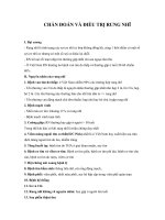
CASE 1:
AN ITCHY, SLOW-GROWING INFANT
History
A 26-week-old baby boy attends your clinic with his mother. He has developed a generalized dry, red, itchy rash over the past seven weeks. His mother has been applying a
regular emollient diligently and using a bath emollient. She reports that he is waking
more and more frequently at night and appears to be troubled by his skin. She is worried
about weaning him. He is currently breast-fed and his mother has an unrestricted diet.
He has been offered a bottle of formula milk, but took only 60 mL before vomiting and
developing a rash. He also developed a rash when his father kissed him, immediately after
eating an egg mayonnaise sandwich.
He is the first baby of his parents; his mother had asthma in childhood and his father is
allergic to shellfish. There are no pets at home. His father is a smoker. The baby was born
at term by normal vaginal delivery and is vaccinated to date.
Examination
His height has reached a plateau over the past eight weeks and now rests on the 9th
centile for his age. He is alert and happy, although he rubs his legs vigorously when
undressed. He has generally dry skin, with widespread low-grade erythema and raised,
poorly defined patches of active eczema; there are widespread excoriations (Fig. 1.1) and
no clinical evidence of impetiginization. He has low-grade generalized shotty lymphadenopathy. The rest of his examination is normal.
INVESTIGATIONS
Skin prick tests
Allergen
Resulting wheal
Interpretation
Positive control
Negative control
Egg white
Egg yolk
Cow’s milk protein
Soya
Wheat
Salmon
Cod
Peanut
5 mm
0 mm
11 mm
4 mm
8 mm
7 mm
0 mm
2 mm
1 mm
9 mm
Functioning assay
Highly likely to be allergic
Possibly allergic
Highly likely to be allergic
Highly likely to be allergic
Not allergic
Not allergic
Not allergic
Highly likely to be allergic
Questions
• What is this eruption?
• What associated condition does he present with?
• What dietary recommendations will
you make for the baby (and mother)?
Figure 1.1
1
ANSWER 1
This eruption is eczema. The history his mother gives makes an associated food allergy
probable – likely to egg and cow’s milk protein (CMP). This, in combination with a positive family history of food allergy and asthma, means we can classify his skin condition
as atopic eczema. His mother is correct to be anxious about weaning him.
It would be appropriate for this baby to be investigated for associated food allergy. Food
allergy is more likely in babies presenting with eczema from a young age, and it is possible that food allergy may be contributing to the activity of his eczema and vice versa.
The first line investigation should be skin prick test (SPT) to the common weaning food
protein allergens (CMP, egg, soya, wheat, and fish). Peanut is commonly added to this
initial panel.
The history suggests that this baby is likely to be allergic to egg and CMP, and this
has been confirmed by SPT. It would be worth restricting his mother’s intake of these
proteins if she intends to continue breast-feeding as this may improve eczema control.
If his mother wishes to stop breast-feeding, the most appropriate alternative at his age
would be an amino acid formula. The incidence of coexisting CMP and soya allergy is
high and the positive SPT would suggest this baby is currently allergic to both. CMP and
egg are nutritionally important and ensuring a balanced diet while restricting both can
be challenging; specialist dietetic advice is important. Low-grade exposure to allergenic
proteins through maternal milk might be contributing to skin signs and his static growth
parameters.
Regular use of topical emollients and avoidance of detergents are essential for maintaining the skin barrier function of infants with eczema. It is unlikely, however, that
emollients and dietary restriction alone will suffice in the management of his eczema.
His parents should be introduced to the practical aspects of topical therapy and a ‘stepup, step-down’ approach to the management of flares. They should be taught to identify
flares early and initiate effective therapy quickly.
The association of early-onset eczema and egg allergy is associated with a three-fold
increased risk of asthma in later childhood. This is an important opportunity to discuss the
potential contribution paternal smoking would have on increasing that risk. Reassuringly,
both egg and CMP allergy are frequently outgrown, although peanut allergy is more likely
to persist.
KEY POINTS
• Atopic eczema frequently presents within the first year of life and early onset is associated
with risk of associated food allergy.
• Eczema before the age of 1 year and egg allergy are associated with an increased risk of
developing asthma.
• Appropriate allergy testing and dietary advice will help prevent unsupervised dietary
manipulation by well-meaning but misguided parents and may help improve eczema
control.
2
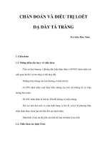
CASE 2:
AN AGITATED ATOPIC CHILD
History
A 5-year-old girl who is well known to your practice attends with her mother. She has been
troubled by worsening pruritus over the last six weeks. She has missed more than ten days of
school in the last month. Her mother reports that she wakes frequently at night and is lethargic and moody during the day. Her bed sheets are covered in flecks of blood in the morning.
The girl is known to be allergic to egg, fish and peanut, and has begun to develop the
symptoms of seasonal allergic rhinoconjunctivitis within the last couple of months. She has
a positive family history of atopy, both parents are allergic to animals and her older brother
has asthma. Her younger brother has been sent home from nursery with impetigo recently.
Her treatments include an emollient as soap and leave-on preparation and various
strengths of topical steroids ranging from very mild to moderately potent depending on
site and eczema severity. On questioning, however, mother
reports that her daughter’s skin is so sore that she is refusing
to bathe or apply her topical treatment.
Examination
A full examination reveals a fractious child; she is unable to
stop scratching her skin once undressed. She is slim, with her
height at the 25th centile and weight at the 4th centile for
her age. She has widespread, mildly tender, shotty lymphadenopathy (cervical, axillary and groin). Her skin is generally
mildly erythrodermic and extensively excoriated, particularly
her limbs (Fig. 2.1), neck and lower back. The excoriations
are covered with haemorrhagic crust and yellowish exudates.
Figure 2.1
INVESTIGATIONS
Haemoglobin
Mean corpuscular volume (MCV)
White cell count
Platelets
Sodium
Potassium
Urea
Creatinine
Albumin
Bilirubin
Alanine transaminase
Alkaline phosphatase
Ferritin
Vitamin D
11.2 g/dL
87 fL
13.7 ϫ 109/L
498 ϫ 109/L
135 mmol/L
4.2 mmol/L
5.7 mmol/L
68 mmol/L
38 g/L
12 mmol/L
26 IU/L
238 IU/L
22 ng/mL
38 ng/mL
Normal
13.3–17.7 g/dL
90–99 fL
3.9–10.6 ϫ 109/L
150–440 ϫ 109/L
135–145 mmol/L
3.5–5.0 mmol/L
2.5–6.7 mmol/L
70–120 μmol/L
35–50 g/L
3–17 mmol/L
5–35 IU/L
30–300 IU/L
20–200 ng/mL
40–80 ng/mL
Questions
• What is the primary diagnosis?
• What secondary complications are exacerbating her pruritus?
• How would you manage this patient?
3
ANSWER 2
The primary diagnosis is atopic eczema associated with a positive family history of atopy
as well as manifestations of IgE-mediated (immediate-type) hypersensitivity (food allergy
and allergic rhinoconjunctivitis). This is clearly a moderate to severe flare of her eczema.
The severity of eczema can be ‘scored’ by various validated subjective (e.g. CDLQI – children’s dermatology life quality index) and objective scoring systems. Crudely, however,
the impact on sleep and school attendance as well as the clinical severity of her eczema
demonstrated in the photograph denotes severe eczema with significant functional
disruption.
There may be several factors contributing to the current flare. It is likely that there is an
element of secondary infection with Staphylococcus aureus or impetiginization of this
child’s eczema. The extensive yellow crusting of her excoriations, her tender lymphadenopathy, and the fact that her brother has impetigo, suggest colonization of the patient
and potentially other family members. Difficulty in adhering to a bathing regime is likely
to contribute. Other potential factors which worsen pruritus include iron deficiency. She
is also vitamin D deficient, presumably due to her dietary restriction (egg and fish are the
main dietary sources of vitamin D).
It is important to gain control of this child’s eczema rapidly. Swabs should be taken for
microbiology culture and sensitivity testing both from the patient and her immediate
family members. As there appear to be at least two members of the family affected by
Staphylococcus aureus it would be worthwhile considering Staphylococcus eradication
protocol for the entire family (i.e. antiseptic washes and antibacterial nasal ointment).
The patient might benefit from a 5–10-day course of antibiotic with good Staphylococcus
aureus coverage (first line: flucloxacillin; second line: erythromycin or co-amoxiclav).
The extensive use of a moderately potent topical corticosteroid ointment for 2–4 weeks
may be required before weaning back to weak preparations or calcineurin inhibitors as
maintenance therapy.
KEY POINTS
• Atopic eczema is by definition eczema associated with a personal and/or family history
of atopy.
• Staphylococcus aureus may cause severe flare of eczema.
• Management of such a flare includes Staphylococcus aureus eradication as well as
appropriate treatment of the eczema.
4
CASE 3:
AN ACUTE MONOMORPHIC ERUPTION IN A
SYSTEMICALLY UNWELL ATOPIC CHILD
History
A 6-year-old boy is brought to the accident and emergency department by his parents
with a 5-day history of worsening eczema associated with malaise and lethargy. In addition to worsening pruritus and sleeplessness he complains of painful skin, particularly
around his face, neck, chest and forearms. He quantifies the level of pain as 8 out of 10.
His current flare is not responding to diligent application of his usual eczema treatments
according to his ‘step-up’ management plan.
The onset of his eczema was at the age of 4 months, and although moderately severe
in infancy it has been reasonably controlled since starting primary school, with regular
use of emollients and mild to moderately potent topical steroids. His background history includes egg allergy (now partially outgrown in that he tolerates well cooked egg in
cakes) and asthma, currently stable. He has never been admitted to hospital before. He
is fully vaccinated to date and had chickenpox at the age of 4 years. His father suffers
from hay-fever and experienced childhood eczema and asthma. He has one older sister
(aged 14) who is well.
His medication includes:
• Regular emollients both as leave-on preparations and soap substitute
• Topical tacrolimus 0.1% twice daily applied to affected areas for the management of
flares
• Hydroxyzine 10 mg nocte during flares and salbutamol inhaler on a prn basis
Examination
He looks unwell and is febrile at 38.5 °C. Systemic examination is normal except for
widespread lymphadenopathy. There is no evidence of conjunctival erythema and his
vision is normal. He has generalized moderate to severe eczema with erythema, dryness,
excoriation and lichenification. He has a superimposed monomorphic eruption over his
lower face, chest and forearms. The eruption is composed of multiple 23-mm monomorphic ‘punched-out’ erythematous lesions in various stages of evolution (Fig. 3.1). Some of
the lesions are vesicular, others pustular, some coalescing, most are eroded and covered
with a golden exudate and others haemorrhagic crust.
Questions
• What are these lesions?
• How would you confirm the diagnosis?
• What complications can be associated
with them?
• What is their management?
Figure 3.1
5
ANSWER 3
These are typical lesions of herpes simplex virus (HSV) infection complicating atopic
eczema. This eruption is called eczema herpeticum, or less commonly Kaposi’s varicelliform eruption.
Diagnosis can be confirmed by viral swab of a blister or eroded area. Many tests can
detect HSV within tissue or blister fluid. HSV can be inferred by positive staining or
electron microscopy or specifically identified as types HSV-1 or HSV-2 by immunofluorescence, culture, or polymerase chain reaction. Bacteriology swab for microscopy and
culture should also be undertaken.
Significant morbidity is associated with eczema herpeticum. The main potential complications include superimposed bacterial infection (Staphylococcus or Streptococcus)
with risk of systemic sepsis, ocular involvement (in particular, HSV keratitis) and, rarely,
systemic HSV infection with risk of spread to the liver, the lungs, the brain, the gastrointestinal tract and even the adrenal glands. In addition pain and discomfort associated
with eczema herpeticum is significant.
The management of widespread eczema herpeticum includes systemic treatment of HSV
infection with aciclovir, identification and treatment of any superimposed bacterial
infection or strategies to prevent superimposed infection, such as antibacterial washes
and creams. Topical tacrolimus should be discontinued in this patient as this may exacerbate the cutaneous spread of HSV. These cases are usually managed as in-patients,
initially with intravenous aciclovir – as oral preparations can be poorly absorbed.
Ophthamological review should be sought in cases of diffuse facial herpes simplex infection or where conjunctival/corneal involvement is suspected.
In a minority of cases recurrences can occur. Rapid treatment of incipient lesions with
topical aciclovir may help prevent disseminated eczema herpeticum.
KEY POINTS
• Herpes simplex infection in patients with eczema can lead to widespread lesions and an
associated risk of superimposed bacterial infection and sepsis.
• It is important to consider and exclude the rare associated complication of herpes
keratitis.
• In-patient management with systemic anti-viral therapy, topical antiseptic measures,
pain relief, and where indicated antibiotic therapy, is required.
6
CASE 4:
A RECURRENT, UNSIGHTLY FACIAL ERUPTION IN A
STRESSED BUT WELL YOUNG ADULT
History
A 29-year-old man attends your clinic with a 4-year history of a recurrent and itchy
facial eruption that he feels is unsightly. He notices the eruption is worse in the winter
and tends to improve over the summer. He is currently studying for business exams and
feels the associated stress has triggered the current flare. He avoids soaps, which make his
face sore, and recently has reduced his alcohol intake in an effort to improve his eruption.
He is otherwise well and on no medication.
Examination
A full examination is unremarkable except for the skin of his face, neck, central chest and
scalp. There are poorly defined erythematous patches with overlying adherent greasy scale
affecting his naso-labial folds and extending onto his cheeks (Fig. 4.1). His eyebrows,
scalp, nape of his neck and central chest are similarly affected.
Questions
• What is this eruption?
• What age groups are affected?
• How would you manage this patient?
Figure 4.1
7
ANSWER 4
This eruption is seborrhoeic dermatitis. It is more common among men and typically
affects the sebum-rich areas of the face, scalp and chest. The pathophysiology of seborrhoeic dermatitis is incompletely understood, however. It is linked with Malassezia yeast,
complement activation and abnormalities of T-cell immunity. It may worsen in individuals infected with HIV or affected by Parkinson’s disease.
The condition usually begins around puberty with a peak of incidence between 25 and
40 years of age. An infantile form of seborrhoeic dermatitis may manifest as cradle cap
(Fig. 4.2), facial greasy scaly dermatitis, napkin dermatitis and, rarely, as an erythroderma.
In predisposed individuals seborrhoeic dermatitis usually recurs. Treatment is aimed,
therefore, at reducing morbidity and preventing flares. Treatment aims are two-fold:
reducing the yeast burden as a secondary preventative measure, and switching off the
resultant secondary dermatitis when it occurs. Although topical corticosteroids may
improve appearances of the dermatitis in the short term, they are thought to hasten recurrences and may foster dependence due to a ‘rebound effect’ and are usually discouraged.
The use of a ketoconazole shampoo, with frequent washing and prolonged lathering often
improves associated dandruff and may improve the facial involvement by depletion of
Malassezia. Use of ketoconazole shampoo as a face wash can be irritating, but if tolerated may improve erythema and scaling. Ketoconazole or miconazole cream, calcineurin
inhibitors in combination with antiseptic emollient washes are recommended. For severe
or refractory seborrhoeic dermatitis systemic itraconazole as a short course or ‘pulsed’
(one week per month) is highly effective at reducing the yeast burden.
Figure 4.2
KEY POINTS
• Seborrhoeic dermatitis is characterized by poorly defined erythematous patches with
overlying greasy, yellowish-brown scale localized to the sebum-rich areas.
• It occurs most commonly among men from adolescence to middle age. Infantile
seborrhoeic eczema can also occur.
• HIV infection and Parkinson’s disease are both associated with refractory seborrhoeic
dermatitis.
8
CASE 5:
BLISTERED HANDS AND FEET IN AN ATHLETIC MAN
History
A 27-year-old man attends your clinic with a 3-day history of a severe burning itch over
his hands associated with localized blistering and similar although less severe changes
on his feet. He is otherwise well, although he did suffer from asthma is childhood and
occasionally still experiences hay fever. He is on no medication. He works as a graphic
designer and his hobbies include cycling and football, he has no exposure to allergens or
irritants. He is unaware of any triggering factor.
Examination
He has diffuse vesicles, coalescing to form tense bullae over the palmar aspects of both
hands extending into the interdigital spaces (Fig. 5.1) and onto the dorsa of his fingers
and hand. In addition he has erythema, maceration, fissuring and peeling between the
4th and 5th toes on the left side and bilateral but asymmetrical (left worse than right)
purulent vesicles over the insteps.
Figure 5.1
Questions
• What is the diagnosis?
• What investigations would you perform?
• What treatments would you initiate?
9
ANSWER 5
The diagnosis is pompholyx or dyshidrotic eczema, the symmetrical and diffuse clear vesicles over the palmar aspect of the hands associated with pruritus are highly suggestive
and the diagnosis is based on clinical features. Other differential diagnoses to consider
include contact dermatitis (irritant or allergic), friction blisters (e.g. epidermolysis bullosa
simplex), herpes simplex infection, and palmoplantar pustular psoriasis.
Atopy appears to be a predisposing factor for pompholyx. There are several potential
triggers of pompholyx including stress and as an ‘id reaction’ to a distant dermatophyte
infection. In this case the features of interdigital maceration associated with inflammatory pustules and vesicles on the instep are suggestive of inflammatory tinea pedis.
Investigations should include scrapings from the feet (interdigital spaces and affected
areas over the plantar aspects) and hands for mycological tests (direct microscopy and
culture). In this case, scrapings from the feet demonstrated hyphae and spores on direct
microscopy with subsequent culture confirming the presence of the zoophilic organism
Trichophyton mentagrophytes var. mentagrophytes. There was no fungal infection of the
hands.
Treatment of a pompholyx ‘id reaction’ involves treatment of the tinea pedis as well as
treatment of the pompholyx itself. Inflammatory tinea pedis is usually managed with systemic antifungal therapy (itraconzole, terbinafine or fluconazole). Infected scales can be
present on clothing or within footwear, so frequent laundering is recommended. Draining
the larger bullae with a sterile needle will reduce the discomfort. Compresses or soaks
with dilute potassium permanganate help to dry the vesicles and prevent secondary bacterial infection. Potent or superpotent topical corticosteroids are the mainstay of therapy.
In the short term a combination preparation of topical corticosteroids and antibacterial
agent is useful. Occasionally, systemic steroids are required.
KEY POINTS
• Pompholyx occurs as a manifestation of hand eczema, irritant or allergic dermatitis and
as an ‘id reaction’ to a distant dermatophyte infection.
• The mainstay of treatment is the prevention of secondary infection and use of potent or
superpotent topical corticosteroids as well as identification and eradication of the trigger.
10
CASE 6:
CHRONIC ERYTHEMATOUS PRURITIC ERUPTION ON
THE LOWER LEGS
History
A 67-year-old woman presents to the vascular surgeons with varicose veins. She had a
history of venous ulceration in the past, which has now healed and she is being considered for bilateral varicose vein surgery. At the consultation she complained of a 3-month
history of skin itching and redness, particularly on the right lower leg, and was noted to
have unilateral erythema and was referred to dermatology for an opinion.
Examination
This patient has obvious dilated and tortuous veins on both lower legs. Confluent background dull erythema is seen on the right lower leg, with small inflammatory superficial
erythematous erosions and excoriations (Fig. 6.1). Palpation revealed warm, dry, rough
skin at the affected site.
INVESTIGATIONS
Vascular studies
Resting pressure right: 174 mmHg, left: 180 mmHg, brachial: 167 mmHg
Resting ankle/brachial pressure index right: 1.042, left: 1.078. Bilateral triphasic pulsatile
waveforms
Venous studies showed bilateral saphenofemoral reflux.
Questions
• What is the diagnosis?
• What treatment would you recommend
for her right leg prior to vein surgery?
• Is this patient suitable for compression
hosiery based on the vascular studies?
Figure 6.1
11
ANSWER 6
This patient has chronic cutaneous changes seen on the right lower leg consistent with
the diagnosis of varicose eczema. This common cutaneous eruption usually has an insidious onset over many weeks to months in patients with a background of venous incompetence. The affected skin is pruritic and dry with marked erythema which may be variable
in intensity depending on its chronicity. In the context of venous insufficiency, pitting
oedema may develop owing to poor venous return leaving the skin tight and oedematous. This results in reduced blood flow to the skin, leading to active dusky erythema and
resultant erosions or even ulceration.
Varicose eczema can be readily distinguished from cellulitis affecting the lower leg.
Varicose eczema usually develops slowly, is frequently bilateral, pruritus is marked, the
skin surface is rough and dry, and there are associated varicose veins. Frequently there is
a background brown discolouration of the affected skin area due to haemosiderin deposition. Haemosiderin pigment is derived from haemoglobin, which is left behind in the skin
when red blood cells extravasate into the tissue.
Management of the skin requires a combination of topical therapy and if possible compression. The leg should be washed with aqueous cream or an antiseptic emollient such
as Dermol 500®. A moderately potent topical steroid should be applied to the eczematous
areas and a rich bland emollient. Compression hosiery or two to four layer bandaging is
essential to ‘squeeze’ the fluid out of the legs and allow skin healing. If the ankle/brachial
pressure index (APBI) is above 0.8 then the arteries are sufficiently patent to permit
compression without compromising the arterial blood supply to the lower extremities.
KEY POINTS
• Varicose eczema can be distinguished from cellulitis by slow onset, pruritus and surface
xerosis.
• Management of the eczema will not succeed without addressing the underlying
oedema.
• Potent topical steroids should be applied to the active eczema areas plus emollients and
compression.
12