Redox state as a central regulator of plant cell stress responses
Bạn đang xem bản rút gọn của tài liệu. Xem và tải ngay bản đầy đủ của tài liệu tại đây (6.26 MB, 387 trang )
Dharmendra K. Gupta
José M. Palma
Francisco J. Corpas Editors
Redox State
as a Central
Regulator of
Plant-Cell Stress
Responses
Redox State as a Central Regulator of Plant-Cell
Stress Responses
Dharmendra K. Gupta José M. Palma
Francisco J. Corpas
•
Editors
Redox State as a Central
Regulator of Plant-Cell Stress
Responses
123
Editors
Dharmendra K. Gupta
Institut für Radioökologie und
Strahlenschutz (IRS)
Gottfried Wilhelm Leibniz Universität
Hannover
Germany
Francisco J. Corpas
Estación Experimental del Zaidín
(EEZ-CSIC)
Granada
Spain
José M. Palma
Estación Experimental del Zaidín
(EEZ-CSIC)
Granada
Spain
ISBN 978-3-319-44080-4
DOI 10.1007/978-3-319-44081-1
ISBN 978-3-319-44081-1
(eBook)
Library of Congress Control Number: 2016947790
© Springer International Publishing Switzerland 2016
This work is subject to copyright. All rights are reserved by the Publisher, whether the whole or part
of the material is concerned, specifically the rights of translation, reprinting, reuse of illustrations,
recitation, broadcasting, reproduction on microfilms or in any other physical way, and transmission
or information storage and retrieval, electronic adaptation, computer software, or by similar or dissimilar
methodology now known or hereafter developed.
The use of general descriptive names, registered names, trademarks, service marks, etc. in this
publication does not imply, even in the absence of a specific statement, that such names are exempt from
the relevant protective laws and regulations and therefore free for general use.
The publisher, the authors and the editors are safe to assume that the advice and information in this
book are believed to be true and accurate at the date of publication. Neither the publisher nor the
authors or the editors give a warranty, express or implied, with respect to the material contained herein or
for any errors or omissions that may have been made.
Printed on acid-free paper
This Springer imprint is published by Springer Nature
The registered company is Springer International Publishing AG Switzerland
Preface
It is known that reactive oxygen species (ROS) are the by-products of aerobic
breakdown and are inescapably formed by a number of metabolic pathways and
electron transport chains. ROS are partially condensed form of molecular oxygen
and normally result from the transfer of electrons to O2 to form, in a succession of
univalent reductions, superoxide radical (O2Á−), hydrogen peroxide (H2O2), and
hydroxyl radical (•OH), respectively, or through an electron-independent energy
transfer till an excited form of oxygen (singlet oxygen) (Gupta et al. 2016; Halliwell
and Gutteridge 2015). Redox signal transduction is a complete feature of aerobic
life enriched through evolution to balance evidence from metabolism and the
environment. Like all other aerobic creatures, plants maintain most cytosolic thiols
in the reduced (−SH) state because of the low thioldisulfide redox potential
imposed by millimolar amount of the thiol buffer including glutathione.
Plants have developed cellular tactics where the endogenous content of
antioxidant enzymes deliver them with amplified defense against harmful effects of
oxidative stress encouraged by heavy metal and other stress sources (Palma et al.
2013). Stress-induced upsurges in ROS level can cause different degree of oxidation
of cell components and a gross change in the redox status. Plant cells generally cope
very well with high rates of generation of superoxide, H2O2, and even singlet
oxygen. When the increment of ROS in plant cells quickly augments and the
scavenging systems of ROS do not operate appropriately, a condition of oxidative
stress and oxidative injury happens (Gupta et al. 2015). In plants, chloroplast is the
most important among the organelles in respect of ROS generation as O2 is constantly provided through the water autolysis and freely available inside the organelle (Gupta et al. 2015). In plant cells, compartmentalization of ROS production in
the different organelles includes chloroplasts, mitochondria, or peroxisomes, and
they also have a complex battery of antioxidant enzymes usually close to the site of
ROS production (Corpas et al. 2015). Plant cells also contain a series of
ROS-scavenging non-enzymatic antioxidants such as ascorbic acid, glutathione
(GSH), and carotenoids, as well as a set of enzymes such as superoxide dismutase
(SOD), catalase, glutathione peroxidase (GPX), peroxiredoxin (Prx), and the
v
vi
Preface
ascorbate–glutathione cycle (Corpas et al. 2015). The total pool of redox-active
complexes which are found in a cell in reduced and oxidized forms generates
cellular redox buffers where NAD(P)H/NAD(P)+, ascorbate/dehydroascorbate
(AsA/DHA), glutathione/glutathione disulfide (GSH/GSSG), and reduced
thioredoxin/oxidized thioredoxin (Trxred/Trxox) are the main pairs. AsA and GSH
are major constituents of the soluble redox shielding system, and they contribute
pointedly to the redox environment of a cell. AsA cooperates tightly with GSH
(c-Glu-Cys-Gly) in the Foyer–Halliwell–Asada cycle (ascorbate–glutathione
cycle), involving three codependent redox couples: AsA/DHA, GSH/GSSG, and
NAD(P)H/NAD(P)+. It undertakes subsequent reduction/oxidation reactions catalyzed by ascorbate peroxidase (APX), monodehydroascorbate reductase (MDAR),
dehydroascorbate reductase (DHAR), and glutathione reductase (GR) that is universally responsible for H2O2 sifting and keeping AsA and GSH in the reduced
state at the outflow of NADPH, this cycle being situated in all cellular partitions in
which ROS detoxification is required.
One of the major consequences of stresses in plant cells is the enhanced generation of ROS which usually damage the cellular components such as membranes,
nucleic acids, proteins, chloroplast pigments, and alteration in enzymatic and
non-enzymatic antioxidants. The molecular mechanisms of signal transduction
corridors in higher plant cells are vital for processes such as hormone and light
sensitivity, growth, development, stress resistance, and nutrient uptake from soil
and water (Gupta et al. 2013).
It is really great achievement for the plant biotechnologists who are working for
years to know how redox state handled by plants. This edited volume will provide the
recent advancements and overview to the plant scientists who are actively involved in
redox signaling states and also a key player for cellular tolerance in plant cells under
different stresses (biotic and abiotic). Other key features of this book are cellular
redox homeostasis as central modulator, redox homeostasis and reactive oxygen
species, redox balance in chloroplasts and in mitochondria, and oxidative stress and
its role in peroxisome homeostasis. Some chapters are also focusing on
glutathione-related enzyme system and metabolism under metal(ed) stress. Abiotic
stress-induced redox changes and programmed cell death are also addressed in the
edition. In summary, the information compiled in this volume will bring depth
knowledge and current achievements in the field of redox state chemistry in plant cell.
Dr. Dharmendra K. Gupta, Prof. José M. Palma, and Dr. Francisco J. Corpas
individually thank all authors for contributing their valuable time, knowledge, and
enthusiasm to bring this book into in the current shape.
Hannover, Germany
Granada, Spain
Granada, Spain
Dharmendra K. Gupta
José M. Palma
Francisco J. Corpas
Preface
vii
References
Corpas FJ, Gupta DK, Palma JM (2015) Production sites of reactive oxygen species (ROS) in
plants. In: Gupta DK, Palma JM, Corpas FJ (eds) Reactive oxygen species and oxidative
damage in plants under stress. Springer Publication, Germany, p 1–22
Gupta DK, Corpas FJ, Palma JM (2013) Heavy metal stress in plants. Springer-Verlag, Germany
Gupta DK, Palma JM, Corpas FJ (2015) Reactive oxygen species and oxidative damage in plants
under stress. Springer-Verlag, Germany
Gupta DK, Peña LB, Romero-Puertas MC, Hernández A, Inouhe M, Sandalio LM (2016) NADPH
oxidases differently regulates ROS metabolism and nutrient uptake under cadmium toxicity.
Plant Cell Environ doi:10.1111/pce.12711
Halliwell B, Gutteridge JMC (2015) Free radicals in biology and medicine. Oxford University
Press, Oxford, UK
Palma JM, Gupta DK, Corpas FJ (2013) Metalloproteins involved in the metabolism of Reactive
Oxygen Species (ROS) and heavy metal stress. In: Gupta DK, Corpas FJ, Palma JM
(eds) Heavy metal stress in plants. Springer Publication, Germany, p 1–18
Contents
1
Cellular Redox Homeostasis as Central Modulator in Plant
Stress Response . . . . . . . . . . . . . . . . . . . . . . . . . . . . . . . . . . . . . . . . . .
C. Paciolla, A. Paradiso and M.C. de Pinto
2
Plant Cell Redox Homeostasis and Reactive Oxygen Species . . . . . .
A. Trchounian, M. Petrosyan and N. Sahakyan
3
Redox Balance in Chloroplasts as a Modulator of Environmental
Stress Responses: The Role of Ascorbate Peroxidase and Nudix
Hydrolase in Arabidopsis . . . . . . . . . . . . . . . . . . . . . . . . . . . . . . . . . . .
T. Ishikawa, T. Maruta, T. Ogawa, K. Yoshimura and S. Shigeoka
4
5
1
25
51
Physiological Processes Contributing to the Synthesis
of Ascorbic Acid in Plants . . . . . . . . . . . . . . . . . . . . . . . . . . . . . . . . .
C.G. Bartoli, M.E. Senn and G.E. Gergoff Grozeff
71
Redox State in Plant Mitochondria and its Role in Stress
Tolerance . . . . . . . . . . . . . . . . . . . . . . . . . . . . . . . . . . . . . . . . . . . . . . .
N.V. Bykova and A.U. Igamberdiev
93
6
Oxidative Stress and its Role in Peroxisome
Homeostasis in Plants . . . . . . . . . . . . . . . . . . . . . . . . . . . . . . . . . . . . . 117
T. Su, Q. Shao, P. Wang and C. Ma
7
Glutathione-Related Enzyme System: Glutathione Reductase
(GR), Glutathione Transferases (GSTs) and Glutathione
Peroxidases (GPXs) . . . . . . . . . . . . . . . . . . . . . . . . . . . . . . . . . . . . . . . 137
J. Csiszár, E. Horváth, K. Bela and Á. Gallé
8
Glutathione Metabolism in Plants Under Metal and Metalloid
Stress and its Impact on the Cellular Redox Homoeostasis . . . . . . . 159
Luis E. Hernández, A. González, A. Navazas, Á. Barón-Sola,
F. Martínez, A. Cuypers and C. Ortega-Villasante
ix
x
Contents
9
Glutathione and Related Enzymes in Response
to Abiotic Stress . . . . . . . . . . . . . . . . . . . . . . . . . . . . . . . . . . . . . . . . . 183
I. Štolfa, D. Špoljarić Maronić, T. Žuna Pfeiffer and Z. Lončarić
10 The Function of Cellular Redox Homeostasis and Reactive
Oxygen Species (ROS) in Plants Tolerance to Abiotic Stresses . . . . 213
Qinghua Shi and Biao Gong
11 Abiotic Stress-Induced Redox Changes and Programmed
Cell Death in Plants—A Path to Survival or Death? . . . . . . . . . . . . 233
S.R. Kumar, G. Mohanapriya and R. Sathishkumar
12 The Role of ROS and Redox Signaling During the Initial Cellular
Response to Abiotic Stress . . . . . . . . . . . . . . . . . . . . . . . . . . . . . . . . . 253
Jos H.M. Schippers and R. Schmidt
13 The Cadmium-Binding Thioredoxin O Acts as an Upstream
Regulator of the Redox Plant Homeostasis . . . . . . . . . . . . . . . . . . . . 275
Moêz Smiri, Sami Boussami, Takwa Missaoui and Amor Hafiane
14 Arsenic Tolerance in Plants: Cellular Maneuvering Through
Sulfur Metabolites . . . . . . . . . . . . . . . . . . . . . . . . . . . . . . . . . . . . . . . . 297
D. Talukdar
15 Regulation of Stomatal Responses to Abiotic and Biotic
Stresses by Redox State . . . . . . . . . . . . . . . . . . . . . . . . . . . . . . . . . . . 331
Y. Murata, S. Munemasa and I.C. Mori
16 The Antioxidant Power of Arginine/Nitric Oxide Attenuates
Damage Induced by Methyl Viologen Herbicides
in Plant Cells . . . . . . . . . . . . . . . . . . . . . . . . . . . . . . . . . . . . . . . . . . . . 349
N. Correa-Aragunde, P. Negri, F. Del Castello, N. Foresi,
J.C. Polacco and L. Lamattina
17 Protein S-Nitrosylation and S-Glutathionylation as Regulators
of Redox Homeostasis During Abiotic Stress Response . . . . . . . . . . 365
J.C. Begara-Morales, B. Sánchez-Calvo, M. Chaki, R. Valderrama,
C. Mata-Pérez, F.J. Corpas and J.B. Barroso
About the Editors
Dharmendra K. Gupta is a senior environmental biotechnology scientist at the
Institut für Radioökologie und Strahlenschutz, Gottfried Wilhelm Leibniz
Universität Hannover in Germany and has published more than 80 research
papers/review articles in peer reviewed journals and has edited nine books. His
research interests include abiotic stress by heavy metals/radionuclides and xenobiotics in plants; antioxidative system in plants, and environmental pollution (heavy
metal/radionuclide) remediation through plants (phytoremediation).
José M. Palma has more than 30 years experience in plant sciences and related
fields. He also served as the deputy director and later director of the Estación
Experimental del Zaidín (EEZ-CSIC), Granada, Spain. He has published more than
100 research papers/review articles in peer reviewed journals and edited five books.
Francisco J. Corpas is a staff member at the Spanish National Research Council
(CSIC) and has more than 24 years of research experience in the metabolism of
antioxidants and nitric oxide in higher plants under physiological and adverse
environmental conditions. At present, he is the head of the Department of
Biochemistry, Cell and Molecular Biology of Plants at the research institute
Estación Experimental del Zaidín-CSIC in Granada, Spain. He has published more
than 120 research papers/review articles in peer reviewed journals and has edited
five books.
xi
Chapter 1
Cellular Redox Homeostasis as Central
Modulator in Plant Stress Response
C. Paciolla, A. Paradiso and M.C. de Pinto
Abstract Plants are frequently exposed to different stressful factors, both of biotic
or abiotic nature, which limit their growth and productivity. To survive under stress
conditions, plants must activate stress-specific signalling pathways, which finally
lead to morphological, physiological, and biochemical changes that allow to adapt
to the adverse environment. Cellular redox homeostasis, determined by a complex
interplay between pathways that produce and scavenge reactive oxygen species
(ROS), plays a key role in the adaptive response. Each deviation in the cellular
redox state, due to an imbalance of ROS production and/or scavenging, is indicative
of environmental disturbance and works as a signal. Under stress conditions, different ROS are produced in many cell compartments. Plants have very proficient,
versatile and flexible antioxidant machinery, which comprises enzymes and
metabolites with distinct biochemical properties and distinct sub-cellular localization. The antioxidant systems play a key role in the control of redox homeostasis,
determining either the extent or the specificity of ROS signals and the downstream
redox-dependent responses. Redox signalling is responsive to a number of environmental cues, and the complex and dynamic pathways of redox regulation occur
in different cell compartments. The redox-dependent modification of sensitive
signalling proteins is proposed as a key mode of redox signal transmission. Each
redox-dependent interaction is opportunely regulated by a restricted environment,
whose change transfers the complex system of information and influences the plant
response to external changes.
Á
Á
Á
Keywords Ascorbate
Antioxidants
Glutathione
Peroxidases
oxygen species Redox homeostasis Redox signalling Stress
Á
Á
Á
Á
Reactive
C. Paciolla Á A. Paradiso Á M.C. de Pinto (&)
Department of Biology, University of Bari “Aldo Moro”,
Via Orabona 4, 70126 Bari, Italy
e-mail:
© Springer International Publishing Switzerland 2016
D.K. Gupta et al. (eds.), Redox State as a Central Regulator of Plant-Cell
Stress Responses, DOI 10.1007/978-3-319-44081-1_1
1
2
1.1
C. Paciolla et al.
Introduction
Plants, as sessile organisms, are frequently exposed to various environmental cues,
which can potentially limit their growth and development. To cope with their sessile
life, plants possess different stress-specific signalling pathways that permit to perceive the external signals and trigger changes in the expression of numerous genes.
Stress-responsive genes may encode both for functional proteins, that protect cells
from damages, and regulatory proteins, such as transcription factors that control
stress signalling and adaptation (Hirayama and Shinozaki 2010; Zhang et al. 2011).
The activation of stress-specific signalling pathways causes morphological, physiological, and biochemical changes that allow plants to adapt to adverse environment. Many studies point out that changes in cellular redox environment play a key
role in the integration of external stimuli and the complex network of
stress-signalling pathways (Fujita et al. 2006; Spoel and Loake 2011; Suzuki et al.
2012; Scheibe and Dietz 2012).
The redox environment of a cell is determined by the global poise of its
oxidation/reduction systems; in this view, the oxidative and reductive reactions
have to be considered together as complementary processes. There is a complex
link between redox state and metabolism: the redox state could be considered an
integrator of cellular and apoplastic metabolism and at the same time is regulated by
different metabolic processes (Geigenberger and Fernie 2014; Noctor et al. 2015).
Thus, redox homeostasis plays a key role for appropriate plant responses to both
developmental and environmental stimuli. Redox changes, due to endogenous or
exogenous inputs, will be sensed, integrated and converted through different signalling pathways, which ultimately will lead to the redox-dependent reprogramming of gene expression.
Two regulated variables are dynamically implicated in maintaining the redox
environment: on the one hand, the production of reactive oxygen species (ROS),
and on the other hand the presence of different redox couples and antioxidant
machinery. The redox homeostasis of the different cellular compartments is
determined by a complex interplay between multiple ROS-producing pathways,
and ROS-scavenging mechanisms. The processes that produce and balance oxidants and antioxidants are useful for the control of plant responses to the changing
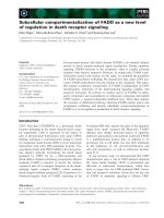
environment (Fig. 1.1).
1.2
ROS Production Pathways
ROS are natural byproducts of the aerobic metabolism, formed either by energy or
by electron transfer to oxygen (Apel and Hirt 2004). Generation of singlet oxygen
(1O2) is due to an energy transfer-dependent mechanism that rearranges the configuration of the unpaired electrons of oxygen, remarkably increasing its oxidising
capability. 1O2 has a half-life of 4 µs in aqueous solution and reacting with
1 Cellular Redox Homeostasis as Central Modulator …
3
Fig. 1.1 Redox homeostasis,
due to the balance among
ROS production and
scavenging, is altered in stress
conditions and activates a
redox-dependent signalling
that trigger the adaptive
response
biological molecules mainly forms endoperoxides and hydroperoxides (Halliwell
2006). The superoxide radical (O2 ÁÀ ) is formed for the transfer of a single electron
to O2; this ROS can reduce quinones and transition metal as copper and iron,
affecting the activity of metal-containing enzymes; however, O2 ÁÀ , being moderately reactive, and having a short half-life (2–4 ls), does not cause extensive
damage by itself, but undergoes transformation into more reactive and toxic
hydroxyl radical (OH) (Halliwell 2006). Because of its high instability at physiological pH, O2 ÁÀ rapidly disproportionates to O2 and hydrogen peroxide (H2O2),
either spontaneously or by the action of superoxide dismutases (SODs, Alscher
et al. 2002). H2O2 can cause inactivation of enzymes by oxidizing their thiol groups
(Møller et al. 2007). However, H2O2, like O2 ÁÀ , is a relative poor oxidant. For this
reason, the abundance of enzymes able to scavenge this ROS may be due to the
requirement to reduce the production of OH, the most reactive and toxic ROS. OH
can be formed at neutral pH through Haber-Weiss or Fenton reactions, catalysed by
redox-active metal ions, especially iron and copper. This ROS is able to damage
different cellular components and, due to the lack of enzymatic systems able to
scavenge this toxic radical, its accumulation can lead to cell death (Møller et al.
2011). On the other hand, H2O2 has been proposed as the most valuable ROS
functioning as second messenger (Petrov and Van Breusegem 2012). Indeed, due to
its significantly longer half-life (1 ms) compared to other ROS members and its
capability to cross cell membranes, being facilitated via aquaporins (Bienert et al.
2007), H2O2 can cover considerable long distances within the cell.
ROS are diffusely produced by a large number of physiological processes,
occurring in both intracellular and extracellular locations. ROS production occurring in the photosynthetic and respiratory electron transport chains has a regulatory
function in alleviating over-reduction, particularly during stress conditions (Noctor
et al. 2014). Chloroplasts and mitochondria, together with peroxisomes, which
4
C. Paciolla et al.
generate O2 ÁÀ and H2O2 through multiple reactions, are the main producing sites of
ROS in plant cells (Foyer and Noctor 2003). ROS overproduction in these organelles has been shown to participate in the responses to different kinds of stress,
both of biotic or abiotic nature (del Río et al. 2006; Rhoads et al. 2006; Miller et al.
2010a, b; Nomura et al. 2012; Suzuki et al. 2012; Sandalio et al. 2013; Huang et al.
2016). The apoplast is another principal site of ROS generation. Cell wall peroxidases (PODs), catalyzing cell wall formation, have been proposed as a source of
pathogen-induced oxidative burst (Daudi et al. 2012). Apoplastic ROS production
during plant–pathogen interaction also occurs via respiratory burst oxidase homologs (RBOHs), localized at the plasma membrane. The pathogen recognition
determines symplastic signals, including calcium influx and protein phosphorylation that activate the protein, which in turn transfers electrons from symplastic
NADPH to apoplastic oxygen, generating O2 ÁÀ at the apoplastic side of the plasma
membrane (Torres et al. 2002; Suzuki et al. 2011). Apoplastic ROS production by
RBOHs is not only involved in pathogen defence but also occurs in response to
abiotic stresses (Zhang et al. 2001; Suzuki et al. 2012).
Other cell compartments have been proposed for ROS production in plant stress
response. For instance, salt stress in Arabidopsis causes ROS production in
endosomes targeted to the central vacuole. The inhibition of the fusion of
H2O2-containing vesicles with the tonoplast leads to the formation of cytoplasmic
H2O2-containing megavesicles and improves plant salt tolerance (Leshem et al.
2006). An example of nuclear ROS production has also been reported. Tobacco BY-2
cells treated with the elicitor cryptogein accumulate ROS firstly in the nucleus and
later in other cell compartments, like endomembranes and cytoplasm. The isolated
nuclei of these cells are able to produce H2O2 in a calcium-dependent manner,
implying that nuclei could be an active source of ROS (Ashtamker et al. 2007).
Many stresses induce ROS production in specific sub-cellular compartments,
which, in turn, results in ROS accumulation in other compartments. Alteration in
ROS production or scavenging in one sub-cellular compartment influences the ROS
level in other compartments (Davletova et al. 2005; Miller et al. 2007;
Vanderauwera et al. 2011). Moreover, it should been considered that a continuous
ROS flow through the cell can be necessary to transmit information between different sub-cellular compartments. The connections between different ROS locations
in the plant cell make it very difficult to study the contribution of a single
sub-cellular compartment in ROS production. These observations could explain
why the mechanisms, by which stress conditions are sensed and integrated, and
how ROS accumulation is interconnected with stress signalling, are not completely
clear (Noctor and Foyer 2016). Further complexity is added by the interactions
between ROS and hormone signalling (Overmyer et al. 2003; Blomster et al. 2011;
Mittler and Blumwald 2015; Berkowitz et al. 2016).
The environmental cues causing ROS overproduction can lead to oxidative
stress that has been generally categorized as a negative condition for the cells.
Indeed, ROS are able to react readily with lipids, proteins, carbohydrates, and
nucleic acids causing significant cell damage and negatively affecting metabolic
activities and integrity of organelles (Foyer and Noctor 2003; Pfannschmidt et al.
1 Cellular Redox Homeostasis as Central Modulator …
5
2007). However, in the last two decades it has become more and more clear that
transient oxidative imbalance can be needed to activate signalling pathways
enabling cells to acclimate to adverse environment (Jaspers and Kangasjarvi 2010;
Suzuki et al. 2012). Thus, ROS, although are involved in the generation of
stress-induced oxidative damages, have an important role in cell signalling, being
able to activate gene expression and to facilitate the development of plant tolerance
to environmental stress.
1.3
ROS-Scavenging Mechanisms
The principal function of antioxidant defences is to control ROS accumulation; the
homeostatic regulation, due to antioxidant redox buffering, determines the extent
and the specificity of ROS signals and ultimately regulates the redox-dependent
signalling pathways, deciding cell fate (de Pinto et al. 2006). However, also
antioxidant systems are finely regulated to permit variations in ROS levels in order
to make easy appropriate signalling functions (Munné-Bosch et al. 2013).
Antioxidants are not inactive spectators, but key compounds that dynamically work
at the cross-point between stress perception and physiological responses.
Plants have a very proficient, versatile and flexible antioxidant machinery
comprising enzymatic and non-enzymatic components, with various biochemical
properties and distinct sub-cellular localization (Foyer and Noctor 2003, 2005).
1.3.1
Non-enzymatic Antioxidants
and Ascorbate-Glutathione Cycle
Tocopherols and carotenoids are key lipophilic antioxidants. Carotenoids, localized in
the plastids, perform their antioxidant activity by protecting the photosynthetic
machinery. For instance, an increase in the number of carotenoid molecules per
chlorophyll unit provides protection from oxidative damages under drought stress
(Munné-Bosch and Alegre 2000). Carotenoids interact with a-tocopherol in the protection of the 1O2-dependent damages of the photosystem II in presence of herbicides
(Trebst et al. 2002). Tocopherols, in particular a-tocopherol, are efficient scavengers of
different ROS, including 1O2 and lipid radicals, thus are indispensable for the protection of biological membranes. Tocopherol deficiency leads to an increase in lipid
peroxidation (Abbasi et al. 2009). The Arabidopsis vte1 and vte4 mutants, deficient in
a-tocopherol, are hypersensitive to salt stress (Ellouzi et al. 2013). On the other hand,
tobacco plants over-expressing Arabidopsis VTE1 subjected to drought stress show
decreased lipid peroxidation and H2O2 content when compared with wild-type plants
(Liu et al. 2008). Under various adverse environmental conditions, tocopherols work
in cooperation with other antioxidants, such as ascorbate (ASC) and glutathione
6
C. Paciolla et al.
(GSH), contributing to the maintenance of a suitable redox state, particularly in
chloroplasts (Munné-Bosch 2005; Szarka et al. 2012).
Among the non-enzymatic antioxidants, the hydrophilic redox couples
ASC/dehydroascorbate (DHA) and GSH/glutathione disulphides (GSSG) have a
key role in maintaining redox homeostasis and in participating to redox signalling
(Foyer and Noctor 2005, 2011, 2013).
ASC is the most abundant hydrophilic antioxidant in plants and is widely distributed
in all cell compartments. ASC takes part in the detoxification mechanisms of chloroplasts, such as the water–water cycle and the xanthophyll cycle, and is a major ROS
scavenger in a wide range of abiotic and biotic stress (Smirnoff and Pallanca 1996;
Asada 1999; Foyer and Noctor 2005; Yabuta et al. 2007; Gallie 2013).
The increase in the enzymes involved in ASC biosynthesis and in the reduction of
its oxidized forms, monodehydroascorbate (MDHA) and DHA, in presence of adverse
environmental cues highlights the important role of this antioxidant in the resistance to
several stress (Urzica et al. 2012; Gallie 2013; Holler et al. 2015). Moreover, the
exogenous treatment of different plant systems with galactone-c-lactone, the biosynthetic precursor of ASC, increases the tolerance to various kinds of abiotic stress
(Maddison et al. 2002; Paradiso et al. 2008; Sgobba et al. 2015). In Arabidopsis plants
inoculated with different RNA viruses, the treatment with ASC is able to alleviate
disease symptoms and inhibit virus replication (Wang et al. 2011).
GSH is a multifunctional tripeptide, containing a sulfhydryl group; it is an
abundant metabolite in plants, and it has been considered a master regulator of
intracellular redox homeostasis (Foyer and Noctor 2011, 2013; Gill et al. 2013).
GSH participates in the reduction of DHA, but also plays a key role in the direct
ROS scavenging and in the protection of the thiol groups of proteins (Zagorchev
et al. 2013). The principal role of GSH in redox regulation occurring during the
response to both abiotic and biotic stress has been recently reviewed extensively
(Frendo et al. 2013; Gill et al. 2013; Zagorchev et al. 2013).
ASC and GSH are linked into a network of reactions, the so-called ASC-GSH
cycle, whose components are essential for the control of redox homeostasis (Foyer
and Noctor 2011, 2013).
ASC acts as specific electron donor for ascorbate peroxidase (APX) that catalyses the conversion of H2O2 to H2O and O2. APX belongs to class I peroxidase
family, which possess a haem prosthetic group. Due to the high affinity for H2O2,
APX is able to efficiently scavenge this ROS, even when it is present at low
concentrations. In plants, cytosolic, mitochondrial, peroxisomal/glyoxysomal and
chloroplastic APX have been identified (Shigeoka et al. 2002; Teixeira et al. 2006;
Najami et al. 2008). The various APX enzymes are differently regulated by several
abiotic stresses in different plant species. However, an enhancement of APX
activity and expression is a good indicator for the acquisition of tolerance to different adverse environmental conditions (reviewed in Caverzan et al. 2012).
Nevertheless, when programmed cell death has to be activated as a defence strategy
a decrease in APX occurs at both transcriptional and post-translational level
1 Cellular Redox Homeostasis as Central Modulator …
7
(de Pinto et al. 2012, 2013). APX down-regulation has been also reported in the
hypersensitive response activated by tobacco plants in response to tobacco mosaic
virus (Mittler et al. 1998).
The scavenging of H2O2 by APX leads to the formation of MDHA that can
spontaneously undergoes dismutation giving ASC and DHA. In chloroplast, particularly, near the thylakoid membranes, the main pathway of ASC regeneration from
MDHA is at expense of ferredoxin. In other sub-cellular compartments, reduction of
MDHA can occur via MDHA reductase (MDHAR), which utilizes NAD(P)H as
electron donors. MDHAR activity has been detected in several cell compartments,
such as chloroplasts, mitochondria, peroxisomes and cytosol (Jiménez et al. 1997;
López-Huertas et al. 1999; Mittova et al. 2004; Kavitha et al. 2010). DHA can be
regenerated by a reductase (DHAR) at the expense of GSH. If not reduced by DHAR,
DHA can undergo irreversible hydrolysis; thus, DHAR has a significant role in
maintaining the reduced ASC pool (Gallie 2013). DHARs have been identified in
cytosol, chloroplasts, mitochondria and peroxisomes (Chew et al. 2003; Kataya and
Reumann 2010). GSH is regenerated from GSSG by the NADPH-dependent glutathione reductase (GR). GR regenerating the reduced form of GSH maintains not
only a high ratio of GSH/GSSG, but also the balance between reduced GSH and ASC
pools (Ding et al. 2009). GR is mainly localized in the chloroplasts, although the
enzyme is also present in cytosol, mitochondria and peroxisomes (Edwards et al.
1990; Jiménez et al. 1997; Romero-Puertas et al. 2006).
Various environmental stresses can differently affect the enzymes of the
ASC-GSH pathway, depending on the plant species, the metabolic and developmental status, and the duration and intensity of the stress (Gill et al. 2013; de Pinto
et al. 2015; Pandey et al. 2015). However, the use of mutants and transgenic plants
over- or under-expressing enzymes of the ASC-GSH cycle has highlighted that a
high correlation exists between the enhancement of the enzymes and metabolites of
this pathway and the stress tolerance (Gill and Tuteja 2010; Gill et al. 2013; Pandey
et al. 2015).
In response to biotic stress, the ASC-GSH cycle is also finely regulated
according to the kind of plant–pathogen interaction, namely compatible or
incompatible, the pathogen life style and the developmental stage of the plants (De
Gara et al. 2003). The different susceptibility to the pathogens among cultivars of
the same plant species correlates with a different activity/expression of the enzymes
of the ASC-GSH cycle. For instance, maize genotypes resistant to the fungus
Fusarium have higher levels of these defence-related enzymes than the susceptible
ones (Lanubile et al. 2012, 2015).
1.3.2
ROS Removal Enzymes
In addition to ASC-GSH cycle, many other enzymatic proteins are involved in ROS
removal (Fig. 1.2). SODs, being involved in the O2 ÁÀ dismutation, avoid the possibility of ÁOH formation and constitute the first line of defence against ROS. SODs,
8
C. Paciolla et al.
based on their metal co-factor, are classified as Mn-SODs, Fe-SODs and
Cu/Zn-SODs that show different cellular localization. Almost all cell compartments
are equipped with this mandatory defence (Alscher et al. 2002). An increase in the
various SOD enzymes occurs in response to different abiotic stress although, also in
this case, this response can take place with different intensity depending on the plant
species, plant developmental stage and stress intensity. Moreover, the improvement
of stress tolerance in plants over-expressing SOD genes underlines the important
role of these enzymes in counteracting the potential negative effects of ROS (Gill
et al. 2015). Recently, it has been reported that the over-expression of a Cu/Zn-SOD
gene in wheat and Arabidopsis enhances the tolerance to salt and oxidative stress.
Interestingly, the improved stress tolerance in these transgenic lines seems to be due
to the modulation of redox homeostasis obtained by the promotion of activity and
expression of NADPH oxidase (Wang et al. 2016). The over-expression of
cytosolic Cu/Zn-SOD is also able to increase disease tolerance against bacterial
pathogens (Faize et al. 2012).
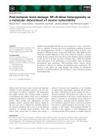
Numerous antioxidative enzymes are involved in the removal of H2O2 (Fig. 1.2).
Catalases (CATs) are haem proteins able to dismutate H2O2, without the need for
reducing cofactors. Since the CAT affinity for H2O2 is much lower than that of other
H2O2 removal enzymes, it seems that CATs function only when this ROS is present at
high levels. At sub-cellular level, CATs are undoubtedly localized in peroxisomes, even
if their presence in other cell compartments, such as cytosol, chloroplasts and mitochondria, cannot be excluded (Mhamdi et al. 2010). Three different classes of CATs
have been found in almost all plant species, and they are expressed in different tissues.
In Arabidopsis, the knockout of CAT1 and CAT3 slightly reduces or has no effect,
respectively, on total CAT activity. On the other hand, cat2 mutants reduce the total
CAT activity by 80 % and show defects not only in photorespiration but also in
response to pathogens (Chaouch and Noctor 2010). CAT genes are highly expressed
even under optimal conditions; thus, exposure to stress not always requires
up-regulation and in many cases some stresses cause down-regulation of CATs
expression and/or activity (Mhamdi et al. 2012).
All the other H2O2-removing enzymes are peroxidases, which require reducing
cofactors. Peroxidases can be divided in haeme-based and thiol-based peroxidases.
Fig. 1.2 Schematic
representation of the enzymes
involved in ROS removal.
APX Ascorbate peroxidase;
PRX Peroxiredoxins; POD
Class III peroxidases; SOD
Superoxide dismutase
1 Cellular Redox Homeostasis as Central Modulator …
9
The first group comprises APX (discussed above) and class III haeme peroxidases
(PODs), which can be involved in both ROS removal and ROS generation. PODs
use different compounds, mainly of phenolic nature, as electron donors, and their
role seem to be correlated principally to the oxidation of the reducing substrate,
rather than to the H2O2 removal (De Gara et al. 2003). PODs are involved in the
stiffening and lignification of the cell wall, which represent an optimal mechanical
barrier for the slowdown of pathogen penetration. Consistently, an increase in POD
activity occurs during different plant–pathogen interactions (Ding et al. 2011;
Lanubile et al. 2012, 2015; Mandal et al. 2014; Oliveira et al. 2014).
The second group of peroxidases, the thiol-based peroxidases, is constituted by
peroxiredoxins (PRX, Tripathi et al. 2009). These proteins, not having a prosthetic
group, remain in an inactive form at the end of their catalytic cycle; the regeneration
of active PRX depends on external electron donors, such as thioredoxins (TRX),
glutaredoxins (GRX), cyclophilins and NADPH-dependent TRX reductase (TR,
Bhatt and Tripathi 2011). TRX and GRX, key proteins involved in the regulation of
cysteine/protein redox state, are generally reduced by TR and GSH, respectively.
Glutathione peroxidases (GPX) belong to the PRX superfamily; although initially
defined as GSH-dependent peroxidases, GPX use only TRX for their regeneration
and do not react with GSH or GRX (Navrot et al. 2006; Bela et al. 2015). Due to the
thiol-dependent activities, GPX isoenzymes, besides detoxification, may be
involved in the regulation of cellular redox homeostasis by maintaining the
thiol/disulphide or NADPH/NADP+ balance (Navrot et al. 2006). GPX proteins are
involved in the response to both biotic and abiotic stress (Navrot et al. 2006; Bela
et al. 2015).
The thiol-based peroxidases, changing the thiol status of TRX and/or GRX, can
have repercussions on redox-sensitive target proteins, thus can be directly involved
in redox-dependent signalling (Foyer and Noctor 2016).
1.4
Redox-Dependent Signalling
Redox homeostasis is a crucial requirement of plant cells: each variation in the
redox state, due to an imbalance of ROS production and scavenging, could be
indicative of environmental disturbance and function as a signal (Fig. 1.1; Potters
et al. 2010). Moreover, any stimulus altering cellular redox homeostasis may
function as an inducer for the same set of defence-related genes. For instance, it has
been reported that both low levels of ascorbate or changes in glutathione pool are
able to induce pathogenesis-related (PR) proteins, acting as elicitors of resistance
response to pathogens (Pastori et al. 2003; Barth et al. 2004; Chaouch et al. 2010;
Han et al. 2013). However, the two hydrophilic redox couples ASC/DHA and
GSH/GSSG seem to function in a different way in the redox signalling (MunnéBosch et al. 2013). ASC, that is the only reductant present at a significant level in
the apoplast, can be oxidized in this compartment by ASC oxidase (Parsons and Fry
2012), which can contribute to create a redox gradient across the plasma membrane,
10
C. Paciolla et al.
connecting intra- and extra-cellular environments. Thus, ASC/DHA redox pair would
principally function in defining opportune thresholds for apoplastic and cytoplasmic
signalling (de Pinto et al. 1999; Pignocchi et al. 2003; de Pinto and De Gara 2004). On
the other hand, GSH, that has a principal role in defining intracellular redox potential,
would be involved mainly in the redox-dependent signalling pathways occurring
inside the cell (Foyer and Noctor 2005; Han et al. 2013). At this regard, the distribution of GSH among distinct intracellular compartments is crucial to define cellular
redox environment in which both metabolism and signalling take place (Zechmann
2014). During oxidative stress, GSH is not only oxidized but also redistributed in
intracellular compartments. In Arabidopsis cat2 mutants producing more H2O2, GSH
levels are higher and more oxidized than in the wild-type plants. Interestingly, the
increase in GSH is higher in the vacuole and chloroplasts than in the cytosol (Queval
et al. 2011). The glutathione compartmentation occurring during oxidative stress
represents a significant aspect of redox homeostasis and signalling, since it is useful to
avoid an excessive oxidising cytosolic redox environment and to allow the signalling
termination (Noctor et al. 2013).
1.4.1
Redox Signalling in Different Cell Compartments
Redox signalling is reactive to innumerable environmental cues, which influence
cellular metabolism and apoplastic environment (Foyer and Noctor 2012). It should
be considered that the content and the redox state of redox-active compounds
greatly vary among different cell compartments; therefore, redox regulation
occurring in various cell compartments can influence differently plant response to
external environment changes (Noctor and Foyer 2016).
The apoplast is as a crucial site for the plant redox-dependent response to external
stimuli, both of biotic and abiotic nature. It has been suggested that in the apoplast
oxidants are not only produced but also perceived. The redox buffering capability of
the apoplast is weaker than that of intracellular compartments, since it is deficient in
NAD(P)H and GSH while it is rich in enzymes that remove antioxidant compounds
(Horemans et al. 2000; Pignocchi et al. 2003; Pignocchi and Foyer 2003;
Ohkama-Ohtsu et al. 2007; Parsons and Fry 2012). For this reason, the ROS lifetime
in the apoplast is longer than inside the cell. Different hypotheses have been issued to
explain the transmission of the redox signal from the apoplast to inside the cell
(Fig. 1.3). One emerging and interesting possibility is that the redox signal can be
transmitted by redox-sensitive proteins on the plasma membrane, such as the K+
channel SKOR or the cysteine-rich receptor-like kinases (García-Mata et al. 2010;
Wrzaczek et al. 2010, 2013). On the other hand, the transmission of the redox signal
could be due to the oxidation of the extracellular ASC pool (Foyer and Noctor 2012).
Finally, it is possible that H2O2 produced in the apoplast migrates inside the cells
through aquaporins and is transduced into the cytosol (Miller et al. 2010a, b). In this
way, the apoplastic oxidative burst can be sensed and transduced also by neighbouring
cells, leading to the formation of the so-called “ROS wave” (Mittler et al. 2011). ROS
1 Cellular Redox Homeostasis as Central Modulator …
11
Fig. 1.3 Schematic model of redox signalling occurring in different cell compartments in response to
stress. After stress, ROS can be generated in the apoplast by respiratory burst oxidase homologs
(RBOHD), which is activated by a calcium influx, which in turn phosphorylates (P) the protein. Redox
signalling in the apoplast can be sensed by changes in ASC oxidative state (#ASC/DHA). In addition,
redox signal can be transmitted by changes in the redox-sensitive proteins, such as the receptor-like
kinases (RLK) and the K+ channel (SKOR), localized on the plasma membrane. It is also possible that
H2O2 migrates inside the cell through aquaporins (AQP) and is transduced into the cytosol. Redox
imbalances occurring in the organelles could participate to retrograde signalling through the action of
proteins with double localization. In particular Whirly1 (WHY1), changing its redox state and
conformation, can move from chloroplasts to the nucleus where it stimulates gene transcription. The
membrane-associated NAC protein, ANAC013, located in the endoplasmic reticulum, in response to
ROS can undergo proteolytic activation and move into the nucleus where it induces the expression of
genes conferring stress tolerance. Moreover, ROS from the organelles can pass in the cytosol. Changes
in cytosolic redox homeostasis, due to interaction between ROS accumulation and antioxidant
systems, can be transduced by redox-dependent modifications of redox-sensitive signalling proteins.
More details are given in the text
generated in the apoplast, as part of the ROS wave, could enter a non-activated cell
and trigger the release of calcium, which in turn phosphorylates and activates the
RBOHD proteins; turning on ROS production, this new activated cell participates to
the activated group of cells involved in the ROS wave (Suzuki et al. 2013; Gilroy et al.
2014). Thus, the ROS wave travels in the apoplast from the initiating tissue to the
whole plant and, together with abscisic acid, is responsible for the activation of
systemic acquired acclimation in response to local environmental stimuli (Suzuki et al.
2013; Mittler and Blumwald 2015).
Different environmental stresses cause redox imbalances principally in the
organelles. Chloroplasts and mitochondria can respond either by rapidly fine tuning
12
C. Paciolla et al.
the altered electron fluxes or by inducing changes in gene expression for long-term
adaptation (Scheibe et al. 2005; Rhoads et al. 2006; Rhoads and Subbaiah 2007). In
the latter case, the imbalance in the electron transport chains must initiate signalling
processes that have to be sensed in the nucleus, with the results of changes in the
expression of genes coding for proteins located in the organelles; such pathway has
been identified as retrograde signalling (Nott et al. 2006; Rhoads and Subbaiah
2007; Leister 2012). Many actors have been proposed to function in retrograde
signalling (reviewed in Kleine and Leister 2016) and among these ROS, and in
particular H2O2, produced in the organelles could represent good intermediates, in
particular for acclimation to stress (Rhoads et al. 2006; Petrov and Van Breusegem
2012; Galvez-Valdivieso and Mullineaux 2010). Another appealing hypothesis
regarding the redox control of retrograde signalling is linked to the possibility that
proteins with double localization move from the organelles to the nucleus, in order
to directly mediate alteration in gene transcription (Fig. 1.3). An interesting
example is the single-stranded DNA-binding protein Whirly1 (WHY1), which has
been identified both in the nucleus and in the chloroplasts, where it is situated
between thylakoids and nucleoids (Krause et al. 2005; Grabowski et al. 2008).
WHY1 is able to move from the chloroplasts to the nucleus where it stimulates the
transcription of PR genes (Isemer et al. 2012). The WHY proteins have a cysteine
residue in a conserved region involved in the formation of disulphide bridges; they
form tetramers and are also able to assemble into oligomers (Desveaux et al. 2002;
Cappadocia et al. 2012); redox regulation has been demonstrated for the chloroplastic WHY3 protein (Stroher and Dietz 2008). Consequently, it has been speculated that stress-induced over-reduction of thylakoidal electron transport chain
could destabilize the oligomeric WHY1, probably acting on cysteine residues, and
release the monomer that will be then translocated to the nucleus. In this way,
WHY1 might work as a redox-regulated element in chloroplastic retrograde signalling, involved in acclimation and immunity responses (Foyer et al. 2014). An
example of this kind of regulation has been also proposed for mitochondrial retrograde signalling. The membrane-associated NAC protein, ANAC013, under
non-stressed conditions is located in the endoplasmic reticulum. This compartment
can be physically connected with mitochondria (Hayashi et al. 2009). Through
these physical connections, mitochondrial ROS, implicated in retrograde signalling
(Rhoads and Subbaiah 2007), could facilitate the proteolytic activation of
ANAC013. In this way, ANAC013 migrates into the nucleus where it binds and
activates the mitochondrial dysfunction motif(s), inducing gene expression that
confers tolerance to oxidative stress (De Clercq et al. 2013).
In addition to the apoplast and the organelles, the cytosol, although not directly
involved in ROS production, plays a key role in the integration of redox signals
(Noctor and Foyer 2016). Indeed, it has been shown that different stresses are able
to render the cytosolic environment more oxidized (Meyer et al. 2007; Jubany-Mari
et al. 2010). Moreover, in cat2 mutants the deficiency in catalase, which is primarily, localized in the peroxisomes, mainly impacts the transcription of cytosolic
antioxidant enzymes (Rahantaniaina et al. 2013). The redox-dependent modification of proteins could be a principal way of redox signalling within this
1 Cellular Redox Homeostasis as Central Modulator …
13
compartment (Fig. 1.3). For instance, redox-dependent modifications of the translational apparatus in the cytosol permit a fast and efficient control of protein synthesis (Moore et al. 2016).
1.4.2
The Role of Redox-Sensitive Proteins in Signal
Transduction
Since different stresses can induce a diverse locally restricted ROS production, it is
possible that the specific redox signalling is determined by microenvironments
(Terada 2006; Zachgo et al. 2013). Changes in the redox state of specific cell
compartments or microenvironments have to be sensed and transduced. A potential
way of perception and signalling engages the redox-dependent modification of
proteins and in particular the modifications of cysteinyl residues, which can be
oxidized to different degrees. The redox-dependent post-translational modifications
of proteins comprise the formation of disulphide bridges, sulphenic, sulphinic and
sulphonic acids as well as S-glutathionylation and S-nitrosylation, due to interaction
of cysteine with GSH and nitric oxide (NO), respectively. These modifications can
determine alteration in protein conformation and activity. Except for the formation
of sulphonic acid, all other redox modifications are virtually reversible (Ghezzi
et al. 2005). The reversible oxidation/reduction of the redox-sensitive proteins can
be mediated directly by ROS or indirectly via the redox-sensitive molecules GSH,
TRX and GRX, which, as discussed above, control the cellular redox environment
(Foyer and Noctor 2005). In this perspective, a key role in the perception of cellular
redox environment has been attributed to peroxiredoxins. For instance, chloroplastic peroxiredoxins, changing their aggregation state in function of their
oxidative state, can act as sensors of oxidative stress and initiators of a signalling
cascade that involves a multiplicity of protein–protein interactions that link redox
changes with the necessary responses (Dietz 2008; Muthuramalingam et al. 2009).
Redox-sensitive proteins comprise metabolic enzymes that directly adjust cellular metabolism to the changing environment and redox-sensitive signalling proteins that perform their tasks through downstream components, such as kinases,
phosphatases and transcription factors (Foyer and Noctor 2005, 2013; Dietz 2008).
Transcription factors (TFs) can also be direct targets of redox-dependent modulation of their activity (Dietz 2014). Among the directly redox-regulated TFs
involved in the stress response, attention has been paid to the study of the heat
shock factors (HSFs), since it was previously shown that these proteins are redox
regulated in animals (Ahn and Thiele 2003; Miller and Mittler 2006). HSFs act by
binding to the highly conserved heat shock element in the promoters of target
genes. A great number of HSF genes are present in the plant genome, and the HSF
network is extremely plastic and controls the response of plants to various stress
conditions (Miller and Mittler 2006). Recently, it has been reported that heat stress
and H2O2 treatment activate the Arabidopsis HSFA1, inducing its binding to the
14
C. Paciolla et al.
promoters of two heat shock proteins; this binding can be reversed by reducing
agents. Thus, it has been proposed that the activation of HSFA1 is redox-regulated,
although the mechanisms of this activation have not been clearly explained (Liu
et al. 2013). A redox-dependent regulation of another Arabidopsis HSF, the
HSFA8, has been described with more details. H2O2 treatment of Arabidopsis
protoplasts causes a translocation of HSFA8 to the nucleus. Interestingly, the
site-directed mutagenesis of two conserved cysteine residues blocks this translocation. Therefore, a role for HSFA8 as a redox sensing TF in the stress-responsive
transcriptional network has been suggested (Giesguth et al. 2015).
One of the best studied events of redox signalling regards the salicylic acid (SA)mediated induction of PR proteins, occurring through the NPR1 (non-expressor of
PR genes1) protein. NPR1 is a key regulator in the plant defence against pathogens
(Pajerowska-Mukhtar et al. 2013); NPR1 is also involved in the response to some
abiotic stress (Jayakannan et al. 2015). Nuclear localization of NPR1 is essential for
the induction of PR genes (Kinkema et al. 2000). Indeed, NPR1 acts as a transcriptional co-activator that, interacting with the TFs of the TGA (TGACG motif
binding factor) family, regulates their DNA-binding activity to the promoters of PR
genes, thus inducing transcription (Despres et al. 2003). NPR1 can be found in the
cytosol as oligomers, linked through intermolecular disulphide bridges, or in the
nucleus as monomers that are the active form of the protein. Pathogen infection or
SA treatment, modulating the cellular redox environment, can control the redox
state of NPR1 (Mou et al. 2003). The reduction of the oligomeric form to the
monomeric one is catalysed by TRX; this reduction permits the nuclear translocation of NPR1; on the other hand, the oligomerization of NPR1 is facilitated by
S-nitrosylation (Tada et al. 2008). The importance of the redox-dependent changes
in NPR1 status and localization has been amply reported. Indeed, in an apx mutant
and in CAT antisense plants that accumulate H2O2, nuclear translocation of NPR1
and induction of PR genes are prevented (Peleg-Grossman et al. 2010). Similarly,
treatment with reduced GSH or oxidizing conditions promotes or inhibits, respectively, the nuclear accumulation of NPR1 (Kovacs et al. 2015). The translocation of
NPR1 into the nucleus is also promoted by the NO donor nitrosoglutathione
(GSNO) (Lindermayr et al. 2010). However, GSNO-induced nuclear translocation
is not due to the S-nitrosylation of NPR1, but rather due to the action of GSNO as a
positive effector upstream of SA. An interesting crosstalk between NO and GSH for
the control of NPR1 activity has been proposed (Kovacs et al. 2015). It is known
that pathogen attack induces an NO burst that dynamically modulates the redox
state of glutathione (Vanacker et al. 2000; Mou et al. 2003; Koornneef et al. 2008).
Since NO fumigation transiently increases the oxidation of GSH and leads to an
increase in the total GSH pool, it has been suggested that the initial oxidation of
GSH promotes de novo GSH biosynthesis and is necessary for SA accumulation
and the NPR1-dependent defence signalling pathway (Kovacs et al. 2015). In
addition to the redox-controlled translocation into the nucleus, there is also evidence for the contribution of a redox control of TFs in the nucleus. For instance, a
redox regulation of the TGAs activity has been proposed. Under oxidizing conditions, TGA1 is in an inactive conformation, due to the formation of intermolecular
1 Cellular Redox Homeostasis as Central Modulator …
15
disulphide bridges between cysteinyl residues (Despres et al. 2003; Lindermayr
et al. 2010). However, TGA1 DNA-binding activity is considerably enhanced in
presence of GSNO, probably for glutathionylation or S-nitrosylation of the Cys 260
and Cys 266; these redox modifications could be responsible of conformational
changes of TGA that enhance its activity (Lindermayr et al. 2010). Interestingly, the
redox-sensitive GRXs have been suggested as possible mediators of the
redox-dependent changes of TGAs (Ndamukong et al. 2007; Li et al. 2011).
1.5
Conclusion and Perspectives
A large number of molecular components are involved in the control of cellular
redox homeostasis. The various ROS, produced in different cell compartments and
a plethora of antioxidants, finely regulate the cellular redox environment. Thus, the
interactions between ROS and antioxidants constitute a strong network, able to give
information on cellular environment. The imbalances in this network, occurring
under stress conditions, act as redox signals that have to be transduced in order to
induce specific adaptive responses. The redox-dependent signalling is not linear; on
the contrary, it is defined by complex and dynamic pathways, which can be connected and sometimes overlapping. Each redox-dependent interaction is opportunely regulated by a restricted environment, whose changes are of primary
importance to transfer the complex system of information. Different cellular compartments are involved in mediating redox-dependent signalling. For this reason, it
is very important to shed light on the role of each compartment and the interaction
among them in the transfer of redox-dependent signals in response to specific
stresses. Undoubtedly, it will be necessary to take into account also the diversity of
ROS and antioxidants that participate in the specific response.
Another important point that needs to be deepened regards the mode of transmission of the redox-dependent signals. Many data suggest that reversible
oxidation/reduction of redox-sensitive proteins could have a key role in signal
transduction. An important step is to understand if these modifications are induced
directly by ROS or indirectly by changes in the oxidative state of the
redox-sensitive molecules GSH, TRX and GRX. Moreover, the identification of
new redox-sensitive signalling proteins involved in specific environmental conditions could help to identify common and divergent points of redox regulation in the
response to different kinds of stress.
References
Abbasi AR, Saur A, Hennig P, Tschiersch H, Hajirezaei M, Hofius D, Sonnewald U, Voll LM
(2009) Tocopherol deficiency in transgenic tobacco (Nicotiana tabacum L.) plants leads to
accelerated senescence. Plant Cell Environ 32:144–157