Nghiên cứu tạo vải chứa vi nang kháng viêm thân thiện môi trường định hướng ứng dụng y dược.
Bạn đang xem bản rút gọn của tài liệu. Xem và tải ngay bản đầy đủ của tài liệu tại đây (3.44 MB, 183 trang )
TABLE OF CONTENT
LIST OF ABBREVIATIONS ................................................................................................................ 4
LIST OF FIGURES ................................................................................................................................ 6
LIST OF TABLES ............................................................................................................................... 10
INTRODUCTION ............................................................................................................................... 12
1. Chapter 1: Literature review .............................................................................................. 16
1.1. Microcapsules and their textile applications .......................................................................... 16
1.1.1. Microcapsules ............................................................................................................................................. 16
1.1.2. Applications and requirements of microcapsules in the textile field .................................. 20
1.1.3. Microcapsules prepared from environment-friendly materials for textile applications
....................................................................................................................................................................................... 24
1.2. Microencapsulation by the solvent evaporation technique .............................................. 25
1.2.1. Techniques of microencapsulation .................................................................................................... 25
1.2.2. Microencapsulation by the solvent evaporation technique .................................................... 26
1.2.2.1. Basic principle of the technique ..................................................................................................................... 26
1.2.2.2. Important parameters of the microencapsulation by the solvent evaporation technique .. 27
1.2.2.3. Using non-halogenated solvents in the microencapsulation by the solvent evaporation
technique .................................................................................................................................................................................. 31
1.2.2.4. The quillaja saponin bio-sourced surfactant ............................................................................................ 32
1.3. The textile substrates ...................................................................................................................... 38
1.3.1. Influence of the textile substrate on the microcapsule loading capability of
microcapsule-treated fabrics ........................................................................................................................... 38
1.3.2. Influence of the textile substrate on the microcapsule distribution of microcapsuletreated fabrics......................................................................................................................................................... 40
1.3.3. Influence of the textile substrate on the active release capability from the
microcapsule-treated fabrics ........................................................................................................................... 41
1.3.4. The interlock knitted fabrics ................................................................................................................ 42
1.4. Textile finishing with microcapsules ......................................................................................... 45
1.4.1. Techniques of finishing textiles with microcapsules ................................................................. 45
1.4.2. Influence of the drying conditions on the microcapsule morphology after finishing
process ....................................................................................................................................................................... 47
1.5. Conclusions of the literature review .......................................................................................... 49
2. Chapter 2: Experimental methods .................................................................................... 50
2.1. Materials ............................................................................................................................................... 50
2.1.1. Interlock knitted fabrics ......................................................................................................................... 50
2.1.2. Chemicals for microencapsulation ..................................................................................................... 51
2.2. Research contents ............................................................................................................................. 53
2.3. Experimental techniques................................................................................................................ 54
2.3.1. Determining the suitable microcapsule size for textile application..................................... 54
1
2.3.2. Evaluating the surface-active properties of quillaja saponin ................................................. 54
2.3.3. Investigating the influence of the microencapsulation parameters on microcapsule
characteristics ........................................................................................................................................................ 56
2.3.3.1. Microencapsulation .............................................................................................................................................. 56
2.3.3.2. Microcapsule characterization ........................................................................................................................ 58
2.3.4. Investigating the influence of textile substrate on the characteristics of microcapsuletreated fabric........................................................................................................................................................... 61
2.3.4.1. Structural parameters of the interlock knitted fabrics ........................................................................ 61
2.3.4.2. Influence of textile material on characteristics of microcapsule-treated fabric ....................... 63
2.3.4.3. Influence of the loop length on the characteristics of the microcapsule-treated fabric ........ 67
2.3.4.4. Influence of the fabric extension on the transdermal drug release capability of fabric ........ 72
2.3.5. Investigating the influences of the drying conditions on the microcapsule morphology
after finishing process ......................................................................................................................................... 74
3. Chapter 3: Results and discussions .................................................................................. 75
3.1. Microencapsulation of ibuprofen ................................................................................................ 75
3.1.1. Determination of the microcapsule size for the targeted textile application ................... 75
3.1.2. Surface-active properties of quillaja saponin S4521 (Sigma Aldrich) ................................ 77
3.1.3. Influence of the microencapsulation parameters on the microcapsule size and
morphology.............................................................................................................................................................. 79
3.1.3.1. Influence of the saponin concentration ....................................................................................................... 79
3.1.3.2. Influence of the stirring rate ............................................................................................................................ 84
3.1.3.3. Influence of the volume of ethyl acetate added to the aqueous phase .......................................... 86
3.1.4. Conclusions on the microencapsulation parameters suitable for textile applications 90
3.1.5. Other characteristics of C0.075 microcapsules ............................................................................ 91
3.2. Influences of the drying conditions on the microcapsule morphology after textile
finishing process ........................................................................................................................................ 93
3.2.1. Influence of the relative humidity during the drying process ................................................ 94
3.2.2. Influence of the drying temperature ................................................................................................. 95
3.3. Influence of the textile substrate on the characteristics of the microcapsule-treated
fabrics ............................................................................................................................................................ 97
3.3.1. Influence of the textile material type ................................................................................................ 97
3.3.1.1. Influence on the microcapsule loading capability .................................................................................. 97
3.3.1.2. Influence on the microcapsule distribution .............................................................................................. 99
3.3.1.3. Influence on the ibuprofen release capability of the microcapsule-treated fabric ............... 102
3.3.1.4. Conclusion ............................................................................................................................................................. 104
3.3.2. Influence of the loop length ................................................................................................................104
3.3.2.1. Influence on the microcapsule loading capability ............................................................................... 105
3.3.2.2. Influence on the microcapsule distribution ........................................................................................... 114
3.3.2.3. Influence on the ibuprofen release capability of the fabric ............................................................. 126
3.3.2.4. Conclusion ............................................................................................................................................................. 127
3.3.3. Influence of the fabric extension.......................................................................................................128
3.4. Conclusion of Chapter 3 ............................................................................................................... 130
4. Final conclusions and future outlook ............................................................................134
2
4.1. Final conclusions ............................................................................................................................ 134
4.2. Future outlook ................................................................................................................................. 135
REFERENCES ...................................................................................................................................136
LIST OF PUBLISHED WORKS OF THE DISSERTATION ......................................................147
3
LIST OF ABBREVIATIONS
Abbreviation
Explanation
Organizations
HUST
Hanoi University of Science and Technology
IMP
Ingénierie des Matériaux Polymères, UMR CNRS
5223
UCBL
University Claude Bernard Lyon 1
ASTM
American Society for Testing and Materials
ISO
International Organization for Standardization
TCVN
Vietnam National Standards
Experimental techniques and equipment
FRSE
Microencapsulation by the solvent evaporation
method with the fast rate of the solvent
evaporation
NRSE
Microencapsulation by the solvent evaporation
method with the normal rate of the solvent
evaporation
FTIR
Fourier-Transform Infrared Spectroscopy
HPLC
High-performance liquid chromatography
SEM
Scanning electron microscopy
UV-Vis
UV visible spectroscopy
Materials and their characteristics
CMC
Critical micelle concentration
E
Microencapsulation efficiency
Eudragit RSPO
Poly(ethyl acrylate-co-methyl methacrylate-cotrimethylammonioethyl methacrylate chloride)
1:2:0.1
HLB
Hydrophilic-lipophilic balance
MLC
Microcapsule loading capability of the
4
microcapsule-treated fabric
L
Drug loading ratio of microcapsule
PCL
Poly-ε-caprolactone
PCM
Phase change material
PLGA
Poly(lactic-co-glycolic acid)
PLLA
Poly (l-lactic acid)
PU
Polyurethane
PVA
Poly (vinyl alcohol)
γ
Surface tension of a solution
Structural parameters of the interlock knitted fabric
l
loop length
Lu
Length of yarn in a structural knitted cell
Pd
Course density
Pn
Wale density
Ps
Area density
SKC
Structural knitted cell
Su
Number of the structural knitted cells per unit area
of the fabric
Mfbr
Mass per unit area of the fabric
t
Fabric thickness
D
Yarn diameter in the fabric
P
Fabric porosity
ρ
Fiber density
5
LIST OF FIGURES
Figure 1.1: Microcapsule classification on the basis of their morphology [31] ........................ 16
Figure 1.2: SEM image of PLGA microsphere containing progesterone [121] ........................ 17
Figure 1.3: An example of the size distribution curve of microcapsules [140]......................... 18
Figure 1.4: Four types of theoretical curves describing the release mechanisms of the active
ingredients from the non-erodible microcapsules [104] .................................................... 19
Figure 1.5: Classification of microencapsulation methods ....................................................... 25
Figure 1.6: Diagram of basic principle of microencapsulation by solvent evaporation
technique [76] .................................................................................................................... 26
Figure 1.7: SEM images of microcapsules with different mass ratio of core/shell: 60:40 (A);
70:30 (B); 75:25 (C) [84] .................................................................................................. 29
Figure 1.8: SEM images of microspheres made by FRSE (on the left) and NRSE (on the right)
[26] .................................................................................................................................... 31
Figure 1.9: Schematic illustration of a surfactant [49] .............................................................. 34
Figure 1.10: Determination of CMC by the curve surface tension-lnC [92]............................. 35
Figure 1.11: Illustration of a spherical micelle [93] .................................................................. 36
Figure 1.12: General molecular structure of quillaja saponin, in which R1, R2, R3 groups
depend on different molecules in the bark extract [98] ..................................................... 37
Figure 1.13: SEM images of fabrics padded with microcapsules: polyester fabric (A) and
cotton fabric (B) [125] ....................................................................................................... 40
Figure 1.14: SEM images of polyester fabric (A) and cotton fabric (B) coated with
microcapsules containing flame - retardant agent [40] ..................................................... 41
Figure 1.15: Structure (A) and notation (B) of interlock knitted fabric [1]............................... 42
Figure 1.16: Structure of knitted loop [1] .................................................................................. 43
Figure 1.17: Model of interlock knitted loop by Dabiryan-Jeddi [27] (A) Front view; (B) Plane
view; (C) Side view; (D) a plain structure ......................................................................... 44
Figure 1.18: SEM images of wet-coated nylon fabrics with and without microcapsules:
without microcapsules (a), with 10% (b), 20% (c) and 30% microcapsules (d) [66] ....... 47
Figure 1.19: SEM images of dry-coated nylon fabrics with and without microcapsules: without
microcapsules (a), with 10% (b), 20% (c) and 30% microcapsules (d) [66] ..................... 47
Figure 1.20: SEM images of microcapsule padded cotton fabrics with different drying
temperature 120 o(11.1-11.2); 140 oC (11.3-11.4); 160 oC (11.5-11.6) [88] ..................... 48
Figure 2.1: Structural formula of ibuprofen (C13H18O2) [14] .................................................... 51
Figure 2.2: Chemical structure of Miglyol 812 [58] ................................................................. 52
Figure 2.3: Chemical structure of eudragit RSPO [147] ........................................................... 53
6
Figure 2.4: Tensiometer SEO-DST30M (Surface & Electro-Optics) ....................................... 55
Figure 2.5: Diagram of microencapsulation of eudragit RSPO loading ibuprofen by solvent
evaporation method ........................................................................................................... 56
Figure 2.6: Equipment system for microencapsulation ............................................................. 57
Figure 2.7: Centrifuge G-16KS of Sigma.................................................................................. 57
Figure 2.8: Optical microscopy Olympus EX 41 ..................................................................... 58
Figure 2.9: Scanning electron microscopy QUANTA FEG 250 ............................................... 58
Figure 2.10: Mastersizer 2000, Malvern Instruments................................................................ 59
Figure 2.11: Ultrasonic equipment Fisher biolock Scientific 750W ......................................... 60
Figure 2.12: Spectrometer UV-Vis Lamda 35 (Perkin Elmer) ................................................. 60
Figure 2.13: Gas chromatograph Agilent Technology 6890N .................................................. 61
Figure 2.14: Experimental washing machine Electrolux........................................................... 62
Figure 2.15: Laboratory conditioning chamber M250-RH ....................................................... 62
Figure 2.16: Electronic scale OHAUS - PA413 ........................................................................ 63
Figure 2.17: Thickness gauge .................................................................................................... 63
Figure 2.18: Vacuum drier France Etuves ................................................................................. 63
Figure 2.19: Fabric samples soaking in the microcapsule suspension ...................................... 64
Figure 2.20: Scanning electron microscopy QUANTA FEG 250 – FEI company ................... 65
Figure 2.21: Interface of Meander 3.1.2 during determining the area of microcapsule aggregate
........................................................................................................................................... 65
Figure 2.22: Glass jar simulating Franz diffusion cell .............................................................. 66
Figure 2.23: Drug release in vitro experiment ........................................................................... 66
Figure 2.24: HPLC system of SHIMADZU .............................................................................. 67
Figure 2.25: Coating equipment Mini Coater (DaeLim Starlet Co.,Ltd) .................................. 68
Figure 2.26: Vacuum drier OV-11 ............................................................................................ 68
Figure 2.27: Scanning electron microscopy JEOL JSM - 7600F .............................................. 70
Figure 2.28: A step in the in vitro experiment of transdermal drug release .............................. 71
Figure 2.29: HPLC system of Merck Hitachi ............................................................................ 72
Figure 2.30: Experimental design to create different levels of fabric extension ....................... 73
Figure 3.1: SEM image of the surface of fabric B3................................................................... 75
Figure 3.2: Distance between fibers in the region of loop legs on cotton interlock fabric B3 .. 76
Figure 3.3: Distance between fibers in the region created by overlapping loops on cotton
interlock fabric B3 ............................................................................................................. 76
Figure 3.4: Surface tension of aqueous saponin solutions according to saponin concentration 78
7
Figure 3.5: Size distributions of microcapsule lots C0.025, C0.050, C0.075 and C0.100 ........ 80
Figure 3.6: Adense/Asurf ratio depending on the saponin concentration ...................................... 81
Figure 3.7: Optical microscope images of microcapsules C0.025 (A), C0.050 (B), C0.075 (C)
and C0.100 (D) .................................................................................................................. 83
Figure 3.8: Optical microscope images of microcapsules R700 (A), R650 (B) and R600 (C) . 85
Figure 3.9: Size distributions of microcapsules R700, R650 and R600 .................................... 86
Figure 3.10: Size distributions of microcapsule lots S0, S8 and S12 ........................................ 87
Figure 3.11: Optical microscope image of microcapsule S8 ..................................................... 88
Figure 3.12: Optical microscope images of the cotton interlock fabrics coated with
microcapsules S0 (A), S8 (B) and S12 (C) after 24 hours of vacuum drying at 25oC ...... 90
Figure 3.13: SEM images of the C0.075 microcapsules (A): overall image; (B): image of the
microcapsule surface ......................................................................................................... 92
Figure 3.14: SEM image of cross-section of microcapsule C0.075 .......................................... 92
Figure 3.15: SEM image of the microcapsule-coated fabric dried at 25oC with the relative
humidity of 65% ................................................................................................................ 94
Figure 3.16: SEM image of the microcapsule-coated fabric dried at 25oC with the relative
humidity of 20% ................................................................................................................ 94
Figure 3.17: SEM image of the microcapsule-coated fabric dried at 25oC with relative
humidity of 0% .................................................................................................................. 95
Figure 3.18: SEM image of the microcapsule-coated fabric vacuum dried at 25oC ................. 96
Figure 3.19: SEM image of the microcapsule-coated fabric vacuum dried at 35oC ................. 96
Figure 3.20: SEM image of the microcapsule-coated fabric vacuum dried at 45oC ................. 96
Figure 3.21: SEM image of the microcapsule-coated fabric vacuum dried at 60oC ................. 96
Figure 3.22: SEM images of the microcapsule-treated fabrics Cot_1 (A), 6535_1 (B) and
Pet_1 (C) .......................................................................................................................... 101
Figure 3.23: Chemical structure of polyester fiber .................................................................. 102
Figure 3.24: Chemical structure of cellulose (main component of cotton fiber) .................... 102
Figure 3.25: Release rate of ibuprofen from microcapsule-treated fabrics according to the type
of the textile material ....................................................................................................... 103
Figure 3.26: Microcapsule loading capability of the fabrics according to the loop length with a
microcapsule concentration of 14 mg/ml ........................................................................ 107
Figure 3.27: Microcapsule loading capability of the fabrics according to the loop length with a
microcapsule concentration of 24 mg/ml ........................................................................ 108
Figure 3.28: Fabric density according to the loop length ........................................................ 111
Figure 3.29: Fabric porosity according to the loop length ...................................................... 112
8
Figure 3.30: SEM images of the lower surface of the fabrics B1 (A) and B5 (B) .................. 113
Figure 3.31: SEM images of the fabrics after microcapsule application by the coating
technique B1 (A), B2 (B), B3 (C), B4 (D) and B5 (E) .................................................... 117
Figure 3.32: SEM images of the fabrics after microcapsule application by the impregnating
technique: B3 (A), B4 (B) and B5 (C) ............................................................................ 125
Figure 3.33: Weight percentage of ibuprofen released into the receptor fluid according to the
fabric extension................................................................................................................ 129
9
LIST OF TABLES
Table 1.1: The values of K for different kinds of fabrics [17] .................................................. 42
Table 2.1: Information of chemicals used for microencapsulation ........................................... 51
Table 2.2: Some properties of ibuprofen [151] ......................................................................... 51
Table 2.3: Doses of ibuprofen for adults and children [14, 65]................................................. 52
Table 3.1: Surface tensions of saponin solutions in distilled water according to the
concentration ..................................................................................................................... 77
Table 3.2: d(0.5) diameter and span values of microcapsule lots C0.025, C0.050, C0.075 and
C0.100 ............................................................................................................................... 79
Table 3.3: d(0.5) diameter and span values of microcapsule lots R700, R650 and R600 ......... 85
Table 3.4: d(0.5) diameter and span values of microcapsule lots S0, S8 and S12 .................... 87
Table 3.5: Structural parameters of the interlock knitted fabrics used to investigate the
influence of the textile material type on the characteristics of the microcapsule-treated
fabric .................................................................................................................................. 97
Table 3.6: Microcapsule loading capability of the fabrics knitted from different materials ..... 98
Table 3.7: Results of two independent samples t tests for fabrics knitted from different
materials ............................................................................................................................ 99
Table 3.8: Statistical results of the area of microcapsule aggregates on different kinds of
fabrics .............................................................................................................................. 101
Table 3.9: Ibuprofen release rate from the microcapsule-treated fabrics having different textile
materials .......................................................................................................................... 103
Table 3.10: Microcapsule loading capability of the fabrics according to the loop length with a
microcapsule concentration of 14 mg/ml ........................................................................ 105
Table 3.11: Microcapsule loading capability of the fabrics according to the loop length with a
microcapsule concentration of 24 mg/ml ........................................................................ 106
Table 3.12: Results of the t tests according to the loop length with a microcapsule
concentration of 14 mg/ml............................................................................................... 106
Table 3.13: Results of the t tests according to the loop length with a microcapsule
concentration of 24 mg/ml............................................................................................... 107
Table 3.14: Practical structural parameters of the fabrics according to the loop length ......... 109
Table 3.15: Microcapsule loading capability of the fabrics according to the loop length with a
microcapsule concentration of 20 mg/ml ........................................................................ 113
Table 3.16: Results of the t tests according to the loop length with a microcapsule
concentration of 20 mg/ml............................................................................................... 114
10
Table 3.17: Area of the microcapsule aggregates according to the loop length with a
microcapsule concentration of 14 mg/ml ........................................................................ 117
Table 3.18: Total cross section area of all microcapsules per cm2 of fabrics with coating
technique.......................................................................................................................... 119
Table 3.19: Calculated values of V, V1, V2 and V3 ................................................................. 121
Table 3.20: Area of the yarn surface might be coated with the microcapsules per cm2 of the
fabrics .............................................................................................................................. 123
Table 3.21: Value of K according to the loop length with a microcapsule concentration of 14
mg/ml ............................................................................................................................... 123
Table 3.22: Area of the microcapsule aggregates according to the loop length with a
microcapsule concentration of 20 mg/ml ........................................................................ 125
Table 3.23: Total cross section area of all microcapsules in an unit area of the fabrics when the
microcapsule concentration impregnated to the fabrics was equal to 20 (mg/ml) .......... 126
Table 3.24: Value of K according to the loop length with a microcapsule concentration of 20
mg/ml ............................................................................................................................... 126
Table 3.25: Weight percentage of ibuprofen on the microcapsule-treated fabrics (by
impregnating) released into the receptor fluid after 24 hours ......................................... 126
Table 3.26: Weight percentage of ibuprofen in the microcapsule-treated fabrics released into
the receptor fluid Iburl according to the fabric extension ................................................ 128
Table 3.27: Content of the microcapsules per cm2 of the fabrics according to the fabric
extension .......................................................................................................................... 129
11
INTRODUCTION
The functional textiles have been researched and developed strongly in recent years,
contributing to the growth of many industrial fields and the social economic development all
over the world. The application of microcapsules is now one of the modern technologies to
manufacture the functional textiles, as shown in a lot of researches and commercial products.
Microcapsules are tiny particles having size of from one to few hundred micrometers,
containing active ingredients packaged within the cores surrounded by the polymer shells. The
main advantages of microcapsules are controlling the release of the active ingredient and
protecting the active ingredient from the surrounding environment. Therefore, microcapsules
have been applied in many textile fields such as thermo-regulating textiles, flame-retardant
textiles, cosmetic textiles, fragrant textiles and medical textiles. In the field of medical textiles,
by using microcapsules, many kinds of medical ingredients have been incorporated into
textiles, including the anti-inflammatory agents such as ibuprofen, dexamethasone and some
herbs.
Using eco-friendly products is a global trend nowadays, so the application of
microcapsules in textile also needs to integrate with this development. The use of
microcapsules made from eco-friendly materials for medical textiles has been mentioned in
many researches with many kinds of natural active ingredients have been encapsulated in the
bio-sourced polymers. However, aside from the polymers and the active ingredients, the
surfactants and the solvents are also two essential components of the microencapsulation, but
the effort to reduce their hazard has not been in concern in the field of textile. Meanwhile, the
natural surfactant quillaja saponin has been approved for human health and been applied
commonly in producing emulsions for food, pharmaceutical and cosmetic industry. Besides, in
recent years, there have been some studies using less toxic non-halogenated solvent ethyl
aceate to replace toxic halogenated sovents in microencapsulation by sovent evaporation
method.
Beside the microcapsules, the textile substrate is also an important component of the
microcapsule-treated fabric. The fabric structure has strong influence on the microcapsule
loading capability and the microcapsule distribution on the fabric, and consequently affects the
active release capability of the fabric. Due to many advantages such as the softness, the high
elasticity, the ability of not curling at edges and the difficulty of unraveling, the interlock
knitted fabric is very suitable for the substrate in the medical textiles using microcapsules,
especially in compressive bandages. However so far in the knitting field in general and on the
interlock fabric in specific, there have been very few researches about the influence of fabric
structural parameters on the characteristics of the microcapsule-treated fabrics. It was reported
that the loop length obviously affected the microcapsule loading capability of the fabric, but
the scientific nature of the influence was not discussed. Moreover, up to now, there have not
been any studies about the influence of other structural parameters yet.
12
Microcapsules either in wet (not dried yet) or in dry (dried completely) type can be
applied to textiles by common finishing techniques such as padding and coating. The drying
and curing steps, which are always required in finishing, may induce the deformation of
microcapsules, resulting in lower active release capability of the fabric. Especially in the case
of using wet microcapsules for the textile finishing, the residual water and solvent in the
microcapsules makes the polymer shell still weak and easier to be deformed during the drying
and curing steps. Therefore, in the progress of applying a new kind of microcapsule to textile,
it is very necessary to investigate the influence of drying conditions on microcapsule
morphology in order to determine the suitable drying condition that keeps the microcapsules
from deformation.
According to the above analyses, the dissertation will discuss about:
"Elaboration of textiles containing eco-friendly anti-inflammatory microcapsules
oriented to medical application".
Research objectives of the dissertation:
•
•
•
Elaborating the microcapsules containing an anti-inflammatory drug and suitable for
textile applications. The solvent evaporation technique is applied for the
microencapsulation, which uses a bio-sourced surfactant and a non-halogenated
solvent.
Finding out the suitable drying conditions for textile finishing with obtained
microcapsules to keep the microcapsules from deformation.
Figuring out the influences of knitted fabric structural parameters (textile material,
loop length, fabric extension) on the important characteristics of the microcapsuletreated fabric (microcapsule loading capability, microcapsule distribution, active
release capability).
Research contents of the dissertation
•
Microencapsulation of ibuprofen, an anti-inflammatory drug, for medical textile
applications:
o Determining the range of microcapsule size that is appropriate for the cotton
interlock knitted fabric used in the dissertation.
o Evaluating the surface active properties of quillaja saponin used in the
dissertation (the lot S4521 of Sigma Aldrich).
o Investigating the influence of microencapsulation parameters on the
microcapsule size and morphology. The parameters investigated were: the
concentration of saponin, the stirring rate, the volume of the solvent added to
the aqueous phase before the emulsification step.
13
•
•
o Choosing the suitable microencapsulation parameters for the medical textile
applications.
Investigating the influences of drying conditions on the microcapsule morphology after
the textile finishing by microcapsules:
o Influence of the relative humidity during drying process.
o Influence of the drying temperature.
Investigating the influences of the knitted fabric structural parameters on the main
characteristics of the microcapsule-treated fabric:
o Influence of the textile material on the microcapsule loading capability, the
microcapsule distribution and the active release capability of the fabric.
o Influence of the loop length on the microcapsule loading capability, the
microcapsule distribution and the active release capability of the fabric.
o Influence of the fabric extension on the active release capability of the fabric.
New contributions of the dissertation
•
•
•
This is the first study using the bio-sourced surfactant quillaja saponin with the nonhalogenated solvent ethyl acetate for the microencapsulation by solvent evaporation
technique.
In the knitting field, the dissertation is the first study investigating the influences of
textile material and loop length on the characteristics of the microcapsule-treated
fabrics.
The dissertation presented a new concept, which was the microcapsule cover ratio, to
explain the effect of the loop length on the microcapsule distribution on the fabrics.
Scientific values of the dissertation
•
•
•
The dissertation proves the surface-active properties of quillaja saponin S4521
(Singma-Aldrich) according to its critical micelle concentration (cmc). The suitable
saponin concentration for the microencapsulation, which is appropriate for the medical
textile application, has been chosen according to the cmc value.
The dissertation indicates the scientific basis to choose the suitable drying conditions
for the textile finishing with elaborated microcapsules. The relative humidity during
drying process was selected basing on the hydration rate of microcapsules. The drying
temperature was selected basing on the glass transition of the polymer membrane.
The dissertation uses the interlock knitted loop model to explain the influence of loop
length on the microcapsule loading capability of fabric. Besides, a new concept that is
the microcapsule cover ratio K is used to explain the effect of loop length on the
microcapsule distribution on fabric.
14
Practical values of the dissertation
•
•
The dissertation contributes to the development of the eco-friendly productions in the
textile industry:
o The eco-friendly microencapsulation by the solvent evaporation technique
using bio-sourced surfactant and non-halogenated solvent.
o The eco-friendly finishing of textile with microcapsules without binders and
auxiliaries.
The dissertation contributes to the development of medical textile products using
microcapsules:
o According to the influence of textile material on the characteristics of the
microcapsule-treated fabric, the textile substrate material could be selected.
o According to the influence of loop length on the characteristics of the
microcapsule-treated fabric, the loop length of the textile substrate could be
determined.
o The influence of fabric extension on the transdermal release of the
microcapsule-treated fabric orient geometrical dimensions of the textile
substrate.
15
Chapter 1: Literature review
1. Chapter 1: Literature review
1.1. Microcapsules and their textile applications
1.1.1. Microcapsules
Definition and classification
Microcapsules are microparticles in which solid, liquid or gaseous active ingredients (the
core) are packaged within the second materials (the shells or the membranes) [31, 61].
Advantages of microcapsules [31]:
-
Controlled release of active compounds.
Separation of incompatible components.
Conversion of liquids to free flowing solids.
Increased stability (protection of encapsulated materials againts oxydation or
deactivation due to reaction in the environment).
Masking of ordour, taste and activity of encapsulated materials.
Microcapsules could be classified on the basis of their size or morphology [31, 61, 104].
By their size, microcapsules can be classified as:
-
Microcapsules: range in size from 1 to few hundred micrometers.
Nanocapsules: range in size below 1 µm (nanometer range).
By their morphology, microcapsules can be categorized into monocored, polycored and
matrix types (Figure 1.1).
Monocore
Polycore
Matrix
Figure 1.1: Microcapsule classification on the basis of their morphology [31]
-
Monocored microcapsules (also called single-core microcapsules) have a single hollow
chamber within the capsule.
Polycored microcapsules (also called multiple-core microcapsules) have a number of
different sized chambers within the shell.
Matrix microcapsules (also called microspheres) have active ingredients integrated
within the matrix of the shell materials.
Important characteristics of microcapsules and their determination
Some important characteristics of microcapsules that often mentioned in literatures are:
-
The morphology
The size
The drug loading ratio and the microencapsulation efficiency
The drug release profile
16
Chapter 1: Literature review
Microcapsule morphology
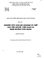
Microcapsule morphology expresses the shape, the features of membrane structure and
the internal structure of a microcapsule [26, 36, 83, 84, 120]. In most studies, microcapsules
have the spherical shapes (Figure 1.2), which help control the drug release much simpler than
other shapes. Depending on microencapsulation parameters, the microcapsule shell could be
smooth or rough, dense or porous, it often contains a system of tiny holes where active
ingredients can be released gradually. The internal structure helps reveal the type of
microcapsule (monocored, polycored or matrix type).
Microcapsule morphology is often observed by scanning electron microscope (SEM). At
low magnification from x100 to x500, SEM images provide information about the shape and
size of microcapsules, and at higher magnification, they give the feature of the shell such as
porosity, the size and the density of tiny holes. The cryo-SEM technique, in which
microcapsules are freezing to keep their original structures prior to observation, help look into
the internal morphology of microcapsules [57, 120, 141]. Besides, the microcapsule samples
using for SEM analysis are often dried microcapsules. For microcapsules suspended in water,
the environmental mode of SEM (with pressure in sample chamber around 2÷3 Torr) is used
in order not to deform microcapsules during observation [15].
Figure 1.2: SEM image of PLGA microsphere containing progesterone [121]
Microcapsule size
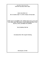
The size is a very important characteristic of microcaspules, since it is in inverse
proportion to surface area, it has very strong influence on the rate of drug release from
microcapsules [31]. Microcapsule size and size distribution are determined by particle size
analysis equipments, which often base on the effect of particle size on the spatial distribution
of scattered light in the laser diffraction. The result of the particle size analysis is often
expressed by the size distribution curve, which indicates the mean diameter of the
microcapsules, the broadness of the size distribution and also provides other size information
(Figure 1.3) [83, 138, 147].
17
Chapter 1: Literature review
Figure 1.3: An example of the size distribution curve of microcapsules [140]
The drug loading ratio and the microencapsulation efficiency
The drug loading ratio is defined as the mass ratio of encapsulated active compound to
microcapsule and often expressed by mass percentage [26, 55, 84, 147, 163]:
m
L (%) = m 𝑎𝑐 x 100 %
mc
(Eq 1.1)
In which:
L (%): the drug loading ratio
mac: the mass of encapsulated active compound
mmc: the mass of microcapsule
The microencapsulation efficiency is defined as the ratio of encapsulated mass to initial
mass used for microencapsulation of active compound, it is also expressed by mass percentage
[26, 42, 55, 147, 163]:
E (%) =
m𝑒𝑛𝑐𝑎𝑝
minit
x 100%
(Eq 1.2)
In which:
E (%): microencapsulation efficiency
mencap: the mass of active compound that is successfully encapsulated into microcapsule
minit: the total mass of active compound initially used for microencapsulation
In order to determine the drug loading ratio and the microencapsulation efficiency, the
active compound needs to be separated from a certain mass of microcapsules and then be
quantified. The separating process is often carried out by using a solvent that can dissolve the
active compound only but can not dissolve the polymer shell of microcapsules. The
dissolution is accelerated by ultrasonic [84, 147], or by using a solvent that can dissolve all the
microcapsules and in this case, a suitable chemical is used to precipitate the polymer to
remove it from the system [26, 42, 55, 163]. The drug loading ratio L(%) and the
18
Chapter 1: Literature review
microencapsulation efficiency E(%) can be deduced from the maximum concentration of
active compound in solvent.
The drug release profile
The drug release rate of microcapsule is often represented by a curve that describes the
change over time of the drug cumulative release. In the medical field, in order to build up the
drug release curve, a certain amount of microcapsules are often immersed in a solvent that
simulates the real release environment. Most studies used the phosphate buffer solution with
pH=7÷7.4 at the temperature of 37 ± 1oC (for oral applications) or at 32 ± 1oC (for topical and
transdermal applications) as the release solvent. After a predetermined period of time, the drug
concentration in release solvent is quantified by UV-Vis or HPLC equipment at suitable
wavelengths and the cumulative release of drug at that time could be deduced [8, 17, 26, 42,
55, 106, 163].
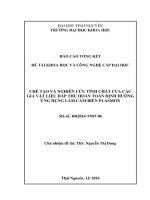
The drug release curve is a very important base to predict the release mechanism of the
active ingredient from the microcapsule [17, 42, 104]. Figure 1.4 represents the four types of
theoretical curves describing the four types of release mechanisms of the active ingredients
from the non-erodible microcapsules.
Figure 1.4: Four types of theoretical curves describing the release mechanisms
of the active ingredients from the non-erodible microcapsules [104]
In which:
-
The curve A describes the release behavior following zero order mechanism.
The curve B describes the release behavior following zero order mechanism with the
burst effect.
The curve C describes the release behavior following the Fick law.
The curve D describes the release behavior following the first order mechanism.
19
Chapter 1: Literature review
1.1.2. Applications and requirements of microcapsules in the textile field
Applications of microcapsules in textile
Microencapsulation technique dates back to 1950s when Green and Schleider produced
microencapsulated dyes from gelatin and arabic gum for the manufacture of carbonless
copying paper. Up to now, microcapsules have been applied widely in many industrial fields
such as chemistry, medicine and pharmacy, agriculture, food, cosmetic, printing and textile
[31]. Since 1990s, microencapsulation technique has been used in textile industry, mainly at
the research and development stage but a few commercial applications. Moving into the 21st
century, microcapsule applications in textile have developed more and more fast and
extensively, especially in Western Europe, Japan and North America, helped produce textile
materials with many new functions that conventional finishing techniques could not
implement [97].
The following are some important applications of the microcapsules in textile:
Application of microcapsules in thermo-regulating textiles
Thermo-regulating fabric is an intelligent textile that offers suitable responses to changes
in temperature of environment surrounding the body. The efficiency of thermal comfort
depends on the heat exchange between the body and surrounding environment. An effective
method that helps fabrics regulate the temperature is integrating phase change materials
(PCMs) into them. The most used PCMs are some popular alkanes such as n-octadecane, nhexadecane, n-eicosane and their mixtures. PCM materials absorb energy during the heating
process as phase change takes place and release energy to the environment in the phase change
during the reverse cooling process. Since PCMs could exist on fabrics in type of liquid when
the environment temperature is higher than their melting point, they need to be
microencapsulated [11, 25, 54, 66, 94, 95].
Application of microcapsules in flame-retardant textile
The mixture of polyurethane-phosphate is known to form an effective flame retardant
intumescent system for textiles. But the intumescent formulation could not be permanent due
to the water solubility of the phosphate. In order to overcome this problem, many researches
try to microencapsulate the phosphate into microcapsules, then apply the combination of
microcapsules with polyurethane resin to fabrics by conventional finishing techniques such as
padding, coating and printing [38-40, 77, 127, 149].
Application of microcapsules in cosmetic textiles
In the cosmetic textiles, microcapsules are often used to encapsulate vitamins, essential
oils, skin moisturizing agents, skin cooling agents, anti-aging agents...to control the release
properties of active ingredients extending the functionality of cosmetic textiles [9, 23, 137].
20
Chapter 1: Literature review
Application of microcapsules in fragrant textiles
Using microcapsules for aroma finishing of fabrics helps improve obviously the fragrant
stability over time and washing cycles. There are many types of fragrance that have been
added to textiles by microencapsulation technique, including mainly perfumes and essential
oils (lemon, citronella, mint, apple, moxa, lavender, grape fruit seed, rose...). Along with the
pleasant by fragrance, essential oils also possess other valuable characteristics such as
antimicrobial and anti-inflammatory activity and insect repellent [41, 85, 87, 88, 139].
Application of microcapsules in printing and dyeing
Applying microcapsules helps overcome some disadvantages of printing techniques.
Without microcapsules, the transfer printing technique was used only for the dyes volatilizing
at the temperature lower than the melting point of textile. By using microencapsulation
technique, two or more microencapsulated dyes can be applied to the surface of a transfer web
that is then laid to the textile, physical pressure can be exerted on the capsules to rupture and
deposit the dyes on the textile. Besides, microencapsulation of dye particles is considered as
the technique most suitable for multicolored speck printing of fabrics [96]. A pigmented
polymer system using sub-micron particles helps reduce the viscosity of the inks under high
loading in a water-based vehicle system, so improves the efficiency of ink jet printing on a
wide variety of textile substrate [74].
Microencapsulation technique has also opened a very fast growth of dyeing technology
using liposomes, which is environmentally friendly, more cost effective than conventional
dyeing technology and does not require specific equipment or skills [97]. Liposomes are
vesicular structures that have an internal aqueous domain entrapped between lipid bilayers.
Lecithin, a bio-sourced phospholipid, is most commonly used for the preparation of liposomes
[115]. Dyeing process using liposomes helps reduce obviously the need of dyeing auxiliries,
so the dyeing waste flow can be handled simply. Liposomes now are applied widely to
encapsulate dispersed dyes for dyeing of polyester and nylon [60, 153, 159], acid dyes for
dyeing of leather and polyamide [114, 115], reactive dyes for dyeing of wool [34].
Application of microcapsules in medical textiles
Microencapsulation technique has contributed considerably to the development of
medical textiles. Microcapsules help protect the therapeutic agents from the oxidation of
environment, control the release and prolong the therapeutic effects by more washing cycles in
comparison to applying therapeutic agents directly to textiles.
Using microcapsules to apply antimicrobial agents to textiles:
-
Powder silver naoparticles were successfully padded to cotton fabrics to give the
fabrics antimicrobial, anti-inflammatory and wound healing capabilities [46].
The microcapsules made from gum acacia that contain geranium leaves extract were
used for combined antimicrobial and aroma finishing treatment of cotton fabrics [143].
21
Chapter 1: Literature review
-
-
-
-
-
The microcapsules made by a natural encapsulation technique with yeast, containing a
mixture of herbs, were applied to cotton and silk fabrics in the antimicrobial finishing
processes [131].
The chitosan microcapsules containing honey were used to elaborate antimicrobial
medical dressings that are air permiable, promote the wound healong, protect the
honey from the oxydation of surrounding environment and reduce the sticky nature of
honey [32].
Some
antibacterials
agents
(clothianidin,
poly(N,N-dimethyl-2hydroxypropylammonium chloride and chlorhexidine gluconate) were encapsulated in
PLA microcapsules for antimicrobial finishing of wool fabric [45].
Ozonated red pepper seed oil was encapsulated in gelatin-arabic gum microcapsules
for antimicrobial finishing of non-woven fabrics [102].
The antifungal therapeutic agent, Terbinafine, was successfully encapsulated in
melamine formaldehyde microcapsules to add the antifungal property to cotton fabrics
[99].
Triclosan was encapsulated in melamine formaldehyde microcapsules for antibacterial
finishing of cotton fabrics [35].
Using microcapsules to apply anti-inflammatory agents to textiles:
-
-
-
-
Traditional Chinese Herbs was successfully encapsulated in chitosan-sodium alginate
microcapsules and grafted to cotton fabrics to aid the treatment of atopic dermatitis
[50].
The antimicrobial and anti-inflammatory finishing was applied to the cotton fabrics by
an eco-friendly natural technique, using sodium alginate microcapsules that containing
marine organisms [33].
The anti-inflammatory drug Dexamethasone was encapsulated in PLGA microspheres
and incorporated to absorbable surgical sutures [73].
The PCL microspheres loading the anti-inflammatory drug, ibuprofen, were padded to
cotton fabrics. The results of investigation showed that after 37 hours the content of
ibuprofen that diffused through the pig skin to enter the receptor fluid from the fabrics
was 5.11 ± 0.86 µg/cm2 [17].
The Eudragit RSPO microcapsules containing anti-inflammatory drug, ibuprofen, were
deposited to cotton fabrics. The mechanical properties of microcapsules deposited on
fabrics were studied thanks to in vitro compression tests and in vivo tests [146].
Requirements of microcapsules for textile applications
Requirements for microcapsule size
Requirements for microcapsule size in textile application depend on many factors,
especially the use of end products and the structures of textile substrates. In general,
microcapsules used in textile often have average diameters in range of 1 ÷ 100 µm. Among
them, the microcapsules containing phase-change materials and flame retardant agents, which
22
Chapter 1: Literature review
are often elaborated by chemical methods, have small size that in range of 1 ÷ 15 µm [25, 38,
94, 124, 126, 127, 149]. The microcapsules used in medical textile field, which are often
elaborated by physico-chemical methods, have bigger size in range of 15 ÷ 100 µm [56, 79,
143, 147]. C.D. Huong et al. [52] have proposed the suitable size of microcapsules, which was
in the range of 15 ÷ 20 µm, for some popular knitted structures (Single, Rib1x1 and Interlock
1x1 fabrics). Their proposal based on the observation on the fabric structure by the optical
microscopy and the scanning electron microscopy in order to determine the width and the
height of the knitted loops.
Requirements for microencapsulation efficiency
According to the definition of microencapsulation efficiency presented at section 1.1.1,
the microencapsulation efficiency exerts strong influence on the cost in the manufacture of
medical textiles using microcapsules. All researches on microencapsulation always aim to get
high microencapsulation efficiency. This value depends closely on the process and
formulation parameters of the microencapsulation. In the field of medical textile, there are
many researches that have reached high microencapsulation efficiency, such as the research on
microencapsulation of ibuprofen from poly-ε-caprolactone by solvent evaporation technique
with microencapsulation efficiency of 84% [17], the research on microencapsulation of
ibuprofen from eudragit RSPO also by solvent evaporation technique with microencapsulation
efficiency of 95% [147], the research on microencapsulation of jojoba oil from ethyl cellulose
by solvent extraction method with microencapsulation of 99% [56]. However, there are still
some researches that were not successful in getting high microencapsulation efficiency, such
as the research on microencapsulation of tamoxifen from gelatin-arabic gum by complex
coacervation technique with microencapsulation efficiency of 53 ÷ 65% [79], the research on
microencapsulation of dexamethasone from PLGA by solvent evaporation technique with
microencapsulation efficiency of only 30% [73].
Requirements for user's safety
The safety for users is highly required for microcapsules applied in medical textiles. This
problem was mentioned in the research of B. Ocepek et al. [99], in which the melamine
formaldehyde microcapsules containing triclosan (an antibacterial agent) were printed to
cotton fabrics. Since both the compositions of microcapsules and printing paste included
formaldehyde, a toxic chemical that had to obey the safety regulations all over the world, the
authors determined the amount of free formaldehyde on the microcapsule treated fabrics.
Their results showed that both microcapsules and printing paste contributed to the presence of
free formaldehyde on fabrics (75÷250 ppm) but the content of formaldehyde decreased
strongly after washing (below 50 ppm). According to Oeko-tex standard 100, the amount of
free formaldehyde in the final fabric should not exceed 20 ppm for baby clothing, 75 ppm for
products that come in direct contact with the skin (with the exception of baby clothing) and
300 ppm for all other products. With regard to these regulations, all the samples that were
23
Chapter 1: Literature review
analyzed were suitable for products that were not in direct contact with the skin, all washed
samples were suitable for clothes worn in direct contact with the skin and just one sample was
appropriate for babies.
The issue of safety was also cared by P. C. Hui et al. [50], when the sodium alginate
microcapsules containing the extract of Penta Herbs were applied to cotton fabrics to aid the
treatment of atopic dermatitis. In order to determine if the microcapsules had toxic effects on
skin, in vitro skin toxicity tests were performed, using the cell membrane integrity test and cell
viability test. Their results showed that elaborated microcapsules did not appear to have toxic
effects on cells and could be suitable for making garments for clinical care.
1.1.3. Microcapsules prepared from environment-friendly materials for textile
applications
Using environment-friendly products is a global trend nowadays. Therefore, the
application of microcapsules in textile also needs to integrate with this trend of development.
So far, there have been many researches in the textile field mentioning about the use of
the bio-sourced active ingredients and bio-sourced polymers for the microencapsulation. Some
bio-sourced active ingredients that have been used include: the herb extracts [33, 50, 131,
143], the plant oils [102, 136], the essential oils [75, 139] and honey [32]... Besides, some biosourced polymers and monomers have been used include: chitosan [32], gum acacia [143],
gelatin - arabic gum [102], sodium alginate [33], silk fibroin [161], isosorbide [9], succinyl
chloride and 1,4-butanediamine [136]...
Aside from polymers and active ingredients, surfactants and solvents are also two
essential components of the microencapsulation. However, up to now, most of the researches
on the microcapsule applications in the textile field have still used the synthetic surfactants
(such as sodium sulphate [143], tween 20 [102], span 80 [50], polysorbate 80 [9]...) and the
toxic solvents (such as cyclohexane [9, 128], dichloromethane [17, 73, 147], chloroform [128]
and toluene [38]...).
Comment:
Microcapsules are very small particles having size in range from one to few hundred
micrometers, with the active ingredients were packaged within the cores surrounded by the
polymer shells. They possess many advantages, especially the ability of controlling the release
of the active ingredients and protecting the active ingredients from the surrounding
environment. Therefore, microcapsules have been applied widely in many industrial sectors,
including the textile industry. In the field of textile, medical textile using microcapsules has
been received much care recently with many types of therapeutic agents have been applied to
textiles, been controlled the release and prolonged the therapeutic effects by washing cycles.
The requirements for microcapsules used for textile in general and for medical textile in
particular include: the size distribution is as narrow as possible, the average diameter is in
24
Chapter 1: Literature review
the range of 1÷100 µm, the microencapsulation efficiency is as high as possible and the
microcapsules need to be safe for users.
The application of microcapsules that made from eco-friendly materials has got much
attention from textile industry. Up to now, many researches have tried to encapsulate the
natural active ingredients in the bio-sourced polymers. Aside from polymers and active
ingredients, surfactants and solvents are also two essential components of the
microencapsulation, however the effort to reduce their hazard has not been concerned in the
field of textile.
1.2. Microencapsulation by the solvent evaporation technique
1.2.1. Techniques of microencapsulation
Techniques of microencapsulation have got more and more diversified recently. The
choice of a technique depends on the properties of active ingredients and the requirements of
the end products. In general, techniques of microencapsulation are divided into three groups
that are chemical, physico-chemical and physico-mechanical methods (Figure 1.5) [31, 61,
104].
Chemical methods
Physico-chemical
methods
Physicomechanical
methods
In situ
polymerization
Solvent
evaporation
Spray drying
Interfacial
polymerization
Solvent extraction
Polymer
precipitation
Coacervation
Co - extrusion
Ionotropic gelation
Layer by layer
deposition
supercritical fluid
Spinning disc
Figure 1.5: Classification of microencapsulation methods
In the chemical methods, the polymer shell is directly formed by the polycondensation of
two reactive monomers surrounding the dispersed core material [36, 123, 160, 162]. The use
25