- Trang chủ >>
- Y - Dược >>
- Sản phụ khoa
Cắt tử cung trong sản khoa
Bạn đang xem bản rút gọn của tài liệu. Xem và tải ngay bản đầy đủ của tài liệu tại đây (582.2 KB, 29 trang )
Cắt tử cung trong sản khoa
I.
II.
III.
IV.
V.
Đại cương
Chỉ định
Kỹ thuật
Biến chứng
Kết luận
Đại cương
Danh pháp: cesarian hysterectomy, peripartum or obstetric hysterectomy Afaf R.A.
Alsayali, DGO; Salah M.A. Baloul, MRCOG(
EMERGENCY OBSTETRIC HYSTERECTOMY: 8-YEAR
REVIEW AT TAIF MATERNITY HOSPITAL, SAUDI ARABIA)
Cắt tử cung trong sản khoa được áp dụng từ thế kỷ 19, nhằm làm giảm tỉ lệ tử vong
và bệnh xuất trong sản khoa trong mổ lấy thai.
Các chỉ định thường gặp:nhiễm trùng huyết, băng huyết sau sanh. Vài thập niên
sau , các chỉ định thường là: đờ tử cung,vỡ tử cung, nhau cài răng lược, ảnh hưởng
bởi các tiêu chuẩn về thực hành và chất lượng chăm sóc tiền sản.
Mổ lấy thai làm tăng nguy cơ nhau cài răng lược, vỡ tử cung
PPH was the commonest indication (60%). Ruptured uterus is the
second most common indication in our study accounting for
36.58% of cases. Incidence reported by Mantri et al 4 is 67.2%,
and by Ambiye and Venkatraman 67.8%. Allahabadia et al 6
reported a lower incidence of 20%.The mortality amongst our
patients was 9.7% comparable to 9.3% reported by Ambiye and
Venkatraman 3. Mantri et al 4 reported 14% mortality and
Allahabadia and Vaidya 632%. Sturdee and Rushton 1 reported no
mortality in their series of 47 cases.( Emergency obstetric
hysterectomy Kant Anita, Wadhwani Kavita Escorts Hospital and Research
Centre, Faridabad - 121 001.)
1. Chỉ định
- Cắt tử cung để cầm máu trong các trường hợp chảy máu từ tử cung do nguyên nhân sản khoa hay
nguyên nhân phụ khoa mà các biện pháp điều trị nội khoa không có kết quả.
- Cắt tử cung vì các thương tổn ở tử cung như: rau bong non, vỡ tử cung, tử cung nhiễm khuẩn nặng
(nhiễm khuẩn huyết, viêm phúc mạc), thủng tử cung, rau cài răng lược, u xơ tử cung to trong khi mổ
lấy thai...
- Chú ý: trong trường hợp có chỉ định cắt tử cung cấp cứu mà người bệnh đang ở trong tình trạng choáng
thì phải khẩn trương tiến hành hồi sức rồi mới thực hiện phẫu thuật.
(Yduocvn.com)
. Phẫu thuật viên
Bác sĩ chuyên khoa sản hay bác sĩ đã được huấn luyện, đào tạo kỹ thuật cắt tử cung.
3. Chuẩn bị
- Người bệnh: được giải thích lý do phải phẫu thuật và khi đồng ý phải ký giấy cam đoan, được sát khuẩn
vùng bụng, vùng vệ, được đặt ống thông tiểu trước khi tiến hành phẫu thuật.
- Phương tiện, dụng cụ: bộ dụng cụ cắt tử cung (có đủ 2 kẹp động mạch tử cung), các thuốc hồi sức,
dịch truyền thay thế máu và máu nếu có
4. Qui trình kỹ thuật cắt tử cung cấp cứu
4.1. Mở bụng theo đường giữa từ bờ trên xương vệ đến rốn, qua da, cân, cơ tới phúc mạc. Mở phúc
mạc lá thành ở phía trên cao gần sát rốn rồi đi dần xuống dưới để tránh gây thương tổn cho bàng
quang. Khẩn trương đặt vải xung quanh mép vết mổ, chèn gạc kỹ đẩy ruột lên cao nhằm bộc lộ rõ
vùng tiểu khung nơi có tử cung.
4.2. Kiểm tra ngay tử cung và các tạng xung quanh. Nếu có thể bộc lộ tử cung ra khỏi ổ bụng qua vết mổ.
Quan sát các tổn thương tại tử cung:
+ Nếu có tổn thương đang chảy máu dùng các loại kẹp thích hợp nhanh chóng cầm máu tạm thời.
+ Nếu tử cung nhiễm khuẩn nặng không được cặp bằng kẹp có răng vào thân tử cung để tránh mủ
trong tử cung chảy vào khoang bụng.
+ Trường hợp thủng tử cung do nạo cần kiểm tra rất kỹ các quai ruột và mạc nối tìm các tổn thương
phối hợp với thủng tử cung để xử trí.
+ Sau đó mới tiến hành các bước khác
4.3. Giải phóng hai cánh bên của tử cung. Bắt đầu cặp dây chằng tròn bằng hai kẹp có mấu, hai kẹp này
cách nhau khoảng 1cm. Cắt giữa hai kẹp. Nếu bảo tồn buồng trứng thì dùng hai kẹp cặp tiếp dây
chằng tử cung - buồng trứng ở gần tử cung và cắt giữa hai kẹp. Nếu không bảo tồn buồng trứng thì
dùng hai kẹp cặp dây chằng thắt lưng - buồng trứng và cắt giữa hai kẹp. Chỉ cắt hai buồng trứng khi
có thương tổn hay người bệnh đã cao tuổi. Tuy nhiên để lại hai buồng trứng thì phẫu thuật khó hơn
đôi chút. Khâu lại các cuống mạch này bằng chỉ tiêu. Riêng cuống mạch của phần phụ nên khâu và
buộc hai lần vì dễ bị tụt gây chảy máu sau mổ.
4.4. Kẹp và cắt động mạch tử cung. Dùng kẹp to có răng kéo tử cung lên cao và để bộc lộ hai cuống
động mạch tử cung. Cặp động mạch tử cung ở vị trí ngang với đoạn dưới tử cung tương ứng eo tử
cung khi không có thai, kẹp kìm vào tận cơ tử cung. Chú ý đến niệu quản chỉ cách cổ tử cung 1,5cm
về phía ngoài. Cắt động mạch tử cung. Khâu và buộc bằng chỉ không tiêu, nên làm hai lần cho mỗi
cuống mạch. Lần lượt cắt hai cuống mạch tử cung ở hai bên.
4.5. Cắt và khâu lại tử cung. Cắt tử cung ở mức ngang đoạn dưới (tương ứng eo tử cung khi không có
thai). Khâu ép mép trước với mép sau của mỏm cắt bằng chỉ tiêu để cầm máu. Nên dùng các mũi
khâu rời bảo đảm cầm máu chắc chắn. Các mũi khâu cách nhau khoảng 1cm là vừa.
4.6. Kiểm tra cầm máu cẩn thận các cuống mạch và mỏm cắt. Chú ý xem tình trạng huyết áp của người
bệnh tại thời điểm kiểm tra cầm máu. Sau đó tiến hành phủ phúc mạc bằng chỉ tiêu với mũi khâu vắt
để che kín mỏm cắt và các cuống mạch.
4.7. Lau, rửa sạch ổ bụng (viêm phúc mạc). Rút bỏ hết gạc chèn. Đếm kiểm tra cẩn thận toàn bộ số gạc
đã bỏ ra. Đóng bụng từng lớp. Chỉ đặt ống dẫn lưu trong trường hợp cần thiết.
5. Tai biến và xử trí tai biến
- Chảy máu sau mổ có thể do tuột chỉ cuống mạch, do chảy máu từ mỏm cắt vì khâu cầm máu không tốt,
do rối loạn đông máu. Biểu hiện bằng choáng tụt huyết áp, tình trạng thiếu máu cấp, ổ bụng có dịch...
phải mổ lại để cầm máu đồng thời với việc hồi sức tích cực, điều chỉnh rối loạn đông máu, bồi phụ
thể tích tuần hoàn.
- Máu tụ ngoài phúc mạc do không kiểm soát tốt tình trạng cầm máu. Thường chỉ cần theo dõi và điều trị
nội khoa, hồi sức tuần hoàn nếu không thấy khối máu tụ to lên thì không cần mổ lại.
- Gây thương tổn đường tiết niệu chủ yếu là thương tổn bàng quang và niệu quản. Phải mổ lại để phục
hồi thương tổn mỗi khi chẩn đoán được.
- Viêm phúc mạc sau mổ cắt tử cung cấp cứu. Phải tiến hành hồi sức, điều trị bằng kháng sinh phối hợp,
liều cao và mổ lại để rửa ổ bụng, dẫn lưu.
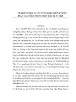
- Mạch máu nuôi tử cung là động mạch tử cung là một nhánh của động
mạch hạ vị dài 13- 15cm, lúc đầu chạy ở thành chậu bên phải, sau đó
hướng xuống dưới vào trong chui vào vùng nền của dây chằng rộng bắt
chéo mặt trước niệu quản khi cách cổ tử cung 1,5cm, sau khi bắt chéo
niệu quản động mạch chạy sát eo tử cung rồi quặt ngược lên chạy dọc bờ
ngoài tử cung tới sừng tử cung động mạch tử cung bắt chéo ở phía sau
dây chằng tròn quặt ngang ra ngoài rồi chạy dưới vòi trứng tiếp nối động
mạch buồng trứng.
Trên đường đi nó phân ra các nhánh bên và nhánh cùng:
- Nhánh niệu quản.
- Nhánh bàng quang- âm đạo.
- Nhánh cổ tử cung - âm đạo.
- Nhánh trên tử cung chạy xiên xoắn ốc vào lớp cơ tử cung.
- Nhánh đáy tử cung phát triển nhiều khi có thai rau thường bám đáy tử
cung.
+ Tĩnh mạch:
- Tĩnh mạch lớp nông chạy cùng động mạch tử cung cùng với động mạch
bắt chéo mặt trước niệu quản.
- Tĩnh mạch lớp sâu đi sau niệu quản nhận máu của bàng quang và âm
đạo cả hai tĩnh mạch nông và sâu đổ vào tĩnh mạch hạ vị.
- Bạch mạch: Tạo thành một hệ thống chi chít ở nền dây chằng rộng đổ
vào hai nhóm mạch chính là nhóm hạch cạnh động mạch chủ bụng và
nhóm hạch dọc theo động mạch hạ vị.
- Thần kinh có nhiều nhánh tách từ đám rối hạ vị chạy theo dây
chằng tử cung cùng đến eo tử cung chi phối tử cung và cổ tử cung.
PERIPARTUM HYSTERECTOMY
T. F. Baskett
HYSTERECTOMY
Emergency peripartum hysterectomy is an unequivocal marker of
severe maternal morbidity and ‘near-miss’ mortality1,2. Reviews
of published data in the past 25 years show a variable incidence,
from one in 3313 to one in 6978 deliveries4. In developed
countries, the incidence is approximately one in 2000
deliveries,with one population-based study in a Canadian province
showing an incidence of 0.53 per 1000deliveries2.Because of the
increasing Cesarean section rate world-wide and the concomitant
rise in placenta previa and placenta previa accreta,the incidence
of emergency peripartum hysterectomy is rising in many
countries. For example,in Canada from 1991 to 2000 the rate rose
from 0.26/1000 deliveries to 0.46/1000 deliveries (relative risk
1.76; 95% confidence interval 1.48–2.08)5. Compared to vaginal
delivery,emergency hysterectomy and delivery by Cesarean
section are strongly associated6,7. In addition, a recent study has
shown that multiple pregnancy had a six-fold increased risk of
emergency peripartum hysterectomy compared to ingleton
pregnancies8. Within this group,higher-order multiple pregnancies
(triplets and beyond) had an almost 24-fold increased risk of
hysterectomy8. It seems logical to conclude that the increase in
multiple pregnancy rates associated with assisted reproductive
technology provides a further contribution to the rising
peripartum hysterectomy rates.Maternal mortality rates
associated with emergency hysterectomy range from 0 to 30%,
with the higher rates in regions with limited medical and hospital
resources9. How valid these rates are today is unclear, as they
were calculated more than a decade ago. Nonetheless,even in
countries with low maternal mortality rates, associated maternal
morbidity can be high due to hemorrhage, blood transfusion,
disseminated intravascular coagulation, infection and potential
injury to the adjacent lower urinary tract7,10,11. This chapter
describes mergency hysterectomy in the immediate postpartum
period following vaginal or Cesarean delivery.
INDICATIONS
By far the most common indication for hysterectomy is
hemorrhage associated with the following conditions7,9–20.
Abnormal placentation In developed countries, placenta previa,
with or without associated accreta, is the most common indication
for hysterectomy. This is due to the rising incidence of these
conditions associated with the increasing number of women
previously delivered by Cesarean section. Despite the fact that
numerous other techniques aimed at preserving the uterus have
been proposed and are discussed in other chapters in this book,
hysterectomy is used to stem the sometimes frightening
hemorrhage associated with placenta previa or accreta in the
majority of hospitals.In addition, on rare occasions, abruptio
placentae, particularly of the concealed variety,may be
associated with such a degree of extravasation of blood into and
through the full thickness of the myometrium (Couvelaire uterus)
as to make it unresponsive to oxytocic drugs, so necessitating
hysterectomy. It must be emphasized, however, that in the
majority of cases of abruptio placentae with Couvelaire uterus the
response to oxytocic drugs is 312 appropriate and the
hemorrhage is due to disseminated intravascular coagulation
rather than
failure of the uterus to contract.
Uterine atony
As outlined elsewhere in this book (Chapter 27),the range of
modern oxytocic drugs has greatly improved the management of
uterine atony. Nonetheless, there are cases in which the uterus is
refractory to all applications of such agents. This is most
commonly found in the prolonged,augmented and/or obstructed
labor: simply stated, the exhausted and infected uterus may
respond poorly to oxytocic agents. The majority of these cases
occur at the time of Cesarean section for dystocia or
cephalopelvic disproportion.
Uterine rupture
The most common cause of complete uterine rupture is within a
previous Cesarean section scar. If the rupture is extensive and
hemorrhage cannot be contained by suture of the ruptured area,
then hysterectomy may be necessary. In addition, rupture of the
intact uterus can occur in multiparous women in response to
inappropriate use of oxytocic agents in the first and second
stages of labor.
Uterine trauma
Traumatic rupture, that is, perforation or laceration of the uterus,
can occur with a variety of obstetric manipulations, including
internal version and breech extraction in bstructed
labor;instrumental manipulation, such as the classical application
of the anterior blade of Kielland’s forceps; manual exploration of
the uterus and manual removal of the placenta or its fragments
after obstructed labor with a ballooned and thin lower uterine
segment; and during curettage for secondary postpartum
hemorrhage.
Cesarean section in the second stage of labor with the fetal head
deeply impacted in the vagina may be associated with lateral
traumatic extension of the lower uterine segment incision into the
major vessels21. On rare occasions, the extent of this tear may
necessitate hysterectomy, especially if one or both uterine
arteries is lacerated and a hematoma obscures the surgical repair.
External traumas, such as assault, a fall or motor vehicle
accident, are relatively rare causes of uterine perforation and
rupture.
Sepsis
In the era of modern antibiotics, sepsis is not a common reason
for emergency hysterectomy.However, it still may be necessary in
cases with extensive uterine sepsis, particularly with clostridial
infections and myometrial abscess formation, in which antibiotic
treatment fails to control the sepsis. Other septic causes of
secondary postpartum hemorrhage include Cesarean scar
infection and necrosis, arteriovenous fistula formation secondary
to uterine trauma and infection, and endomyometritis associated
with hemorrhage. All may rarely require hysterectomy.
SURGICAL PRINCIPLES
Although the technique of obstetric hysterectomy is similar in
principle to that of abdominal hysterectomy in gynecology,
numerous anatomical and physiological changes in pregnancy
create potential surgical difficulties.
(1) The uterine and ovarian vessels are enlarged and distended,
often markedly so, and the adjacent pelvic tissues are edematous
and friable.
(2) Abdominal entry may have been via Pfannestiel or lower
midline incision,depending on the urgency and speed required.
(3) Maneuvers to obtain immediate hemostasis will depend on the
cause of the hemorrhage. In cases of uterine rupture,Green–
Armytage clamps or sponge forceps can be used to compress the
bleeding edges of torn uterine muscle. The uterus should be
eventrated from the abdominal wound. The structures of the
adnexa on each side are pulled laterally by an assistant and the
surgeon applies straight clamps adjacent to the top sides of the
uterus to include the round ligament, the Fallopian tube and the
utero-ovarian ligament.This serves to control the collateral 313
Peripartum hysterectomy
blood flow to the uterus from the ovarian arteries. Using
transillumination, the avascular spaces in the broad ligament,
roughly opposite the level of a transverse lower Cesarean incision,
should be identified and a catheter passed through on each side
to encircle the lower uterine segment just above the cervix. This
should be twisted tightly closed with a clamp and should serve to
compress the uterine arteries. These two maneuvers should
occlude the main collateral ovarian and uterine artery supply to
the uterus.
(4) The vascular pedicles are thick and edematous and should be
double clamped.Remove the proximal clamp first and apply a free
tie and then replace the distal clamp with a transfixing suture.
The proximal free tie should ensure that there is no hematoma
formation in the base of the pedicle.
(5) If the cervix and paracolpos are not involved as the source of
hemorrhage,subtotal hysterectomy should be adequate to
achieve hemostasis and is safer, faster and easier to perform than
total hysterectomy.
However, if the lower segment and paracolpos are involved in the
hemorrhage,such as in cases of placenta previa and/or accreta,
total hysterectomy will be necessary for emostasis.
(6) Avoid the ureters by placing all clamps medial to those used to
secure the uterine arteries.
(7) It can be difficult to identify the cervix,particularly when the
hysterectomy is being done at full cervical dilatation. If there is a
Cesarean incision, a finger can be placed through this and the
cervical rim palpated. It is safest to enter the vagina posteriorly,
identify the rim of the cervix and then proceed anteriorly.
(8) The bladder is particularly vulnerable in cases previously
delivered by Cesarean section, as it may be adherent to the lower
uterine segment and cervix. It is therefore essential to check the
integrity of the bladder intraoperatively. This can be done by
manipulating the bulb of the Foley catheter to see if it is visible
through the bladder wall. The bladder also can be filled with a
colored fluid such as methylene blue or sterile milk taken from the
neonatal nursery.The latter is preferable as it does not cause
permanent staining of the tissues.
Thus, after repair of any bladder injury, it is easier to see that this
has been successful with subsequent installation of milk in the
bladder. Any tear in the bladder should be repaired with two
layers of 3/0 polyglactin (vicryl) or equivalent suture.
Otherwise,No. 1 polyglactin (vicryl) or equivalent is used
throughout the procedure.
(9) If there is any doubt about the integrity of the bladder wall or
ureters, and after repair of any bladder injury, it is wise to perform
a postoperative cystoscopy to confirm that they are intact. This
can be done by observing urine come from each ureteric orifice;
this may be facilitated by giving intravenous indigo carmine and
waiting 10–15 min.
(10) Perioperative antibiotic prophylaxis should be continued for
24–48 h.Thromboprophylaxis with heparin should be instituted as
soon as one is satisfied that hemostasis is ecure.
(11) Detailed notes should be made to include the preoperative
events, indications for hysterectomy and the surgical details.After
the initial postoperative recovery, the woman should receive a
comprehensive outline of events from an experienced
obstetrician.
In a number of series, as many as 25% of women who received an
emergency obstetric hysterectomy were primigravid, for whom
the fertility-ending nature of the procedure can
be devastating7. Therefore, particularly in this group of women,
obstetricians should be familiar with and be prepared to perform
alternative procedures to control the emorrhage. The application
of other techniques to arrest hemorrhage that can be both lifesaving and uteruspreserving are outlined in several chapters in
this book. When conditions are recognized in the antenatal period
that lead to increased risk 314
POSTPARTUM HEMORRHAGE
of severe obstetric hemorrhage, such as placenta previa and/or
accreta, referral of these cases to hospitals with the equipment
and personnel to provide the alternative techniques to
hysterectomy should be undertaken where feasible.Ultimately,
however, one has to strike a balance between spending excessive
time on alternative techniques that are proving ineffective,leading
to delay, further hemorrhage and probable disseminated
intravascular coagulation, and moving to the definitive and lifesaving hysterectomy.Such is the art of obstetric judgement in
trying circumstances.
References
1. Baskett TF, Sternadel J. Maternal intensive care and ‘near-miss’
mortality in obstetrics. Br JObstet Gynaecol 1998;105:981–4
2. Baskett TF, O’Connell CM. Severe obstetric maternal morbidity:
a 15-year population-based study. J Obstet Gynaecol 2005;25:7–9
3. Korejo R, Jafarey SN. Obstetric hysterectomy –five years
experience at Jinnah Postgraduate Medical Centre, Karachi. J
Pakistan Med Assoc1995;45:86–8
4. Yamamoto H, Sagae S, Nishik WA, Skuto R.Emergency
postpartum hysterectomy in obstetric practice. J Obstet Gynecol
Res 2000;26:341–5
5. Wen SW, Huang L, Liston RM, Heaman M,Baskett TF, Rusen ID.
Severe maternal mortality in Canada, 1991–2001. Can Med Assoc
J2005;173:759–63
6. Kacmar J, Bhinmai L, Boyd M, Shah-Hosseini R, Piepert J. Route
of delivery as a risk factor for emergency peripartum
hysterectomy: a casecontrol study. Obstet Gynecol 2003;102:141–
5
7. Baskett TF. Emergency obstetric hysterectomy.J Obstet
Gynaecol 2003;23:353–5
8. Francois K, Ortiz J, Harris C, Foley MR, Elliott JP. Is peripartum
hysterectomy more common in multiple gestations? Obstet
Gynecol 2005;105:1369–72
9. Ozumba BC, Mbagwu SC. Emergency obstetric /hysterectomy in
Eastern Nigeria. Int Surg 1991;76:109–11
10. Bakshi S, Meyer BA. Indications for and outcomes of
emergency peripartum hysterectomy.A five-year review. J Reprod
Med 2000;45:733–7
11. Engelsen IB, Albrechsten S, Iverson OE. Peripartum
hysterectomy – incidence and maternal morbidity. Acta Obstet
Gynecol Scand 2001;80:409–12
12. Lau WC, Fung HY, Rogers MS. Ten years experience of
cesarean and postpartum hysterectomy in a teaching hospital in
Hong Kong. Eur J Obstet Gynecol Reprod Biol 1997;74:133–7
13. Stanco LM, Schrimmer DB, Paul RH, Mishell DR. Emergency
peripartum hysterectomy and associated risk factors. Am J Obstet
Gynecol 1993;168:879–83
14. Tuncer R, Erkaya S, Sipahi T, Kara F. Emergency postpartum
hysterectomy. J Gynecol Surg1995;11:209–13
15. Sebitloane MH, Moodley J. Emergency peripartum
hysterectomy. East Afr Med J 2001;78:70–4
16. Sheiner E, Levy A, Katz M, Mazor M. Identifying risk factors for
peripartum cesarean hysterectomy.A population-based study. J
Reprod Med2003;48:622–6
17. Abu-Hei JA, Jawlad FM. Emergency peripartum hysterectomy
at the Princess Badeea Teaching Hospital in North Jordan. J Obstet
Gynaecol Res1999;25:193–5
18. Bai SW, Lee HJ, Cho JS, Park YW, Kim SK,Park KH. Peripartum
hysterectomy and associated factors. J Reprod Med 2003;48:148–
52
19. Chew S, Biswas A. Caesarean and postpartum hysterectomy. J
Singapore Med 1998;39:9–13
20. Castaneda S, Karrison T, Ciblis LA. Peripartum hysterectomy. J
Perinat Med 2000;28:472–81
21. Allen VM, O’Connell CM, Baskett TF. Maternal and perinatal
morbidity of caesarean delivery at full cervical dilatation
compared with caesarean delivery in the first stage of labour. Br
JObstet ynaecol 2005;112:986–90315 Peripartum
J Obstet Gynecol India Vol. 58, No. 6 : November/December 2008 pg 504-506
Original Article
Peripartum hysterectomy – A five year study
Marwaha Parveen 1, Kaur Manjeet 2, Gupta Anju 3
1 Professor 2 Associate Professor 3 Senior Resident Department of Obstetrics and
Gynecology, Government Medical College, Patiala Paper received on
19/12/2006 ; accepted on 19/09/2008
Correspondence :
Dr. Marwah Parveen # 42, Officers Colony,Patiala,Tel. 0175 2213735 Email :
Introduction
Peripartum hysterectomy has a definite role in developing countries. Inspite of
advancements in obstetrics, dai handling of obstructed labor and its complications
are quite revalent in rural India. The present study was carried out to find out the
risk factors leading to peripartum hysterectomy.Methods A retrospective analysis
of 30 cases of emergency peripartum hysterectomy was done over a period of 5
years from January 1999 to December 2003. All the risk factors, indications for
hysterectomy, fetal and maternal outcome, and operative and postoperative
complications were analyzed. Most of these cases were referred from periphery to
our tertiary institute.
Observations
There were 30 cases of cesarean hysterectomy amongst 9526 deliveries over the 5
years giving an incidence of 0.31%. The youngest woman to undergo hysterectomy
was 22 years old and the oldest was 40 years old.Twenty one (70%) of the women
were in the age group of 26 to 35 years, three (10%) were primigravidas, 15 (50%)
were primiparas and 12 (36%) were multiparas.Seventeen women (56.6%)
belonged to poor socioeconomic status. Twelve (40%) were booked cases who paid
regular visit to the hospital and had pregnancy complications like placenta previa
and fibroid uterus. Eighteen (16%) were unbooked and all 505 of them reported
were referred from periphery in /unstable condition with rupture uterus and absent
fetal heart. All the 18 had preoperative hemorrhagic shock and three of them
developed renal failure. All the booked patients were clinically stable.
Indications
Rupture uterus was the most common indication for cesarean hysterectomy seen in
18 (60%) women, all of whom were referred from peripheral rural areas within a
radius of 15 to 18 km. Out of these 18 cases, seven had previous one cesarean
section and were handled by dais with oxytocin abuse, five were in obstructed
labor,and six had prolonged and intravenous oxytocin administration by the dai.
There were three cases of bladder rupture among the 18 with rupture uterus and all
the three had a scarred uterus. In cases with previous lower segment cesarean
section rupture had occurred along the line of previous incision and had extended
laterally into the broad ligament. Of the remaining 11cases of uterine rupture, five
had vertical tear on the left side extending upto the vaginal portion of the
cervix,and in six cases left side of the uterus was involved with broad igament
hematomas and massive hemoperitoneum with the uterus lying on one side and
/the fetus lying high up in the abdominal cavity often below the
diaphragm.Morbidly dherent placenta was the second most common indication in
six (20%) women. Two of them had previous one cesarean section, one had
placenta previa with previous one lower segment cesarean section, two had
placenta accreta, and one had history of manual removal of placenta in previous
pregnancy.Atonic postpartum hemorrhage was the third indication in three (10%)
women with placenta previa. All of them were booked cases, and had major degree
type IV placenta previa. There was one (3.3%) case of traumatic postpartum
hemorrhage due to extension of previous uterine incision which ended in cesarean
hysterectomy. There were two (6.6%) cases of fibroid uterus complicating
pregnancy that were taken up for elective cesarean section with concurrent
hysterectomy.There were three maternal deaths, one because of disseminated
intravascular coagulation and two
because of irreversible hemorrhagic shock and renal failure, in cases who had
rupture bladder. There was 60% fetal mortality all of it in the 18 patients of rupture
uterus with fetus death. Thus in the rupture uterus group there was 100% fetal
mortality. In 29 cases, subtotal hysterectomy was done and in one case total
hysterectomy was performed. In two cases of cesarean section uterine artery
ligation followed by internal iliac artery ligation was performed to control
hemorrhage but ultimately hysterectomy had to be done. In three cases bladder
repair was done. Number of blood transfusions required ranged form 3 to 11
depending upon the blood loss.
Postoperative Complication: Nineteen patients had febrile morbidity, four had
paralytic ileus, six had wound infection, two had endotoxic shock, two had
renalfailure, one had deep vein thrombosis and 13 had urinaryinfection. Such a
high maternal morbidity is self explanatory.
Table 1. Reported incidences of obstetric hysterectomy.
Author Incidence
Mesleh et al (1998) 1 0.03%
Bakshi and Meyer (2002) 2 0.27%
Kastner et al (2002) 3 0.14%
Mukherjee et al (2002) 4 0.15%
Sheiner et al (2003) 5 0.048%
Baskett (2003) 6 0.53%
Parmeshwari Devi et al (2004)7 0.07%
Sahu et al (2004) 8 0.20%
Kwee et al (2005)9 0.03%
Kant and Wadhwani (2005) 10 0.26%
Present study 0.31%
Discussion
Peripartum hysterectomy is a major operation almost always an emergency one
with significant blood loss and high maternal and fetal morbidity and mortality.
Our incidence of 0.31% is comparable to other studies as shown Table 1. Ours is a
tertiary institute for referral and most of the cases are referred late. The rupture
uterus is the most common indication in our study. The comparison of indications
in various studies is shown in Table 2.
Peripartum hysterectomy
506
Table 2. Reported indications.
Gupta Mukherjee Kastner Baskett Sahu Praneshwari Kwee Kant Present
and et al 4 et al 3 (2003) 6 et al 8 Devi et al 9 and study
Ganesh et al 7 Wadhwani
(1994) 11 (2002) (2002) (2004) (2004) (2005) (2005) 10 (2005)
Rupture uterus — 38.3% — — 38.8% 23% — 36.58% 60%
Morbidly adherent — 8.4% 48.9% 50% 13.88% 26.9% 50% 12.19% 20%
placenta
Atonic PPH — 10.3% 29.8% 32.8% — 19.2% 27% 41.46% 10%
Traumatic PPH 39.4% 6.5% 4.3% — — 7.6% — — 3.3%
Pregnancy with — 0.9% — — — — — — 6.6%
fibroid uterus
The second most common indication is morbidly adherent placenta followed by
atonic PPH, traumatic PPH and term pregnancy with fibroid uterus. Rupture uterus
is a serious obstetric emergency with high maternal and perinatal mortality.
Though a common obstetric problem in developing country, it is preventable.
Occurrence of uterine rupture is significantly associated with grand multiparity,
scarred uterus, lack of antenatal care, unsupervised labor at home, injudicious use
of oxytocin, and low socioeconomic status of the women. These factors are largely
preventable. Postoperative complications like febrile morbidity, paralytic ileus,
wound infection,endotoxic shock renal failure and deep vein thrombosis are
common because of prolonged labor intrauterine manipulations, and dormant
sepsis4,5,7,8,10,12.No maternal deaths were reported by Basket6, and Mesleh et
al1 while 10% maternal deaths were reported by others 5,9,10. Emergency
obstetric hysterectomy is no doubt a life saving procedure for managing life
threatening obstetric hemorrhage and uterine rupture. This is one situation when
the surgeon is in a dilemma, in deciding about emergency hysterectomy, as a last
resort to save the life of the mother, the fetus being already lost and the mother still
young, often a primigravida or of low parity with no living child. This operation
should be made rarer by good antenatal care, of active anagement of labor, early
recognition of complications and timely performance of cesarean section when
indicated. But every obstetrician should be conversant with obstetric hysterectomy.
References
1. Mesleh R, Ayoub H, Algwiser A et al. Emergency peripartum hysterectomy. J
Obstet Gynaecol1998;18:533-7.
2. Bakshi S, Meyer BA. Indications for and outcomes of emergency peripartum
hysterectomy. A five-year review. J Reprod Med 2000;45:733-7.
3. Kastner ES, Figueroa R, Garry D et al. Emergency peripartum hysterectomy:
experience at a community teaching hospital. Obstet Gynecol 2002;99:971-5.
4. Mukherjee P, Mukherjee G, Das C. Obstetric hysterectomy – A review of 107
cases. J Obstet Gynecol India 2002;52:34-6.
5. Sheiner E, Levy A, Katz M et al. Identifying risk factors for peripartum cesarean
hysterectomy. A population based study. J Reprod Med 2003;48:622-6.
6. Baskett TF. Emergency obstetric hysterectomy. J Obstet Gynaecol 2003;23:3535.
7. Praneshwari Devi RK, Singh NN, Singh D. Emergency obstetric hysterectomy.
J Obstet Gynecol India2004;54:343-5.
8. Sahu L, Chakravertty B, Panda S. Hysterectomy for obstetric emergencies. J
Obstet Gynecol India2004;54:34-6.
9. Kwee A, Bots ML, Visser GH et al. Emergency peripartum hysterectomy. A
prospective study in The Netherlands. Eur J Obstet Gynecol Reprod
Biol2006;124:187-92.
10. Kant A, Wadhwani K. Emergency obstetric hysterectomy. J Obstet Gynecol
India 2005;55:132-34.
11. Gupta U, Ganesh K. Emergency hysterectomy in obstetrics: review of 15 years.
Asia Oceania J ObstetGynaecol 1994;20:1-5.Marwaha Parveen et al
Articles
Peripartum hysterectomy: a ten-year experience at a tertiary care hospital in a
developing country
Ferha Saeed MBBS FCPS * Roha Khalid MBBS
Abdullah Khan MBBS
*
MD
Shazia Masheer MBBS FCPS
Javed H Rizvi MBBS FACS *
*
Department of Obstetrics and Gynecology, Aga Khan University Hospital,
Karachi; Medical College, Aga Khan University Hospital, Karachi, Pakistan
Correspondence to: Shazia Masheer, Department of Obstetrics and Gynecology,
Aga Khan University Hospital, Karachi, Pakistan Email:
Acute bleeding after delivery can be a life-threatening complication. Emergency
hysterectomy is usually undertaken as a last resort. This study was conducted in
order to estimate the incidence, indications, risk factors and complications
associated with peripartum hysterectomy performed at a tertiary care hospital. We
retrospectively analysed 39 of 45 cases of emergency peripartum hysterectomy
performed at the Aga Khan University Hospital from 1997–2006. Peripartum
hysterectomy was defined as one performed for a haemorrhage after delivery which
is unresponsive to other treatments. The most frequent indications for peripartum
hysterectomy were morbidly adherent placenta (46%) and uterine atony (23%). The
duration of surgery was shorter (P = 0.045) but the complications were higher (P =
0.029) in total compared with subtotal hysterectomies. Our results suggest that
caesarean deliveries are associated with an increased risk for peripartum
hysterectomy, which is of concern given the increasing rate of caesarean deliveries.
Subtotal hysterectomy is a reasonable alternative in emergency obstetric
hysterectomy.
Department of Epidemiology, University of Washington, Seattle, Washington,
USA.
Obstetrics and Gynecology [2009, 114(1):115-23]
Type: Journal Article
Abstract
Highlight Terms
Gene Ontology(1)
Diseases(6)
OBJECTIVE: To identify factors associated with peripartum hysterectomy performed within 30
METHODS: This was a population-based case-control study using Washington State birth certifi
and mode of delivery and 95% confidence intervals (CIs) were computed.
RESULTS: There were 896 hysterectomies. Incidence rates ranged from 0.25 in 1987 to 0.82 per
not. As compared with vaginal delivery, vaginal delivery after cesarean (27 cases compared with
CONCLUSION: Incidence rates of peripartum hysterectomy are increasing over time. The most
LEVEL OF EVIDENCE: II.
Current studies
Peripartum hysterectomy
Background
Severe obstetric haemorrhage is a leading cause of severe maternal morbidity in Australia and
remains a cause of maternal death. One-off studies on peripartum hysterectomy have shown that
the incidence is increasing1 and that there is an association between prior caesarean delivery and
the need for peripartum hysterectomy.2 The caesarean section rate in Australia continues to
increase, with the most recent figures showing that over 30% of women gave birth by this mode
in 2005, compared with less than 20% in 1996.3 There is an urgent need to explore the
epidemiology and management of peripartum hysterectomy in Australia.
Research Questions
1.
What is the current incidence of peripartum hysterectomy in Australia?
2.
What are the risk factors for peripartum hysterectomy in Australia?
3.
How is severe obstetric hemorrhage resulting in peripartum hysterectomy managed in
Australia?
4.
What are the outcomes for both the woman and the infant when a pregnancy results in
peripartum hysterectomy in Australia?
Method
Prospective, case-control study using monthly negative surveillance system of all birthing
services in Australia (>50 births) – AMOSS. Nominated clinicians and midwives within each
maternity unit will be e-mailed a simple tick-box to indicate whether a case occurred or whether
there is ‘nothing to report’. If a case arose, the reporting clinician will complete a case form
using the secure web-based data system. The clinician/midwife will also complete two control
forms using the secure web-based data system. Only non-identifiable data will be collected.
Surveillance Period
January 2010 - June 2011
Case Definition
The cases will be all women in Australia identified as having a peripartum hysterectomy using
the following definition:
EITHER any woman whose pregnancy terminates and who has a hysterectomy in the same
clinical episode or within six weeks postpartum when the indication for hysterectomy is related
to the pregnancy e.g. secondary postpartum haemorrhage
OR any woman giving birth and undergoing a hysterectomy in the same clinical episode or
within six weeks postpartum when the indication for hysterectomy is related to the birth e.g.
secondary postpartum haemorrhage
Control Selection
The two births immediately prior to the case, in the same hospital.
Study Size
The study will run for 18 months. The estimated sample size for this time duration is 330 cases
based on the Victorian obstetric haemorrhage and associated hysterectomy study conducted for
the years 1999 – 2002.1 The incidence of peripartum hysterectomy was shown to be increasing
consistently over these years, with 48 hysterectomies performed in Victoria in 2002, from a pool
of 61 959 maternities; 7.7 per 10,000 maternities. This is a larger sample estimate than that made
based on the UKOSS results: between 100 and 130 cases based on the UKOSS results of 4.1
(95%CI 3.6 to 4.5) per 10,000 maternities.2
References
1.
Haynes, K., C. Stone, and J. King, Major Conditions Associated with Childbirth in
Australia: Obstetric Haemorrhage and Associated Hysterectomy. 2004, Department of
Human Services: Melbourne.
2.
Knight, M., et al., Cesarean delivery and peripartum hysterectomy. Obstetrics &
Gynecology, 2008. 111(1): p. 97-105.
3.
Laws, P.J., et al., Australia's mothers and babies 2005, in Perinatal statistics series no. 20.
Cat. no. PER 40. 2007, AIHW National Perinatal Statistics Unit: Sydney.
EDITORIAL COMMENT
Peripartum Hysterectomy Risk Factors in Taiwan
Ming-Jie Yang, Peng-Hui Wang*
Department of Obstetrics and Gynecology, Taipei Veterans
General Hospital, National Yang-Ming UniversityHospital, and
Institute of Clinical Medicine, National Yang-Ming University
School ofMedicine, Taipei, Taiwan, R.O.C.© 2010 Elsevier Taiwan
LLC and the Chinese Medical Association. All rights reserved.
*Correspondence to: Dr Peng-Hui Wang, Department of Obstetrics
and Gynecology, Taipei Veterans General Hospital, 201, Section 2,
Shih-Pai Road, Taipei 112, Taiwan, R.O.C.
E-mail: ● Received: March 28, 2010 ●
Accepted: May 25, 2010
In addition, nearly half of all deliveries (46.7%) in this study were
by cesarean section,5 which may also be a risk factor, although
the authors did not mention it.
The presence of an attendant at every birth and access to
emergency obstetric care are key to reducing maternal morbidity
and mortality in the developing world,although esource-rich
countries have a rising cesarean section rate with its consequent
effect on the incidence of abnormal placentation and its link with
peripartum hysterectomy.9 In fact, cesarean section is the single
most important factor resulting in peripartum
hysterectomy,because women undergoing primary cesarean
section had the highest peripartum hysterectomy rate, with an
adjusted odds ratio of 12.13 (95% confidence interval, 8.30–
17.14), compared with women undergoing vaginal delivery.2 The
risk from primary cesarean section is even more severe than that
of repeated cesarean sections. This may indicate that the
incidence of peripartum hysterectomy may increase significantly
in the near future; if the age of women during pregnancy
continues to increase, the primary cesarean section rate will
continue to rise.
References
1. Wise A, Clark V. Challenges of major obstetric
haemorrhage.Best Pract Res Clin Obstet Gynaecol 2010;24:353–
65.
2. Jou HJ, Hung HW, Ling PY, Chen SM, Wu SC. Peripartum
hysterectomy in Taiwan. Int J Gynaecol Obstet 2008;101:269–72.
3. Turner MJ. Peripartum hysterectomy: an evolving picture. IntJ
Gynaecol Obstet 2010;109:9–11.
4. Ozden S, Yildirim G, Basaran T, Gurbuz B, Dayicioglu V.Analysis
of 59 cases of emergent peripartum hysterectomies during a 13year period. Arch Gynecol Obstet 2005;271:363–7.
5. Yalinkaya A, Guzel AI, Kangal K. Emergency peripartum
hysterectomy:16-year experience of a medical hospital. J Chin
MedAssoc 2010;73:360–3.
6. Leridon H, Slama R. The impact of a decline in fecundity and of
pregnancy postponement on final number of children and demand
for assisted reproduction technology. Hum Reprod2008;23:1312–
9.
7. Schmidt L. Should men and women be encouraged to start
childbearing at a younger age? Expert Rev Obstet Gynecol
2010;5:145–7.
8. Cheng MH, Wang PH. Placentation abnormalities in the
pathophysiology of preeclampsia. Expert Review Mol
Diagn2009;9:37–49.
9. Hsu TY. Abnormal invasive placentation—placenta previa
increta and percreta. Taiwan J Obstet Gynecol 2009;48:1–2.400
Abstract
The aim of this study was to estimate incidence, indications and complications of peripartum
hysterectomy in Apex Hospital of the Kashmir valley. We analyzed 100 cases of emergency
cesarean hysterectomies performed in our hospital from January 2001 to December 2002. The
incidence of emergency hysterectomy was 2.6 per thousand deliveries. Most common indication
for emergency hysterectomy was uterine rupture (30%), followed by placenta previa (25%),
uterine atony (21%) and placenta increta/accerta/percreta (8%). Majority of uterine rupture cases
were late referrals from rural areas. The commonest postoperative complication was fever (27%),
followed by lower respiratory tract infection (10%), wound infection (8%), and bladder injury
(8%). Maternal mortality following emergency hysterectomy was 3%, and the cause of death was
related to complications like shock, septicemia and disseminated intravascular coagulation
(DIC).
Introduction
Emergency hysterectomy is carried out as life saving procedure. Even today, 8-10% of maternal
mortality, in developing countries, directly occurs due to massive obstetrical hemorrhage 1.
Emergency peripartum hysterectomy that occurs after vaginal delivery, or at the time of cesarean
births, is usually reserved for situations where conservative measures do not control hemorrhage.
Most common indication for emergency peripartum hysterectomy has been uterine atony and
uterine rupture2,3. Recently the most common reported indication is placenta accerta, and is most
likely related to increase in number of cesarean deliveries observed over the past two decades
4 5 6 7 8
, , , , . The purpose of our study was to estimate incidence, indications, and postoperative
complications associated with emergency hysterectomy in this part of the world.
Methods
The present prospective study was carried out in Lalla-Ded Hospital, which is one of the main
hospitals associated to Government Medical College Srinagar, Kashmir and is the only referral
maternity care hospital at present catering the whole Kashmir valley. A random sample of first
100 women who underwent caesarean hysterectomy for various indications between January
2001 to December 2002 was studied. Maternal characteristics like age, parity, residence, and any
previous cesarean delivery were recorded. The indication for surgery, type of hysterectomy,
postoperative complications, any need for blood transfusion, and pregnancy outcome were
obtained. Data thus collected was subjected to appropriate statistical analysis.
Results
From January 2001 to December 2002, 45460 normal vaginal deliveries, 10139 cesarean
deliveries, and 146 emergency cesarean hysterectomies were performed. We analyzed 100
randomly selected samples out of 146 cases of emergency cesarean hysterectomy. The incidence
of emergency cesarean hysterectomy was 2.6 per thousand deliveries. A total of 36% patients
were from urban areas while 64% belonged to rural areas (Table 1).
Table 1: Incidence of cesarean hysterectomies
The proportion of patients requiring emergency hysterectomy increased from 5% in 20-24 years
age group to 42% in 35-39 years, and then showed decline at or above 40 years (Table 2).
Of the patients who underwent emergency cesarean hysterectomy, 64 were of Para 3 (40%) and
para 4 (24%); parity distribution showed a fall in the number of hysterectomies from 40% in Para
3 to 2% in Para 7 (table3). There were 3% primigravida who underwent cesarean emergency
hysterectomy.
Table 2: Age distribution of patients requiring emergency hysterectomy (n=100)
The most common indication for emergency cesarean hysterectomy was rupture of the uterus
(30%), all the cases being rural referrals, followed by placenta previa (25%), atonic uterus
(21%), accidental hemorrhage (12%), and placenta increta/acreta (8%)
Table 3: Parity distribution of patients who underwent emergency Cesarean hysterectomy
(n=100)
In 97% of cases subtotal hysterectomy was carried out, whereas 3% patients required total
hysterectomy. Average length of hospital stay in those patients who underwent cesarean
hysterectomy was 10-15 days.
Table 4: Indications for emergency hysterectomy (n=100)
Fever was the most common complication (27%), followed by urinary tract infection (12%),
bladder injury (8%), wound infection (8%), lower respiratory tract infection(10%), and life
threatening septicemia (1%). Perinatal mortality was 43%, and maternal mortality was 3% in
present study.
Discussion
Cesarean hysterectomy has undergone tremendous change, both in terms of the indications and
frequency of the procedure. Obstetric cesarean hysterectomy is mostly done for indications
deemed to be serious and life threatening to the patient, and not amenable to conservative
management.
Present study describes maternal mortality, morbidity, etiology, and fetal outcome of 100 patients
who underwent cesarean hysterectomy in our hospital in a period of about 2 years.