Nghiên cứu căn nguyên, đặc điểm dịch tễ học lâm sàng, cận lâm sàng và yếu tố tiên lượng bệnh viêm não cấp ở trẻ em Việt Nam (TTTA)
Bạn đang xem bản rút gọn của tài liệu. Xem và tải ngay bản đầy đủ của tài liệu tại đây (251.85 KB, 27 trang )
1
INTRODUCTION
Acute encephalitis is an acute inflammatory condition of the brain
parenchyma, presents as diffuse or focal neuropsychological dysfunction.
It occurs in all parts of the world, at any age but the incidence is higher in
children. This is a serious medical condition that is life-threatening and a
serious public health problem because of the high morbidity and
mortality. The diagnosis of encephalitis in the world and Vietnam in the
past was difficult because there is no clear standard so in 2013 the
international encephalitis association has officially agreed on the
diagnosis of encephalitis.
In Vietnam, there has been no research has been carried out by the
new diagnostic criteria for encephalitis of “ the consensus statement of
international encephalitis consortium 2013” and not much research on
comprehensive assessment of the causes and predictors of acute
encephalitis in children. On the other hand, thanks to the advances in
molecular biology testing of infectious diseases in Vietnam, the etiology
of acute encephalitis has been determined more and more accurately. So
we conducted this thesis “The study of etiology, clinical epidemiology,
subclinical characteristics, and prognostic factors of acute
encephalitis in Vietnamese children” with the following objectives:
1. Identification of microbiological causes of acute encephalitis in
children ≥ 1 month at the Vietnam National children’s hospital from
1/2014 to 12/2016.
2. Describe the clinical epidemiological characteristics of acute
encephalitis in children according to some common causes.
3. Identify some of the major predictors of acute encephalitis due to
common causes in children.
THE NECCESITY OF THE THESIS
Acute encephalitis is a disease caused by a variety of causes, in
which the causes are largely determined by viral infections. However, the
percentage of undetermined causes remains high even in the developed
world.
Early diagnosis as well as proper identification of causal factors and
prognostic factors of acute encephalitis in children contributes to proper
monitoring and treatment, reducing the mortality and sequelae of acute
encephalitis. It also helps policymakers develop effective disease
prevention plans. That is why this thesis is urgent and of practical value.
2
NEW CONTRIBUTIONS OF THIS THESIS
This is the first time the thesis applies the international consensus
criteria for the diagnosis of encephalitis in 2013 and provides relatively
comprehensive information on the etiology, clinical epidemiology and
prognostic factors of acute encephalitis in children. Research results
show that:
+ The rate of definite cause of acute encephalitis has reached 57,6%
and the possible etiology is 6,7%. In this thesis for the first time in
Vietnam we mentioned the causes of encephalitis that are found outside
the cerebrospinal fluid (CSF)
+ There are many causes of encephalitis for the first time in Vietnam
such as Rickettsia, Human herpes Virus 6 (HHV6) and possible causes
such as influenza B, M. pneumonia, Rotavirus, Respiratory syncytial
virus (RSV)
+ The cause of acute encephalitis at the youngest age is
S.pneumoniae and the largest is Japanese encephalitis virus
+ The most localized seizure is encephalitis due to Herpes simplex
virus (HSV), major febrile seizures are mainly caused by Japanese
encephalitis (JE)
+ Cause of disease is one of the important predictors in which unknow
causes of acute encephalitis has the highest mortality rate with 15,6%.
Encephalitis caused by HSV had the highest rate of sequelae with 46,8%.
+ The thesis investigated five major predictors of JE: mechanical
ventilation, glasgow score at admission ≤ 8, glasgow decrease after 24
hours of hospitalization, muscle tone dysfuntion, abnormal on Magnetic
resonance imaging (MRI) brain and can not find independent factor in
multivariate analysis.
+ The study identified four major predictors for Herpes simplex
encephalitis: mechanical ventilation, glasgow score at admission ≤ 8,
muscle tone dysfuntion, convulsions > 5 times/day.
Factor of
convulsions > 5 times daily is independent factor after multiple
regression analysis
+ There were 5 severe prognostic factors in patients with
pneumococcal encephalitis: mechanical ventilation, glasgow score at
admission ≤ 8, muscle tone dysfuntion, platelet counts in the blood <
150 (G/l), protein in CSF > 5g/l and no independent factor was found in
multivariate analysis.
+ There were five major predictors of unidentified encephalitis:
mechanical ventilation, Glasgow score at admission < 8 points, glasgow
3
decrease after 24 hours, seizure > 5 times/day, muscle tone dysfuntion,
abnormal images on computed tomography (CT) scan. No independent
factor was found in multivariate analysis.
THESIS LAYOUT
There are 139 pages in this thesis, including: 2 pages of
introduction, 3 pages of conclusions, 1 page of recommendations, and 4
Chapters: Literature review (32 pages), Subjects and Methods (22 pages),
Results (36 pages), and Discussions (43 pages). The Thesis contains 38
Tables, 4 Pictures, 4 Firgures, 2 Algorithms, and 158 Referrences (13
Vietnamese and 145 English documents).
CHAPTER 1: LITERATURE REVIEW
1.1. Epidemiology and causes of acute encephalitis
The incidence of encephalitis in the world is difficult to assess due to
differences in definitions and reporting systems. Geographic factors such
as climate, the presence of disease or vectors as well as local vaccination
programs affect the incidence of acute encephalitis in each part of the
world. However, due to the lack of diagnostic criteria, the rate of acute
encephalitis and acute encephalitis in even the United States is not yet
clear and certain.
In Vietnam, the incidence of encephalitis at community level is not
accurate, the prevalence is higher in children than in adults and males
than in females, and usually occurs in the summer.
Other causes of acute encephalitis have been identified such as those
identified as JE, HSV, EV, measles, rubella, Cytomegalovirus (CMV),
Epstein–Barr virus (EBV), Varicella-zoster virus (VZV), mumps, Human
immunodeficiency virus (HIV)bacteria, and some parasites,
autoimmune ... However, the number of cases of acute encephalitis has
not determined the root cause is still relatively high proportion.
Since 2014, in Vietnam national children’s hospital applied the
international consensus criteria for the diagnosis of encephalitis in 2013
from which many bacterial encephalogenic causes have been identified
such as pneumococcal, H. influenzae, Staphylococcus, Escherichia coli.
1.2. Clinical, subclinical, and prognostic factors of acute encephalitis
Symptoms of acute encephalitis are usually age-related, the smaller
age the symptom are more nonspecific symptoms with fever and other
symptoms such as headache, nausea may be encountered in both
4
bacterial and viral causes of encephalitis, acute encephalitis as well as
meningitis.
All patients with suspected acute encephalitis should do puncture the
cerebrospinal fluid as soon as possible after admission. MRI scans should
be performed within 24 hours of admission. With changed CSF, clinical
symptoms and suggestive images on MRI can diagnosis acute
encephalitis.
The prognosis factors of acute encephalitis patients depends on
many factors such as the timing of the diagnosis, the patient's immune
status, the level of modern medicine, the cause of the disease, the age,
clinical and subclinical symptoms, as well as genetic characteristics of
the patient.
CHAPTER 2: MATERIALS AND METHODS
2.1. Study subjects
Study subjects included 186 children > 1 month that diagnosed acute
encephalitis at the Vietnam National children’s hospital from January
2014 to December 2016.
2.1.1. Inclusion criteria
2.1.1.1. Tiêu chuẩn chẩn đoán viêm não cấp (adapted from International
Encephalitis Consortium 2013)
Major inclusion criteria (required)
Patients presenting to medical attention with altered mental status
(defined as decreased or altered level of consciousness, lethargy or
personality change) lasting ≥24 h with no alternative cause identified.
Minor inclusion criteria: (2 required for possible encephalitis; ≥3
required for probable or confirmed encephalitis)
- Documented fever ≥38° C (100.4°F) within the 72 h before or after
presentationb
- Generalized or partial seizures not fully attributable to a
preexisting seizure disorderc
- New onset of focal neurologic findings
- CSF WBC count ≥5/cubic mmd
- Abnormality of brain parenchyma on neuroimaging suggestive of
encephalitis that is either new from prior studies or appears acute
in onset
2.1.1.2. Criteria diagnosis the cause of acute encephalitis
a./ The determined cause of acute encephalitis
5
There is evidence of viruses, bacteria, immune factors based on
PCR or ELISA specific IgM positive results for each virus, bacterium
and specific antibodies in CSF positive.
b./ The possible cause of acute encephalitis
Identification of causative agents based on specimens outside CSF
by culture methods, PCR, ELISA, autoimmune factors in body fluids:
blood, endotracheal fluid, urine, stool…
2.1.1.3. Exclusion criteria: with one of the following criteria
Acute encephalitis due to poisoning
Acute encephalitis due to metabolic disorders
Brain injury in patients with renal failure
Brain injury in patients with liver failure
Cases of insufficient data
2.2 Methods
A cross-sectional descriptive study of all eligible pediatric acute
encephalitis patients admitted to hospital from January 2014 to
December 2016 was included in the study.
2.3. Statistical analysis
SPSS software 22.0 was used to analyze these data.
Chi - square test was used to compare the ratios and correlations
between two quantitative variables. For quantitative variables with
standard distribution: Using Studen's t test, One way ANOVA to compare
the differences. For non-standard distribution quantitative variables: the
Mann-Whithney U test, the Kruskal-Walis H test was used. Comparison
paired test was used to compare quantitative data for the same patient.
The use of logistic regression and multivariate logistic regression
was used to find the relationship between risk factors and treatment
outcomes.
2.4. Research ethics
Conducting research does not affect the diagnosis and treatment
process; do not have any harm to the patient, but only conduct additional
etiological tests on the patient's specimen - if further confirmation of the
6
cause is beneficial for diagnosis, treatment and prognosis. patient.
The research work was approved by the Vietnam national
children’s hospital and HaNoi medical university.
All personal information of research subjects are kept
confidentially
CHAPTER 3: RESULTS
Over 3 years of study, we collected 861 encephalitis patients eligible for
the study
3.1. The causes of acute encephalitis
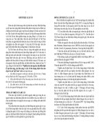
3.1.1. The ratio define the cause
Confirmed cause
35.7
Probable cause
Unknow cause
57.6
6.7
Figure 3.1. The ratio define the cause of acute encephalitis
Comment: 496 (57,6%) patients identified the cause of acute
encephalitis, 6,7% probable cause and 35,7% unknow cause of acute
encephalitis
Table 3.1: Distribution of causes of acute encephalitis
Causes
Confirmed
n
%
Probable
n
%
Total
n
%
7
Virus
403
81,3
26
44,8
429
Bacteria
89
17,9
16
27,6
105
Parasite
4
0,8
0
0
4
Autoimmune
0
0
16
27,6
16
Total
496
100
58
100
554
Comment: The virus accounted for the highest rate of 77,5% of
81,3% of the confirmed causes and 44,8% of the probable causes.
77,5
18,9
0,7
2,9
100
which
8
3.1.2. Distribution of causes of microbiology in acute encephalitis
Table 3.2: Distribution the causes of encephalitis by virus
Confirmed
Probable
Total
(n=403)
(n=26)
(n=429)
Causes
n
%
n
%
n
%
JE
312
77,4
0
0
312
72,7
HSV
75
18,6
2
7,7
77
17,9
EV
5
1,2
1
3,8
6
1,4
VZV
1
0,2
5
19,2
6
1,4
EBV
3
0,7
1
3,8
4
0,9
Mumps
0
0
4
15,4
4
0,9
Rabbit
3
0,7
0
0
3
0,7
CMV
0
0
3
11,5
3
0,7
Rotavirus
0
0
3
11,5
3
0,7
Measles
1
0,2
1
3,8
2
0,5
RSV
0
0
2
7,7
2
0,5
HIV
0
0
2
7,7
2
0,5
Dengue virus
0
0
1
3,8
1
0,2
HHV6
1
0,2
0
0
1
0,2
Influenzae B
0
0
1
3,8
1
0,2
VNNB/VZV
1
0,2
0
0
1
0,2
VNNB/EV
1
0,2
0
0
1
0,2
Comment: JE virus is the most common cause of acute encephalitis
among viruses accounting for 72,7%, HSV is the second virus cause
acute encephalitis accounts for 17,9%
Table 3.3: Distribution the cause of encephalitis by bacteria
Confirmed
Probable
Total
(n=89)
(n=16)
(n=105)
Causes
n
%
n
%
n
%
S.pneumoniae
56
62,9
1
6,2
57
54,3
Tuberculosis
23
25,9
8
50
31
29,5
S.aureus
4
4,5
2
12,5
6
5,7
H.influenzae
3
3,4
1
6,2
4
3,8
Rickettsia
1
1,1
1
6,2
2
1,9
M.pneumonia
0
0
2
12,5
2
1,9
e
Syphilis
1
1,1
0
0
1
0,9
E.coli
1
1,1
0
0
1
0,9
9
M.catahalis
0
0
1
6,2
1
0,9
Comment: S.pneumoniae is the most common cause bacteriae of acute
encephalitis 54,3%. Tuberculosis is the second leading cause bacteriar of
encephalitis with 29,5%.
3.2. Clinical epidemiological characteristics of acute encephalitis in
children by some common causes
3.2.1. Some epidemiological characteristics by common causes
3.2.1.1. Distribution of the cause of acute encephalitis by month
160
140
120
100
80
60
40
20
0
Unknown cause (n=307)
JE
HSV (n=77)
S.pneumoniae (n=57)
Jan
Fer
Mar Apr May
Jun
Jul
Aug
Sep
Oct Now Dec
Figure 3.2: Distribution of the cause of acute encephalitis by month
Comment: Acute encephalitis caused by JE virus causes seasonal illness
with the highest number of patients in June, July and August, especially
in June each year. The others cause encephalitis causes sporadic all
months by year.
3.2.1.2. Distribution of causes of acute encephalitis by sex
100%
80%
60%
40%
20%
0%
36.1
46.8
31.6
35
63.9
53.2
68.4
65
Male
Female
10
Figure 3.3: Distribution of causes of acute encephalitis by sex
Comment: The causes of encephalitis caused by JE, pneumococcus and
unknown cause are more common in male than female.
3.2.1.3. Age distribution of causes of acute encephalitis
Table 3.4: The average age of patients with acute encephalitis by cause
The average age
Median
Min-Max
n
Causes
(Year)
(Year)
JE
312
5,7
0,13-15,75
HSV
77
1,3
0,29-9,58
S.pneumoniae
57
0,7
0,21-11,25
Unknown cause
307
4,0
0,13-15,29
Total
861
3,5
0,13-15,75
Comment: JE has the highest median age was 5,7 years old, S.pneumonia
and HSV has the lowest median age of 0,7 years and 1,3 years.
3.3.2. Clinical characteristics of acute encephalitis by cause
3.3.2.1. Glasgow score by cause at admission
Table 3.5: The average Glasgow score by cause at admission
Causes
n
The average Glasgow score
JE (n=312)
312
10,12 ± 1,64
HSV (n=77)
77
10,25 ± 1,51
S.pneumoniae (n=57)
57
9,39 ± 1,64
Unknown cause (n=307)
307
10,01 ± 2,07
Comment: Patients with acute encephalitis due to S.pneumonia has the
lowest Glasgow score at admission 9.39 ± 1.64 scores.
3.3.2.2. Signs of convulsions by cause
Bảng 3.6: The characteristic of convulsion by cause
Convulsions
Generalized
Local
5 times/day
Causes
n
%
n
%
n
%
JE (n=222)
162
51,9
60
28
8,9
HSV (n=76)
21
27,3
55
35
S.pneumoniae (n=43)
15
26,3
28
19,
2
71,
5
49,
1
45,
5
5,3
3
11
Unknown cause (n=226)
149
48,5
77
25,
53
17,
1
3
p
<0,001
<0,001
<0,001
Comment: Generalized convulsions has the highest rate in JE group,
accounting for 51,9%. Local convulsions are most common in acute
encephalitis due to HSV accounting for 71,5%. Differences in signs of
generalized convulsions, localized convulsions and times of convulsions
> 5 times daily by the causes were different with p < 0,001.
3.3.2.3. Other neurological signs
75.7
80
70
60
50
40
30
20
10
0
%
74.4
54.4
36.8
JE
12
=3
n
(
)
e
nia
o
m
eu
pn
.
S
7)
=5
n
(
U
n
ow
n
nk
e
us
ca
7)
30
=
(n
V
HS
7
=7
(n
)
Figure 3.4: Signs of neck stiffness by cause
Comment: 75,7% JE, 74,4% S.pneumoniae, 36,8% HSV has signs of
neck stiffness
Table 3.7: Symptoms of muscle tone dysfunction by cause
Signs
Normal
Hypertonic
Hypotonic
n
%
n
%
n
%
JE (n=312)
158
50,6
134
42,9
20
6,4
HSV (n=77)
26
33,8
42
54,5
9
11,7
Causes
12
S.pneumoniae (n=57)
21
36,8
32
54,3
5
8,8
Unknown cause (n=307)
150
48,9
134
43,6
23
7,5
p
<0,05
<0,05
<0,05
Comment: Signs of hypertonic are the most common in patients with
acute encephalitis, HSV and S.pneumoniae with 54,5% and 54,3%,
respectively. Signs of hypotonic is the most common in patients with
acute encephalitis due to HSV with 11,7%
%
80
70
60
50
40
30
20
10
0
59.7
58.4
39
36.1
18.8
8
2.9
Normal
Hemiplegia
25.6
Quadriplegia
Paraplegic
Unknown
cause
(n=307);
2.3 1
Unknown
cause
(n=307);
HSVHSV
(n=77);
(n=77);
S.pneumoniae
0 75.9
S.pneumoniae
0
(n=57);
(n=57);
71.20 0
Figure 3.5: Signs of paralysis by cause
Comment: Signs of hemiplegia are the most common in the group of
acute encephalitis due to HSV 59,7%, JE 36,1%.
3.3.2.4. Management of respiratory failure by cause
Table 3.8: Management of respiratory failure by cause
Causes
JE (n=312)
HSV (n=77)
S.pneumonia (n=57)
Unknown cause (n=307)
p
Mechanical
ventilation
n
%
57
18,3
15
19,5
21
36,8
88
28,7
< 0,001
Oxygen by
mask
n
%
46
14,7
21
27,3
22
38,6
39
12,7
< 0,001
Total
n
%
103
33
36 46,8
43 75,4
127 41,4
<0,001
13
Comment: Patients with S. pneumonia had the highest rate of respiratory
failure with 36,8% mechanical ventilation and 38,6% oxygen. The
difference between respiratory distress, mechanical ventilation and
oxygen intake among groups different with p < 0,001.
14
3.3.3. Subclinical signs of acute encephalitis
3.3.3.1. The ratio of changed CSF by cause
Table 3.9: The ratio of changed cells in CSF by cause
Cells in CSF
(cells/mm3)
Normal
5-100
Causes
JE (n=312)
n
%
n
68
HSV (n=77)
25
20
8
50
S.pneumoniae (n=57)
5
21,
8
32,
5
8,8
Unknown cause (n=307)
18 60,
5
3
<0,001
p
>100-500
>500
%
n
%
n
%
66,
7
64,
9
21 36,
8
10 34,
6
5
<0,001
35
11,
2
2,6
1
0,3
0
0
28,
1
3,9
15
26,
3
1,3
2
16
12
<0,001
4
<0,001
Comment: acute encephalitis with unknown cause had 60,3% with no
changed the number of cell in CSF. The number cells in CSF > 500
cells/mm3 were found mainly in patients with pneumococcal at 26,3%.
The number of cells with different causes in CSF with p < 0,001.
Table 3.10: The ratio of changed protein in CSF by cause
Protein CSF
(g/l)
Normal
>0,45 – 1
>1 – 5 g/l
> 5g/l
Causes
JE (n=310)
n
%
n
%
n
%
n
%
78
32
0
33,8
10
10,
3
13
0
41
20
0
26
64,5
HSV (n=77)
0
0
S.pneumoniae (n=57)
1
25,
2
53,
2
1,8
3
5,3
39
14
Unknown cause (n=298)
17
6
59,
1
82
27,5
37
68,
4
12,
4
24,
6
1
3
15
p
<0,001
<0,001
<0,001
<0,001
Comment: Unknown cause acute encephalitis had 59,1% without
changed protein in DNT. Protein in CSF increased > 5g/l are the most in
the group of pneumococcal accounted for 24,6%. Protein changes in CSF
in different cause were different with p < 0,001.
3.3.3.3. Imaging of lesion in CT scans by cause
b./ Imaging of lesion in CT scans by cause
Table 3.11: Imaging of lesion in CT scans by cause
Causes
JE
HSV
S.pneumoniae Unknow
n=92
n=24
n=25
n causes
p
n=108
Lesion
n
Unnormal (≥ 1 27
location)
Cerabral
15
edema
Temporal lobe 2
injury
Parietal lesion
1
Frontal
lobe 1
injury
Occipital
0
lesions
Basal ganglia 2
lesions
Thalamic
6
injury
Brain
stem 1
lesion
Abcess
0
Dilated
0
ventricular
Infarction
1
Hemorrhage
0
%
29,3
n
20
%
83,3
n
13
%
52
n
39
%
36,1 < 0,001
16,3
6
25
1
4
16
14,8
<0,05
2,2
10
41,7
0
0
2
1,9
<0,05
1,1
1,1
2
2
8,3
8,3
0
4
0
16
1
1
0,9
0,9
>0,05
>0,05
0
1
4,2
0
0
2
1,9
>0,05
2,2
0
0
1
4
4
3,7
>0,05
6,5
1
4,2
1
4
4
3,7
>0,05
1,1
0
0
0
0
1
0,9
>0,05
0
0
0
0
0
0
1
4
4
16
0
7
0
6,5
>0,05
>0,05
1,1
0
0
3
0
12,5
1
0
4
0
0
2
0
1,9
>0,05
>0,05
16
Comment: The cerebral edema was the highest in patients with HSV and
JE accounted for 25% and 16,3%. temple lobes, parietal lobes, frontal
lobes and occipital lobes lesion are mainly affected by HSV encephalitis:
41,7%, 8,3%, 8,3% and 4,2%. Brain cerebral edema and temporal lobe
lesions on CT scans differed between groups with p < 0,05.
c./ Imaging of lesion in MRI by cause
Table 3.12: Imaging of lesion in MRI by cause
Causes
Lesion
JE
n=235
HSV
n=72
S.pneumonia
e
n=42
n
%
Unknown
causes
n=225
n
%
n
%
n
%
Unnormal (≥ 1 15
location)
3
Temporal
lobe 29
injury
Parietal lesion
20
65,
1
12,
3
8,5
57,1
138
61,3
3
7,1
45
20
3
7,1
24
10,7
12
12
11
26,2
15
6,7
Occipital lesions
10
4,3
0
0
12
5,3
Brain stem lesion
5
2,1
97,
2
70,
8
29,
2
13,
9
13,
9
0
24
Frontal lobe injury
7
0
5
1
2
1
1
0
1
0
0
0
0
6
2,7
Cerebellum lesion
0
0
0
0
0
0
8
3,6
Basal
ganglia lesions
32
1
1,4
3
7,1
31
13,8
Thalamic injury
1
7
2
23,
6
2,8
2
4,8
37
16,4
White matter lesion
11
4
4
13,
6
48,
5
1,7
5
11,9
14
6,2
Grey matter lesion
1
4
0
0
1
2,4
0
0
9,4
8
11,
1
1
2,4
31
13,8
Cortical brain lesion 22
p
<0,00
1
<0,00
1
<0,00
1
<0,00
1
>0,00
5
>0,00
5
<0,00
1
<0,05
<0,00
1
<0,00
1
<0,00
1
>0,05
Comment: Temple lobes and parietal lobes lesion are common in patients
with HSV encephalitis accounted for 70,8% and 29,2%. Thalamic lesion
was the most common in JE accounted for 48,5%. Lesion of temporal
lobes, parietal lobes, frontal lobes, cerebellum, central gray nucleus,
thalamus and white matter differ between different groups by causes.
17
3.4. Predictor of factors of acute encephalitis in children
3.4.1. Treatment results by cause
Table 3.13: Treatment results by cause
Results
Severe
Mild
Died
sequalae sequalae
n
% n %
n
%
Causes
Good
recover
n
%
JE (n=312)
10 3,2 78 25
70 22,4 154 49,4
HSV (n=77)
3
3,9 36 46,8 18 23,4 20 26
S.pneumoniae (n=57)
8 14,0 17 29,8 5
8,8 27 47,4
Unknown
cause 48 15,6 59 19,2 60 19,5 140 45,6
(n=307)
p
< 0,001
Comment: Acute encephalitis with unknown cause had the highest
mortality rate of 15,6%, HSV encephalitis had the highest mortality
46,8%. Mortality rates, sequelae and recovery among the etiologic
groups were different with statistically significant at p < 0,001.
3.4.2. Prognosis factors with acute encephalitis by cause
3.4.2.1. Prognosis factors with acute Japanese encephalitis
Table 3.14: Univariate regression analysis of the prognosis factors
with JE
Factors
Sex (Male)
> 1 month - ≤ 1 year
The
> 1 month - ≤ 5 years
> 5 years - ≤ 10 years
age
> 10 years
The time from onset to admission
≤ 3 days
Fever ≥ 390C
Mechanical ventilation
Glasgow score on admission ≤ 8
Glasgow reduced after 24 hours
Mild
139/224
32/224
66/224
90/224
36/224
78/224
Sever
e
61/88
8/88
28/88
40/88
12/88
27/88
OR
95%CI
p
1,38
0,6
1,12
1,24
0,82
0,83
0,82 - 2,34
0,26 - 1,36
0,66 - 1,90
0,75 - 2,04
0,41 - 1,67
0,49 – 1,41
0,23
0,22
0,68
0,39
0,59
0,49
175/224 71/88 1,17 0,63 – 2,17
0,62
12/224 45/88 18,4 9,03 – 37,84 < 0,0001
9
20/224 30/88 5,27 2,79 – 9,97 < 0,0001
30/224 49/88 8,12 4,59 – 14,37 < 0,0001
18
Convulsion
Convulsion ≥ 5 times/day
Paralysis
Hypertonic/hypotonic
154/224
17/224
86/224
76/224
68/88
11/88
42/88
78/88
Sodium on admission < 130 mmol/l
Changed CSF
Abnormalities on CT
Abnormalities on MRI
49/119
205/224
15/224
97/224
23/88
79/88
14/88
54/88
1,54 0,87 – 2,74
0,14
1,74 0,78 – 3,88
0,18
1,47 0,89 – 2,41
0,13
15,1 7,44 – 31,02 < 0,0001
9
0,51 0,28 – 0,92
0,02
0,81 0,35 – 1,87
0,63
2,28 0,92 – 5,68
0,07
3,29 1,64 – 6,61 0,0008
Comment: Severe prognostic factors in patients with JE were:
mechanical ventilation, glasgow score on admission ≤ 8 points, glasgow
score decreased after 24 hours, hyper/hypotonic abnormal images on
MRI. Multivariate regression analysis failed to find independent
predictors
3.4.2.2. Prognosis factors with acute Herpes simplex encephalitis
Table 3.15: Univariate regression analysis of the prognosis factors with
Herpes simplex encephalitis
Factors
Sever
e
OR
95%CI
p
17/3 24/39
8
1,97
0,79-4,90
0,14
> 1 month - ≤ 1 17/3 18/39
year
8
0,59
0,43-2,59
0,90
> 1 year - ≤ 5 years
17/3 19/39
8
1,17
0,48-2,88
0,73
Fever ≥ 390C
23/3 25/39
8
1,16
0,46-2,93
0,75
Mechanical ventilation
1/38 14/39 20,7
2
2,56167,74
0,0045
Glasgow score at admission ≤ 8
1/38
Glasgow reduced after 24 hours
3/38 17/39 2,08
Convulsion
37/3 39/39
Sex (Male)
The age
Mild
9/39
11,10 1,33-92,60
0,02
0,85-5,11
0,11
3,16 0,12-80,02
0,49
19
8
Convulsion ≥ 5 times/day
11/38 24/39 3,93 1,51-10,18 0,0049
Paralysis
23/3 24/39
8
1,04
0,42-2,61
Hypertonic/hypotonic
17/3 34/39
8
8,4
2,69-26,17 0,0002
Sodium on admission < 130 mmol/l
14/3 15/39
6
0,98
0,39-2,49
0,96
Changed CSF
26/3 30/39
8
1,54
0,56-4,23
0,40
Abnormalities on MRI
34/3 36/39
8
1,06
0,06417,61
0,97
Treatment Acyclovir ≥ 4 ngày
31/3 33/39
8
1,24
0,38-4,11
0,72
0,93
Comment: Severe prognostic factors in patients with HSV encephalitis in
univariate regression analysis: mechanical ventilation, glasgow score on
admission ≤ 8 points, convulsions > 5 times/day, hyper/hypotonic
abnormalities on MRI. Multivariate regression analysis found
convulsions > 5 times/day is independent predictor.
3.4.2.3. Prognosis factors with acute encephalitis due to S.pneumoniae
Table 3.16: Univariate regression analysis of the
prognosis factors with pneumococcal encephalitis
Factors
Sex (Female)
The
> 1 month - ≤ 1 year
> 1 year - ≤ 5 years
age
The time from onset to admission
≤ 3 days
Sever
e
26/32 14/25
22/32 16/25
9/32 8/25
19/32 13/25
Mild
OR
95%CI
p
0,29
0,81
1,20
0,74
0,09 – 0,96
0,27 – 2,45
0,38 – 3,76
0,26 – 2,13
0,04
0,71
0,75
0,58
20
Fever ≥ 390C
Mechanical ventilation
Glasgow score on admission ≤ 8
Glasgow reduced after 24 hours
Convulsion
Convulsion ≥ 5 times/day
Paralysis
Hypertonic/hypotonic
Sodium on admission < 130 mmol/l
CRP in blood >100 mg/l
Platelet < 150 G/l
Cells in CSF > 500 cells/mm3
Protein in CSF > 5 g/l
Abnormalities on CT
Abnormalities on MRI
26/32 23/25 2,65 0,49 – 14,47 0,26
4/32 17/25 14,8 3,88 – 56,98 0,000
8
1
1/32 9/25 17,4
2,03 –
0,009
4
150,05
2
10/32 14/25 2,80 0,94 – 8,31 0,06
24/32 19/25 1,06 0,31 – 3,57 0,93
5/32 3/25 0,74 0,19 – 3,43 0,69
10/32 4/25 0,42 0,11 – 1,54 0,19
16/32 20/25 6,5 2,04 – 20,76 0,001
6
16/32 16/25 0,67 0,23 – 1,92 0,45
24/32 16/24 0,67 0,21 – 2,14 0,49
1/32 7/25 12,0
1,37 –
0,02
6
106,05
8/32 7/25 1,17 0,36 – 3,81 0,79
3/32 10/25 6,44 1,54 – 27,01 0,01
8/12
9/12 1,50 0,25 – 8,84 0,65
11/26 10/15 2,73 0,72 – 10,27 0,14
Comment: Severe prognostic factors in patients with pneumococcal
encephalitis univariate regression analysis: mechanical ventilation,
lasgow score on admission ≤ 8 points, hyper/hypotonic, platelet count
<150 G/l, protein in CSF > 5/l. Multivariate regression analysis failed to
find independent predictors.
21
3.4.2.4. Prognosis factors with acute encephalitis due to unknown cause
encephalitis
Bảng 3.17: Univariate regression analysis of the prognosis factors with
unknown cause encephalitis
Factors
OR
95%CI
p
134/200
35/200
65/200
61/200
39/200
85/200
Sever
e
67/107
26/107
44/107
26/107
11/107
46/107
0,83
1,51
1,45
0,73
0,47
1,02
0,51 – 1,35
0,85 – 2,68
0,89 – 2,36
0,43 – 1,25
0,23 – 0,97
0,63 – 1,64
0,44
0,16
0,13
0,25
0,04
0,93
105/200
18/200
14/200
33/200
142/200
23/200
62/200
75/200
33/195
60/107
70/107
40/107
55/107
84/107
30/107
27/107
82/107
21/106
1,16 0,72 – 1,85
19,13 10,22 – 35,81
7,93 4,06 – 15,49
5,35 3,14 – 9,11
1,49 0,86 – 2,59
2,99 1,64 – 5,49
0,75 0,44 – 1,28
5,47 3,21 – 9,30
1,21 0,66 – 2,23
Mild
Sex (Male)
> 1 month - ≤ 1 year
The > 1 year - ≤ 5 years
age
> 5 years - ≤ 10 years
> 10 years
The time from onset to
admission
≤ 3 days
Fever ≥ 390C
Mechanical ventilation
Glasgow score on admission ≤ 8
Glasgow reduced after 24 hours
Convulsion
Convulsion ≥ 5 times/day
Paralysis
Hypertonic/hypotonic
Sodium on admission < 130
mmol/l
Changed CSF
Abnormalities on CT
Abnormalities on MRI
113/200 59/107 0,91
16/66 23/44 3,42
88/154 46/70 1,44
0,59 – 1,52
1,51 – 7,74
0,79 - 2,58
0,55
<0,0001
< 0,0001
< 0,0001
0,16
0,0004
0,29
< 0,0001
0,53
0,82
0,003
0,22
Comment: Severe prognostic factors in patients with unknown cause
encephalitis in unvariate regression analysis: mechanical ventilation,
glasgow score on admission ≤ 8 points, glasgow score decreased after 24
hours, hyper/hypotonic > 5 times/day, abnormal images on CT.
Multivariate regression analysis failed to find independent predictors
Chapter 4: DISCUSSION
4.1. The causes of acute encephalitis
4.1.1. The ratio of defined cause
Studying 861 pediatric patients with acute encephalitis from
January 2014 to December 2016, 496 patients with confirmed causes
(57,6%) and 58 patients with probable causes (6,7 %) and 307 patiens
with unknow causes (35,7%)
4.2.2. Distribution of causes of microbiology in acute encephalitis
22
Of the causes of acute encephalitis virus accounted for 77,5%,
bacteria accounted for 18,9%, autoimmune 2,9% and only 0,7% by
parasite.
Among the causes of viral encephalitis, JE remains the leading cause
of 72,2% of total patients, of which 294 were identified as positive for
find ELISA IgM JE in CSF and 18 were identified by serum.
Encephalitis caused by HSV accounted for 17.9% of viral encephalitis
caused and is the second cause virus. HSV causes sporadic encephalitis
and is recognized as the leading cause of encephalitis worldwide in
Europe. HSV is the leading cause of infection in 19% of all patients.
acute encephalitis in the United Kingdom and 42% of all patients
confirmed cause. Acute encephalitis caused by other causes such as EV,
CMV, EBV, VZV, mumps, measles and coinfection was also reported in
our study at low rates.
S.pneumonia is the most common bacteria that cause acute
encephalitis accounting for 54,3% of all bacterial causes, next to acute
tuberculosis encephalitis accounted for 29,5%. In this study we only
diagnosed tuberculosis encephalitis when the patient had evidence of the
presence of tuberculosis in CSF or gastric fluid. In 23 patients we
identified tuberculosis in CSF by PCR and 8 patients found tuberculosis
in gastric fluid.
Some of the causes of acute encephalitis were first identified at the
Vietnam National children’s hospital such as rickettsia, HHV6 and some
possible causes such as influenza B, rotavirus were found in our study.
4.3. Clinical epidemiological characteristics of acute encephalitis in
children by some common causes
4.3.1. Some epidemiological characteristics by cause
4.3.1.1. Distribution of acute encephalitis by month
JE is only cause that has seasonal encephalitis, the disease is high in
summer, especially in June every year. According to a study by Nguyen
Thu Yen, a study of JE in Vietnam from 1998 to 2007, found that June
was the most number of patients admitted. Other causes of acute
encephalitis such as S.pneumoniae, HSV, others are not seasonal
encephalitis as other studies in the world.
4.3.1.2. Distribution of acute encephalitis by sex
Japanese encephalitis are more common males than females.
Similarly, other studies on JE in the world have also shown the results
studied in India in 2011: JE in male accounted for 67,8% and females
32,3% respectively. Encephalitis caused by HSV did not differ by sex in
our study. According to Le Trong Dung, the proportion of boys with HSV
encephalitis was 1,16 with girls/boys, but according to Elbers the ratio of
boys to girls was 1/1.
23
Encephalitis caused by S.pneumoniaw in our study also had gender
differences with the rate of male 68,4% and female with 31,6%
equivalent to 2,2 / 1. This finding is similar to Stockmann and Arditi that
studied of pneumococcal meningitis in children with a higher proportion
of male than female.
4.3.1.3. Age distribution of causes of acute encephalitis
JE has median age of 5,7 years. In our study, the youngest patient
was 1,5 months and the oldest was nearly 16 years old. According to
Pham Nhat An, the average age of JE is 64,84 ± 43,67 months. The
average age in JE in Cambodia is similar to previous Vietnamese studies
of 6,2 years. Acute pneumococcal encephalitis was the lowest for the age
with median is 0,7 years, equivalent to 8,4 months. In the world, the
average age of pneumococcal meningitis is about 9 months. Acute HSV
encephalitis is also prevalent in young children with a median age of 1,3
years higher than pneumococcal encephalitis. Le Trong Dung also found
that the most common age was under 1 year old accounted for 48,7 %,
followed by 1 to 5 years accounted for 41,1 %
4.3.2. Clinical characteristics of acute encephalitis by cause
4.3.2.1. Glasgow score on admission by cause
The average Glasgow score at the time of admission was the
lowest in the pneumococcal encephalitis with 9.39 ± 1.64 points.
According to the Thailan study, the average Glasgow score at the time of
admission was 12 points higher than our study by the study population
including meningitis patients.
4.3.2.2. Signs of convulsions by cause
The localized convulsion were the highest in the HSV encephalitis
with 71,5% similar to previous studies at the Vietnam National children’s
hospital with localized convulsion was 81%.
Generalized convulsion accounted for 51,9% of JE, this is lower
than the study by Pham Nhat An with 75% JE had generalized
convulsion, followed by unknown causes encephalitis accounted for
48,5%, according to Thailan study also found that the rate of generalized
convulsion up to 50%.
4.3.2.3. Other neurological signs
Signs of stiff neck were seen in 75,7% of patients with JE and 74,4%
of patients with pneumococcal encephalitis and only 36,8% of patients
with HSV encephalitis. According to a study in the United Kingdom
accounted for 46% of the total number of patients.
24
Signs of hypertonic are more common in patients with acute
encephalitis due to HSV accounted 54,5% and S.pneumonia 54,3%.
According to Le Trong Dung study of acute HSV encephalitis had
74,36% patients with hypertonic. Unknown cause encephalitis and JE
met 43,4% and 42,8% of the patients with hypertonic.
Signs of hemiplegia with the highest rate of HSV encephalitis 59,7%
and the second of JE with 36,1%. According to Pham Nhat An, HSV
encephalitis also had the highest hemiplegia (35.1%) and JE (27,1%).
4.3.2.4. Management of respiratory failure by cause
The patients needed mechanical ventilation or oxygen was
highest in the group of acute encephalitis due to S.pneumonia 75,4%, the
lowest group of JE with 33%. According to Le Trong Dung also
commented respiratory distress symptoms in 20,51% of patients with
HSV encephalitis. Stockmann found that 79% to 88% of children with
pneumococcal menigitis were admitted to intensive care unit when
hospitalized and 39-65% needed mechanical ventilation.
4.3.3. Subclinical signs of acute encephalitis by cause
4.3.3.1. The ratio of changed CSF by cause
The variation in the number of CSF cells in different causes. The
number of CSF cells was the highest in the pneumococcal encephalitis
group with 26,3% of patients had cells in CSF > 500 cells/mm3, 28,1%
of patients with cells from > 100 to 500 cells/mm3. The number of CSF
cells in patients with acute viral encephalitis varies from 5 to 100 cells/
mm3 in 66,7% of patients with JE and 64,9% of patients with HSV. The
average CSF cells count in viral encephalitis in the United State project
was 70 cell /mm3 and the average CSF cell count in 76 patients / mm3 of
HSV.
Proteins in CSF were the highest in the pneumococcal encephalitis
with 68,4% from > 1 - 5 g/l, 24,6% with > 5 g/l. According to the study
of acute encephalitis in California, the average protein concentration in
bacterial encephalitis group was 0,92 g/l. Encephalitis HSV was 53,2%
of patients with normal range of protein concentration, similar to the
study of Pham Nhat An 76% of patients with protein DNT from 0,4-1g/l.
4.3.3.3. Imaging of cerebral lesion by cause
a./ Imaging of lesion on CT scan by cause
JE had 29,3% that detected abnormalities on CT and the most
common lesions were cerebral edema (16,3%) and thalamic lesions
(6,5%). The first studies on imaging in JE found that the rate of abnormal
detection on CT is low. Patients with HSV encephalitis had the highest
rate of abnormal findings in CT scans in our study of 83,3%, 41,7% with
temporal lobe lesions, 25% with cerebral edema, 125% hemorrhage,
8,3% parietal and frontal lobes and 4,2% occipital lesions.
25
b./ Imaging of lesion on MRI by cause
JE detected 65,1% total patients and thalamic lesions up to 48,5%.
Localized lesions included temporal lobes (12,3%), parietal lobes (8,5%),
frontal lobes (5,1%), occipital lobes (4,3%), gray matter (4% ) and white
matter (1,7%).
Encephalitis caused by HSV detected abnormalities up to 97,2% of
total patients in which the most common lesions are temporal lobe injury
(70,8%), parietal lobe (29,2%), frontal lobe (13,9%), occipital lobe
(13,9%).
Encephalitis caused by S.pneumoniae accounted for 57,1% abnormal
on MRI. Image lesions on MRI are not specific for bacterial encephalitis,
such as white matter, infarction, thalamus, frontal lobes, temporal lobes,
dilated ventricular...
4.4. Predictor of factors of acute encephalitis in children
4.4.2. Treatment results by cause
Unknown causes encephalitis has the most mortality rate of 15,6%
and severe sequelae of 19,2%, studies in the world have also reported
similar results in unknown causes encephalitis such as the French study
the rate of mortality was 23%, in the United Kingdom was 9%, but UK
studies show that the rate of severe sequelae in this group is 23%.
Encephalitis due to S.pneumoniae has a much higher mortality rate than
viral encephalitis with a mortality rate of 14,0% which is similar to that
of the unknown causes group. JE and HSV encephalitis had the mortality
rate were 3,2% and 3,9% respectively. Previous studies of pneumococcal
meningitis have reported very high mortality rates with 79%. Now many
antibiotics are available to treat meningococcal meningitis but the
mortality rate is still up to 25%.
The mortality rates in patients with JE and HSV encephalitis have
been significantly reduced compared with previous studies
4.4.3. The prognosis factors of acute encephalitis by cause
4.4.3.1. The prognosis factors of JE
In our study, by unvariate regression analysis, the severe prognosis
factors in JE included: mechanical ventilation, glasgow score on
admission ≤ 8, glasgow score decreased after 24 hours of hospitalization,
hyper/hypotonic, abnormal images on MRI.
Low Glasgow score, patients requiring mechanical ventilation, was a
major predictor of JE in the most studies due to involvement of thalamic
lesions and brainstem. Studies in the world have also found that the
severe lesion on MRI is associated with a higher incidence of JE, as
reported by Shoji and Misra.
4.4.3.2. The prognosis factors of HSV encephalitis
Results of logistic regression analysis revealed that factors related to
severe prognosis included: mechanical ventilation, glasgow score on