Nghiên cứu kiểu hình và kiểu gen ở bệnh nhi beta thalassmia tt tiếng anh
Bạn đang xem bản rút gọn của tài liệu. Xem và tải ngay bản đầy đủ của tài liệu tại đây (440.35 KB, 27 trang )
MINISTRY OF EDUCATION & TRAINING MINISTRY OF HEALTH
HANOI MEDICAL UNIVERSITY
NGUYEN HOANG NAM
PHENOTYPE AND GENOTYPE STUDY
IN Children WITHBETA-THALASSEMIA
Speciality: Pediatrics
Code: 62720135
SUMMARY OF THESIS
HÀ NỘI - 2019
The thesis was carried out at
HANOI MEDICAL UNIVERSITY
Scientific Supervisors ;Ass.Prof. PhD. Dr Bui Van Vien
PhD. Dr. Duong Ba Truc
Critic 1: ...................................................................................
Critic 2: ...................................................................................
Critic 3: ...................................................................................
The thesis was defended at the Thesis Evaluation Council,
Hanoi Medical University, at
The thesis can be found at:
- The National Library
- Library of Hanoi Medical University
1
INTRODUCTION
Beta-thalassemia is a hereditary disease that reduces or does not
synthesize β-globin chain in hemoglobin due to β-globin gene
mutations.This isrecessive hereditary disease in autosomal chromosomes.
Clinical β-thalassemia is very heterozygous, from mild with no
symptoms to severe. The severity of the disease depend on the imbalance
ofalpha-globin and β-globin chains, the mutations and theβ-globin
genotypes. Studies the phenotypes and genotypes of β-thalassemia are the
scientific basis for prenatal diagnosis. Sudieson the β-thalassemia gene
mutation in Vietnam are not enough, especially there is no study on the
phenotype-genotype correlation of β-thalassemia. Hence that, we study the
topic: "Phenotype and genotype study in children with β-thalassemia."
Study objectives :
1. To describe clinical and hematologic phenotypes in patients
withβ-thalassemia at the National Hospital of Pediatrics;
2. To determinegene mutations in patients with β-thalassemia;
3. To compare phenotype with genotype ofβ-thalassemiamajor and
intermedia.
NECESSITY OF THE THESIS
β-thalassemia is a common genetic disease in Vietnam. The treatment
ofthalassemiamajor and intermedia are mainly by blood transfusions, iron
chelation whole of life, and bone marrow transplant, that is a burden for
the families and society. Prevention of major and intermediary thalassemia
is therefore important. In order to have the scientific basis for prevention,
to know the characteristics of the mutations is needed. So, this study is
necessity, scientific and practically.
NEW CONTRIBUTIONS OF THE THESIS
- More mutation forms were found in the study than the previous
published studies in, such as -88.
- The β-globin gene mutations aremorecommon in the RNA
2
translation process than RNA processing and RNA transcription, in the
exon than in the intron and the promoter region, so the majority of
mutations have the β0 phenotype. It has been concluded that in Vietnam β 0thalassemia are more common than β+ -thalassaemia.
- Comparisons the phenotypes with genotypes ofmajorand
intermediarβ-thalassemia showed thatCD41/42, CD17, CD71/72 mutations
and their combined genotypeswith other mutations related to severe βthalassemia. Since then,abortinggestation indication can be suggested in
prenatal diagnosis.
FRAME OF THE THESIS
The thesis is presented in 112 pages, including: - 3 pages of
Introduction, - 36 pages of Literature overview, - 12 pages of Subjects
andMethods, - 28 pages of Results, - 29 pages of Discussions, - 2 pages of
Conclusions, - 1 page of Recommendation. The thesis has 47 tables, 15
figures, 2 diagrams. There are 143 references, including 24 in Vietnamese
and 119 in English.
CHAPTER 1
LITERATURE OVERVIEW
1.1. Epidemiology
Distribution of β-thalassemia in the world
β-thalassemia is a genetic disease that is closely related to the
national origin, distributed globally, but is geographically distinct.
According to the International Federation of Thalassemia (2005) it is
estimated that 1.5% of the world population, 80-90 million people carry
the β-thalassemia gene, each year there are 60,000 new cases of disease. In
Southeast Asia alone, the number of people carrying the β-thalassemia
gene is up to 50% of global gene carriers, about 40 million.
Distribution of β-thalassemia in Vietnam
The common hemoglobinpathies are α-thalassemia, β-thalassemia and
HbE. Hemoglobinpathies are prevalent in all provinces, in many different
ethnic groups , and more common in mountainous and highland ethnic
3
minority. β-thalassemia is popular in the North, hemoglobin E is more
common in central and southern of Vietnam. In Vietnam, β 0-thalassemia is
more common than β+ -thalassaemia.
1.2. Genetic basis of β-thalassemia
Normal hemoglobin
Hemoglobin (Hb) consists of heme and globin. Globin consists of four
polypeptide, two α, two β. In humans there are 6 types of normal Hb. Hb
in the embryonic stage is Hb Gower 1, Hb Gower 2 and Hb Portland. Hb
in the fetus to adulthood is HbA1, HbA2 and HbF. The globin structure of
HbA1 is α2β2. The HbA2 is α2δ2 and HbF is α2γ1.
The coding genes of globin chains for hemoglobin
The genes coding for the synthesis of globin of Hb are arranged in
two clusters. The α genes in chromosome 16, while the β gene is found in
chromosome 11. The alpha globin gene cluster includes three functional
genes, one of which is the ξ2 gene coding for ξ chain of the HbGower 1 in
embryo, the other two genes are α1 and α2 genes encoding for α-globin.
The globin β gene cluster consists five functional genes, the ε gene coding
for ε-globin is in Hb Gower 1 and Hb Gower 2, the γ gene encodes for γ
globin in HbF, the other two genes are δ for δ-globin and β for β-globin.
β -globin gene mutations causingβ-thalassemia
β-thalassemia mutations are specific heterogeneous changes in DNA.
Mutations can be changed in a single base; or loss of one or more
nucleotides; either invert or re-arrange the DNA sequence. The β-globin
gene mutations affect one of several stages of gene expression, such as
transcription, RNA processing and RNA translation, affecting globin
production, altering the rate of globin synthesis, hemoglobin patterns in
different clinical conditions. The forms of β-thalassemia depend on the
genemutations.More than 200 mutations of β-thalassemia have been
detected recently, different distribution on various regions and nations.
Almost of mutations have been described, of which only about 20
mutations are common, accounting for 80% of the mutations in the
thalassemia genes in the world. Each region with a high frequency of
4
thalassemia has 4 - 6 common mutations. The β-thalassemia gene
mutations are classified into 3 classes, in different positions.
(1) Transcriptional mutations, at promoterregulator elements and 5'UTR (5' untranscriptional region);
(2) RNA processing mutations at splice juntions, consensus splice
sites, intron, exon and 3'-UTR
(3) RNA
translation
mutations,
at
initiation
codon,
nonsensecodonsandframeshift.
There are also mutant deletion and mutation excluded.
Transcriptional mutations affect to the promoter of transcriptional
process, reducing the β-globin synthesis, creating β+ -thalassaemia.
RNA translation mutations affect to termination the disruption RNA
β-globin chain, resulting complete absence of β-globin production,
creating β0-thalassemia.
RNA processing mutations affect to finishing information process of
mRNA altered nucleotides, resulting in β 0-thalassemia or β+-thalassemia.
Mutations in splice junctions, in introns or exonscauses β 0-thalassemia,
also in 3'-UTR causes β+ -thalasemia
Frequency of β-thalassemia gene mutations in Vietnam
Studies on β-globin gene mutation causing β-thalassemia in
Vietnamese people are still incomplete. The published results showed that
eight common types of mutations cause 95% of β-thalassemia cases,
including CD17(AAG-TAG), CD 41/42(-TCTT), -28 (A> G), IVSI-1 G>
T), IVSI-5(G> C), IVSI2-654(C> T) and CD26(GAG> AAG) ofHbE.
1.3. Phenotype–genotype correlation in β-thalassemia
β-thalassemia is classified into 4 clinical categories: silent, minor,
intermedia, and major. Clinical and hematologic patterns depend on the
mutant genotype, on the combination of β0 or β+
5
CHAPTER 2
STUDY SUBJECTS AND METHODS
Study subjects
104 children, 55 β-thalassemia and 49 β-thalassemia / HbE were
enrolled in the National Hospital of Paediatrics, of which 50 were under 1
year of age, 39 were 1- to 5-year-old, 12 to 5 and 10 years respectively. -15
years old,. 59 male, 49 female; 71 are Kinh, 33 are ethnic minorities (12 of
whom are Thai, 10 are Tay, 11 are 5 other ethnic groups including Muong,
San Diu, Dao, Bo Y), 14 are residents in Hanoi , scattered in other 28
provinces and cities from Ha Tinh back to the northern border.
Study Methods
Descriptive, analysis, collation and prospecting studies.
Clinical assessment by a doctor and a specialist. Hematologic,
biochemical and genetic tests performed at the National Hospital of
Pediatrics.
The process of detecting and analyzing β-globin gene mutations is as
follows:
- Separation of DNA from peripheral blood with German commercial
QIA kits.
Detecting 9 common point mutations in Southeast Asia, CD41 / 42,
CD17, IVS 1-1, -28, IVS 2-654, CD 71/72, IVS 1-5, CD95 and CD26
( HbE) Multiplex ARMS -PCR technique.
- Gene sequence of β-globin when no mutation is detected by
Multiplex ARMS - PCR
- Carry out GAP PCR to detect deletion mutations as needed.
6
Study Designing
Patients
Anemia,Splenomegal
y
Clinical Evalution
Hematology:
MCV,MCH,
HbA1, HbA2, HbF
Beta-Thalassemia
Classcification:
Major,Intermediain
termidiate, minor
Ethnic group
Detection of β–globin
gene mutations
mutations
Mutant distribution
distributiodistribud
istributiodistributio
nn Genotype
distribution
Compare phenotype
and genotype
Thalassemia type
Severity
Gene function, location:
-Transcriptional mutation
- RNA processing
- RNA Translation
- Exon, Intron,Frameshift
Clinical,
Hematology
7
CHAPTER 3
STUDY RESULTS
3.1. Clinical and hematological phenotype of β-thalassemia
Table 3.1. Clinical manifestations at hospitalization
β–
Clinical
thalassemia
Symptoms
(n = 55)
n
%
Age of disease: <1 year
41
74.6
1-3years old 12
21.8
Anemia
55
100
+ Previous blood transfusion 52
+ Age of blood transfusion
- <1 year
34
65.4
- 1-3 years old
13
25.0
Blood transfusion> 5 40
76.9
times / year
Jaundice
7
12.7
Splenomegaly
48
87.3
Hepatomegaly
35
63.6
Thalassemic face
32
58.2
Dark skin
14
25,5
Weight - 2SD
13
23,6
Height - 2SD
12
21,8
β–
thalassemia/HbE
(n = 49)
n
%
17
34.7
22
44.9
49
100
42
Total β –
thalassemia
(n = 104)
n
%
58
55.7
34
32.7
104
100
94
10
20
21
23.8
47.6
50.0
44
33
61
46.8
35.1
64.0
14
36
24
21
4
12
12
28.5
73.5
49.0
42.9
8.2
24.5
24.8
21
84
59
53
18
25
24
20.2
80.8
56.7
51.0
17.3
24.0
23.0
Comments: - Early manifestation, 55.7% before 1 year old, 88.4%
under 3 years old.
- Clinical symptoms are diversir\ty: 100% -anemia, 81.9%- blood
transfusion before age 3, 64%- dependent blood transfusion, 20.2% jaundice, 80.8% - splenomegaly, 51% -thalassemicface,56.7%hepatomegaly, 17.3%- dark skin and 24%- growth retardation..
- Clinical manifestations of β-thalassemia andβ- thalassemia /HbEare
similar, only different in severeity.
Table 3.2 - Classification of β-thalassemia
8
Type
Major
β – thalassemia
n
%
β – thal. (n = 55)
48
87.3
β – thal./HbE (n =
25
51.0
49)
Total (n = 104)
73
70.2
Intermedia
n
% n
6
22
10.9 1
44.9
2
28
26.9 3
Minor
%
1.8
4.1
2.9
Comment: Most patients were β-thalassemia major and intermedia
Table 3.3. Classification of β-thalassemiaintermedia.
β – thalassemia intermedia
n
(%)
Group I
7
25
Group II
5
17.9
Group III
Total
16
28
57.1
100
Comments :57.1% of β-thalassemia intermedia were in group III, which
were closely to thalassemia major.
Table 3.4. Hemoglobin, Hematocrit, RBC index
β–
Total
β–
thalassemia/Hb
β–
Full blood count
thalassemia
E
thalassemia
(n = 55)
(n = 49)
(n = 104)
2.53 ± 0.73
RBC (T/l)
3,15 ± 0,87
2.85 ± 0.88
60.77±16.6
Hb (g/l)
69.08±20.40
65.50±10.3
18.23±4.73
Hematocrit (%)
21.52±6.26
20.05±5.92
77.18±6.42
MCV (fl)
66.88±8,07
70.77±8.07
24.68±3.29
MCH (pg)
21.23±3.23
23.08±3.64
324.05±30.2
MCHC (%)
310.65±25.22
318.16±28.17
1
RDW
24.40±2.85
23.78±3.39
23.11±3.70
Comments: The number of red blood cells, Hb, hematosrit decreased,
MCV decreased below 70.77fl, MCH decreased below 23.08 pg. MCHC
9
was normal and large RDW.
Table 3.5.Hemoglobin patternsin β-thalassemia
β–
thalassemia
(n = 55)
β–
thalassemia/Hb
E
(n = 49)
Total
β–
thalassemia
(n = 104)
36.04 ± 26.2
34.06 ± 28.82
35.03 ± 27.30
- Range
Hb A2
0 - 78.2
0 - 61.5
0- 78.2
- Median
3.88 ± 5.20
3.92 ± 4.80
3.90 ± 4.96
- Range
1.4 - 9,9
1.8 - 9.2
1.4 - 9.9
37.12 ± 18.50
40.52 ± 20.60
6.8 - 85.2
6.8- 95.0
40.32 ± 17.30
18.36 ± 10.60
12 - 63.1
12 - 63.1
Hb patterns (%)
Hb A1
- Median
Hb F
- Median
- Range
47.83 ±
30.52
14.0 - 95.0
Hb E
- Median
- Range
-
Comments:
- β-thalassemia :HbA1 decreased significantly, possibly 0%, HbF
increased, highest up to 95%, normal or slightly increased HbA2, up
to 7.9%
- β-thalassemia / HbE : HbA1 decreased, lowest may be 0% and
increased of
HbFhighest was 85.2%, with high HbE, up to 63.1%, normal HbA2
10
3.2.β -globin gene mutations in patients with β-thalassemia
Table 3.6. β -globin gene mutations in β-thalassemia patients
β – globin mutations
Phenotyp Numbermutationale
%
in β – thalassemia
e
n
63
30.3
CD 41/42 (-TCTT)
0
β
62
30
CD 17 (AA – TAG)
0
β
49
23.5
CD 26 (GAG – AAG)
+
β
10
4.8
CD 71/72 (+ A)
0
β
6
2.9
IVS 2 -654 (C – T)
0
+
β/β
6
2.9
- 28 (A – G)
+
β
- 88 (C – T)
3
1.4
++
β
CD95 (TAC – TAA)
2
1
0
β
IVS 1 – 1 (G – T)
2
1
0
β
IVS 1- 5 (G – C)
2
1
0
β
Rare mutation
3
1.4
+
β
-140 (C – T)
1
0.5
c.441-c442 ins AC
1
0.5
2.3kb – deletion
1
0.5
Total
208
100
Comments: - 13 mutation types were identified, 4 common
mutationsare CD41/42, CD17, CD26, CD 71/72.6 less common IVS2654, -28, - 88, CD95, IVS1-1, IVS1-5, and 3 rare mutations are -140,
c.441-c442ins AC, 2.3kb deletion. Most mutations have β 0phenotype, β+
phenotype is less common.
Distribution of gene mutations by ethnicgroupes
There are no difference in mutations in all ethnic groups except for
CD26 and -28.CD26 mutations are more common in Thai (50%) than Kinh
(23.2%) and Tay (5%) (p <0.01). -28 mutations were more common in the
Tay (5%) than Kinh (p <0.05)
Distribution of mutations by location and function of β-globin gene
Study on the distribution of gene mutations by location and
11
functionhas important implications for β-thalassemia phenotype, results as
following.
- Mutations are much higher in exon 2 (124/208 - 59.6%), exon 1
(62/208 - 30%), lessthan at intron 2 (6/208 2.9%), intron 1 (4/208 - 1.9%)
and promoterarea (9/208 - 4.3%).
Table 3.7.Distribution of the β-globin gene mutations by gene function.
Functions of gene
Transcriptional mutations ( Phenotype β+ and
9
β++)
- Promoter
-28 (A – G)
-88 (C – T)
n
Tỷ lệ %
4.3
RNA processing( Phenotype β0 or β+)
10
4.8
- Splice junction
IVS 1 – 1 (G – T)
IVS 1 – 5 (G – C)
IVS 2 – 654 (C – T)
RNA translation(Phenotye β0)
186
89.4
- Nonsense codon
CD17 (AAG – TAG)
CD26 (GAG – AAG)
CD95 (TAC – TAA)
- Frameshift
CD 41/42 (- TTCT)
CD71/72 (+A)
Other rare mutations
3
1.4
Total
208
100
-Comment : Mutations were more in RNA translation tha RNA
processing and transcription.
Distribution of the β-globin gene mutations by genotypes
12
There are 25 mutant combinations, with 5 genotype groups β0β0,
β+β+, β0β+ , β0βE, β+βE.
- β0β0genotype has 40 patients (38.46%), 17 homozygotes with 2
combinationsCD41/42-CD41/42, CD17-CD17, and 23 compound
heterozygous of with 5 types of combinationsCD41/42–CD17, CD17–
CD71/72, CD41/42–CD71/72, CD41/42–CD95, CD41/42–IVS1-5
- β+β+ genotype has 1 patient (0.96%) with a combination IVS2-6542.3kb
- β0β+genotype has 14 patients (13.46%) with 9 combinations,
including-28-CD17,-28-CD41/42,-88-CD41/42,CD17-IVS2-65, CD41/42IVS2654, CD71/72-IVS2-654, IVS1.1-IVS2-654, -140-CD17, CD71/72c.441-442insAC.
- β0βE genotype has47 patients (45.2%) with 6 combinations,
including CD17-CD26, CD41/42-CD26, CD71/72-CD26, IVS1-1-CD26,
IVS1-5-CD26, CD95- CD26.
- β+βEgenotype has two patients (1.92%) with two combinations
- 28-CD26 and -88-CD26
3.3.Comparison phenotype-genotype of β-thalassemia major and
intermedia
Table 3.8.Comparison β-globin gene mutations with clinical severity
Mutations
CD41/42
CD17
CD26
CD71/72
IVS 2-654
-28
-88
CD95
Num
ber
63
62
49
10
6
6
3
2
Major
n
51
48
25
9
3
4
1
%
81
77.4
51
90
Intermedia
n
%
12
19
14
22.6
22
44.9
1
10
2
1
2
1
Minor
n
%
2
1
1
1
4.1
13
IVS 1-1
IVS 1-5
C-140
C.441-C442ins AC
2.3 kb del
Total
2
2
1
1
1
208
1
1
143
1
1
1
1
68.8 59 28.4
1
62.8
Table 3.9.Compatisonmutant combination genotypes with clinical
severity
Combination of
mutations
CD17-Cd26
CD41/42-CD26
CD41/42-CD17
CD41/42-CD41/42
CD17-CD17
CD17-CD71/72
CD41/42-CD71/72
CD71/72-CD26
-28-CD17
-28-CD41/42
-88-CD41/42
CD17-IVS2-654
CD41/42-Cd95
CD41/42-IVS1-5
IVS2-654-2.3
kb
deletion
nCD41/42-IVS2-654
CD71/72-IVS2-654
IVS1-1-IVS2-654
-140-CD17
Cd17-c.441-c442
insAC
IVS1-1-CD26
IVS1-5-CD26
n
21
20
15
9
8
3
3
3
3
2
2
2
1
1
1
1
1
1
1
1
1
1
Major
n
10
15
13
9
8
3
2
2
3
1
1
1
1
1
Intermedia
n
11
5
2
Minor
n
1
1
1
1
1
1
1
1
1
1
1
1
1
14
CD95-CD26
-28-CD26
-88-CD26
Total
1
1
1
104
1
1
73
1
3
28
Comments: - The mutations CD17, CD41 / 42, CD71 / 72 and the
type of combination of them with other mutations were related with
majpor and andintermedia types..
- CD26 mutations and combination genotypes with other mutations
may be associated with major or intermedia, and less with minor types.
Table 3.10. Comparison of clinical phenotype with genotypes
Clinical phenotypes
Age of detection (years)
Age of blood transfusion
(years)
Level of anemia (%)
- Heavy
- Medium
- Light
Spleen (%)
Gan to (%)
Bone deformation (%)
Slow growth (%)
- Weight
- Height
β0β0
(n = 40)
0.97 ± 1.22
1 ± 1.4
β0β+
(n = 14)
1.28 ± 0.87
1.32 ± 0.76
β0βE
(n = 47)
2.77 ± 0.72
2.48 ± 2.1
50
50
90
60
32.5
28.6
35.7
35.7
78.6
71.4
42.8
29.8
61.7
8.5
76.6
51
23.4
57.5
60
57.1
64.3
42.6
42.6
Comment: Clinical manifestation phenotype of β0β0 gene were
earlier, more severe anemia, earlier transfusion than β 0β+ and β0βE.
Clinical manifestations and transfusion of β0βE genotype were later.
Table 3.11. Comparison of β-globin genotype with some red blood cells index
RBC characteristic
β0β0
β0β+
β0βE
15
MCV (fl)
MCH (pg)
(n = 40)
74.26 ± 7.5
24.87 ± 3.6
(n = 14)
73.81 ± 6.8
23.72 ± 3.1
(n = 47)
66.96 ± 5.6
21.24 ± 3.2
Comment: - All β0β0,β0β+, β0βE genotypes showed MCV < 75 fl,
MCH< 25pg.
Table 3.12. Comparison genotypes with hemoglobin pattern phenotypes
Composition Hb (%)
HbA1
HbA2
HbF
HbE
β0β0
(n = 40)
0
6.04 ± 2.1
94.5 ± 3.2
-
β0β+
(n = 14)
64.8 ± 15.2
3.68 ± 1.9
40.02 ±14.3
-
β0βE
(n = 47)
0
2.4 ± 1.6
51.9 ± 12.8
40.2 ± 11.5
Comments:
- β0β0: HbA1 was absent, almost wereHbF, HbA2 increased.
- β0β+: HbA1decreased, HbF increased, HbA2 normal or increased.
- β0βE:HbA1was absent, HbF increased, HbEpresented,
HbA2 increased
16
CHAPTER 4
DISCUSSIONS
4.1.Clinical, hematologic phenotype of β- Thalassemia
4.1.1. Characters of clinical phenotype in β - thalassemia
Most patients in the study were β-thalassemia major and
intermediaThe results of the study (Table 3.1) showed that the clinical
manifestation of β-thalassemia are very early with three major syndromes:
chronic hemolytic anemia, iron overload, and physical growth retardation.
Anemia is severe, almost patients are dependent blood transfusions, 64.9%
of patients need to have blood transfusion over 5 times per year. Severe
anemia due to hemolysis and ineffective production of red blood cells
production in bone marrow. Iron infiltration is a consequence of multiple
blood
transfusions
and
increased
intestinal
ironabsorption.Growthretardation is the result of chronic severe anemia,
systemic
iron
overload,
particularly
in
endocrine
systems,
and
malnutrition.
Clinical manifestations of β-thalassemia are earlierthaninβ-thal/HbE(p
<0.001); more severe in anemia, in blood transfusion <1year (p <0.01), and
over 5 times per year (p <0.05).Most patients with β-thalassemia are major
and intermedia types. Classificationβ-thalassemia intermediaaccording to
ShubbaPhadke(Table 3.3.) showed that 57.1% of them in group III, that
were more severe clinical manifestations, like major form.
Therfore we can conclude thatclinical phenotype of β-thalassemia in
Vietnam β-thalassemia is moresevere.
4.1.2. Characters of hematological phenotype inβ-thalassemia.
RBC, hematocrit, Hb reductions were significantly lower in βthalassemia than β-thalassemia / HbE (p <0.05).
Red blood cell are markedly different, small MCV, normal MCHC,
large RDW with poikylocytosis.
17
Hemoglobin patterns are quite specific. HbA1 increased
significantly, the lowest was 0%, HbF increased, the highest was 95% Hb,
Hb A2 slight increased , andHbE appeared in β-thalassemia / HbE. The
mechanism of hemoglobin alteration is due to the absence or reduction of
β-globin synthesis resulting by β-globin gene mutations, depending on β0thalassemia or β+ -thalassaemia.
4.2. β -globin gene mutations in β-thalassemia
4.2.1. Detected β -globin gene mutations
208 mutant alleles were detected in 104 patients with β-thalassemia,
with 13 various mutations. The rate of detected mutation is 100%, because
ofalmoststudysubjects are β-thalassemia major and intermedia
The four most common mutations are CD41/42, CD17, CD26, and
CD71/72, six less common mutations are IVS2-654, -28, -88, CD95,
IVS1-1, IVS1-5, and 3. rare mutations are - 140, c.441-c142ins AC and
2.3 kb deletion. This study has found one more mutations forms than
previous studies, that is -88. Comparison to previous studies in the
country, the common mutations in this study are similar.
18
Table 4.1.Β-globin gene mutations in β-thalassemia in Vietnam
North
2000
Midle
2013
South
2002
South
1988
CD41/42 (-TCTT)
North
(This
study)
30,3%
34,5%
+
35,7%
43,5%
CD17 (AAG--TAG)
30%
48,3%
+
25%
13%
CD26 GAG--AAG)
23,5%
-
+
-
-
CD71/72 (+A)
4,8%
3,5%
-
7,3%
8,7%
IVS 2-654 (C--T)
2,9%
13,8%
-
7,3%
13%
-28 (A—G)
2,9%
-
7,3%
-
-88 (C—T)
1,4%
-
-
-
-
CD95 (TAC--TAA)
1%
-
-
-
-
IVS 1-1 (G--T)
1%
-
+
6%
4,4%
IVS 1-5 (G—C)
1%
-
-
-
-
c-140 (C—T)
0,5%
-
-
-
-
c.441-c442 ins AC
0,5%
-
-
-
-
2.3 kb deletion
0,5%
-
-
-
-
-
-
-
11,8%
17,4%
Mutations
Others
Compared with other countries, the common mutations found in
Vietnam are quite similar to that in some Southeast Asian countries; but
different from European countries. In Europe and the Mediterranean, the
most common mutations are CD39, IVS1-110, IVS1-6, and IVS2-745.
19
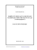
VIỆT NAM
Codon 41/42 30.3%
Codon 17
30.0%
Codon 26 23.5%
Codon 71/72 4.8%
Khác
11.1%
Figure 4.1. Distribution of common β-thalassemia gene mutation in Asia
4.2.2. Distribution of β-globin gene mutations by function and, location
Gene mutation sites have significant implications for gene
expression. Research results show that most of the mutations occur in the
RNA translation, less in RNA processing and transcription; more in exon
than intron and promoter area. . From that it can be concluded that in
Vietnamβ0-thalassemia is more common than β+-thalassemia. This review
is in line with previous studies in Vietnam and is consistent with the
characteristics of β-thalassemia in Southeast Asia. Clinical profiles, βthalassemia in Vietnam are more severe.
4.2.3.Distribution of gene mutations by genotypes
In the 208 mutation alleles, 25 mutation genotypes were identified,
the most common combinations genotypes were CD17-CD26, CD41/42CD17, CD41 /42-CD41/42, CD17-CD17, that were classified into 5 broad
genotypes β0β0, β+β+, β0β+, β0βE and β+βE.. The most common was β0βE
genotype, followed by β0β0, β0β+, β+βE,β+β+. That were similar with two
20
recent studies, in 2018, in southern and northern of Vietnam. Βetathalassemia is a very heterogeneous with molecular and clinical features.
Clinical and hematologicalmanifestations, also the severity of the disease
depend on the genotype.
4.3. Phenotype–genotype comparison of β-thalassemia major and
intermedia
Comparison of clinical phenotype with genotype of β-thalassemia
Results of the study showed that CD41/42, CD17, CD71/72
mutations and their combination genotypes with other mutations were
highly related to severe clinical presentations. These results canbe
explainedby these mutations were β0-thalassemia phenotypes, that did not
synthesize β-globin chains.CD26 mutations and their combination
genotypeswith other mutations may be seen in major, intermedia and mild
β-thalassemia.
Clinical manifestations varied by 3 genotype groups, β 0β0, β0β+ and
β0βE. Clinical manifestations of β0β0 genotype were more severe than those
of β0β+and β0βE, indicating at onset age, early blood transfusion age, and
the severity of anemia (p <0.05). The reason is that the β 0β0 gene does not
synthesize of β-globin chains, and the imbalance of alpha / non-alpha
chain is greater than the two β0β+ and β0βEgenotypes.Clinical
manifestations of β0β+ and β+βEgenotypewere not differently, due to still a
partial β-globin synthesis, less imbalance of alpha / non-alpha chain. From
there, it can be concluded that there is a clear correlation between clinical
phenotype and genotype of β-thalassemia.
Comparison of hematological phenotype with genotype of βthalassemia
Almost of MCV, and MCH of β 0β0,β0β+ and β0βE were <75fl and
<28pg. Microcytosis and hypochrome are the characters of β–
thalassemia, that are used to screen thalassemia in community.
Mechanisms
of
this
characters
are
the
results
21
ofhemoglobinhyposynthesisand ineffective erythropoiesis in bone
marrow due toβ–globin gene mutations.
Hemoglobin patternsvarriedby genotypes (table 3.12).
In
β0β0genotype, HbA1areabsent,almost of Hb are HbF and small part of
HbA2. Inβ0β+ genotype HbA1are decreased with high HbF and normal or
slightly highof HbA2. In β0βE genotype, HbA1 are absent with high HbF
and HbE. The alteration of hemoglobin patterns depend on the β–globin
gene mutations with β0 or β+ phenotype, absent or decreased production
of
β–globin chains. Due to absent of production of β–globin chains
inβ0β0and β0βE, therefore HbA1 are absent.Due to still hypoproduction
ofβ–globin chains in β0β+ phenotype, therefore HbA1are decreased.
Because of absent or decreasingof β–globin chains, the residue α–globin
chains combine with gamma or delta chains, HbF and HbA 2are
increased.Like that, there is the correlation closely between hematological
phenotype and genotype.
22
CONCLUSIONS
Study 104 patients with β-thalassemia we may conclude:
1.Clinical and hematological phenotypes of β-thalassemia
areratherspecific
Clinical manifestations are early, 88.4% under 5 years of age and
55.7% under 1 year of age. Clinical manifestations are diversity with three
syndromes : severe chronic haemolytic anemia (64.9% of dependent on
blood transfusion), iron over;oad and growth retardation. Almost of
patients were β-thalassemia major (70.2%) and intermedia (26.9%). Over
half of β-thalassemia intermedia manifested as β-thalassemia major.βthalassemia was more severe than β-thalassemia/HbE.
Hematological phenotype is quite specific, with low Hb,
microcytosis (MCV=70.7 ± 8fl), hypochromic(MCH=23 ± 3.6 pg).
Hemoglobin patternvarries by disease types. In β-thalassemia, HbA1
decreased or absent, HbF increased, HbA2 increased slightly. In βthalassemia/HbE, HbA1 decreased, HbF and HbE increased.
2.Βeta-globin gene mutations in patients with β-thalassemia are diversity
- 208 mutation alleles were detected in 104 patients with βthalassemia, the mutation rate was100%, with 13 mutations. The four
most common mutations were CD41/42, CD17, CD26 and CD71/72 with
30.3%, 30%, 23.5% and 4.8%, respectively. There are 9 less common
types of mutations, IVS2.654, - 28, - 88, CD95, IVS 1.1, IVS 1-5, -140,
c.441-442insAC, and 2.3kb deletion with 1-2,9%. There was no significant
difference in the distribution of mutations among ethnic groups except
CD26 and -28. CD26 mutations were more common in Thai (50%) than in
Kinh (23.2%) and Tay (5%). -28mutationin Tay is differenced inKinh
people.
Most of the mutations occurred more in RNA translation (89.4%)
than in RNA processing (4.8%) and RNA translation (4.3%); more
23
inexonthan in intron and promoter area. More mutations have the
β0phenotype (68%), than β+ phenotype. The most common mutation
combinations are CD17-CD26, CD41/42-D26, CD41/42-CD17, CD41/42CD41/42 and CD17-CD17. Combinations of mutations were divided into
five groups of genotypes: β0β0 (38.46%) with 5 mutation combinations, of
which 17 were homozygotes and 23 were compound heterozygotes, β +β+
(0.96%) with one type of combination, β0β+ (13.46%), with 9 mutation
combinations, β0βE (45.2%) with 6 mutation combinations and β+βE (1.92
%), with two mutation combinations.
3.There are the correlations between phenotype and genotype of βthalassemiamajor and intermediate
- CD41/42, CD17, CD71/72 mutations and their combinations with
other mutations related to clinical phenotype major and intermedia. CD26
mutations and their combinations with other mutations cn be seen both in
clinical phenotype major andintermedia, and also can be seen in mild
phenotype.
- Clinical phenotype of β0β0genotype is more severe than of β0β+,
β0βE. There is no different on clinical phenotype between β 0β+ and β0βE
genotypel
- Hemoglobin patterns depend on genotype, HbA1 is abent in the
β0β0, β0βE genotype, decrease in the β0β+ genotype, HbE is only present in
β0βE and β+βEgenotype.