Nghiên cứu điều trị ung thư thực quản bằng phẫu thuật nội soi ngực phải kết hợp mở bụng tt tiếng anh
Bạn đang xem bản rút gọn của tài liệu. Xem và tải ngay bản đầy đủ của tài liệu tại đây (315.85 KB, 30 trang )
MINISTRYOFEDUCATIONANDTRAINING MINISTRYOF NATIONAL DEFENCE
108 INSTITUTE OF CLINICAL MEDICAL ANDPHARMACEUTICALSCIENCES
HO HUU AN
RES EARC H ON TREATMENT O F ESO PHAGEAL CANCER
BY RIGHT THO RACOSCOPIC ESOPHAGECTO MY
CO MBINED W ITH LAPARO TO MY
Spe ciality: Gastrointe stinal surgery
Code : 62720125
SUMMARY O F TH E TH ESIS
Ha Noi - 2019
THE THESIS WAS COMPLETED AT
108 INSTITUTE OF CLINICAL MEDICAL AND
PHARMACEUTICAL SCIENCES
Name of supervisor:
1. Associate Professor & Ph. D. Trieu Trieu Duong
2. Ph. D. Nguyen The Truong
Re viewe r 1:
Re viewe r 2:
Re viewe r 3:
The thesis will be defened on …..date……month…..2019
The thesis can be found in:
1. Nat ional library of Viet Nam.
2. Library of 108 Institute of
Pharmaceutical Sciences.
Clinical
Medical and
1
ABSTRAC T
Esophageal cancer is the 4th most common cancerous diseases of the
gastrointestinal tract, with increasing incidence rates. In 2005 there were
497,700 new cases and the rate may increase up to 140% in 2025. It also
causes high death rate with 416,500 deaths in the US in 2005.
Treatment of esophageal cancer is a multimodality, including surgery,
chemotherapy and radiotherapy, of which surgery plays the most important
role. Transthoracic esophagectomy (TTE) (by Ivor Lewis or McKeownAkiyama) or trans-hiatal esophagectomy (THE) (by B. Orringer) are the
most common surgeries to t reat esophageal cancer. However, convetional
open surgery has high rates of complications of 23 - 40%, with 1.2 – 8.8% of
mortality rate. With the fast advances of minimally invasive surgery in
almost the last decades, minimally invasive esophagectomy has been applied
and quickly advanced with such benefits as reduced complications,
especially pulmonary comlications, and shortened hospitalization and
recuded costs for patients. Some recent research reports have proven the
safety and feasibility of the surgery. However, there are still controversies
about the safety, feasibility and outcomes of cancer treatment study of
minimally invasive surgery in the treatment of esophageal cancer.
In Viet Nam, minimally invasive surgery for treating esophageal cancer
has been implemented since 2003 at Big centers such as Cho Ray Hospital,
Viet Duc Hospital and 108 Military Central Hospital. However, there has not
been any research with sufficient long-term outcome review of the approach.
With the above-mentioned matters, we would like to study the topic,
“Research on Treatment of esophageal cancer by right thoracoscopic
esophagectomy combined wit laparotomy” with the objectives as follows:
1. to describe some clinical and subclinical features of patients with
esophageal cancer having been treated by right thoracoscopic
esophagectomy combined with laparotomy.
2. to review the outcomes of esophageal cancer treatment with right
thoracoscopic esophagectomy combined with laparotomy.
2
DISSERTATIO N
The research was conducted on 71 patients with esophageal cancer
treated with right thoracoscopic surgery combined with laparotomy at the
108 Military Central Hospital from January 2010 to December 2017.
1. Some clinical and subclinical features: Common symptoms
include dysphagia (81,7%) and weight loss (80,3%). Adenocarcinoma
is most common comprising 67.6%. Squamous cell carcinoma
comprises 97.2%. The tumor in the middle third of esophagus are
57.1% and in t he lower third 47.9%. T he sensitivity and specificity of
CT scans t o T1, T 2 and T3 are (38%; 95%), (50%; 79%) and (74%;
75%) respectively. The rate of nodal metastases is 33.8% (24/71). T he
avrage number of metastatic nodes are 2.8 ± 2.6 (1-13). Stage 0 are of
4.2%, stage I of 14.1%, stage II of 59.2%, stage III of 22.5%, and stage
IV of 0%.
2. Outcomes of surgeries
- Intraoperative outcomes: Mean surgical time 193.9 ± 49.3 minutes,
average number of node removed is 10.1 ± 8.6. The rate changing to
open technique is 1.4%. Complications are of 7.0%.
- Early complications include pneumonia of 12.9%, respiratory
distress of 7.1%, anastomotic leaks of 11.4%, chylothorax of 4.3%, and
mortality of 0%.
- Long-term outcomes: Long-term monitoring 21.7 ± 19.4 months
long. T he rate of postoperative complications is 33.3%, delayed
complication rate is 24.6%. Overall survival rate is 45.7 months (95%
CI:35.9-55.4). The overall survival rate after one, two, three and four
years are 79.7%, 62.3%, 52.3%, and 43.6% respectively.
As a result, the study has made some new contributions, confirmed
the safety, feasibility, efficacy, reduction of complications and ensured
the oncological principles of the right thoracoscopic esophagectomy
combined with laparotomy. for treatment of esophageal cancer.
TH E S TRUC TUR E O F TH E DISSERTATIO N
The dissertation consists of 123 pages, including abstract of 2 pages,
3
overview of 36 pages, study subject s and method of 20 pages, study
results of 26 pages, discussion of 39 pages and conclusion of 1 page.
T wo research with 40 t ables, 07 charts and 22 pictures. 133 reference
materials, including 13 in Vietnamese language and 120 in foreign
languages.
Chapte r 1
O VERVIEW
1.1. ANATO MY O F THE ESO PHAGUS - STO MACH
1.1.1. Shape
1.1.2. Structure
The esophageal wall is composed of four layers from outermost to
innermost, including the outermost adventit ia, muscularis propia,
submucosa and mucosa.
1.1.3. Relations
1.1.4. Blood supply and innervation
The esophagus has art erial supply, including inferior thyroid artery,
bronchial branch of aortic artery, left gastric artery (55%) and left
inferior phrenic artery.
Vagus nerve (nerve X) supplies nerves to esophagus. The upper
segment of the esophagus is coordinated by the branches of the
recurrent laryngeal nerve.
1.1.5. Lymphatics
Below the esophageal mucosa is a lymphatic drainage system of
mainly longitudinal vessels. The lymphatic drainage system drains into
bigger lymph nodes and form the surficial lymphatic plexus and then
connect to the lymph nodes along the esophagus.
1.1.6. Gastric arte ries: The branches supplying blood to the
stomach which originate from the celiac trunk include branches for the
lesser gastric curvature, fundic and cardiac part s of the stomach, and
short gastric arteries.
1.2. DIAGNOS IS
4
1.2.1. Clinical
Common symptoms include dysphagia, vomiting, pain in the
retrosternal area, hoarseness, weight loss, malnourishment and tylosis.
1.2.2. Subclinical: Pre-treatment diagnosis is highly significant for
esophageal cancerous diseases. Ho wever, there are still a lot of
challenges for pre-operative diagnosis. Therefore, in order to have an
accurat e diagnosis, not only one approach but various approaches
should be incoporated.
1.2.2.1. Endoscope: Endoscope combined with biopsy may have the
sensitivity up to 96%. Advantages: Low cost, widely applicable even at
lower levels of the health services; noninvasive, may apply for
treatment interventions such as mucosa or submucosa removal for very
early stages.
1.2.2.2. Endoscopic ultrasound: is a significant subclinical approach to
assess tumor invasion especially the invasion of the esophageal wall.
1.2.2.3. Computed tomography scan: is an important subclinical
investigation to assess the invasion of the mediastinum and nodal
disease, and detect distant metastases. This is considered a good
investigation for preoperatively staging esophageal cancer.
1.2.2.4. Magnetic resonance imaging (MRI) scan: With technological
advances, MRI scans combined between T2W and DWI sequences have
been reported with detection rates in assessing tumor invasion of T1 33%,
T2 58%, T3 96% and T4 100%.
1.2.2.5. Positron emission tomography (PET/CT) scan: Multiple
analytic studies have shown the sensitivity and specificity of FD-PET
for local metastatic lymph node detection are 51% [95% CI, 34%–69%]
and 84% (95% CI, 76%–91%) respectively.
Other studies have shown t hat FDG-PET has higher sensitivity for
determination of distant metastases than other methods such as CT
scan, ultrasound and SPECT . Luketich found that FDG-PET has the
sensitivity of 88% (7/8) and the specificity of 93% (25/27) for distant
metastasis detection.
5
1.3. HISTO PATHO LO GY AND STAGES
1.3.1. Histopathology
1.3.1.1. Macroscopy: Esophageal cancer has three common patterns:
fungating comprises more than 60%, ulcerative (20 - 30%), infiltrating
is rare about 10%.
1.2.1.2. Microscopy: According to WHO classification in 1977, there
are: squamous cell carcinoma (more than 90%), adenocarcinoma (~
9%), melanoma, Sarcoma (rare, about l%)
1.3.2. Stage classification: There are many different methods of
classification proposed by various cancer associations, however, the
classifications by the Union for International Cancer Control (UICC)
and American Joint Committee on Cancer (AJCC) have been widely
applied. The staging of esophageal cancer is based on 3 factors,
including T (primary tumor), N (regional lymph nodes) and M (distant
metastasis).
1.4. TREATMENT
1.4.1. Flowchart of treatment of esophage al cance r
1.4.2. Surgery
Such methods as transthoracic esophagectomy, Ivor-Lewis (via the
thorax) and the 3-incision approach is the most common method in
countries in North America, whilst transthoracic esophagectomy and
extensive nodal dissect ion (the three-dimensional t echnique) is widely
employed in Asian countries such as Japan and South Korea.
* Akiyama method in esophagectomy: in 1971, Akiyama introduced
the procedure: firstly, open and expose the chest cavity for
esophagectomy, then open the abdomen to create a gastric conduit,
followed by opening the neck to create an esophagogastric anastomosis.
The procedure is performed in three approaches: thoracic, abdominal
and left neck incisions.
- Advantages: Extensive nodal dissect ion in the mediastinum,
abdomen and neck; high resection of the esophagus ensures safet y of
6
the resection surface, cervical anastomosis is easy for anastomotic leak
revision, if any, and lowers the rate of reflux.
- Disadvantages: cervical anastomosis may increase the risk of
postoperative anastomotic leak and stricture.
1.4.3 Esophageal substitute
Esophageal substitutes are of two types: auto-transplanted tissues,
including stomach, jejunum or colon, and synthesis (composite
combined with collagen, plastic tubes).
Stomach is the ideal esophageal replacement for alimentary
reconstruction after esophagectomy, because of its sufficient vascular
supply, sufficient length for mobilization for creating either thoracic or
cervical anastomosis, and only one anastomosis required so shorter
operation time. T he main disadvantage of gastric conduit is
inflammatory gastroesophageal stricture developed from acid or bile
reflux.
1.4.4. Minimally invasive surgery
Minimally invasive surgery in treating esophageal cancer has been
widely applied with the benefits of small incisions, less intraoperative
blood loss, less postoperative complications, shortened intensive care
and hospitalization and better postoperative respiratory recovery.
1.4.5. Nodal disse ction in surgery for e sophage al cance r
In 1994, at t he International Society for Diseases of the Esophagus
(ISDE) held in Munich, Germany, a concept of the area for
lymphadenectomy. T he term “2-level lymphadenectomy” is
accordingly used for abdominal and mediastinal nodal dissection whilst
the term “3-level lymphadenectomy” is used for abdominal, mediastinal
and cervical nodal dissection.
1.4.6. Supportive treatment
1.4.6.1. Radiotherapy
1.4.6.2. Chemotherapy
1.4.6.3. Chemoradiotherapy
7
1.5. REVIEW O F MINIMALLY INVASIVE ESO PHAGECTO MY
O UTCO MES
1.5.1. Internationally
In 1992, Dallemagne B. et al described the first esophagectomy
using both thoracoscopy and laparoscopy with gastrict conduit and
cervical anastomosis for treating esophageal cancer.
There have been many other studies proving that thoracoscopic
esophagectomy is a safe and feasible procedure for treating esophageal
cancer:
Duration of thoracoscopic phase: 90- 281 minutes.
Average blood loss: 200 - 536 ml
Number of nodes dissected in thoracoscopic phase: 7 – 29 nodes.
Rate of transfer to open surgery of thoracoscopic phase: 0 - 20%.
The studies have also shown the outcomes of right thoracotomy
approach in esophagectomy are very encouraging with reduced
postoperative complication rates, especially respiratory complications.
T able 1. 3: Complications in the studies
Postope rative
Authors
complications
Smit her Jakhmola Kinjo
Kubo
n=309
n=48
n=34
n=28
General complications
62%
?
58%
?
(%)
Respiratory
35.4%
38%
17.8%
complications (%)
Thoracic conduit leaks
2.1 %
0
10.7%
(%)
Recurrent
laryngeal
?
?
25%
nerve injury
Anastomosis leak (%)
5.5%
6.3%
8.8%
3.5%
Hospitalization
13
?
32
?
Mortality
2.3%
6.3%
0
0
8
1.5.2. Viet Nam
T he studies of Minimally invasive surgery in treating esophageal
cancer by Nguyễn Minh Hải (2003), Triệu T riều Dương (2003), Phạm
Đức Huấn (2006), Lê Lộc (2017) with sample size from 20 - 150
patients resulted in:
Average operation durat ion: 330 - 395 minutes.
Average ICU care time: 1 day.
Postoperative complications: 10 - 20%.
T he authors have also concluded that thoracoscopic esophagectomy
has wider operating field and better vision, is easier to control bleeding
and can be safely performed at medical centers where there are good
anesthetic and recovery facilities and competent Minimally invasive
surgeons. In addition, mediastinal lymphadenectomy can be performed
during the Minimally invasive surgeries on candidates for
esophagectomy, but for those contraindicated for esophagectomy due t o
extensive tumor invasion and metastasis, thoracoscopic surgery also
assists in more accurate determination of esophageal cancer stages to
avoid unnecessary thoracic opening.
Chapter 2
RESEARC H SUBJECT AND METHO DO
2.1. RESEARC H TIME AND LO CATIO N
The research was conducted at 108 Military Central Hospital from
January 2010 to December 2017.
2.2. RESEARC H SUBJECT
2.2.1. Selection crite ria
- Patients diagnosed with esophageal cancer, having undertaken right
thoracoscopic esophagectomy combined with laparotomy..
- Patients diagnosed with t horacic segment esophageal cancer stages IIII.
9
- Patients with ASA ≤ 3, without any contraindication against
endotracheal anesthesia.
2.2.2. Exclusion criteria
- Patients having undertaken other procedures of esophagetomy.
- Patients combined with other malignent pathologies, and those of
insufficient patient profile.
2.3. RESEARC H METHO D
2.3.1. Re search design
- T he descriptive retrospective cohort plus prospective cohort methods
are used.
• The retrospective group: patients having been operated from
February 2010 to December 2014
• The prospective group: patients having been operated from
January 2015 to November 2017.
- Indicate for surgery for stage I-III esophageal cancer.
- Indicate for Van Hagen chemoradiotherapy [54].
- Evaluate pathological stages in accordance with AJCC - 2010
- Collect Histological specimens as the golden standard for diagnosis.
- Monitor and evaluate early outcomes within 30 days posoperatively.
Schedule for follow-up visits or telephone to evaluate Long-time
outcomes at 3 months, 6 months, 1 year, and 3 years, etc.
- Collect data in line with consistently agreeable patient files.
2.3.2. Re search chart
2.3.3. Sample size: T he minimal sample size is calculated with an
equation of 95% liability. The number of patients in the prospective
group is 60 patients.
2.3.4. Procedures applied in the research
2.3.4.1. Right thoracoscopic esophagectomy
* Patient and surgical equipement preparation
* Patient positioning and surgical team location
* Steps of the surgery
- T horacic phase: Right thoracoscopic surgery
10
Dissect the esophagus and posterior mediastinum
Dissect and mobilize the esophageal part s superior and inferior
to the t umor, lymphadenectomy.
Insert a drain in the right pleural cavity.
- Abdomino-cervical phase:
Open the abdomen to create a gastric conduit
Dissect the nodal system in the upper abdominal quadrants.
Creat e the cervical gastroesophageal anastomosis.
2.3.5. Evaluation standards
2.3.5.1. AJCC-2010 stage evaluation
2.3.5.2. ASA pre-operative anesthetic classification
2.3.5.3. Evaluation of tumor reponse to pre-operative
chemoradiotherapy
2.3.6. Re search crite ria
2.3.6.1. General information of the patients: Age, gender, pathological
history, time of detection, reasons of admission and clinical symptoms.
2.3.6.2. Pre-treatment assessment: Assess tumor location and invasion,
nodal metastasis by preoperative gastroesophageal endoscopy and CT
scan, and assess respiratory functions.
2.3.6.3. Evaluate pre-operative chemoradiotherapy outcomes
2.3.6.4. Rate and record intraoperative scores: number of trocars used
during the thoracic phase, surgery durat ion, intraoperative
complications, number of nodes dissected
2.3.6.5 Postoperative evaluation parameters
- Early outcomes: T ime at recovery, hospitalization, removal of
thoracic, abdominal and anastomosis drains, postoperative mortality,
early complications, postoperative histology.
- Long-time outcomes: Long-time complications, recurrence,
survival time and factors affecting postoperative survival time.
2.3.7. Data management and processing
2.3.8. Re search ethics
11
Chapte r 3
RESULTS
3.1. CO MMO N CHARACTERISTICS
- Mean age 55.8 ± 8.3 years old (40 - 76). 100% male.
- 13 patients (18.3%) with combined internal diseases. 3 patients (4.2%)
with previous surgeries, however, most of them had been operated in the
lower abdominal quadrants and the extraperitoneal space.
3.2. CLINICAL AND SUBCLINICAL PRESENTATIO N
3.2.1. Clinical
- Average detection t ime 2.05 ± 1.59 months, mostly from 1-3 months
accounting for 73.2 %.
- Reasons for admission are mainly difficulty swallowing + obstruction
accounting for 81.7%.
- Dysphagia (77.5%) and weight loss (80.3%) are the two most
common symptoms.
3.2.2. Subclinical
3.2.2.1. Endoscopy
- Macroscopy: Fungating pattern comprises most 67.6 % (48/71).
- Microscopy: Squamous cell carcinoma mostly common (97.2%).
3.2.2.2. Respiratory functions: Most patients have normal respiratory
funct ions (88.7%), none of them have any severe ventilation disorder.
3.2.2.3 CT scan
- Tumor location: 100% tumors in the middle third and lower third of
the esophagus.
- Invasion shown in the CT scans: A majority of patients have invasion
to cT2-cT3 comprising 83.1% of which cT3 accounts for 56.3%.
- Values for diagnosis of invasion by CT scan:
+ Sensitivity to T1, T2 and T 3 are 38%, 50% and 74% respectively
+ Specificity to T1, T 2 and T3 are 95%, 79% and 75% respectively
+ Accuracy for T 1, T2 and T 3 are 85%, 69% and 75% respectively.
- Values for diagnosis of mediastinal nodal metastases of CT scan:
sensitivity 53%, specificity 46%, accuracy 48%.
12
- Values for diagnosis of abdominal nodal metastases of CT scan:
sensitivity 80%, specificity 85%, accuracy 84%.
3.2.2.4. Postoperative pathology
- Microscopy: Squamous cell carcinoma accounts for most 93.0 %.
There are 3 patients (4.2%) whose specimens are not observed with any
tumor cells following preoperative chemoradiotherapy.
- Invasion:
+ Patients without any preoperative chemoradiotherapy have majority
of pT 2-pT3 (81.5%)
+ Patients with preoperative chemoradiotherapy have pT1-pT2 of
52.9%, especially 6 patients (35.7%) no longer have any tumor cells.
T able 3.1: Rates of nodal metastases
NonTotal
Metastatic
mestastatic
nodes
nodes
Metastatic nodes
n
%
n
%
Mediastinum
17
23.9
54
76.1
Abdomen
13
18.3
58
81.7
24
33.8
47
66.2
Ge neral
metastases
nodal
n
%
71
100
71
100
71
100
Comments: 24 patients (33.8%) with nodal metastases, of whom the
mediastinal nodal metastasis rat e is 23.9%, abdominal nodal metastasis
rate is 18.3%.
- Nodal m etastasis from pathologylogy: pN0, pN1, pN2 and pN3 rates
are 66,.2%, 21.1%, 11.3 % and 1.4% respectively. Mean number of
metastatic nodes is 2.8 ± 2,6 (1-13)
13
Stages
Stage I
Stage II
Stage III
Stage IV
Neither tumor
cells nor nodes
found
Total
T able 3.2. Cancer stages
Chemoradi
Surgery
otherapy (n =54)
PT (n=17)
Total
(n =71)
n
3
9
2
0
%
17.6
52.9
11.8
0
n
7
33
14
0
%
13.0
61.1
25.9
0
N
10
42
16
0
%
14.1
59.2
22.5
0
3
17.6
-
-
3
4.2
17
100
54
100
71
100
Comment: T he group without any preoperative chemoradiotherapy:
Patients of stage II account for most (61.1%). T he group with
preoperative chemoradiotherapy: mostly at stages I and II (64.7%).
There are 3 patients (17.6%) who are found without any more tumor
cells after chemoradiotherapy.
3.3. TREATMENT O UTCO MES
T able 3.3. Treatment
Tre atment
Quantity (n) Rate (%)
Surgery
29
40.9
Surgery
+
postoperative
24
33.8
supportive treatment
Preoperative chemoradiotherapy
17
23.9
+ Surgery
Missing information *
1
1,4
Total
71
100
Comment: There are 23.9% of the patients treated with preoperative
chemoradiotherapy + surgery, 40.9% of the patients only treated with
surgery.
14
3.3.1. Outcome s of pre operative chemoradiotherapy
- Assess tumor invasion before and after preoperative
chemoradiotherapy:
The rate of reduced invasion cT 2 after chemoradiotherapy is 66.7%
(2/3) patients
The rate of reduced invasion of cT3 after chemoradiotherapy of is
83.3% (10/12) patients
The rate of reduced invasion of cT4 after chemoradiotherapy is
100% (2/2) patients
- Evaluate histological response of the tumor after preoperative
chemoradiotherapy according to Ryan et al: There are 4 patients
(17.6%) with full response, 8/17 (47.1%) patients with average
response, 23.5% with limited response and 2 patients (11.8%) without
any response to preoperative chemoradiotherapy.
3.3.2. Intraope rative outcomes
One out of 71 patients is transferred to open surgery therefore we do
not include this one in the outcomes of Minimally invasive surgery.
- Quantity of trocars (thoracic phase): There are 53 patients (75.7%)
who used 3 trocars, 17 patients (24.3%) who used 4 trocars.
- Average surgery duration 193.9 ± 49.3 minutes, of which the time for
thoracic phase is 74.8 ± 29.5 minutes. There is no surgery time
difference between the groups with and without preoperative
chemoradiotherapy (p>0.05, t test)
- Surgical complications: The rate of general complications 7.0%,
including 1 patient (1.4%) with bleeding, 2 patients (2.8%) with
thoracic tubes, 1 patient (1.4%) with tracheal injury and 1 patient
(1.4%) with pulmonary tissue damage, 1 patient (1.4%) t ransferred to
open surgery. There is no difference in complication rates between
between the groups with and without preoperative chemoradiotherapy.
- T otal mean nodes dissect ed is 10.1± 8.6, with the biggest number of
40 nodes. T he mean mediastinal nodes dissect ed is 5.5± 5.5, with the
biggest number of 33 nodes. T he mean abdominal nodes dissect ed is
4.5 ± 4.6. Total mean nodes dissected in the prospective group is higher
15
than that in the retrospective group with statistical significance (12.0 ±
9.4 vs 7.4 ± 6.7, p < 0.05)
3.3.3. Early outcome s
- Early complications: Rate of pneumonia is 12.9%, respiratory distress
7.1%, pleural effusion 7.1%, anastomosis leak 11.4%, chylothorax
4.3%, bleeding 2.9%, recurrent nerve injury 18.6%. No mortality within
the first 30 days postoperatively. There is no difference in the rates of
postoperative complications between the groups with and without
preoperative chemoradiotherapy (p > 0.05).
T able 3. 4: T ime for ICU, drain removal and postoperative care (n=70)
Shortest Result
Mean
Longest
ICU care (day)
Thoracic drain removal
(day)
Abdominal drain removal
(day)
Cervical drain removal
(day)
Postop care (day)
1.4 ± 1.6
0 -8
6.5 ± 2.0
3 - 14
6.1 ± 1.7
3 - 14
10.3 ± 5.6
3 – 30
16.4 ± 6.2
7 - 40
3.3.4. Long - time outcome s and some relate d factors
70 patients are successful with Minimally invasive surgery,
however, during the distant monitoring 1 patient has gone missing. This
patient is not evaluated for long-time outcomes. For cum survival time,
it is considered still alive (according to Kaplan Meier method).
- Mean monitoring time: 21.7 ± 19.4 months
- Distant complications: The rate of anastomosis stricture monitored for
3 months is 23.2% (16/69), recurrent nerve injury 15.9% (11/69), 1.4%
patient with intestinal obstruction.
- Immediate and distant recurrence: T he total rate of postoperative
recurrence is 33.3%, of which there is 10.1% of immediate recurrence ,
16
including recurred cervical nodes of 4.3%, mediastinal and abdominal
nodes of 5.8%, no patient with any recurred anastomosis. T he rate of
distant recurrence is 24.6%, of which the most common location is
pulmonary recurrence of 7.2%.
T able 3.5. Cum survival
Monitoring time
Characte ristics
1 year
2 years
3 years
4 years
Cum survival
79.7%
62.3%
52.3%
43.6%
(CI: 95%,
(CI:95%,
(CI:95%,
(CI:95%,
67.5-87.7) 47.5-74.0) 36.2-66.2) 26.7-59.4)
Mean
cum
45.7 months (CI:95%, 35.9- 55.4)
survival
Mean
21.7± 19.4 months (1.9 – 81.8)
monitoring time
Comment: Mean cum survival 45.7 months (CI:95%, 35.9- 55.4). T he
rates of first, second, third and fourth year survival are 79.7%, 62.3%,
52.3%, and 43.6% respectively.
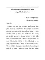
Chart 3.1. Survival functions in accordance with invasion level.
17
Comment: Survival functions between different levels of invasion are
different, and the difference is statistically significant with χ 2 -= 11.92,
p=0.0179 <0.05.
T able 3.6. Comparison of mean survival between the groups with and
without nodal metastasis
Nodal
Mean survival
p
95% C I
metastasis
(month)
N (-)
51.0
39.9-62.2
0.0357
N (+)
20.2
14.1-26.2
Comment: Survival of the group without nodal metastasis is longer
than that of the group with nodal metastasis, the difference is
statistically significant with p<0.05
- Cum survival in line with quantity of m etastatic nodes: The rates of
survival after one year of N0, N1, N2 and N3 are 85,6%, 70,9%, 68,6%
and 50% respectively.
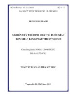
Chart 3.2. Survival functions in line with cancer stages.
Comment: T here are statistically significant differences of survival
funct ions in line with cancer stages, Log Rank (Mantel-cox), χ 2 -=
12.1, p <0.05. The rates of survival after t wo years of stages 0-I, II, and
III are 100%, 65.9% and 32.7% respectively.
18
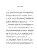
Chart 3.3. Survival functions of groups with and without preoperative
chemoradiotherapy.
Comment: There is no difference of survival functions time between
the groups with and without preoperative chemoradiotherapy, p>0.05.
Chapte r 4
DISCUSSIO N
4.1. CO MMO N CHARACTERISTICS
4.1.1. Age & gender: Mean age 55.8 ± 8.3 years old (40-76). The most
common age group is from 40-60 years old accounting for 71.8 %
(49/71). It is similar t o the research by Phạm Đức Huấn, Luketich, and
Beasley with the mean age of 53 - 66 years old. Esophageal cancer is
more common in male than female, the male/female ratio in other
research is from 3.5-5/1. In our study we have 100% male patients.
4.1.2. Combined diseases: In the research, there are 13 patients
(18.3%) with combined internal medicine diseases. The authors think
that patients with high indicators of combined diseases (cardiovascular,
respiratory, malnourish) have higher risks of anastomosis leak (odds
rat io, 6.564; 95% CI, 1.676 to 25.716) and respiratory complications
(odds rat io, 2.732; 95% CI, 1.317 to 5.666). Other research have also
19
shown the correlation between combined diseases and the rates of
postoperative complications.
4.2. CLINC AL AND SUBCLINICAL
4.2.1. Clinical
The research shows that the mean time from symptom detection till
admission is 2.05 ± 1.59 months (0.2 - 9). T he main reasons for going
to hospital is difficult swallowing, obstruction swallowing accounting
for 81.7%. Early diagnosis of esophageal cancer is often difficult as the
disease has no symptoms or very vague symptoms which can be easily
missed. Of the 2418 cases of early esophageal cancer, Kodama and
Kakegawa found that 55% patients without any symptoms, 9.8% with
difficulty swallowing, 3.6% with obstruction swallowing.
4.2.2. Subclinical
4.2.2.1. Endoscopy: The research finds fungating pattern as the most
common accounting for 67.6%, ulcerative pattern 11.3%, combined
pattern 14.1%, infiltrating pattern 4.2%. In addition, there are 2 patients
(2.8%) with superficial and protruding mucosa. T his is type 0
(superficial pattern) in the classification by Japan Society for
Esophageal Cancer. Similar results have been found in other research.
4.2.2.2. Measure respiratory functions: 88.7% patients with normal
respiratory functions, 11.3% with mild and moderate obstructive
ventilation disorders, no patient with severe obstructive ventilation
disorder. Preoperative assessment of respiratory functions is a very
important factor for esophageal cancer surgery, it allows us to choose
suitable patients, as well as to have appropriate measures in place to
minimize postoperative respiratory complications.
4.2.2.3. Computed tomography scan
Nowadays, CT scan is a common and effective method for stage
determination before t reatment of esophageal cancer. The research has
100% t umor injury in t he middle – lower two t hirds, including middle
third of 57.1% and lower third of 47.9%. The results are similar to other
reports of domestic authors. Some overseas research also shows the
20
most common rates of middle – lower t wo t hirds, however, a majority
of lower third.
Kết quả nghiên cứu thấy độ nhạy và độ đặc hiệu của CLVT đối với
chẩn đoán T1, T2, T3 lần lượt là (38%;95%), (50%; 79%) và (74%;
75%). T ác giả Li báo cáo độ nhạy và đặc hiệu của CLVT lần lượt là
77.4%, 74.8% đối với phân biệt T1- 2 và T3 trong UT tế bào vảy.
Nodal evaluation in CT scan depends on the size of nodes. In
general, the research indicates that the standard size of general
metastatic node is 1 cm. The research shows the sensitivity, specificity
and accuracy of CT scan for mediastinal nodal metastases are 53%,
46% and 48% respectively, and for abdominal nodal metastases are
80%, 85% and 84% respectively. The results are similar to research by
other authors.
4.2.2.4. Histopathology
- Microscopy: T wo major types of esophageal cancer are squamous
cell carcinoma and adenocarcinoma. T here is also a low rat e of
cancerous sarcoma and melanoma. In Viet Nam and other Asian
countries, squamous cell carcinoma accounts for a majority, whilst in
western countries, adenocarcinoma accounts for more. T he research
results show t hat squamous cell carcinoma accounts for 97.2% (69/71)
patients, the rest are adenocarcinoma 2.8% (2/71) patients.
- Level of tumor invasion: The research show a major rate of pT2
pT3 cancer stages accounting for 71.8% (51/71 patients). The rates of
pT1, pT2, pT3 and pT4 in the group without preoperative
chemoradiotherapy are 16,7%, 35,2%, 46,3%, 1,9% and 23,5%, 29,4%,
11,8%, 0% in the group with preoperative chemotherapy repestively. In
addition, in the group with chemoradiotherapy, t here are 6 out of 17
patients (35.3%) pT0 (histopathology shows no tumor cell aft er
chemoradiotherapy).
- Characte ristics of nodal metastasis: The research shows the rates
nodal metastasis of 33.8% (24 out of 71), including mediastinal nodal
metastasis of 23.9% and abdominal nodal metastasis of 18.3. T he
average number of metastatic nodes is 2.8 ± 2.6 (1-13).
21
- Characteristics of disease stages: The research finds a majority of
patients with stages II and III accounting for 81.7 %. Compared with
other domestic authors, our rate of stage I patients (14,1%) is higher. In
addition, 17 patients in the studied group may have been treated with
preoperative chemoradiotherapy, and following that t here is a relatively
high rate of patients of stages II-III drops to stage I. The reduction of
tumor invasion after chemoradiotherapy for T2 is 66.7% (2/3), T3 of
83.3% (10/12) and T4 of 100% (2/2) BN. The group of stage I and II
with preoperative chemoradiotherapy account for most (64.7%), Three
patients (17.6%) no longer has any tumor cell after preoperative
chemoradiotherapy. T he results in the group without preoperative
chemoradiotherapy show a majority of stage II of 61.1%, stage III of
25.9% and stage I of 13%.
4.3. SURGERY O UTCO MES
In our research 70 patients with esophageal caner are succesfully
treated with right thoracoscopic esophagectomy combined with
laparotomy., including 17 patients having had preoperative
chemoradiotherapy, 29 patients with surgery only and 24 patients with
surgery plus postoperative supportive treatment, 1 patient has gone
missing.
4.3.1. Outcome s of pre operative chemoradiotherapy
In the research, 17 patients are treated with preoperative
chemoradiotherapy. Analysis shows that chemoradiotherapy helps reduce
tumor invasion with the reduced rat es at cT2, cT3 and cT4 of 66.7%,
83.3% and 100% respectively. The rates of total response is 17.6%
(3/17), moderate response 47.1% (8/17), poor response 23.5% (4/17) and
no response 11.8% (2/17).
4.3.2. Intraope rative outcomes
4.3.2.1. Operation time: T he research finds that the average operation
time is 193.9 ± 49.3 minutes (120-300), including thoracic phase of
74.8 ± 29.5 minutes (25-150). Five patients had operation t ime of 300
minutes. These cases had cancerous lesions invading adjacent organs,
the dissect ion damaged the trachea and the dissect ion for esophagus
22
removal was difficult. Compared with other authors, the average
operation t ime in this research is shorter. The reason may be that, in the
abdominal phase, we applied open surgery at the same time with the
cervical phase as we use two surgical teams for each surgery. T he
analysis shows no difference in operation time between t he group with
preoperative chemotherapy and the group with surgical t reatment only.
4.3.2.2. Patient positioning and quantity of trocars used: In the
research, most patients, during the thoracoscopic phase, are put in
lateral-prone position at 30-45 degree angle with the table. This
positioning conbines the advantage of both lateral position (easy for
changing position) and prone position (minimizing blood loss, shorter
operstion time, better operation field and reducing postoperative
respiratory complications). 53 out of 70 patients (75.7%) used 3 trocars
in the surgeries at t he locations as follows: 1 x 10 mm trocar ở at t he
posterior axillary intercostal VIII-IX space for the camera, 1 x 10 mm
trocar at middle axillary intercostal VI space and 1 x 5 mm trocar at the
intercoastal IV cavity with middle axillary approach for surgical
equipment. However, different surgeons have their own reference of
location of trocar insertion. The key principle is t o ensure the working
angle of about 60-70 degree which is better for performance. If there is
any difficulty, insert another 5 mmm trocar. Recently, patients are often
have extensive lymphanodectomy (nodal dissect ion around the right
recurrent nerve), we therefore insert 4 trocars for easier dissection
performance.
4.3.2.3. Intraoperative pyloric reconstruction: 100% of our patients
received pyloric stent placement. Previous research have proven the
role of pyloric reconstruction or pyloromyotomy to reduce
postoperative gastric conduit blockage. However, recently, there have
been reports showing that either pyloric reconstruction or
pyloromyotomy compared with none pylory reconstruction (pyloric
stent intervention) in esophagectomy and esophageal substitute with
gastric conduits is of no different outcomes.
23
4.3.2.4. Quantity of nodes dissected: Most patients in the research
received two region lymphanodectomy of the mediastium and upper
abdominal quadrants. Average quantity of nodes dissected is 10.1 ± 8.6
(0-40), including mediastinal nodes of 5.5 ± 5.4 (0-33), abdominal
nodes of 4.5 ± 4.6 (0-18). Minimum quantity of nodes dissect ed in a
radical dissect ion of esophageal cancer has not been announced.
However, the authors believe that the more nodes dissect ed may ben
the better, the larger quantity of nodes dissect ed related to better
survival functions.
4.3.2.5. Operative complications: We have an overall rate of general
complication of 7.0%, including blleding of 1.4%, tracheal injury of
1.4%, thoracic t ube damage of 2.8% and pulmonary t issue damage of
1.4%. The rate for transfer to open surgery is 1.4%. There is no
difference in operative complications between t he two groups with and
without preoperative chemoradiotherapy with p>0.05. T he research
results are similar to those from Trần Phùng T iến Dũng, with the
complication rate of 6.9%, including 3 cases with thoracic tube damage
(3.45%) and 3 cases with tracheobronchial tear (3.45%). Luketich
studying 222 patients found 2 patients (0.9%) with intraoperative
tracheal break.
4.3.3. Early outcome s
4.3.3.1. Early complications: T he overall complication rate of
esophageal cancer surgery of other research are from 40-70%. Our
overall complication rate is 42.9%. There is no difference between t he
groups with and without preopreative chemoradiotherapy. A statistics
collating 57 studies (n=15790) compared between Minimally invasive
surgery and open surgery by Yibulayin et al found an overall
complication rate of Minimally invasive esophagectomy of 41.2%
4.3.3.2 Recovery time and drain removal
The research finds an average ICU stay of 1.4 ± 1.6 days (0-8),
average postoperative hospitalization of 16.4 ± 6.2 (7-40) days. 20% of
the patients didn’t stay at the recovery postoperatively. ICU stay of
patients in the research is similar to other authors. However, the