Nghiên cứu đặc điểm lâm sàng, cận lâm sàng và hiệu quả điều trị can thiệp nội mạch bệnh động mạch chi dưới mạn tính khu vực dưới gối tt tiếng anh
Bạn đang xem bản rút gọn của tài liệu. Xem và tải ngay bản đầy đủ của tài liệu tại đây (308.35 KB, 27 trang )
MINISTRY OF EDUCATION AND TRAINING MINISTRY OF DEFENCE
108 INSTITUTE OF CLINICAL MEDICAL AND PHARMACEUTICAL SCIENCES
--------------------------------------------------------
LUONG TUAN ANH
THE CLINICAL CHARACTERISTICS ANDEFFICACY
OFINFRAPOPLITEAL PERCUTANEOUS
TRANSLUMINAL ANGIOPLASTY IN PATIENT WITH
LOWER EXTREMITY ARTERIAL DISEASE
Speciality: Cardiology
Code: 62.72.01.41
ABSTRACT OF MEDICAL PHD THESIS
Hanoi – 2019
THE THESIS WAS DONE IN:108 INSTITUTE OF CLINICAL
MEDICAL AND PHARMACEUTICAL SCIENCES
Supervisor:
1. Ass.Prof.PhD. Le Van Truong
2. Ass.Prof.PhD. Vu Dien Bien
Reviewer:
1.
2.
3.
This thesis will be presented at Institute Council at:108 Institute of
Clinical Medical and Pharmaceutical Sciences
Day
Month
Year 2019
The thesis can be found at:
1. National Library of Vietnam
2. Library of 108 Institute of Clinical Medical and
Pharmaceutical Sciences
1
INTRODUCTION
Lower extremity arterial disease (LEAD) is very common,
prevalence 3-7% of the population, 20% in people over 75 years old.
Ulcers and gangrenelower limb is the end- stages of the disease,
threatened amputation, loss of limb functiondue to infrapopliteal
arterial lesions. Below the knee revascularizationis the most
important in limb salvage for this disease.
There
are
two
methods
of
infrapopliteal
arterial
revascularization: bypass surgery and percutaneous angioplasty, so
bypass surgery is difficult due to below the knee artery small, long
lesions, bad run-off, elderly patients, many serious diseases
combined. Percutaneous transluminal angioplasty (PTA)is becoming
as important treatments for this area.
Currently LEAD with infrapopliteal lesions was concerned,
innitial step was deployed in Vietnam, yet researchs on medium and
long-term effectiveness, small sizes, should we proceed subject with
two purposes:
1. Study on clinical characteristics of lower extemity arterial
disease with infrapopliteal lesions.
2. Evaluate mid-term outcomes and factors influencing clinical
outcomes of infrapopliteal angioplasty in patient with lower
extremity arterial disease.
2
Chapter 1
OVERVIEW
1.1. LEAD Concept
Lower extremity arterial disease (LEAD) is only partially or
entirely in the lower limbs is not provided with adequate blood,
responding to physiological activities, with a duration of time
more than two weeks. This concept excludes acute limb
ischemia, vessel wounds, vascular complications.
The cause of LEAD is the development of atherosclerotic
plaques, which cause a narrowing or complete blockage of the
limb vessels.
Below the knee (BTK) arteriesincludes tibial artery (aterior
tibial artery, posterior tibial artery, peroneal artery), pedal artery
(dorsal pedal artery, medial plantar artery, lateral plantar artery).
1.2. Clinical Characteristics of LEAD
LEAD progresses through several stages, from asymptomatic,
claudication, rest pain, ulcer and gangrene. Critical limb ischemia
(CLI, including rest pain, ulcer and gangrene lower limb) with
infrapopliteal arterial lesion, considered the end stage of the LEAD,
threaten to limb losss.
LEAD is a common chronic cardiovascular disease caused by
atherosclerosis, with coronary artery disease and stroke, the
prevalence of 3-7% of the population (20% in people over 70 years of
age), of which the rate of CLI is 1 % population.
Common risk factors of LEAD are elderly age (> 50 years),
smoking, diabetes, hypertension, and dyslipidemia.
3
Table 1.2. Rutherford classification of PAD
Grade
Category
Clinical
0
0
Asymptomatic
I
1
Mild claudication
I
2
Moderate claudication
I
3
Severe claudication
II
4
Rest pain
III
5
Minor tissue loss
IV
6
Major tissue loss
1.3. LEAD Diagnostics
Hemodynamic tests
Imaging Diagnostics
ABI index
Doppler and Duplex Ultrasound
TBI index
CTA
Treadmill test
MRA
Segmental systolic pressure
Angiography
TcPO2, SPP
In which the diagnostics tests are used in Vietnam are measuring
ABI index, ultrasound of lower extremities arterial lesions, CTA
before percutaneous transluminal angioplasty, and angiography in
intervention procedure.
1.4. PTA of LEAD with Infrapopliteal lesions
1.4.1. Treatment Purposes
+ Reduce symptoms of limb ischemia.
+ Limb salvage.
4
1.4.2. Indications
+ Clinical stage
. CLI
. Moderate claudication or severe claudication does not respond
to medical treatment.
+ Arterial lesion classification
TASC B, C, D (TASC 2000).
+ Multi-level of lower extremity arterial lesion
. Aortoiliac lesions: aorto-iliac occlusion, aorto-iliac stenosis
in patient when life expectancy is not over 2 years.
. Femoro-popliteal lesions: short lesions (< 25cm), long
lesions (≥ 25cm) in patient when life expectancy is not over 2 years.
1.4.3. Techniques of infrapopliteal revascularization
There
are currently two
techniques
of infrapopliteal
revascularization are: balloon angioplasty (plain balloon, drug coated
balloon), stenting (covered and uncovered stent), with specified is:
+ Balloon angioplasty is the priority technique.
+ Stenting if the ballooning is not effective.
BTK intervention is considered to be a revascularization method
with a high effectiveness of limb preservation, less complications than
bypass surgery. In which, plain ballooning is the priority method,
assessing the effectiveness of normal ballooning with different types of
infrapopliteal lesions as well as combining with additional techniques
(drug-coated balls, atherectomy, ...) in order to reduce the rate of
restenosis is still needing further research to confirm the effect.
5
Chapter 2
SUBJECTS AND METHODS
2.1. RESEARCH SUBJECTS
85 patients with 91 infrapopliteal arterial lesions, were
reperfusioned by PTA in 108 hospital from May 2011 to June 2016.
2.1.1. Selection criteria
- There are clinical symptoms of lower limbs ischemia, duration of
time more than 2 weeks.
- Infrapopliteal arterial stenosis is over 50% diameter or total
occlusion (angiography), correspond to clinical symptoms of lower
limbs ischemia.
- Patients agree to participate in the research.
2.1.2. Exclusion criteria
- Acute limb ischemia (ALI).
- Non-atherosclerosis LEAD (Takayasu, Bueger, Raynaud,...).
- Infrapopliteal arterial stenosis or occlusion due to external causes of
vessel (tumor, trauma,...).
- Venous disease of lower limbs (varicose veins, venous
thrombosis,...).
- Peripheral neuropathy of lower limb (peripheral neuritis, peripheral
neuropathy due to diabetes,...).
- Severe disease (liver failure, renal failure, heart failure, acute
myocardial infarction, stroke, severe infections).
2.2. RESEARCH METHOD
2.2.1. Study design: prospective, intervention, follow-up.
6
2.2.2. Research steps
2.2.2.1. Before lower limb PTA
Patients should be screened and tested for investigation
eligibility. Patients meet inclusion criteria will be asked to participate
in this research.
+ Clinical examination: finding limb ischemia, duration of illness,
cardiovascular risk factors (old age, diabetes, hypertension, smoking,
metabolic lipid disorders, coronary artery disease, stroke,...).
+Laboratory tests:
- Blood tests: blood formulation, coagulation tests (Prothrombin,
INR, APTT, Fibrinogen), blood biochemistry tests (Ure, Creatinine,
Lipid, Protid, Albumin, Bilirubin, SGOT, SGPT, electrolytes),
immunity tests (HBsAg, anti-HIV, anti-HCV).
- Cardiopulmonary X-ray, ECG, echocardiography.
- ABI index.
ABI measured by Doppler handheld smartdrop 45 (Japan), from
2011 to 2013, when we did not have automatic ABI meter and by
ABI automatic ABI meter VP1000 Plus (OMRON, Japan), from
2013 to 2016.
+Lower limb arteries ultrasound by GE Vivid 7 (GE, USA), in
cardiology department (108 hospital).
+Lower extremity artery imaging byMSCT 16 slices Brivo 385 (GE,
USA), in imaging diagnostic department (108 hospital).
2.2.2.2. Lower Limb PTA
7
Iliac artery lesion and femoral artery lesion were revascularized
before infrapopliteal artery lesion. It will be possible to open lesions in
one or two sessions, depending on each patient.
+ Patient preparation: patients being screened, tested and explained
deeply about disease and treatment method. Patients was asked to
sign an informed consent, do not eating and drinking at least 6h
before procedure.
+ Interventional procedure of iliac and femoral artery occlusion
- Anesthesia: local anesthesia with 5-10ml lidocaine 2% in vascular
access.
- Patient posture: lying on the back.
- Vascular access: common femoral artery or brachial artery.
- Giving the catheter to the iliac and femoral artery occlusions, taking
assessments the lesion, collateral branches and run-off.
- Going through the occlusion by guidewire 0.035 inches, with
intraluminal technique or subintimal technique (in case CTO over 3
months). We could use additional support catheters to increase the
ability to pass through the complicated occlusion.
- Open the occlusion by dilating balloon 6F, 6atm pressure, keeping
30s, then we do angiography after deflating balloon.Finishing
procedure if the recurrence stenosis under 50% diameter, on the
otherwhile choosing the bigger balloon or stenting when balloon
failure.
+ Infrapopliteal PTA
- Anesthesia: spinal anesthesia at L4-L5.
8
- Patient posture: lying on the back.
- Vascular access: femoral artery in the side of infrapopliteal lesion.
- Evaluating the lesions of iliac and femoral artery, collateral
circulation and run-off before intervention.
- Revascularization femoral and popliteal artery occlusion (see
above).
- Giving guiding catheter to popliteal artery. Going through
antegrade the infrapopliteal artery lesions by support catheter
TrailBlazer 4F (Boston, USA), Controlwire 18 (Boston, USA). When
failure, we could go retrograde from tibial artery or pedal artery, with
sheath 4F.
- Dilating the occlusions by balloon 3-3.5 mm diameter for tibial
artery and balloon 2-2.5 mm diameter for pedal artery, with 100-200
in length. Keeping dilation from 30s to 2 minutes, from 6 to 14 atm
pressure.
- We do angiography after deflating balloon. Finishing procedure if
the recurrence stenosis under 50% diameter, on the otherwhile
ballooning again with a more suitable size balloon.
2.2.2.3. Follow-up after PTA
- Clinical follow-up (FU), re-do all biochemical, hematological after
PTA. Well treatment for patient until being discharged.
- Measuring ABI and lower extremity arteries ultrasound after 1 day.
Amputation gangrene and discharge.
- Periodic follow-up 1, 3, 6 and 12 months after procedure include
clinical examination, ABI measurement, ultrasound, and assessment of
9
risk factors.
- Reintervention if there is clinical symptoms of lower limb
ischemia, restenosis or reocclusion on ultrasound.
2.2.3. PTA Assessment
- Technical success rate, ABI, clinical symptoms improvevement
(Rutherford classification).
- Effectiveness on pain relief, improve walking distance.
- Wound healing (WH), limb salvage.
- Complications: hematoma, distal thrombosis, peritoneal bleeding,
acute renal failure,...
- Mortality: death rate after procedure 1 month, 3 months, 6 months
and 12 months.
- Restenosis and reocclusionafter procedure 1 month, 3 months, 6
months and 12 months.
- Factors influencing clinical outcomes of infrapopliteal angioplasty
risk factors, lower extremity artery lesions, techniques and
strategies.(direct / indirect angiosome (DR/IR), 1-tibial / 2-tibial
arteries revascularization).
2.3. DATA PROCESSING
- The research data is processed by medical statictic method
with SPSS 20.0 software for Window.
10
Chapter 3
RESEARCH RESULTS
3.1. GENERAL CHARACTERISTICS
3.1.1. Clinical characteristics
- Mean age was 75.6, the age group ≥80 was 40%. Male 67.1%,
female 32.9%.Risk factors werehypertension (64.7%), diabetes
(25.9%), metaboliclipid disorder (25.9%), smoking (24.7%).Clinical
stages were Rutherford 5 (45.1%) and Rutherford 4 (30.8%). Ulcers
and gangrene in toes (45.1%) wasmost common.
3.1.2. Subclinical characteristics
- Mean ABI was 0.56, group ABI 0.4-0.75 was most common (38%).
The artery lesion was majority arefemoral, popliteal–infrapopliteal
level (53.8%) and infrapopliteal level (38.5%). Mean tibial artery
lesion length was 20.4 cm. Infrapopliteal artery lesion classification
was TASC D (97.8%).
3.2. TECHNICAL CHARACTERISTICS AND CLINICAL
OUTCOMES OF INFRAPOPLITEAL PTA
3.2.1. Technical characteristics
- Most common vascular access was from the ipsilateralfemoral at
the infrapopliteal lesion side (97.8%). Antegrade revascularization
was 86.8%. Intraluminal technique was 54.6% (subintimal technique
was 45.2%). 1 tibial artery revascularization rate was 58.2%, 2 tibial
artery revascularization was 35.2%. Direct angiosome was 70.2%
- Complications rate was 3.3% (3 cases with mild clinical symptom,
recovered rapidly. 1 case hematoma, 1 case distal thrombosis and 1
case peritoneal bleeding must be open sugery).
3.2.2. Clinical Outcomes of Infrapopliteal PTA
11
Table 3.19.Success rate
Success accessment
Number of success
%
Technical success
129
79.6
(n=162*)
Hemodynamic success
69
75.8
(n=91)
Clinical success
88
96.7
(n=91)
Conclusion: Technical success was 79.6%. Hemodynamic success was
75.8%. Clinical success rate was 96.7%.
Table 3.22.Wound healing
WH 1 monthFU (n=48)
WH 3monthsFU (n=47)
WH 6 monthsFU (n=46)
WH 12 monthsFU (n=46)
Number of legs
13
34
46
46
%
27.1
72.3
100
100
Duration of wound healing(month) = 3.1 1.8
Conclusion: Wound healing time was 27.1%, 72.3% and 100% after
1 month, 3 months and 6 months respectively.Mean duration of wound
healingwas 3.1 1.8 months.
Table 3.26.Wound healing and reperfusion
WH
1monthFU (2)
Yes
No
WH 3
monthsFU
(3)
Yes
No
Reperfusion (1)
DR
IR
(n, %)
(n, %)
13
0
(37.1)
(0)
22
13
(62.9)
(100)
27
7
(77.1)
(58.3)
8
5
(22.9)
(41.7)
p
p 1-2 < 0.05
p 1-3 > 0.05
Conclusion: Reperfusion properties (direct/indirect angiosome)
affectthe rate of woung healing after 1 month (p < 0.05).
12
Table 3.27.Wound healing time and reperfusion
Reperfusion
DR (1)
IR (2)
Wound healing time(month)
2.6 ± 1.7
4.4 ± 1.7
p
p 1-2 < 0.05
Conclusion: The woung healing time of direct angiosome group and
indirect angiosome group significantly differentiated (p < 0.05).
Table 3.28.Lower limb amputationrate
No. of lower
limb (n=91)
12
10
2
0
Amputation rate
Amputation
Toe
degree
Foot
Leg
%
13.2
11.0
2.2
0
Conclusion: Amputation rate was 13.2%, no patient must be
amputated leg.
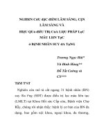
Chart 3.6.InfrapoplitelPTA restenosis
Conclusion: Restenosis rate of infrapopliteal PTA was 34.1%, 50%,
65.9% after 3 months, 6 months, 12 months respectively.
Bảng 3.30.Restenosis and Clinical stages
Clinical stages(1)
Restenosis
rate – 3
months
FU
Yes
No
p
Rutherford2
(n, %)
Rutherford
3
(n, %)
Rutherford
4
(n, %)
Rutherford
5
(n, %)
Rutherford
6
(n, %)
1
(100)
0
(0)
3
(25)
9
(75)
11
(39.3)
17
(60.7)
10
(25.6)
29
(74.4)
5
(62.5)
3
(37.5)
p 1-2
>
0.05
13
(2)
Sum
1
12
28
39
8
Restenosis
rate – 6
months
FU
(3)
Yes
1
(100)
0
(0)
1
4
(33.3)
8
(66.7)
12
13
(46.4)
15
(53.6)
28
19
(48.7)
20
(51.3)
39
7
(87.5)
1
(12.5)
8
p 1-3
>
0.05
1
(100)
0
(0)
1
4
(33.3)
8
(66.7)
12
19
(67.9)
9
(32.1)
28
26
(66.7)
13
(33.3)
39
8
(100)
0
(0)
8
p 14<
0.05
Restenosis
rate – 12
months
FU
(4)
No
Sum
Yes
No
Sum
Conclusion: The more severe clinical stages, the higher infrapopliteal
restenosis rate. Restenosis rate of infrapopliteal PTA after 12 months
was 33.3%, 67.9%, 66.7%, 100% of Rutherford 3, Rutherford 4,
Rutherford 5, Rutherford 6 respectively.
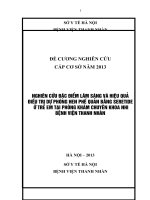
Chart 3.7.Infrapoplitel PTA reocclusion
Conclusion: Reocclusion rate of infrapopliteal PTA was 18.2%,
25%, 35.6% after 3 months, 6 months, 12 months respectively.
Table 3.34.Infrapoplitealreintervention
Re-intervention rate
3 months FU
Re-intervention rate
Patient (n= 88)
%
Patient (n= 88)
5
5.7
13
14
6 months FU
%
14.8
Re-interventionrate
Patient (n= 87)
17
12 months FU
%
19.8
Mean duration time of reintervention (month) = 6.0 2.5
Conclusion:Re-intervention rate of infrapopliteal PTA was 14.8%,
19.8% after 6 months, 12 months respectively. Mean duration time
of re-intervention was 6.0 2.5 months.
Table 3.35.Mortality of infrapopliteal PTA
Mortality 1 month FU
(n=91)
Mortality 3 months FU
(n=91)
Mortality 6 months FU
(n=91)
Mortality 12 months FU
(n=91)
Patient
%
Patient
%
Patient
%
Patient
%
1
1.1
1
1.1
2
2.2
3
3.3
Conclusion: Mortality of infrapopliteal PTA was 1.1%, 3.3% after 1
month, 12 months respectively (1 acute pneumonia case after 1 month, 2
intracereberalhaemorrahge cases).
3.3. FACTORS INFLUENCING CLINICAL OUTCOMES
3.3.1. Affection of clinical factors
Table 3.36.Clinical stages and clinical outcomes
Clinical stages(1)
Hemodynamic
success
(2)
Clinical
success
(3)
p
Rutherford
2
(n, %)
Rutherford
3
(n, %)
Rutherford
4
(n, %)
Rutherford
5
(n, %)
Rutherford
6
(n, %)
1
(100)
1
(8.3)
3
(10.7)
12
(29.3)
5
(55.6)
Yes
0
(0)
11
(91.7)
25
(89.3)
29
(70.7)
4
(44.4)
Sum
1
12
28
41
9
No
0
(0)
1
(100)
0
(0)
12
(100)
0
(0)
28
(100)
2
(4,9)
39
(95.1)
1
(11,1)
8
(88.9)
No
Yes
p12<
0.05
p13>
0.05
15
Complications
(4)
Sum
1
12
28
41
9
No
1
(100)
0
(0)
12
(100)
0
(0)
28
(100)
0
(0)
38
(92.7)
3
(7,3)
9
(100)
0
(0)
1
12
28
38
9
Yes
Sum
Conclusion: The more severe clinical stages, the lower success rate
of hemodynamics. Success rate of hemodynamics was 91.7%,
89.3%, 70.7%, 44.4% of Rutherford 3, Rutherford 4, Rutherford 5,
Rutherford 6 respectively.
Table 3.39.Arterial lesion levels and outcomes
Hemodynamic
success
Yes
(2)
No
Clinical success
Sum
Yes
(3)
No
Complications
Sum
Yes
(4)
No
Reintervention 3
months FU
(5)
Reintervention 6
months FU
(6)
Reintervention 12
months FU
(7)
Sum
Yes
No
Sum
Yes
No
Sum
Yes
No
Arterial lesion levels(1)
Single- level
Multi- level
(n, %)
(n, %)
21
48
(60)
(85.7)
14
8
(40)
(14.3)
35
56
34
54
(97.1)
(96.4)
1
2
(2.9)
(3.6)
35
56
1
2
(2.9)
(3.6)
34
54
(97.1)
(96.4)
35
56
0
5
(0)
(9.3)
34
49
(100)
(90.7)
34
54
1
12
(2.9)
(22.2)
33
42
(97.1)
(77.8)
33
53
2
15
(6.1)
(28.3)
31
38
(93.9)
(71.7)
p
p 1-2 < 0.05
p 1-3 > 0.05
p 1-4 > 0.05
p 1-5 > 0.05
p 1-6 < 0.05
p 1-7 < 0.05
p14>
0.05
16
Sum
33
53
Conclusion: Multi-level arterial lesion was higher than single-level
arterial lesion in hemodynamic success rate (OR = 4),
reintervention6 months FU (OR = 17.3), reintervention 12 months
FU (OR = 6.1).
3.3.2. Affection of revascularization strategy
Table 3.40.Number of tibial artery revascularization and outcomes
Number of tibial artery
revascularization(1)
Wound healing 1
month FU
(2)
Wound healing 3
months FU
(3)
Restenosis 3
months FU
(4)
Restenosis 6
months FU
(5)
Restenosis 12
months FU
(6)
Yes
No
Sum
Yes
No
Sum
Yes
No
Sum
Yes
No
Sum
Yes
No
Sum
1 tibial artery
(n, %)
8
(50)
8
(50)
16
13
(81.2)
3
(18.8)
16
8
(25.8)
23
(74.2)
31
13
(41.9)
18
(58.1)
31
17
(54.8)
14
(45.2)
31
≥2tibialartery
(n, %)
5
(15.6)
27
(84.4)
32
21
(67.7)
10
(32.3)
31
22
(38.6)
35
(61.4)
57
31
(54.4)
26
(45.6)
57
41
(71.9)
16
(28.1)
57
p
p 1-2 < 0.05
p 1-3 > 0.05
p 1-4 > 0.05
p 1-5 > 0.05
p 1-6 > 0.05
Conclusion: Wound healing rate after 1 month of 1 tibial artery
group was higher than ≥ 2 tibial artery group, 50% vs 15.6%
respectively, OR = 5.4
17
Chapter 4
DISCUSSION
4.1. GENERAL CHARACTERISTICS
4.1.1. Clinical characteristics
- Mean age was 75.6, the age group ≥80 was 40%. Male 67.1%,
female 32.9%. Risk factors were hypertension (64.7%), diabetes
(25.9%), metabolic lipid disorder (25.9%), smoking (24.7%). The
proportion of patients with diabetes is lower than that reported in
other studies, more men than women are due to differences in
smoking rates.
- Clinical stages were Rutherford 5 (45.1%) and Rutherford 4
(30.8%).
Ulcers
and
gangrene
in
toes
(45.1%)
wasmost
common.Less common patients with ulcers and gangrene spread the
feet and legs (Rutherford 6), is the stage where reperfusion
intervention is more difficult.
4.1.2. Subclinical characteristics
- Mean ABI was 0.56, group ABI 0.4-0.75 was most common (38%).
- The artery lesion was majority arefemoral, popliteal–infrapopliteal
level (53.8%) and infrapopliteal level (38.5%). Mean tibial artery
lesion length was 20.4 cm. Infrapopliteal artery lesion classification
was TASC D (97.8%).
We believe that the above indicators are due to the proportion of
patients with diabetes is not so high, the disease has a long time of
development, causing arterial lesions was relatively severe.
4.2. TECHNICAL CHARACTERISTICS AND CLINICAL
18
OUTCOMES OF INFRAPOPLITEAL PTA
4.2.1. Technical characteristics
- Most common vascular access was from the ipsilateralfemoral at the
infrapopliteal lesion side (97.8%), antegrade revascularization was
86.8%. This is the characteristics of infrapopliteal PTA with the
predominant arterial lesion level is the femoral, popliteal – infrapopliteal
and infrapopliteal alone, high rate of chronic total occlusion.
- Reperfusion of 1 tibial artery was 58.2%, reperfusion of ≥ 2 tibial artery
was 35.2%. Fernandez's (2010) study shows that the rate of
revascularization of 1tibial artery was 80%. Reperfusion of1 tibial artery to
ischemia area is satisfactory, only when reperfusion was failed, it is
necessary to reconstructing from 2 or more tibial arteries, in order to
increase the effect. Maximum indirect perfusion. Our direct angiosome rate
reached 70.2%; This result is even higher than some reports, like that of
Lida (2014) with 63.4% of Soares (2016) only reaching 52.2%.
- Complications rate was 3.3% (3 cases with mild clinical symptom,
recovered rapidly. 1 case hematoma, 1 case distal thrombosis and 1 case
peritoneal bleeding must be open sugery).Research by Romiti (2008) this
rate was 7.8%; Okamoto (2016) announced that 12.3% had complications.
This rate was lower because the patient was younger, the disease was less
coordinated, the clinical level was less severe. The reports all showed that
the reperfusion below the knee was safer than the surgery.
4.2.2. Clinical Outcomes of Infrapopliteal PTA
- Technical success was 79.6%. The technical success rate in Romiti's
study (2008) was 89%,Kok's study (2017) was 75%. This ratio
depends on the level of the patient's disease and the skill of the
19
physician to intervene.
- Wound healing rate was 27.1%, 72.3% and 100% after 1 month, 3
months and 6 months respectively.Mean duration of wound healingwas
3.1 1.8 months. This rate in Kawarada’ study (2014) was 36.8%
and 57.5% after 3 months, 6 months respectively. Shiraki’s study
(2015) found an average duration of wound healing was 4.2 months.
Our wound healing rate was higher and the duration was shorter than
these studies because our patients often only had ulceration or
gangrene in toes.
- Amputation rate was 13.2%, limb salvage after 12 months was
100%. Limb salvage after 12 months of Sadek’ study (2009) was
81%, of Alexandrescu (2009) was 89%. Limb salvage is high is the
advantage of infrapopliteal PTA.
- Restenosis rate of infrapopliteal PTA was 34.1%, 50%, 65.9% after
3 months, 6 months, 12 months respectively. High restenosis rate is
the “Achille heel” of infrapopliteal PTA, which being studied to
improve with other techniques (eg drug-coated balloon, stent,
atherectomy,…). The restenosis rate after 12 months in Giles (2008)
was 61%,Liistro (2013) was 74%.
- Reocclusion rate of infrapopliteal PTA was 25%, 35.6% after 6
months, 12 months respectively. Mustapha's study (2016) found that
re-occlusion rate after 12 months was 36.9%. In general, reocclusion
rate is about half of restenosis rate in the same time.
- Re-intervention rate of infrapopliteal PTA was 14.8%, 19.8% after
6 months, 12 months respectively.The re-intervention rate after 12
20
months of the study of Lida (2013) was 34%, of Mustapha (2016)
was 18.2%. The re-intervention depends on lower limb arterial
lesions, risk factors control, and most importantly is the level of
treatment compliance of patients.
- Mortality of infrapopliteal PTA was 1.1%, 3.3% after 1 month, 12
months respectively (1 acute pneumonia case after 1 month, 2
intracereberalhaemorrahge cases). Giles (2008) found that the mortality
rate after 12 months was 19%, this rate in Romiti's study (2008) was
2.7%. The cause of death is due to the severity and associated diseases
caused.
4.3. FACTORS INFLUENCING CLINICAL OUTCOMES
4.3.1. Affection of clinical factors
Clinical stages
- The more severe clinical stages, the lower success rate of
hemodynamics. Success rate of hemodynamics was 91.7%, 89.3%,
70.7%, 44.4% of Rutherford 3, Rutherford 4, Rutherford 5,
Rutherford 6 respectively. Tsuchiya's (2015) study found that lower
extremity arterial disease with Rutherford stage 4 was less amputated
(12.3%) and death rate after 1 month (6.7%) than in the Rutherford
stage 5 or 6 (rates are 22.7% and 33.3%, respectively).
- The more severe clinical stages, the higher infrapopliteal restenosis
after 12 months rate. Restenosis rate of infrapopliteal PTA after 12
months was 33.3%, 67.9%, 66.7%, 100% of Rutherford 3,
Rutherford 4, Rutherford 5, Rutherford 6 respectively. Lida (2012)
found that without using cilostazol and statins, completely chronic
21
total occlusive lesions were factors that increased the restenosis rate.
Arterial lesion levels
- Multi-level arterial lesion group was higher than single-level
arterial lesion group in hemodynamic success rate (85.7% vs 60%,
OR = 4), re-intervention rate after 6 months (22.2% vs 2.9%,OR =
17.3) and 12 months (28.3% vs 6.1%, OR = 6.1). Fernandez (2011)
found that the multi-level arterial lesion group was higher than the
single-level arterial lesion group with the wound healing rate after 6
months (87% vs 69%), the duration time of completely woung
healing was faster (7.7 ± 6.6 months vs 11.5 ± 8.8 months) and the
limb salvage after 12 months (95% vs 81%)
4.3.2. Affection of revascularization strategy
Number of tibial artery revascularizations
- Wound healing rate after 1 month of 1 tibial artery group was
higher than ≥ 2 tibial artery group (50% vs 15.6% respectively, OR =
5.4). Darling's study (2016) found that the wound healing rate were
similar between the two groups, while Kobayashi (2016) found that
the reperfusion group of ≥ 2 tibial artery had higher rates of wound
healing (87%vs 79%), the time is shorter (83 days vs 142 days). The
cause of the difference of the studies is due to intervention strategies,
we choose to prioritize reperfusion directly ulcers and gangrene area,
if failure new reperfusion 2 or more tibial artery, when some other
authors choose to reperfusion as much of the tibial artery as possible.
Reperfusion properties
- Direct angisome reperfusion improved compared to indirect
22
angiosome reperfusion of the rate of wound healing after 1 month
(37.1% vs 0%) and duration time of completely woung healing (2.6 ±
1.7 months vs 4.4 ± 1.7 months). The study of Kabra (2013) found that
the rate of wound healing after 1 month, 3 months and 6 months was
higher in the direct reperfusion group than the indirect reperfusion group
(the corresponding ratios were 7.9% vs 5%, 57.6% vs 12.5%, 96.4% vs
83.3% respectively). The fact that direct reperfusion is the first priority
in below the knee revascularization, has the greatest effect on the results
of wound healing and limb salvage.
CONCLUSIONS
1. GENERAL CHARACTERISTICS
1.1. Clinical characteristics
. Mean age was 75.6, the age group ≥80 was 40%.
. Male 67.1%, female 32.9%.
. Risk factors were hypertension (64.7%), diabetes (25.9%),
metabolic lipid disorder (25.9%), smoking (24.7%).
. Clinical stages were Rutherford 5 (45.1%) and Rutherford 4
(30.8%). Ulcers and gangrene in toes (45.1%) wasmost common.
1.2. Subclinical characteristics
. Mean ABI was 0.56, group ABI 0.4-0.75 was most common (38%).
. The artery lesion was majority arefemoral, popliteal–infrapopliteal
level (53.8%) and infrapopliteal level (38.5%).
. Mean tibial artery lesion length was 20.4 cm. Infrapopliteal artery
lesion classification was TASC D (97.8%).
2. CLINICAL OUTCOMES OF INFRAPOPLITEAL PTA AND
23
FACTORS INFLUENCING CLINICAL OUTCOMES
2.1. Clinical outcomes of infrapopliteal PTA
. Technical success was 79.6%.
. Wound healing rate was 27.1%, 72.3% and 100% after 1 month, 3
months and 6 months respectively.Mean duration of wound healingwas
3.1 1.8 months.
. Amputation rate was 13.2%, limb salvage after 12 months was
100%.
. Restenosis rate of infrapopliteal PTA was 34.1%, 50%, 65.9% after
3 months, 6 months, 12 months respectively.Reocclusion rate of
infrapopliteal PTA was 25%, 35.6% after 6 months, 12 months
respectively. Re-intervention rate of infrapopliteal PTA was 14.8%,
19.8% after 6 months, 12 months respectively.
. Complications rate was 3.3%.
. Mortality of infrapopliteal PTA was 1.1%, 3.3% after 1 month, 12
months respectively
2.2. Factors influencing clinical outcomes
Clinical factors
. The more severe clinical stages, the lower success rate of
hemodynamics (Hemodynamic success rate was 91.7%, 89.3%,
70.7%, 44.4% of Rutherford 3, Rutherford 4, Rutherford 5,
Rutherford 6 respectively) and the higher infrapopliteal restenosis
after 12 months rate (12 months restenosis rate of infrapopliteal PTA
was 33.3%, 67.9%, 66.7%, 100% of Rutherford 3, Rutherford 4,
Rutherford 5, Rutherford 6 respectively).