Gastric juice-based real-time PCR for tailored Helicobacter pylori treatment: A practical approach
Bạn đang xem bản rút gọn của tài liệu. Xem và tải ngay bản đầy đủ của tài liệu tại đây (426.56 KB, 7 trang )
595
Int. J. Med. Sci. 2017, Vol. 14
Ivyspring
International Publisher
International Journal of Medical Sciences
2017; 14(6): 595-601. doi: 10.7150/ijms.18996
Research Paper
Gastric Juice-Based Real-Time PCR for Tailored
Helicobacter Pylori Treatment: A Practical Approach
Xianhui Peng1, Zhiqiang Song2, Lihua He1, Sanren Lin2, Yanan Gong1, Lu Sun1, Fei Zhao1, Yixin Gu1,
Yuanhai You1, Liya Zhou2, Jianzhong Zhang1
1.
2.
State Key Laboratory of Infectious Disease Prevention and Control, Collaborative Innovation Center for Diagnosis and Treatment of Infectious Diseases,
Chinese Center for Disease Control and Prevention, Beijing, China;
Department of Gastroenterology, Peking University Third Hospital, Beijing, China.
Corresponding authors: Prof. Jianzhong Zhang, Tel.: 86-10-58900707, Fax: 86-10-58900700, E-mail: ; and Prof. Liya Zhou, Tel.:
86-18910192576, Fax: 86-10-62357303. E-mail address:
© Ivyspring International Publisher. This is an open access article distributed under the terms of the Creative Commons Attribution (CC BY-NC) license
( See for full terms and conditions.
Received: 2016.12.31; Accepted: 2017.03.15; Published: 2017.05.15
Abstract
A gastric juice-based real-time polymerase chain reaction (PCR) assay was established to identify Helicobacter
pylori infection, clarithromycin susceptibility and human CYP2C19 genotypes and to guide the choice of proton
pump inhibitor (PPI), clarithromycin and amoxicillin treatment for tailored H. pylori eradication therapy. From
January 2013 to November 2014, 178 consecutive dyspeptic patients were enrolled for collection of gastric
biopsy samples and gastric juice by endoscopy at the Peking University Third Hospital; 105 and 73 H.
pylori-positive and -negative patients, respectively, were included in this study. H. pylori infection was defined
as samples with both a strongly positive rapid urease test (RUT) and positive H. pylori histology. A series of
primers and probes were distributed into four reactions for identifying the H. pylori cagH gene coupled with
an internal control (Rnase P gene), A2142G and A2143G mutants of the H. pylori 23S rRNA gene, and
single-nucleotide polymorphisms (SNPs) G681A of CYP2C19*2 and G636A of CYP2C19*3. The E-test and
DNA sequencing were used to evaluate the H. pylori clarithromycin susceptibility phenotype and genotype.
The SNPs CYP2C19*2 and CYP2C19*3 were also evaluated by nucleotide sequencing. The sensitivity,
specificity, positive predictive value (PPV), and negative predictive value (NPV) of this gastric juice-based
real-time PCR assay were evaluated by comparing with the same measures obtained through gastric
biopsy-based PCR and culture. The H. pylori diagnostic sensitivities of the culture, PCR, and gastric biopsy- and
gastric juice-based real-time PCR assays were 90.48% (95/105), 92.38% (97/105), 97.14% (102/105) and 100%
(105/105), respectively; the specificities of the above methods were all 100%. Higher false-negative rates were
found among the gastric biopsy samples assessed by culture (10.48%, 11/105), PCR (7.62%, 8/105) and
real-time PCR (2.86%, 3/105) than in gastric juice by real-time PCR. Regarding clarithromycin susceptibility, a
concordance of 82.98% (78/94) and discordance of 17.02% (16/94) were observed among the different
methods, discrepancies that mainly represent differences between the H. pylori clarithromycin susceptibility
phenotype and genotype. Three coinfections of susceptible and resistant strains were detected, with
resistant-to-susceptible ratios of 1.16, 3.44, and 8.26. The CYP2C19 genotyping results from gastric juice by
real-time PCR were completely in accordance with those obtained from biopsy samples by conventional PCR.
This gastric juice-based real-time PCR assay is a more accurate method for detecting H. pylori infection,
clarithromycin susceptibility and CYP2C19 polymorphisms. The method may be employed to inform the
choice of proton pump inhibitor (PPI), clarithromycin and amoxicillin treatment for tailored H. pylori
eradication therapy.
Key words: gastric juice; real-time PCR; tailored H. pylori eradication.
Introduction
The global consensus statement on the
management of Helicobacter pylori eradication
recommends standard triple therapy based on a
proton pump inhibitor (PPI), clarithromycin and
amoxicillin (metronidazole) as the first-line treatment
[1]. However, several studies have reported that the
first eradication rate was much lower than 80% [2-4].
In the Maastricht IV consensus report, clarithromycin
resistance was the most important reason for
eradication failure based on the standard triple
596
Int. J. Med. Sci. 2017, Vol. 14
therapy. Indeed, PPI-clarithromycin-containing triple
therapy without prior susceptibility testing should be
abandoned if the clarithromycin resistance rate in the
region is greater than 15-20% [5]. A multi-centre
randomized trial reported a primary resistance rate of
H. pylori to clarithromycin ranging from 0 to 40%
(average 23.9%), with the resistance rate varying
among people from different regions; in addition, a
higher secondary drug resistance rate was noted in
the same population [6]. A study evaluating H. pylori
antibiotic resistance in Beijing from 2000 to 2009
revealed an increase in the clarithromycin resistance
of H. pylori, with an average rate of 37.2% [7]. Thus,
more attention should be paid to tailored H. pylori
eradication therapy, which may help to improve
eradication rates and reduce H. pylori resistance.
Culture and standard susceptibility testing to
antimicrobial agents should be performed in
populations with a high clarithromycin resistance rate
if standard clarithromycin-containing therapy is being
considered [5]. However, culture and antibiotic
resistance testing methods have many disadvantages,
including requirements of time and laboratory
equipment as well as strong technology and heavy
workloads, limiting their widespread application in
clinical practice. When standard susceptibility testing
based on H. pylori isolation is not possible, molecular
tests, such as polymerase chain reaction (PCR) or
real-time PCR, can be used to detect H. pylori infection
and clarithromycin resistance using gastric biopsy
samples [8,9]. As H. pylori has a focal distribution in
different parts of the gastric mucosa, false-negative
results often occur with single-site gastric
biopsy-based detection. In contrast, gastric juice,
which contains constantly shed gastric epithelial cells
and bacteria from the entire stomach, should be more
suitable for detecting actual H. pylori infection [10].
Point mutations in the peptidyl transferase
region of the 23S rRNA gene frequently confer
macrolide resistance. The most common point
mutations are A2143G (69.8%) and A2142G (11.7%),
which account for more than 80% of clarithromycin
resistance [11]. Other mutations, such as A2142C,
A2115G, G2141A, T2717C, A2115G, G2141A and
A2142T, are rarely observed [12, 13]. Another
important
effect
on
eradication
may
be
polymorphisms of the cytochrome P450 (CYP2C19)
gene, the genotype of which determines the metabolic
rate of PPI in the human liver. For example, the
CYP2C19 wild-type allele (CYP2C19*1) has high
enzymatic activity compared to the mutant-type
CYP2C19*2 and CYP2C19*3 alleles. CYP2C19*2 and
CYP2C19*3 are located in the fifth (G681A) and fourth
(G636A) exons, respectively. Accordingly, the
CYP2C19 phenotype has been classified into three
groups:
homozygous
extensive
metabolizers
(Hom-EMs), heterozygous extensive metabolizers
(Het-EMs) and poor metabolizers (PMs) [14,15].
In this study, we established an easy and
accurate diagnostic technology based on gastric juice
to identify H. pylori infection, H. pylori clarithromycin
susceptibility and CYP2C19 gene polymorphisms in
patients.
Materials and Methods
Bacterial strains
A total of 44 DNA samples, including 28 samples
from common non-H. pylori bacteria isolated from the
gastric mucosa, 15 enterobacterial samples and one
human tissue sample, were provided by the
Department of Communicable Disease Diagnostics,
National Institute for Communicable Disease Control
and Prevention, Chinese Centre for Disease Control
and Prevention (see the supplement, Table S1).
Patients and specimens
Patients at Peking University Third Hospital
with dyspeptic symptoms were enrolled from January
2013 to November 2014. In total, 178 patients were
randomly selected in this trial, including 105 cases
that were both rapid urease test (RUT) strongly
positive (becoming red within 2 min) and histology
test (Warthin-Starry silver staining) positive and 73
cases that were negative for both RUT (no colour
change within 2 hours) and histology. We considered
the 105 cases as the HP-positive group and the
remaining 73 cases as the HP-negative group. Of the
subjects, 90 were women and 88 men, with ages
ranging from 19 to 68 years (mean±SD, 41.6±12.8).
Four gastric mucosa biopsies and 5-10 mL of fasting
gastric juice specimens were collected by
gastrointestinal endoscopy examination. None of the
patients received any H. pylori eradication therapy,
including antibiotics and acid-suppressive drugs
(PPIs, H2-receptor antagonists, bismuth agent, or
antacids). For details, refer to the supplementary
information (Table S2).
Ethical considerations
The study was approved by the independent
Ethics Committee of Peking University Health Science
Centre (IRB00001052-0709) and by the Research Ethics
Committee
of
the
National
Institute
for
Communicable Disease Control and Prevention (No:
ICDC-2013001) and was performed in accordance
with the ethical guidelines of the Declaration of
Helsinki, Good Laboratory Practices and Good
Clinical Practices. Written informed consent was
obtained from each patient prior to study enrolment.
597
Int. J. Med. Sci. 2017, Vol. 14
Sample DNA extraction, H. pylori isolation and
E-test
One piece of a gastric biopsy sample was
homogenized using a sterile glass homogenizer. Half
of the gastric biopsy tissue homogenate was directly
used for DNA extraction. Approximately 1 mL of
gastric fluid was neutralized with an equivalent
amount of Tris-HCl (0.67 mol/L, pH 7.4). The mixture
was mixed well and centrifuged at 13,000 rpm for 10
minutes. The supernatants were removed, and the
pellets were reserved. Genomic DNA was extracted
using the QIAamp DNA Mini Kit (QIAGEN,
Germany).
The other half of the homogeneous solution was
uniformly coated onto the surface of a Karmali agar
plate supplemented with 7% defibrinated sheep blood
(Biotek Medical Device Co., Ltd., Beijing, China) and
an appropriate H. pylori selective supplement
(OXOID, England). The plates were incubated for 2-7
days in a microaerophilic environment (5% O2, 10%
CO2 and 85% N2) at 37°C. The isolates were identified
by Gram staining, and positivity was confirmed by
urease, oxidase and catalase traits. Clarithromycin
susceptibility was assessed by the E-test, with the
addition of 200 μL of inoculum onto plates, which was
equivalent to the McFarland 2 opacity standard (8.8
×107), and incubation for 2 days in a microaerophilic
environment at 37°C. Isolates were considered
resistant when the minimal inhibitory concentration
(MIC) value was more than 2 μg/mL.
Conventional PCR
Four pairs of specific primers focused on target
genes were used to confirm the presence of H. pylori,
H. pylori 23S rRNA gene mutations and CYP2C19*2
and CYP2C19*3 genotypes in the gastric biopsy
specimens. The primer sequences are shown in table
1. All of the PCR reactions were performed in a 25-µL
volume containing 12.5 μL 2× Easy Taq® PCR
SuperMix (Transgene, Beijing, China), 0.5 μL forward
and reverse primers (2 μL each), 2 μL template DNA
and 9.5 μL nuclease-free water. The PCR
amplifications were performed under the following
conditions: denaturation at 94°C for 5 min, 40 cycles of
denaturation at 94°C for 30 seconds, annealing at 55°C
for 30 seconds, and extension at 72°C for 30 seconds,
and a final extension at 72°C for 7 min. The
amplification products were analysed by 1.5%
agarose gel electrophoresis. Positive products were
sequenced using both forward and reverse primers.
Table 1. Primer sequences used for conventional PCR and sequencing
Target gene
ureB
H. pylori 23S rRNA
CYP2C19*2
CYP2C19*3
PCR primer (5'-3')
F: AAAGAGCGTGGTTTTCATGGCG
R: GGGTTTTACCGCCACCGAATTTAA
F: AGCGATGTGGTCTCAGCA
R: CAAGGGTGGTATCTCAAGG
F: AATTACAACCAGAGCTTGGC
R: TATCACTTTCCATAAAAGCAAG
F: AAATTGTTTCCAATCATTTAGCT
R: ACTTCAGGGCTTGGTCAATA
Product (bp)
217 bp
Reference
[16]
444 bp
This study
168 bp
[17]
271 bp
[18]
Table 2. Primer and probe sequences and their distribution for multiple real-time PCR
Distribution
Reaction 1
Target gene
RnaseP
cagH
Reaction 2
HP23SrRNA
Reaction 3
CYP2C19*2
Reaction 4
CYP2C19*3
Primer
RnaseP-F
RnaseP-R
RnaseP-P
cagH-F
cagH-R
cagH-P
HP23S-F
HP23S-R
HP23S-AA
HP23S-GA
HP23S-AG
CY2-F
CY2-R
CY2-G
CY2-A
CY3-F
CY3-R
CY3-G
CY3-A
Sequence (5’–3’)
5’-AGATTTGGACCTGCGAGCG-3’
GAGCGGCTGTCTCCACAAGT
VIC-TTCTGACCTGAAGGCTCTGCGCG-MGB
TTATGTTAGAAATCGCTTGAGTGTCA
CGCTTCTCAAATGATACTTAATCAATC
FAM-AGGTGCTAGTAGCTAATC-MGB
TTCAGTGAAATTGTAGTGGAGGTG
TCCCATTAGCAGTGCTAAGTTGTA
FAM-AGACGGAAAGACC-MGB
VIC-AGACGGGAAGACC-MGB
VIC-AGACGGAGAGACC-MGB
GCTTGGCATATTGTATCTATACCTT
GATTCTTGGTGTTCTTTTACTTTCT
FAM-ATTTCCCGGGAACC-MGB
VIC-ATTTCCCAGGAACC-MGB
AATTGAATGAAAACATCAGGATTG
ACTGTAAGTGGTTTCTCAGGAAGC
FAM-CTGGATCCAGGTAAG-MGB
VIC-CCTGAATCCAGGTAAG-MGB
Product
71
GenBank No.
U77665.1
98
FR666857.1
98
NR_076155.1
85
NG_008384.2
88
NG_008384.2
598
Int. J. Med. Sci. 2017, Vol. 14
Real-time PCR
Five primers and nine matching Taqman probes
targeting the H. pylori cagH and 23S rRNA genes and
the human RnaseP, CYP2C19*2 and CYP2C19*3 genes
were designed for this assay. All sequences obtained
from
NCBI
Entrez
Nucleotide
Database
( />were
aligned using Vector NTI alignment software
( />-science/cloning/vector-nti-software.html).
The
primers and probes were designed using Primer
Express 3.0 software (Applied Biosystems). The
sequences of the primers and probes used in this
study are summarized in table 2.
The reaction mixture (20 μL) was prepared as
follows: 2 μL 10× PCR buffer, 2 mM MgCl2
(Platinum® Taq DNA Polymerase, Invitrogen,
Thermo Fisher, USA), 0.4 μL dNTPs (Promega, USA),
0.5 μM forward and reverse primers (Sangon Biotech,
Shanghai, China), 0.2 μM probe (ABI, USA), 0.2 μL
Taq DNA polymerase (Platinum® Taq DNA
Polymerase, Invitrogen, Thermo Fisher, USA), 2 μL
DNA template, and up to 20 μL nuclease-free water.
Evaluation of multiple real-time PCR
performance
The specificity of the cagH probe in the real-time
PCR was assessed using bacterial DNA from 28
common bacteria in addition to H. pylori from gastric
mucosa and 15 enterobacteria.
To assess the sensitivity of this assay, we
constructed recombination plasmids containing a
target gene or point mutation from the reference
strains. To evaluate the detection limit of each probe
in this assay, a series of 10-fold dilutions of the
recombination plasmids ranging from 1×10⁹
copies/μL to 1×100 copies/μL were used as the
template. Simultaneously, the correlation coefficient R
and amplification efficiency of each primer/probe
were determined using standard curves based on
10-fold serial dilutions of the recombinant plasmids.
To evaluate assay precision, the intra- and
inter-assay variability were evaluated to reveal the
corresponding repeatability and reproducibility,
respectively. High, medium and low plasmid
concentrations (1×10⁷ copies/μL, 1×10⁵ copies/μL,
and 1×10² copies/μL, respectively) were used as the
template. To estimate intra-experimental variation,
nine positive standard plasmids with different copy
numbers were detected three times in the same
experiment.
To
determine
inter-experimental
variation, the same plasmids were tested on different
days in three different experiments. All 178 gastric
juice samples were detected by this multiple real-time
PCR approach, and the results were compared with
those obtained by culture and gastric biopsy-based
PCR.
Statistical analysis
A P value <0.05 was considered significant. All
statistical tests and figures in our study were prepared
using R statistical software version 3.3.1 (http://
www.r-project.org/).
Results
Real-time PCR assay development
Bacterial DNA from 28 common gastric bacteria
and 17 enterobacteria were detected with our four
assays, and no positive amplification was observed.
The cagH and 23S rRNA assays were only positive for
H. pylori and were negative for the other bacteria. The
CYP2C19*2 and CYP2C19*3 assays were only positive
for the human genome. A series of 10-fold dilutions of
plasmid DNA (ranging from 1×10⁹ to 1×100
copies/μL) was used as the template and tested in
four multiple real-time PCR reactions, with each
plasmid concentration repeated three times. The limit
of detection (LOD) of all probes was 102 copies/μL of
plasmid DNA. The LODs of the cagH-prob and
RnaseP-prob in the first-group PCR were 101
copies/μL, with average Ct values of 36.66 and 36.57,
respectively. The LODs of the HP23S-AA, HP23S-AG,
and HP23S-GA probes in the second-group PCR were
100, 100, and 101 copies/μL, with average Ct values of
37.30, 37.91, and 37.33, respectively. The LODs of the
CY2-G and CY2-A probes in the third-group PCR
were 101 and 102 copies/μL, with average Ct values of
35.03 and 35.20, respectively. The LODs of the CY3-G
and CY3-A probes in the fourth-group PCR were 101
and 102 copies/μL, with average Ct values of 37.27
and 38.00, respectively. No significant differences
were found in repeatability and reproducibility
evaluations (P>0.05).
Real-time PCR performance for H. pylori
infection diagnosis
In the H. pylori-positive group, the positive rates
obtained using culture, PCR, and gastric biopsy- and
gastric juice-based real-time PCR assays were 90.48%
(95/105), 92.38% (97/105), 97.14% (102/105) and 100%
(105/105), respectively. The consistency rate for all
four H. pylori infection diagnostic methods was
89.52% (94/105). Four discrepancies occurred
between the culture and PCR methods, including
three cases with positive PCR results but negative
culture results and one case with a positive culture
result but a negative PCR result. Ten false-negative
results were found by cultures, eight by PCR and
three by gastric biopsy-based real-time PCR.
599
Int. J. Med. Sci. 2017, Vol. 14
Table 3. Comparison of the performances of culture, PCR and real-time PCR for the diagnosis of H. pylori infection
Parameter
Sensitivity
Specificity
Positive predictive value
Negative predictive value
Result (%)
Culture
(gastric biopsy-based)
90.48% (95/105)*
100% (73/73)
90.48% (95/105)*
87.95% (73/83)*
PCR
(gastric biopsy-based)
92.38% (97/105)*
100% (73/73)
92.38% (97/105)*
90.12% (73/81)*
Real-time PCR
(gastric biopsy-based)
97.14% (102/105)**
100% (73/73)
97.14% (102/105)**
96.05% (73/76)***
Real-time PCR
(gastric juice-based)
100% (105/105)
100% (73/73)
100% (105/105)
100% (73/73)
*: P<0.05, **: P=0.081, ***: P=0.086.
For these false-negative cases, both the H.
pylori-specific ureB and the 23S rRNA gene fragments
could be amplified from the corresponding gastric
juice specimen, and these PCR products were
confirmed by nucleotide sequencing. In the H.
pylori-negative group, no false-positive results were
found in the gastric biopsy or gastric juice samples by
PCR or real-time PCR. The significance levels for
sensitivity, specificity, positive predictive value (PPV)
and negative predictive value (NPV) among the
different methods are displayed in table 3.
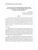
Additionally, we compared the distribution of Ct
values for real-time PCR between the gastric biopsy
and gastric juice specimens. As shown in figure 1, the
Ct values obtained from the gastric juice samples
(24.27±3.05) were significantly higher than those
obtained from the gastric biopsy samples (25.75±3.32).
discordance among the methods. These discrepancies
suggest inconsistency between the genotype and
phenotype. In three cases, both resistant and
susceptible genotypes were detected simultaneously
by PCR and real-time PCR using both gastric biopsy
and gastric juice specimens, whereas E-test results
showed only phenotypic resistance or susceptibility.
The resistant-to-susceptible ratios detected by
real-time PCR for gastric juice were 1.16, 3.44, and
8.26.
Additionally, we found 11 cases that were
culture-negative but PCR- or real-time PCR-positive.
In three cases, the clarithromycin genotype obtained
by real-time PCR using gastric juice was in complete
agreement with the genotype based on gastric
biopsies from the same patients determined by either
PCR or real-time PCR. The clarithromycin genotype
in another five cases identified by gastric juice-based
real-time PCR was confirmed by gastric biopsy-based
real-time PCR, though the PCR results were negative.
Moreover, three cases were only detected by gastric
juice-based real-time PCR. The details are provided in
table 4.
Table 4. Comparison of clarithromycin susceptibility testing by
E-test, PCR and real-time PCR
Figure 1. Comparison of Ct value distributions between gastric biopsy and
gastric juice specimens.
Clarithromycin susceptibility testing
Regarding clarithromycin susceptibility, we
found 82.98% (78/94) concordance among the
different methods for the 94 H. pylori-positive cases,
which consisted of 40 clarithromycin-susceptible
cases and 38 clarithromycin-resistant cases. The
discrepancies accounted for 15.24% (16/94) of the
Gastric biopsy
E –test
PCR
S
S
R
R
S
H
R
H
R
S
S
R
—
R
—
—
—
—
—
S
—
—
—
—
Total
Real-time PCR
S
R
H
H
S
R
R
R
—
S
S
—
Gastric juice
Real-time PCR
S
R
H
H
S
R
R
R
R
S
S
S
No. of gastric
specimens (%)
40 (38.10%)
38 (36.19%)
1 (0.01%)
2 (0.02%)
12 (11.43%)
1 (0.01%)
1 (0.01%)
3 (0.03%)
2 (0.03%)
2 (0.02%)
2 (0.01%)
1 (0.01%)
105
S, susceptible; R, resistant; H, heterogeneous.
Human CYP2C19 genotyping
The CYP2C19 genotype in all 178 patients was
determined by gastric biopsy PCR coupled with
nucleotide sequencing. Gastric biopsy PCR results
600
Int. J. Med. Sci. 2017, Vol. 14
and gastric juice-based real-time PCR provided
identical
results
regarding
CYP2C19*2
and
CYP2C19*3 mutations.
Discussion
A real-time PCR method was developed to guide
tailored H. pylori therapy through easy and accurate
detection of H. pylori 23S rRNA gene mutations and
the human CYP2C19 genotype from gastric juice
samples. The gastric juice-based real-time PCR results
were compared with results acquired using
conventional diagnostic methods with strongly
positive RUT and histology-positive biopsy
specimens. Gastric juice-based real-time PCR
demonstrated a higher sensitivity and NPV compared
to culture and PCR using gastric biopsy samples for
the diagnosis of H. pylori. Although no remarkable
significance was determined regarding specificity,
based on PPV and NPV evaluations for gastric
biopsy-based real-time PCR (P>0.05), we can
speculate that significance may be apparent when a
larger sample size is assessed. Taking health
economics into account, RUT and histology were
performed for the diagnosis of H. pylori infection. For
RUT-positive cases, gastric juice-based real-time PCR
showed 100% concordance, whereas false-negative
results were obtained by culture, PCR and gastric
biopsy-based real-time PCR. Such false negatives may
have occurred for the following reasons: ① the ‘focal
distribution’ of H. pylori in the gastric mucosa may
lead to low-level colonization or absence in some
gastric niches; ② contamination by other bacteria that
suppress H. pylori overgrowth; ③ the presence of
non-culturable coccoid forms; ④ loss of viability
during transport; and ⑤ reduced sensitivity in
patients with bleeding peptic ulcers detected using
classical diagnostic methods with gastric biopsies [19,
20]. Compared with these conventional diagnostic
methods, our gastric juice-based real-time PCR
exhibits the following prominent advantages: gastric
juice reflects the real H. pylori infection status in the
entire gastric environment; there is no need for viable
bacteria and critical transport conditions for culture;
Taqman-MGB probe real-time PCR has higher
sensitivity than traditional methods [21]; and gastric
fluid specimens appear to be more suitable for
patients with bleeding tendencies.
Because tailored treatment based on 23S rRNA
mutations and CYP2C19 polymorphisms yield a
higher H. pylori eradication rate than the empirical
standard triple therapy, H. pylori susceptibility to
clarithromycin and the human CYP2C19 genotype
should be evaluated. Culture and the E-test are often
employed
to
determine
the
clarithromycin
susceptibility phenotype, whereas PCR with
nucleotide sequencing is used to identify 23S rRNA
and CYP2C19 genotypes. However, these classic
genotype methods are usually time-consuming and
have notable laboratory equipment requirements,
which are not applicable in daily clinical practice. In
our study, the gastric juice-based real-time PCR was
completed within 1 hour and 40 minutes (not
including DNA extraction). We found 82.97% (78/94)
concordance and 17.02% (16/94) discordance among
the three methods using gastric biopsy or gastric juice
samples. These discrepancies were most likely
because the A2142G to A2143G ratio accounts for
approximately 80% of all mutations causing
clarithromycin resistance. The results are in
agreement with the results reported in the literature
[11]. Additionally, three mixed infections of
susceptible and resistant strains were simultaneously
detected by real-time PCR and PCR, whereas one
infection was classified as susceptible and another
two as resistant by culture. The resistant-tosusceptible ratios tested by real-time PCR were 1.16,
3.44, and 8.26. Thus, resistant strains play a major role
in the entire gastric microenvironment, and we
should avoid using clarithromycin when devising an
administration scheme.
Despite the superior performance of our gastric
juice-based real-time PCR, inevitable shortcomings
exist. Some hot-spot mutations associated with
clarithromycin resistance should be added to improve
the detection accuracy. To enhance patient
compliance and reduce discomfort, the string test can
be adopted, instead of endoscopy, for collecting
gastric juice. Indeed, obtaining gastric fluid specimens
using the string test is suitable for large-scale
population screening.
Conclusions
In summary, we established a gastric juice
Taqman-MGB-based real-time PCR method that
could conveniently and accurately determine the
A2142G or A2143G mutation associated with
clarithromycin resistance and the human CYP2C19
genotype. In this manuscript, we show that our
method can overcome many flaws and deficiencies
compared to the use of gastric biopsy specimens
tested using various traditional detection methods.
Four obvious advantages were observed: ① higher
sensitivity of H. pylori diagnosis; ② low false-negative
results caused by focal distribution; ③ precise
instructions to assess H. pylori clarithromycin
susceptibility, especially for coinfections with
clarithromycin-resistant and susceptible strains; and
④ easier operation and a shorter time requirement.
This gastric juice-based real-time PCR method
demonstrated better performance than culture and
Int. J. Med. Sci. 2017, Vol. 14
gastric biopsy-based PCR. Thus, gastric juice-based
real-time PCR is a more accurate method that can be
used to guide individualized H. pylori eradication.
Supplementary Material
Supplemental table s1, table s2.
/>
Acknowledgements
This work was supported by funding from the
China
Mega-Project
for
Infectious
Disease
(2011ZX10004-001), a grant from the National
Technology R&D Program in the 12th Five-Year Plan
of China (2012BAI06B02) and a grant from the State
Key Laboratory of Infectious Disease Prevention and
Control (SKLID) (2014SKLID102).
601
15. Kuo CH, Lu CY, Shih HY, Liu CJ, Wu MC, Hu HM, Hsu WH, Yu FJ, Wu DC,
Kuo FC. CYP2C19 polymorphism influences Helicobacter pylori eradication.
World J Gastroenterol. 2014; 20: 16029-36.
16. Liu J, He L, Haesebrouck F, Gong Y, Flahou B, Cao Q, Zhang J. 2015.
Prevalence of Coinfection with Gastric Non-Helicobacter pylori Helicobacter
(NHPH) Species in Helicobacter pylori-infected Patients Suffering from
Gastric Disease in Beijing, China. Helicobacter. 2015; 20: 284-90.
17. Griese EU, Läpple F, Eichelbaum M. 1999. Detection of CYP2C19 alleles *1, *2
and *3 by multiplex polymerase chain reaction. Pharmacogenetics. 1999; 9:
389-91.
18. De Morais SM , Wilkinson GR, Blaisdell J, Meyer UA, Nakamura K, Goldstein
JA. Identification of a new genetic defect responsible for the polymorphism of
(S)-mephenytoin metabolism in Japanese. Mol Pharmacol. 1994; 46: 594-8.
19. Colin R, et al. Low sensitivity of invasive tests for detection of Helicobacter
pylori infection in patients with bleeding ulcer. Gastroenterol Clin Biol. 2000;
24: 31-5.
20. Lo CC, et al. Polymerase chain reaction: a sensitive method for detecting
Helicobacter pylori infection in bleeding peptic ulcers. World J
Gastroenterol. 2005; 11: 3909-14.
21. P. Narayanasamy. Molecular Biology in Plant Pathogenesis and Disease
Management: Microbial Plant Pathogens. In: P. Narayanasamy. Molecular
Biology in Plant Pathogenesis and Disease Management: Microbial Plant
Pathogens. Germany: Springer Netherlands; 2008: 7-158.
Competing Interests
The authors have declared that no competing
interest exists.
References
1.
2.
3.
4.
5.
6.
7.
8.
9.
10.
11.
12.
13.
14.
Group EHpS. Current European concepts in the management of Helicobacter
pylori infection. The Maastricht Consensus Report. Gut 1997; 41:8-13.
Graham DY, Fischbach L. Helicobacter pylori treatment in the era of
increasing antibiotic resistance. Gut 2010; 59:1143-53.
Malfertheiner P, Bazzoli F, Delchier J. Helicobacter pylori eradication with a
capsule containing bismuth subcitrate potassium, metronidazole, and
tetracycline given with omeprazole versus clarithromycin-based triple
therapy: a randomised, open-label, non-inferiority, phase 3 trial (vol 377, pg
905, 2011). Lancet 2011; 377:905-13.
Zhiqiang Song, Liya Zhou, Jianzhong Zhang, et al. Hybrid Therapy as
First-Line Regimen for Helicobacter pylori Eradication in Populations with High
Antibiotic Resistance Rates. Helicobacter 2016; 21:382-8.
Malfertheiner P, Megraud F, O'Morain CA, Atherton J, Axon AT, Bazzoli F,
Gensini GF, Gisbert JP, Graham DY, Rokkas T, El-Omar EM, Kuipers EJ,
European Helicobacter Study G. Management of Helicobacter pylori
infection--the Maastricht IV/ Florence Consensus Report. Gut 2012; 61:646-64.
Hong Cheng, Fulian Hu, Yong Xie, et al. Prevalence of Helicobacter pylori
Resistance to Antibiotics and its Influence on the Treatment outcome in China:
A Multicenter Clinical Study. Chin J Gastroenterol 2007; 12: 525-30.
Gao W, Cheng H, Hu F, Li J, Wang L, Yang G, Xu L, Zheng X.
The evolution of Helicobacter pylori antibiotics resistance over 10 years in
Beijing, China. Helicobacter 2010; 15:460-6.
Chisholm SA, Owen RJ, Teare EL, Saverymuttu S. PCR-based diagnosis of
Helicobacter pylori infection and real-time determination of clarithromycin
resistance directly from human gastric biopsy samples. J Clin Microbiol. 2001;
39: 1217-20.
Kargar M, Ghorbani-Dalini S, Doosti A, Souod N. Real-time PCR for
Helicobacter pylori quantification and detection of clarithromycin resistance
in gastric tissue from patients with gastrointestinal disorders. Res Microbiol.
2012; 163: 109-13.
Yakoob J, Rasool S, Abbas Z, Jafri W, Abid S, Islam M, Ahmad Z. Gastric
juice for the diagnosis of H pylori infection in patients on proton pump
inhibitors. World J Gastroenterol. 2008; 14: 1539-43.
Megraud F. H pylori antibiotic resistance: prevalence, importance, and
advances in testing. Gut. 2004; 53: 1374-84.
Saez J, Belda S, Santibanez M, Rodriguez JC, Sola-Vera J, Galiana A,
Ruiz-Garcia M, Brotons A, Lopez-Girona E, Girona E, Sillero C, Royo G.
Real-time PCR for diagnosing Helicobacter pylori infection in patients with
upper gastrointestinal bleeding: comparison with other classical diagnostic
methods. J Clin Microbiol. 2012; 50: 3233-7.
Van der Ende A, Van Doorn LJ, Rooijakkers S, Feller M, Tytgat GN, Dankert J.
Clarithromycin-susceptible and -resistant Helicobacter pylori isolates with
identical randomly amplified polymorphic DNA-PCR genotypes cultured
from single gastric biopsy specimens prior to antibiotic therapy. J Clin
Microbiol. 2001; 39: 2648-51.
Ozdil B, Akkiz H, Bayram S, Bekar A, Akgöllü E, Sandikçi M. Influence of
CYP2C19 functional polymorphism on Helicobacter pylori eradication. Turk J
Gastroenterol. 2010; 21: 23-8.