High quality in vitro expansion of human endothelial progenitor cells of human umbilical vein origin
Bạn đang xem bản rút gọn của tài liệu. Xem và tải ngay bản đầy đủ của tài liệu tại đây (1.16 MB, 8 trang )
Int. J. Med. Sci. 2017, Vol. 14
Ivyspring
International Publisher
294
International Journal of Medical Sciences
2017; 14(3): 294-301. doi: 10.7150/ijms.18137
Research Paper
High quality in vitro expansion of human endothelial
progenitor cells of human umbilical vein origin
Yan Mou1, 2, Zhen Yue1, Haiying Zhang1, Xu Shi1, 3, Mingrui Zhang2, Xiaona Chang1, Hang Gao1,
Ronggui Li1 and Zonggui Wang2
1.
2.
3.
Key Laboratory of Pathobiology, Ministry of Education, Norman Bethune College of Medicine, Jilin University, Changchun, China;
The Second Hospital of Jilin University, Changchun, China;
The First Hospital of Jilin University, Changchun, China.
Corresponding authors: Dr. Ronggui Li, The Key Laboratory of Pathobiology, Ministry of Education, Norman Bethune College of Medicine, Jilin University,
Changchun, 130021, P.R. China. Tel.: 86-43185619481; E-mail: and Dr. Zonggui Wang, The Second Hospital of Jilin University, Changchun, P.R.
China. Tel.: 86-43188796114; E-mail:
© Ivyspring International Publisher. This is an open access article distributed under the terms of the Creative Commons Attribution (CC BY-NC) license
( See for full terms and conditions.
Received: 2016.10.30; Accepted: 2017.01.14; Published: 2017.02.25
Abstract
The limited availability of qualified endothelial progenitor cells (EPCs) is a major challenge for
regenerative medicine. In the present study, we isolated human EPCs from human umbilical vein
endothelial cells (HUVECs) by using magnetic micro-beads coated with an antibody against human
CD34. Flow cytometric assay showed that majority of these cells expressed VEGFR2 (KDR),
CD34 and CD133, three molecular markers for early EPCs. It was also found that a bioreactor
micro-carrier cell culture system (bio-MCCS) was superior to dish culture for in vitro expansion of
EPCs. It expanded more EPCs which were in the early stage, as shown by the expression of
characteristic molecular markers and had better angiogenic potential, as shown by matrix-gel
based in vitro angiogenesis assay. These results suggest that HUVECs might be a novel promising
resource of EPCs for regenerative medicine and that a bio-MCCS cell culture system might be
broadly used for in vitro expansion of EPCs.
Key words: endothelial progenitor cells; micro-carrier; angiogenesis; cell therapy.
Introduction
Endothelial progenitor cells (EPCs) are stem/
progenitor cells with the potential to differentiate into
mature endothelial cells [1]. In contrast with mature
endothelial cells, EPCs have a greater ability to
proliferate and to contribute to angiogenesis [2-5].
Accumulated evidence suggests an importance of
EPCs for neovascularization and vascular remodeling
[6-8]. Moreover, EPCs have been used to treat
vascular diseases [9], promoting reconstruction of
ischemic regions [10], and have the potential for
regenerative medicine therapy [11, 12]. However, the
limited availability of EPCs has been the major
restriction to their broad application for cell research
and regenerative medicine.
Early and late stage EPCs can be characterized
by surface markers and biological properties [13], but
no unique definitive marker for EPCs has been
described. However, three molecular markers, CD133,
VEGFR2 (KDR), and CD34 are widely accepted as
characteristics of early stage EPCs [13]. EPCs have
been mainly isolated from bone marrow (BM) and
peripheral blood (PB), as well as umbilical cord blood.
BM-derived EPCs express CD133, VEGFR2 (KDR),
and CD34, representing more immature progenitors
in an early stage [13, 14]. On the other hand,
PB-derived EPCs can be obtained through repetitive
collection, which is not possible with BM sources.
However, EPCs isolated from PB lose CD133 and
CD34, representing more mature EPCs in late stage
[14]. Thus, more work is required to find alternative
EPCs sources with abundant cell numbers in the early
stage. Among these alternative sources are human
Int. J. Med. Sci. 2017, Vol. 14
umbilical vein endothelial cells (HUVECs) which
represent an earlier stage of development, and have
also been widely used for experimental research [15].
Furthermore, it has been shown that HUVECs can be
passaged for about 40 population doublings in vitro.
More importantly, it has been reported that
populations of HUVECs include EPCs [16]. However,
to our knowledge, until now the means for isolating
EPCs from HUVECs has not been described.
EPCs are adhesive cells which occupy the
bottom of the culture dish. Conventionally, EPCs
cultured on dishes require repetitive passaging once
proliferating to confluence, which is time consuming
and expensive. Also, culture procedures may cause
cell differentiation and reduce angiogenic potential
[17]. Therefore, a strategy to provide a robust source
of functional EPCs would be highly advantageous.
The aim of this study was to isolate human EPCs from
HUVECs, to expand them in vitro on a large scale, and
to analyze their angiogenesis capacity.
Materials and Methods
Materials
Endothelial cell medium (ECM, Cat. No. 1001)
and endothelial cell growth supplement (ECGS, Cat.
No. 1052) were purchased from the ScienCell
Research Laboratories (San Diego, USA). bFGF (Cat.
No. ZG-DGFYL-7-02) was purchased from ZeGuang
Bio (Beijing, China). CollagenaseⅡ (Cat. No.
17101015) was purchased from Gibco BRL (Rockville,
USA). Human CD34 MicroBead Kit (Cat No.
130046702) was purchased from Miltenyi Biotec.
(Bergisch Gladbach, Germany). Fetal bovine serum
(FBS, Cat. No. SH30071.03) was from HyClone Inc.
(Logan, USA). Fluorescent antibodies anti-KDR-PE
(Cat. No. 560494) and anti-CD34-FITC (Cat. No.
555821) were from BD Pharmingen (San Jose, USA),
and anti-CD133-APC (Cat. No. 130090826) was
purchased from Miltenyi Biotec. (Bergisch Gladbach,
German). In vitro Angiogenesis Assay Kit (Cat. No.
ECM625) was purchased from Millipore (Billerica,
USA). Calcein-AM (Cat. No. sc-203865) was
purchased from Santa Cruz Biotechnology, Inc.
(Dallas, USA). Porcine gelatin micro-beads (Culcell
tispher-G, Cat. No.1001296469) were purchased from
Percell Biolytica AB (Åstorp, Sweden).
Isolation of HUVECs
Human umbilical cords were collected from
healthy volunteers according to a protocol approved
by the Ethics Review Board of the Second Hospital of
Jilin University. HUVECs were obtained from human
umbilical cord veins by a chemical digestion method
as reported previously [18]. The cells were cultured in
295
ECM supplemented with 5% FBS, 1% ECGS and
2ng/ml bFGF. The cells were plated in 6 cm diameter
dishes, at a seeding density of 5×10⁵ cells/dish,
incubated for 24 h with a change of culture medium,
and cultured for 7 days, with medium change every
other day on tissue culture dishes in the presence of
5% CO2 and 37°C.
Separation of EPCs
The CD34-positive EPCs were separated from
primary HUVECs by using magnetic micro-beads
coated with an antibody against human CD34
following the manufacture’s guideline (Cat. No.
130046702, Miltenyi). Briefly, a single-cell suspension
was prepared and the cell density of each sample was
2×10⁶ cells every separation. The cells were added to
100 µL of magnetic micro-beads coated with an
antibody against human CD34 and incubated for 30
minutes at 4°C. Cells were washed and resuspended
in 500 µl buffer (a solution containing PBS, pH 7.2,
0.5% FBS and 2 mM EDTA). The suspension was
placed into a column in the magnetic field of a cell
separator. CD34 negative cells (which passed through
the column) were discarded. After removing the
column from the separator, the magnetically isolated
CD34-positive cells were collected into a suitable
collection tube.
EPCs in vitro expansion in the Bio-MCCS and
dish culture
A bioreactor micro-carrier cell culture system
(bio-MCCS) was used to expand the EPCs in vitro.
Briefly, separated EPCs were digested with 0.25%
trypsin when they became confluent, harvested by
centrifugation, and counted. Approximately 1×10⁶
cells were evenly inoculated onto 0.25 g of rehydrated
micro-beads. In vitro culture was performed in the
bioreactor using 50 ml ECM, supplemented with 5%
FBS, 1% ECGS and 2ng/ml bFGF at 37°C and 5% CO2.
The Cellspin was set at 20 rpm, with a 5-min running
time/ 59-min stop intervals. One-third of the medium
was exchanged with fresh medium every 3 days for a
total of 12 days expansion culture. As a control, the
cells were plated in tissue culture dishes of 6 cm
diameter at an initial seeding density of 5×10⁴ cells per
dish and incubated for 24 h with a change of culture
medium, every other day. The cells were passaged
every 4 days during the 12 days culture period.
Flow cytometric analysis
The surface markers of the cells were analyzed
using flow cytometry. Cells were detached with 0.25%
trypsin and incubated for 20 minutes at 4°C at
manufacturer-recommended concentrations with
fluorescent antibodies: anti-KDR-PE (20μl per test, a
test=1×10⁶ cells in a 100-μl experimental sample),
Int. J. Med. Sci. 2017, Vol. 14
anti-CD34-FITC (20μl per test), and anti-CD133-APC
(10μl per test) as EPCs markers [13]. Fluorescent
isotype-matched antibodies were used as negative
controls. Cells were analyzed with a flow cytometer
with ≥10, 000 events stored. The emitted green
fluorescence of anti-CD34-FITC (FL1) and red
fluorescence of anti-KDR-PE, anti-CD133-APC (FL3)
were detected at excitation wavelengths of 488 and
546 nm, respectively, and at emission wavelengths of
525 and 647 nm, respectively.
In vitro angiogenesis assay
The angiogenic potential of the cells was
evaluated by a Matrix-gel in vitro angiogenesis assay
technique. The assay was performed with a detailed
procedure as described previously [19]. For
quantification, the values for the pattern recognition,
branch point and total capillary tube length were
determined following the manufacturer’s guidelines
(ECM625; Millipore).
Statistical analysis
SPSS 19.0 software was utilized to analyze the
data. Student’s t test was used to analyze the
significance of any differences between two groups. χ2
was used to analyze the qualitative data. The
statistical significance was defined as p<0.05.
Results
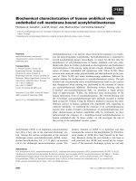
Isolation of EPCs from human umbilical vein
We obtained HUVECs from human umbilical
veins using a conventional chemical digestion
296
method. Fig. 1 shows representative microscopic
appearances. Freshly isolated cells attached to the
bottom of the culture dish and appeared as spindle or
elliptical shapes. Endothelial cell islands remained
compact after culture for 1 day. Subsequently, they
expanded and a monolayer of the endothelial lineage
occupied the plastic surface by day 7. This had a
characteristic cobblestone-like morphology (Fig. 1).
These results are consistent with traits of endothelial
lineage cells [14, 20], indicating that the isolated cells
are HUVECs.
VEGFR2 (KDR), CD34 and CD133 expression in
isolated HUVECs were analyzed with flow cytometry
and these data appear in Fig. 2. Most HUVECs
expressed VEGFR2 (KDR) and CD133 with lesser
expression of CD34 (Fig.2, HUVECs). Based on the
accepted standard, that early stage EPCs express
VEGFR2 (KDR), CD133, and CD34 as molecular
markers [13, 14]. These results indicate that these
cultures contained early EPCs, although these were
not the majority of cells in HUVECs cell cultures. To
isolate the EPCs in an early stage, magnetic
micro-beads coated with an antibody against human
CD34 were applied. As expected, the proportion of
CD34 positive cells separated this way was
significantly increased from about 8% before
(HUVECs in Fig. 2) to 79% after separation (EPCs in
Fig. 2). The majority of the EPCs expressed VEGFR2
(KDR), CD34 and CD133, as shown in Fig. 2. Thus,
human EPCs were successfully isolated from
HUVECs and majority of them belong to the early
stage.
Figure 1. Microscopic appearance of isolated HUVECs in primary culture. HUVECs were isolated from human umbilical vein by classic collagenase
digestion method. Phase-contrast microscopic appearance are shown.
Int. J. Med. Sci. 2017, Vol. 14
297
Figure 2. Expression of KDR, CD34 and CD133 in HUVECs and EPCs. The CD34+ cells were separated from CD34- cells by using magnetic micro-beads
coated with an antibody against human CD34. The expression of molecular markers was analyzed by flow cytometry. Representative data are shown in A and
statistical data are shown in B. N=3, **p < 0.01 versus HUVECs.
EPCs expansion in vitro
To obtain abundant and high quality human
EPCs, we expanded the cells in vitro using a
bio-MCCS culture. The conventional dish culture was
used as a control to compare the efficiency of the two
expansion methods. Fig. 3A shows that EPCs cultured
with bio-MCCS could attach to and proliferate on
micro-beads. Growth curves for each method were
plotted (Fig. 3B). EPCs cultured with bio-MCCS
generated more cells without passaging for 12 days of
culture. Whereas EPCs cultured on dishes were not as
abundant by day 12 and these had been passaged 3
times. One expanded culture with MCCS is
equivalent to fourteen dishes with dish culture
(11.7×10⁶ from one bottle versus 0.8×10⁶ from one
dish). Thus, the bio-MCCS method is superior to
Int. J. Med. Sci. 2017, Vol. 14
conventional dish culture in the total harvest cell
numbers and expansion efficiency.
We next measured early EPC marker expression
after expansion by using flow cytometry. Fig. 4A
shows representative data and Fig. 4B shows the
statistical results. The percentage of cells expressing
VEGFR2 (KDR), CD34 and CD133 in EPCs expanded
with the bio-MCCS (MCCS in Fig.4) was significantly
higher than that expanded with dish culture (Dish in
Fig.4). The results indicated that the bio-MCCS
culture technique had great advantage over dish
culture in maintaining the cells in the early stage
when they were used to expand EPCs. This
percentage also decreased after expansion with both
methods, when compared with freshly isolated EPCs,
as shown in Fig.2.
An in vitro angiogenesis assay was used to
evaluate the angiogenic potential of expanded EPCs,
298
based on their ability to form tubular networks [21,
22]. Fig. 5A shows representative microscopic
appearances. Statistical data are shown in B, C and D.
Closed polygons and/ or complex mesh-like
structures formed in both cell types (Fig. 5B),
indicating that both methods offered cells with
angiogenic traits. However, EPCs expanded with
bio-MCCS formed more branch points (p<0.01, Fig.
5C) and had longer tubes (p<0.01, Fig. 5D) compared
with EPCs harvested in dish culture. These data
indicate that the bio-MCCS technique preserves
potent angiogenesis compared with dish culture.
Taken together, these results indicate that a bio-MCCS
culture was superior to the dish culture for in vitro
expansion of EPCs, by its efficiency, maintaining the
cells in early stage and supporting more angiogenesis
of the cells, suggesting its importance in the in vitro
expansion of the cells.
Figure 3. In vitro expansion of EPCs with bio-MCCS and dish culture. The bio-MCCS and dish culture methods were used for EPCs expansion.
Representative microscopic appearance of bio-MCCS culture are shown in A. Cell growth curves for two methods are shown in B. Data are presented as the mean
± SD. N=3.
Int. J. Med. Sci. 2017, Vol. 14
299
Figure 4. Expression of KDR, CD34 and CD133 of expanded EPCs. EPCs were expanded for 12 days and flow cytometry was used to quantify marker
expression. Representative data are shown in A and statistical data are shown in B. N=3, **p < 0.01 versus dish cultured cells.
Discussion
Here, we obtained HUVECs from human
umbilical veins, and these cells had growth features,
morphology and surface markers characteristic of
endothelial lineage cells. Most of these freshly isolated
HUVECs expressed VEGFR2 (KDR), consistent with
previous reports that HUVECs expressed VEGFR2
(KDR), as a molecular marker for endothelial cells [23,
24]. Interestingly, these cells also expressed CD133, a
molecular marker for early stem/ progenitor cells. It
has been reported that established HUVECs cell lines
do not express CD133 [23, 24] and this result was also
noted in our previous study (unpublished data). Until
now, no report on CD133 expression in primary
cultured HUVECs has been reported. Our results
indicate that CD133 is expressed in freshly isolated
HUVECs, but that this molecular marker is lost in
established cell lines, suggesting that freshly isolated
HUVECs have some characteristics of stem/
progenitor cells, but that these are gradually lost
during passage in culture.
Int. J. Med. Sci. 2017, Vol. 14
300
Figure 5. In vitro angiogenesis of expanded EPCs. EPCs were expanded for 12 days and angiogenesis was measured by a Matrix-gel based in vitro angiogenesis
assay. The cell staining and the values quantification for the pattern recognition, branch point and total capillary tube length are described in the Methods section. Data
are expressed relative to dish cultured cells. Representative microscopic appearances are shown in A. Statistical results are shown in B, C and D, respectively. N =
5, **p < 0.01 versus dish cultured cells.
By using magnetic micro-beads coated with an
antibody against human CD34, we successfully
isolated human EPCs. The majority of them expressed
three molecular markers, VEGFR2 (KDR), CD34 and
CD133, indicates that they belong to the early stage of
EPCs. To our knowledge, this study is the first report
on isolating human EPCs from primary cultures of
HUVECs, suggesting that HUVECs might be a novel
promising resource of EPCs for regenerative
medicine.
Our results show that the bio-MCCS culture was
superior to the dish culture for in vitro expansion.
First, the expansion was more efficient. Secondly,
more of the expanded cells were maintained in the
early stage. Finally, the cells expanded with
bio-MCCS technique were more capable of in vitro
angiogenesis. The results indicated that the bio-MCCS
culture technique had great advantage over dish
culture in maintaining the cells in the early stage
when they were used to expand EPCs. The percentage
of cells expressing VEGFR2 (KDR), CD34 and CD133
was also less after expansion with the bio-MCCS
culture, compared with freshly isolated EPCs,
indicating further studies still required to optimize
the culture condition, for example, supplementing the
media with specific growth factors or cytokines, and
coating the micro-beads with extracellular matrix.
Even with this weak point, in the expanded EPCs
described here, the percentages of cells positively
expressing VEGFR2 (KDR), CD133 and CD34 is
comparable to those of freshly isolated bone marrow
[25], umbilical cord blood [23], and peripheral blood
[26] derived human EPCs. These expanded EPCs
described here formed complex tube-like structures in
Int. J. Med. Sci. 2017, Vol. 14
6 hours in a Matrix-gel based in vitro angiogenesis
assay. It has been reported that 12 hours are required
to form capillary-like structures for the EPCs derived
from umbilical cord blood in the same assay system
and in 6 hours only the cells line up with each other
could be seen [27]. EPCs from BM [28] could adhere to
and incorporate into the tube-like structures. In
addition, EPCs from PB could attach to protrusions of
endothelial cells around the tube-like structures [29].
Conclusions
We successfully isolated human EPCs from
HUVECs, which belong to the early stage of the cells,
by the expression of VEGFR2 (KDR), CD133 and
CD34. The results suggest that EPCs from HUVECs
might be a novel resource of cells for regenerative
medicine. We also found that a bio-MCCS culture was
superior to the dish culture for in vitro expansion of
EPCs, by producing more cells, maintaining the early
stage and supporting more angiogenesis of the cells.
The result suggests that a bio-MCCS culture system
described here might be broadly used for in vitro
expansion of EPCs, or other cells of attached growth.
Acknowledgments
This study was supported in part by the National
Natural Science Foundation of China (Grants: NSFC
No. 21277057) and National Science Foundation of
Jilin Province (No. 20130624003JC). We would like to
express our great appreciation to Professor F. William
Orr from the University of Manitoba in Canada for his
great help in revising the manuscript.
301
9.
10.
11.
12.
13.
14.
15.
16.
17.
18.
19.
20.
21.
22.
23.
Competing Interests
24.
The authors have declared that no competing
interest exists.
25.
References
26.
1.
2.
3.
4.
5.
6.
7.
8.
Asahara T, Murohara T, Sullivan A, Silver M, van der Zee R, Li T, et al.
Isolation of putative progenitor endothelial cells for angiogenesis. Science
(New York, NY). 1997; 275: 964-7.
Kirton JP, Xu Q. Endothelial precursors in vascular repair. Microvascular
research. 2010; 79: 193-9.
Briasoulis A, Tousoulis D, Antoniades C, Papageorgiou N, Stefanadis C. The
role of endothelial progenitor cells in vascular repair after arterial injury and
atherosclerotic plaque development. Cardiovascular therapeutics. 2011; 29:
125-39.
Griese DP, Ehsan A, Melo LG, Kong D, Zhang L, Mann MJ, et al. Isolation and
transplantation of autologous circulating endothelial cells into denuded
vessels and prosthetic grafts: implications for cell-based vascular therapy.
Circulation. 2003; 108: 2710-5.
Urbich C, Dimmeler S. Endothelial progenitor cells: characterization and role
in vascular biology. Circulation research. 2004; 95: 343-53.
Caballero S, Sengupta N, Afzal A, Chang KH, Li Calzi S, Guberski DL, et al.
Ischemic vascular damage can be repaired by healthy, but not diabetic,
endothelial progenitor cells. Diabetes. 2007; 56: 960-7.
Melchiorri AJ, Bracaglia LG, Kimerer LK, Hibino N, Fisher JP. In Vitro
Endothelialization of Biodegradable Vascular Grafts Via Endothelial
Progenitor Cell Seeding and Maturation in a Tubular Perfusion System
Bioreactor. Tissue engineering Part C, Methods. 2016; 22: 663-70.
Tongers J, Roncalli JG, Losordo DW. Role of endothelial progenitor cells
during ischemia-induced vasculogenesis and collateral formation.
Microvascular research. 2010; 79: 200-6.
27.
28.
29.
Schmidt DE, Manca M, Hoefer IE. Circulating endothelial cells in coronary
artery disease and acute coronary syndrome. Trends in cardiovascular
medicine. 2015; 25: 578-87.
Peplow PV. Growth factor- and cytokine-stimulated endothelial progenitor
cells in post-ischemic cerebral neovascularization. Neural regeneration
research. 2014; 9: 1425-9.
Foster WS, Suen CM, Stewart DJ. Regenerative cell and tissue-based therapies
for pulmonary arterial hypertension. The Canadian journal of cardiology.
2014; 30: 1350-60.
Wei L, Zhu W, Xia L, Yang Y, Liu H, Shen J, et al. Therapeutic effect of
eNOS-transfected endothelial progenitor cells on hemodynamic pulmonary
arterial hypertension. Hypertension research : official journal of the Japanese
Society of Hypertension. 2013; 36: 414-21.
Asahara T, Kawamoto A, Masuda H. Concise review: Circulating endothelial
progenitor cells for vascular medicine. Stem cells (Dayton, Ohio). 2011; 29:
1650-5.
Hristov M, Weber C. Endothelial progenitor cells: characterization,
pathophysiology, and possible clinical relevance. Journal of cellular and
molecular medicine. 2004; 8: 498-508.
Peng H, Chen L, Huang X, Yang T, Yu Z, Cheng G, et al. Vascular peroxidase 1
up regulation by angiotensin II attenuates nitric oxide production through
increasing asymmetrical dimethylarginine in HUVECs. Journal of the
American Society of Hypertension : JASH. 2016; 10: 741-51.e3.
Ingram DA, Mead LE, Moore DB, Woodard W, Fenoglio A, Yoder MC. Vessel
wall-derived endothelial cells rapidly proliferate because they contain a
complete hierarchy of endothelial progenitor cells. Blood. 2005; 105: 2783-6.
Kalka C, Masuda H, Takahashi T, Kalka-Moll WM, Silver M, Kearney M, et al.
Transplantation of ex vivo expanded endothelial progenitor cells for
therapeutic neovascularization. Proceedings of the National Academy of
Sciences of the United States of America. 2000; 97: 3422-7.
Baudin B, Bruneel A, Bosselut N, Vaubourdolle M. A protocol for isolation and
culture of human umbilical vein endothelial cells. Nature protocols. 2007; 2:
481-5.
Mou Y, Yue Z, Wang X, Li W, Zhang H, Wang Y, et al. OCT4 Remodels the
Phenotype and Promotes Angiogenesis of HUVECs by Changing the Gene
Expression Profile. International journal of medical sciences. 2016; 13: 386-94.
Bouis D, Hospers GA, Meijer C, Molema G, Mulder NH. Endothelium in vitro:
a review of human vascular endothelial cell lines for blood vessel-related
research. Angiogenesis. 2001; 4: 91-102.
Yoon CH, Hur J, Park KW, Kim JH, Lee CS, Oh IY, et al. Synergistic
neovascularization by mixed transplantation of early endothelial progenitor
cells and late outgrowth endothelial cells: the role of angiogenic cytokines and
matrix metalloproteinases. Circulation. 2005; 112: 1618-27.
Sieveking DP, Buckle A, Celermajer DS, Ng MK. Strikingly different
angiogenic properties of endothelial progenitor cell subpopulations: insights
from a novel human angiogenesis assay. Journal of the American College of
Cardiology. 2008; 51: 660-8.
Cheng CC, Chang SJ, Chueh YN, Huang TS, Huang PH, Cheng SM, et al.
Distinct angiogenesis roles and surface markers of early and late endothelial
progenitor cells revealed by functional group analyses. BMC genomics. 2013;
14: 182.
Yin AH, Miraglia S, Zanjani ED, Almeida-Porada G, Ogawa M, Leary AG, et
al. AC133, a novel marker for human hematopoietic stem and progenitor cells.
Blood. 1997; 90: 5002-12.
Quirici N, Soligo D, Caneva L, Servida F, Bossolasco P, Deliliers GL.
Differentiation and expansion of endothelial cells from human bone marrow
CD133(+) cells. British journal of haematology. 2001; 115: 186-94.
Rehman J, Li J, Orschell CM, March KL. Peripheral blood "endothelial
progenitor cells" are derived from monocyte/macrophages and secrete
angiogenic growth factors. Circulation. 2003; 107: 1164-9.
Duan HX, Cheng LM, Wang J, Hu LS, Lu GX. Angiogenic potential difference
between two types of endothelial progenitor cells from human umbilical cord
blood. Cell biology international. 2006; 30: 1018-27.
Walter DH, Rittig K, Bahlmann FH, Kirchmair R, Silver M, Murayama T, et al.
Statin therapy accelerates reendothelialization: a novel effect involving
mobilization and incorporation of bone marrow-derived endothelial
progenitor cells. Circulation. 2002; 105: 3017-24.
Loomans CJ, Wan H, de Crom R, van Haperen R, de Boer HC, Leenen PJ, et al.
Angiogenic murine endothelial progenitor cells are derived from a myeloid
bone marrow fraction and can be identified by endothelial NO synthase
expression. Arteriosclerosis, thrombosis, and vascular biology. 2006; 26:
1760-7.