Ebook Critical observations in radiology for medical students: Part 2
Bạn đang xem bản rút gọn của tài liệu. Xem và tải ngay bản đầy đủ của tài liệu tại đây (41.66 MB, 143 trang )
Chapter 7
Spine imaging
Joana N. Ramalho1,2 and Mauricio Castillo2
Department of Neuroradiology, Centro Hospitalar de Lisboa Central, Lisboa, Portugal
Department of Radiology, University of North Carolina, Chapel Hill, USA
1
2
Introduction
Spine pathology can be grossly divided into degenerative and nondegenerative diseases that may be clinically indistinguishable as
symptoms commonly overlap. Patients with spine disorders may
present with focal or diffuse back pain, radiculopathy, or myelopathy. Myelopathy describes any neurologic deficits related to disease
in the spinal cord while radiculopathy generally results from
impingement of the spinal nerves along their course. Focal back
pain without neurologic compromise or fever is not usually an
emergency and does not require emergent imaging. However,
vertebral metastases or infectious discitis may cause isolated focal
back pain, and if neurological deficits accompany them, immediate
imaging is indicated. When the history and physical findings are
nonspecific, as frequently they are in clinical practice, imaging findings become central to the diagnosis and treatment.
Imaging modalities
Conventional radiography was the initial imaging procedure in
spine evaluation, but with computed tomography (CT) and magnetic
resonance imaging (MRI) now widely available, radiographs are no
longer considered adequate. Radiographs are still useful for acute
trauma screening, for localization purposes during surgery procedures
(plain films and fluoroscopy), and for dynamic imaging (flexion and
extension). CT myelography and MRI with myelographic and neurographic sequences have also replaced conventional myelography.
Spinal CT is the modality of choice for evaluation of the bone
structures and calcifications, while MRI is better to evaluate the
details of spinal anatomy, including the intraspinal contents (spinal
cord, conus medullaris and cauda equina, dural sac epidural, subdural and subarachnoid spaces), neural foramina, joints, ligaments,
intervertebral discs, and bone marrow. Sagittal and axial images
should be acquired through the cervical, thoracic, and lumbar segments of the spine, as they are generally considered complementary.
The addition of coronal images may also be useful, especially in
patients with scoliosis.
A standard spine MRI protocol comprises sagittal and axial T1‐
and T2‐weighted sequences and fluid‐sensitive MR images (which
include short tau inversion recovery (STIR) or fat‐saturated T2‐
weighted sequences), complemented by postcontrast T1‐WI if
tumor, inflammation, infection, or vascular diseases are suspected.
Diffusion‐weighted imaging (DWI) is challenging in the spine,
largely due to physiological cerebrospinal fluid (CSF) flow‐induced
artifact and distortion from magnetic susceptibilities. It has been
used in the diagnosis of spinal cord infarct. Similar to the brain,
spinal cord infarcts show restricted diffusion, seen as bright lesions
on DWI with low signal on apparent diffusion coefficient (ADC)
maps. It has also been used to distinguish benign from pathologic
vertebral body compression fractures, but its usefulness and efficacy
in this setting remains controversial.
Diffusion tensor imaging (DTI) evaluates the direction and magnitude of extracellular water molecules movement within the white
matter fibers and enables the visualization of the major white matter
tracts in the brain and spine. Spine DTI has been used to evaluate
the integrity of the extent of neural damage in patients with acute or
chronic spinal cord injury and also to distinguish between infiltrative and localized tumors because the latter are easier to resect.
Nuclear medicine bone scans and PET/CT are used to screen the
entire skeleton for metastasis. They are highly sensitive but nonspecific, since degenerative and nondegenerative processes may show
increased uptake.
Ultrasound (US) has limited applications in adults, except during
surgery after removal of the posterior elements. In this setting, it
may be used to image the spinal cord. However, in neonates, the
nonossified posterior elements provide the acoustic window
through which the spinal anomalies can be readily evaluated.
Conventional digital subtraction angiography (DSA) can be performed for spinal vasculature evaluation, since spinal CT and MR
angiography are difficult to interpret and have limited application.
The major indications for spinal DSA are evaluation of suspected
arteriovenous fistulas (AVF), arteriovenous malformations, and
localization of the arterial cord supply before surgery.
Critical Observations in Radiology for Medical Students, First Edition. Katherine R. Birchard, Kiran Reddy Busireddy, and Richard C. Semelka.
© 2015 John Wiley & Sons, Ltd. Published 2015 by John Wiley & Sons, Ltd.
Companion website: www.wiley.com/go/birchard
116
Spine imaging 117
Appearance of the normal spine study
Vertebral anatomy varies somewhat by region, but the basic components are the same as follows:
• Vertebral body with vertebral end plates that define the intervertebral space, which contains the intervertebral disc
• Posterior vertebral arch that includes a pair of pedicles, a pair of
laminae, and 7 processes: 2 superior articular processes, 2 inferior
articular processes, 2 transverse processes, and 1 posterior midline spinous process
The cervical spine comprises the first seven superior vertebrae of the
spinal column. C1, also known as the atlas, and C2, also known as the
axis, are unique. The other cervical vertebrae are similar in size and
configuration. C1 is a ring‐shaped vertebra, composed of anterior
and posterior arches and two lateral articular masses, without a
central vertebral body. The vertebral arteries commonly traverse the
lateral masses of C1. C2 is also a ring‐shaped vertebra but has a
central body and a superiorly oriented odontoid process, also known
as the dens, which lies posterior to the anterior arch of C1. The
normal distance between the dens and anterior arch of C1 is approximately 3 mm in adults and 4 mm in children as they are held
together mainly by the transverse ligament. Exclusive to the cervical
spine are bilateral uncovertebral joints, also named Luschka joints
formed by the articulation of the uncinate process between two adjacent vertebral bodies. The transverse foramen (also known as the
foramen transversarium) located in the transverse processes of the
cervical vertebrae gives passage to the vertebral artery, the vertebral
vein, and a plexus of sympathetic nerves generally from C6 up to C1.
The discs of the cervical and thoracic spine are much thinner
compared with the lumbar discs. In the lumbar spine, the posterior
margins of the discs tend to be slightly concave at upper levels, straight
at L4/5 level, and slightly convex at the lumbosacral spinal junction.
This appearance should not be confused with pathologic bulging.
The main ligaments of the spine are the anterior longitudinal
ligament (ALL), posterior longitudinal ligament (PLL), and posterior
ligamentous complex (PLC) that include the supraspinous and interspinous ligaments, articular facet capsules, and ligamentum flavum.
The spinal canal contains the thecal sac formed by the dura mater
and surrounded by the epidural space, which contains epidural fat and
a large venous plexus. The thecal sac houses the spinal cord, conus
medullaris, and cauda equina (lower lumbar and sacral nerve roots),
surrounded by freely flowing CSF within the subarachnoid space.
The spinal cord is composed of a core of gray matter surrounded
by the white matter tracts. In the axial plane, the gray matter has a
“butterfly shape” given by its anterior and posterior horns joined in
the midline by a commissure. The conus medullaris normally ends
around L1–L2 vertebral level. The filum terminale is a strand of
pial–ependymal tissues, proceeding downward from the apex of the
conus medullaris to the coccyx.
Throughout the spine, the intervertebral foramina, or neural
foramina, contain the nerve roots and its sleeve, the dorsal root
ganglion, fat, and blood vessels.
On MRI, the appearance of different structures varies according to
the sequence used. The vertebral body contains bone marrow, which
signal varies with age, reflecting the gradual conversion of red marrow
to fatty marrow. The normal mature bone marrow shows high T1‐WI
and fairly high T2‐WI signal intensity, related with the presence of fat.
Tumor infiltration, radiation therapy, increased hematopoiesis, or any
disease that affects the bone marrow may alter the normal bone
marrow signal. Peripherally, the bone marrow is surrounded by low
T1‐ and T2‐WI signal of the cortical bone. Intervertebral discs demonstrate slightly less signal than the adjacent vertebral bodies on T1‐WI,
but the differentiation of the centrally located nucleus pulposus and
peripheral annulus fibrosis of the discs is difficult on this sequence. On
T2‐WI, the normally hydrated nucleus pulposus composed of water
and proteoglycans shows high signal centrally with lower signal from
the less hydrated annulus fibrosis. CSF demonstrates low signal on T1‐
WI and high signal on T2‐WI that provides contrast with the adjacent
spinal cord and nerve roots within the spinal canal, which show
intermediate signal on both sequences. The periphery of the spinal
canal is lined by high T1 signal intensity epidural fat. The spinal ligaments and dura show low signal intensity on T1‐ and T2‐WI.
As elsewhere in the body, bones and calcifications appear hyperdense on CT. Paraspinal muscles have intermediate density, while
CSF spaces are hypodense. As stated before, the differentiation
between intraspinal contents cannot be made on CT.
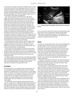
CT and MRI scans of the normal spine are shown in Figure 7.1.
Critical observations
Myelopathy
Myelopathy results from compromise of the spinal cord itself, generally
due to compression, intrinsic lesions, or inflammatory process known
as “myelitis.” It is most commonly caused by compression of the spinal
cord by intradural or extradural tumors (most frequently bone metastases), trauma (spinal cord injury), and degenerative cervical or dorsal
spondylosis. Many primary neoplastic, infectious, inflammatory, neurodegenerative, vascular (arteriovenous malformation, dural fistulae,
infarct, or hematoma), nutritional (vitamin B12 deficiency), congenital
(neural tube defects), and idiopathic disorders result in myelopathy,
though these are very much less common. Despite the clinical situation,
MRI is the procedure of choice for spinal cord evaluation.
In an acute setting, imaging evaluation is primarily focused on
extrinsic cord compression or presence of intramedullary spinal
cord hematoma, since the resultant myelopathy may be reversible,
particularly if treated earlier and aggressively. With regard to
imaging of myelopathy, the following should be kept in mind:
• MRI shows mass effect upon the cord and sometimes areas of
high T2‐WI signal inside the cord (Figure 7.2).
• Keep in mind that this T2‐WI sign is inconstant, may appear late,
and, when present, is associated with poor prognosis even after
therapy. DTI has been used recently to overcome this limitation,
by showing abnormalities of the white matter tracts before the
T2‐WI abnormalities being evident but is generally not used
routinely in clinical practice.
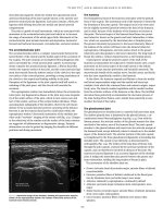
Epidural abscess
Epidural abscess represents a rare but important neurosurgical
emergency requiring immediate action. Most result from hematogenous spread from infections elsewhere in the body and are primarily located in the posterior aspect of the spinal canal. Abscesses
from direct spread from neighboring structures, such as spondylodiscitis, are often located in the anterior aspect of the spinal canal.
The following are imaging features of abscesses (Figure 7.3):
• On MRI, abscesses typically display intense peripheral rim
enhancing surrounding a heterogeneous nonenhancing central
zone of necrosis, and/or pus, with restricted diffusion.
• The dura represents a relative mechanical barrier, so infections
tend to spread in a craniocaudal fashion within the epidural space.
• Epidural abscesses have little room to expand axially and
compression of the thecal sac and spinal cord may be seen. Spinal
cord high T2‐WI signal may develop representing edema, myelitis,
or ischemia secondary to cord compression.
CC
P
SP
SP
NF
VB
CC
ID
L5
L5
SAP
L5
L5
IAP
S1
(a)
(b)
(c)
(d)
SC
SC
CM
*
*
CE
*
(e)
(f)
(g)
Nerve root
Surrounding fat
Vessels
NF
*
(h)
SC
*
Disc herniation
(I)
Figure 7.1 Normal anatomy of the spine on CT and MRI. CT of the lumbar spine: coronal bone window (a), midsagittal bone window (b), and soft tissue
window(c) at the level of the central canal (CC) and sagittal bone window (d) at level of the neural foramina (NF). MR of the lumbar spine: midsagittal
T1‐WI (e) and T2‐WI (f), coronal T2‐WI (g), and sagittal T1‐WI (h) at the level of the neural foramen. Axial T2‐WI at the level of the cervical spine (i),
conus medullaris (j), and cauda equina (k). CE, cauda equina; CM, conus medullaris; * CSF; IAP, inferior articular process; ID, intervertebral disc; P,
pedicle; SAP, superior articular process; SC, spinal cord; SP, spinous process; VB, vertebral body.
Spine imaging 119
(j)
(k)
Figure 7.1 (continued)
(a)
(c)
(e)
(b)
(d)
(f)
Figure 7.2 Cord compression. Sagittal and
axial cervical T2* (a and b) show a disc
herniation with cord compression. Sagittal
STIR (c) and axial postcontrast T1‐WI (d)
show a cervical spine metastatic tumor.
Sagittal STIR (e) and axial T2‐WI (f) show a
thoracic burst fracture.
Trauma
The screening examination for low‐risk traumatic spine injuries
consists of radiographs, supplemented by CT to further characterize
or detect fractures. After severe trauma however, CT should be
immediately performed, since unstable fractures can compromise the
diameter of the central canal leading to cord compression. MRI is used
to assess the nerve roots, soft tissues, and the spinal cord itself, particularly in patients who have neurologic symptoms unexplained by CT.
MRI can detect posterior ligamentous injuries, traumatic disc herniation, and spinal epidural hemorrhage difficult to visualize on CT.
120 Chapter 7
(a)
(b)
(c)
(d)
(e)
Figure 7.3 Epidural abscess. Axial T2‐WI (a) and axial (b) and sagittal (c) postcontrast T1‐WI show a posterior epidural abscess. Sagittal postcontrast
T1‐WI (d) and postcontrast FS T1‐WI (e) show an anterior epidural abscess (arrows).
Mechanical stability is a critical factor for treatment planning in
patients with traumatic spine injury. Spine stability is defined as the
ability to prevent the development of neurologic injury and progressive deformity in response to physiologic loading and a normal
range of movement. Spine stability relies on the integrity of both
bone and ligamentous components, and injury to either or both can
result in instability and require surgical stabilization.
Basion
BAI
BDI
ADI
Cervical spine
The cervical spine is highly susceptible to traumatic injury, because
it is extremely mobile with relatively small vertebral bodies and supports the head, which is heavy and acts as a lever. Different
classification systems have been developed in an attempt to predict
instability, to standardize injury nomenclature and to define a consistent therapeutic approach. Regardless of the classification used,
the cervical spine is usually divided between the upper cervical
spine, with its unique anatomy and the subaxial cervical spine.
Upper cervical spine
Atlanto‐occipital dissociation injuries are severe and include both
atlanto‐occipital dislocations and atlanto‐occipital subluxations.
On imaging studies, a gross disruption of the normal alignment of
the atlanto‐occipital joints may be seen. A number of lines and
distances on the cervical spine plain films and CT may help the
diagnosis: (i) basion‐dens interval (BDI) greater than 12 mm in
adults, (ii) basion‐axial interval (BAI) greater than 12 mm in adults,
and atlantodental interval (ADI) greater than 3 mm (adults males)
and greater than 2.5 mm (adults females; Figure 7.4).
Occipital condyle fractures may be divided into (i) type I, an
impaction fracture, which is a result of axial loading and lateral
bending; (ii) type II, a basilar skull fracture that extends into the
occipital condyle; and (iii) type III, a tension injury, resulting in an
avulsion of the occipital condyle.
Atlas fractures are common (representing 10% of all cervical
fractures) and usually associated with other cervical spine fractures.
Figure 7.4 Normal distances on the craniocervical junction. Midsagittal CT
demonstrates the posterior axial line drawn along the posterior cortex of the
body of the axis and extended cranially. The BAI is the distance between the
basion and this line. BDI is the distance from the most inferior portion of
the basion to the closest point of the superior aspect of the dens. ADI is the
distance between the posterior aspect of the anterior arch of C1 and the
most anterior aspect of the dens.
These fractures are classified based upon their location. Posterior
arch fractures are typically bilateral, are the most common, and are
stable. Lateral mass fractures are usually unilateral and may have
instability if there is associated ligamentous injury. The burst fracture is commonly called a Jefferson fracture and has a characteristic
pattern of fractures in both the anterior and posterior arches, which
widen rather than narrow the spinal canal (Figure 7.5).
Spine imaging 121
*
*
(a)
(b)
(c)
Figure 7.5 Atlas (C1) fractures. Axial (a) and coronal (b) CT show a right lateral mass fracture (*). Axial (c) CT show a Jefferson fracture (fractures in the
anterior and posterior arches) (arrows).
(a)
(b)
(c)
(d)
Figure 7.6 Hangman’s fracture. Axial (a), sagittal at the level of the right pars interarticularis (b), midsagittal (c), and sagittal at the level of the left pars
interarticularis (d) CT scans (arrows).
Odontoid fractures also known as the dens fractures are
common fractures (representing 20% of all cervical fractures), usually classified as (i) type I, a fracture of the upper part of the odontoid process; (ii) type II, a fracture at the base of the odontoid,
usually unstable and with a high risk of nonunion; and (iii) type III,
a fracture of the odontoid, which extends into the body of C2.
Hangman’s fracture is a term frequently used to describe traumatic spondylolisthesis of the axis. The fracture involves both pars
interarticularis of C2 and is as a result of hyperextension and distraction. Despite the name, which hearkens to the era of judicial
hangings, this fracture is virtually never seen in suicidal hanging,
and major trauma such as high‐speed motor vehicle accident is in
fact the most common association. It is the most severe cervical
fracture that can be sustained with preservation of life (Figure 7.6).
Subaxial cervical spine
Subaxial cervical spine injuries represent a broad of injury patterns
and degrees of instability. The most accepted classification systems
are based on the mechanism of injury.
Flexion–compression injuries represent a continuum of injury
patterns, with minor degrees of trauma producing simple vertebral
body compression fractures and more severe injuries resulting in a
triangular “teardrop” fracture (fracture of the anteroinferior
vertebral body—teardrop sign) or a quadrangular fracture with
posterior ligamentous disruption. The most severe pattern results
in posterior subluxation of the posterior vertebral body into the
canal, acute kyphosis, and disruption of the ALL, PLL, and posterior ligaments, associated with a high incidence of cord damage.
Flexion–distraction injuries also represent a spectrum of pathology
from mild posterior ligamentous strains to bilateral facet dislocations.
Facet dislocation refers to anterior displacement of one vertebral body
onto another and may occur in variable degrees as follows (Figure 7.7):
• Facets subluxation—the superior facet slides over the inferior
facet.
• Perched facets—the inferior facet appears to sit “perched” on the
superior facet of the vertebra below.
• Locked facets—when one facet “jumps” over the other and
becomes locked in this position.
• The naked facet sign refers to the CT appearance of an uncovered
facet when the facet joint is completely dislocated.
Complications include cord injury (especially with bilateral involvement or in the setting of canal stenosis) or vertebral artery injury,
such as dissection or thrombosis.
Vertical compression‐type injuries are most commonly manifested as a cervical vertebral burst fracture. Axial loading of the
cervical spine results in compression of the vertebral body with
resultant retropulsion of the posterior wall into the canal.
Hyperextension injuries also represent a continuum of injury
patterns with mild trauma resulting in widening of the disc space
with disruption of the ALL and disc injury. In more severe cases, a
teardrop fracture, characterized by the avulsion of the anteroinferior corner of the vertebral body, may be seen. Extension teardrop
is not as severe as its counterpart, the flexion teardrop fracture.
However, posterior ligaments disruption with displacement of the
cephalad vertebrae into the spinal canal may also occur.
Thoracolumbar spine
Three different biomechanical regions can be defined: (i) the upper
thoracic region (T1–T8) that is rigid and stable due to the ribcage;
(ii) the transition zone (T9–L2) between the rigid and kyphotic
122 Chapter 7
(d)
(a)
(b)
(c)
(e)
Figure 7.7 Facets dislocation. Sagittal at the level of the right articular processes (a), midsagittal (b), and sagittal at the level of the left articular processes
(c) show locked facets (arrows). Axial (d and e) CT shows normal facet joints and naked facet, respectively.
upper thoracic part and the flexible lordotic lumbar spine, where
most injuries occur; and (iii) the L3–sacrum zone, a flexible
segment where axial loading injuries usually occur.
Numerous thoracolumbar spine injury classification systems
have been developed, most of them based on the three‐column
concept devised by Denis.
According to Denis’ classification, the anterior column comprises
the ALL and the anterior half of the vertebral body, the middle column
comprises the posterior half of the vertebral body and the PLL, and the
posterior column comprises the pedicles, the facet joints, and the
supraspinous ligaments. In his model, stability is dependent on at least
two intact columns. The Denis system also classifies spinal trauma as
minor (fractures of transverse processes, articular processes, pars
interarticularis, and spinous processes that do not lead to acute instability) and major injuries (compression fracture, burst fracture, seat
belt injury, and fracture–dislocation), according with injury morphology and mechanism. As of lately, this classification has fallen out
of favor with neurosurgeons and spine surgeons.
Recently, the Spine Trauma Study Group proposed the thoracolumbar injury classification and severity score (TLICS). The TLICS
is both a scoring and a classification system, based on three injury
categories that are independently critical and complementary for
appropriate treatment recommendations: (i) injury morphology,
(ii) integrity of the PLC, and (iii) neurologic status of the patient.
Within each category, subgroups are arranged from least to most
significant, with a numeric value assigned to each injury pattern.
Point values from the three main injury categories are totaled to
provide a comprehensive severity score (Table 7.1). One distinguishing feature of the TLICS is its emphasis on injury morphology
rather than the mechanism of injury, since various mechanisms can
lead to similar injury patterns.
Independently of the different classifications systems, morphologic description of the fractures seen on imaging studies must be
reported as follows:
• Compression fracture—vertebral collapse, defined as a visible loss
of vertebral body height or disruption of the vertebral end plates.
Less severe compression injuries may involve only the anterior
portion of the vertebral body.
Table 7.1 The thoracolumbar injury classification and severity
score with its subcategories and respective scoring.
Injury category
Injury morphology
Compression
Burst
Translation and rotation
Distraction
PLC status
Intact
Injury suspected or indeterminate
Injured
Neurologic status
Intact
Nerve root involvement
Spinal cord injury
Incomplete
Complete
Cauda equine syndrome
1
2
3
4
0
2
3
0
2
3
2
3
Source: From Khurana et al. (2013).
• Burst fractures—a type of compression fracture with disruption
of the posterior vertebral body, varying degrees of retropulsed
fragments in the spinal canal and bone shards of the vertebra
penetrating surrounding tissues (Figure 7.8).
• Translation injuries—defined as a horizontal displacement or
rotation of one vertebral body with respect to another. These
injuries are characterized by rotation of the spinous processes,
unilateral or bilateral facet fracture–dislocation, and vertebral
subluxation. Anteroposterior or sagittal translational instability is best seen on lateral images, while instability in the
mediolateral or coronal plane is best seen on anteroposterior
views.
• Distraction injuries—identified as anatomic dissociation along
the vertical axis that can occur through the anterior and posterior
supporting ligaments, the anterior and posterior osseous elements, or a combination of both.
A basic description of injury features includes the degree of
comminution, percentage of vertebral height loss, retropulsion
Spine imaging 123
(b)
(c)
(a)
(d)
(e)
Figure 7.8 Burst fractures. Lateral plain film (a) shows an L2 compression fracture (arrow). Axial (b), coronal (c), and sagittal (d) CT scans and sagittal
T1‐W MRI (e) of the same patient.
distance, percentage of canal compromise, and other contiguous or
noncontiguous vertebral injuries. Osseous retropulsion alone does
not imply neurologic injury. In the thoracic spine, retropulsion may
cause significant neurologic injury because the spinal canal is
narrow and blood supply to the cord is sparse. In contrast, in the
lumbar spine, a burst fracture may cause marked displacement of
the cauda equina without neurologic deficits since the central canal
is wide and the spinal cord generally ends at the level of L1.
The PLC serves as the posterior “tension band” of the spinal
column and protects it from excessive flexion, rotation, translation,
and distraction. Disruption of the PLC is seen on radiographs and
CT or MR images as follows:
• Splaying of the spinous processes (widening of the interspinous
space), avulsion fracture of the superior or inferior aspects of
contiguous spinous processes, widening of the facet joints, empty
(“naked”) facet joints, perched or dislocated facet joints, or
vertebral body translation or rotation.
The PLC must be directly assessed at MRI regardless of the
severity of vertebral body injury seen at CT, because there is an
inverse relationship between osseous destruction and ligamentous
injury (Figure 7.9). With respect to spinal soft tissue injuries, keep
in mind the following:
• On MRI, the ligamentum flavum and supraspinous ligament are
seen as a low‐signal‐intensity continuous black stripe on sagittal
T1‐WI or T2‐WI. Disruption of these stripes indicates a supraspinous ligament or ligamentum flavum tears.
• Fluid in the facet capsules or edema in the interspinous region on
fluid‐sensitive MR images (which include STIR or fat‐saturated
T2‐weighted sequences) reflects a capsular or interspinous
ligament injury, respectively.
Spinal cord injury
Spinal cord injury usually occurs at sites of fractures, secondary
to bony impingement and cord compression. However, cord
injury may also occur in the absence of bone fractures, caused by
hyperflexion and hyperextension mechanism and associated
vascular insults.
124 Chapter 7
(a)
(b)
(c)
Figure 7.9 Hyperflexion cervical injury. Sagittal T2‐WI (a and b) shows disruption of the posterior ligamentous complex (arrows), cord edema and
hemorrhage, better depicted on axial T2* (c) (arrow).
There are two types of spinal cord injury:
• Nonhemorrhagic—seen on MRI as areas of high T2‐WI signal
that represents edema
• Hemorrhagic—seen on MRI as areas of low signal intensity on
T2‐/T2*‐WI within the area of edema that represents hemorrhage (see Figure 7.9)
There is a strong correlation between the length of the spinal cord
edema and the clinical outcome with patients who have over two
vertebral segments doing poorly. However, the most important
prognostic factor is the presence of hemorrhage, which has an
extremely poor outcome.
Specific types of trauma, such as sudden distracted forces along
the long axis, may lead to cord avulsion, more common at the
junction of the cervical and thoracic cord. These injuries are more
common in children.
Extramedullary hematomas
Extramedullary hematomas can follow trauma or be spontaneous.
Subdural hematomas are rare and are usually related to coagulopathies. Epidural hematomas are more common, since the ventral epidural space contains a rich venous plexus susceptible to tearing
injuries, even without vertebral fractures. MRI is the modality of
choice to depict epidural and subdural hematomas.
Nerve root avulsion
The traumatic lesions described earlier may also affect nerve roots
and result in radiculopathies. An additional form of direct trauma
to the nerve roots is avulsion from their connection to the cord.
Brachial plexus nerve roots are most commonly affected resulting
in upper extremity neurologic deficits. Birth trauma is a classic
example of nerve root avulsion at the cervicothoracic junction. CT
myelography or MRI may confirm the diagnosis as follows:
• MRI allows the direct visualization of nerve roots, CSF leaks
through avulsed nerve roots sleeves, and associated cord injuries
(edema and cord hematoma in acute stage, myelomalacia in the
chronic stage).
• Postcontrast enhancement of nerve roots suggests functional
impairment even if the nerve appears continuous and is due
to disruption of the nerve–blood barrier. Abnormal enhancement of paraspinal muscles is also an indirect sign of root
avulsion.
• The steady‐state coherent gradient echo sequences (MR myelography) can easily identify nerve roots and the meningocele sac as
do T2‐weighted images.
• Diffusion‐weighted neurography is a new MRI technique that
may also show postganglionic injuries, as a discontinuation of the
injured nerves. It is not currently used in routine clinical practice.
Vascular lesions
Spinal cord infarct
Spinal cord infarct is uncommon, but it is usually associated
with devastating clinical symptoms and poor prognosis. It can be a
complication of aortic aneurysm surgery or stenting; however, in
the majority of patients, no obvious cause is identified. Patients
usually present with acute paraparesis or quadriparesis, depending
on the level of the spinal cord involvement.
Spine imaging 125
(d)
(a)
(b)
(c)
(e)
Figure 7.10 Spinal cord infarct. Sagittal T1‐WI (a), T2‐WI (b) and STIR (c), and axial T2‐WI (d) show a spinal cord infarct (arrows) with restricted
diffusion (e) (arrows).
MRI should be obtained in all patients with suspected spinal cord
infarction, not only to confirm the diagnosis but also to exclude
other more readily treated causes of cord impairment, such as compression. The following are the imaging features of cord infarctions
(Figure 7.10):
• The hallmark of spinal cord infarction is a high T2‐WI signal
lesion within the cord, most commonly located centrally (anterior spinal artery territory). On axial images, a characteristic
snake‐eye appearance may be seen due to the prominent high
signal involving the anterior gray matter horns. Central involvement can be more extensive and the white matter can also be
affected.
• Restricted diffusion, when present, establishes the diagnosis.
• Spinal cord enlargement may be seen during the acute phase,
while cord atrophy may be seen during the chronic phase.
Cord ischemia due to venous hypertension or arterial steal can be
seen in spinal vascular malformations.
Spinal vascular malformations
Spinal arteriovenous malformation is a generic term used to cover
any abnormal vascular complex that has a direct connection between the arterial system and the venous system without intervening
capillaries.
Intramedullary AVMs have a congenital nidus of abnormal
vessels within the spinal cord. Hemorrhage or ischemia (related
with steal phenomenon) may be seen. Flow voids may be depicted
on MRI within the substance of the spinal cord. They are exceedingly rare.
Extramedullary AVMs are located in the pia (intradural AVMs,
located outside the substance of the spinal cord) or in the dura
(spinal dural AVF). An AVF represents an abnormal connection
between an artery and a vein in the dura of the nerve root sleeve.
They are the most common type of AVMs in adults and the symptoms are related with venous hypertension and cord congestion
with edema. The dilated venous plexus can be visualized on MRI as
multiple flow voids and the cord shows high T2 signal and contrast
enhancement.
Degenerative conditions
Degenerative disease of the spine
CT continues to be used widely in the examination of degenerative
spinal disorders, and only a few differences between CT and MRI
have been noted concerning diagnostic accuracy in the lumbar spine.
CT remains superior in the evaluation of osseous features, such as
osteophytes, spinal stenosis, facet hypertrophy, or sclerosis associated
with degenerative disorders. MRI is the preferred procedure for
evaluating the cervical spine as well as intervertebral disc disease.
As disc degeneration progresses, the water content of the disc
decreases and fissures develop in the annulus. This results in decreased
disc space height, posterior bulging of the disc annulus, and low
signal of the disc on T2‐WI. Further degeneration results in disc space
collapse, misalignment, and nitrogen accumulation within the disc.
Alterations in adjacent vertebral body marrow often occur with disc
degeneration and appear as bands of altered signal intensity on MRI
paralleling the narrowed disc (Figure 7.11).
The nomenclature of disc disease is controversial. Different definitions have been given to disc bulges, herniations, protrusions,
extrusions, sequestrations, and migrations. The recommendations
126 Chapter 7
*
*
(a)
(b)
(c)
(d)
(e)
Figure 7.11 Degenerative disease of the spine. Sagittal T1‐WI (a) and T2‐WI (b) show decreased disc space height and low signal of the disc on T2‐WI (*).
Sagittal CT (c) shows disc space collapse and nitrogen accumulation within the disc (arrow). Sagittal T1‐WI (d) shows a parallel band of low T1 signal
adjacent to the end plates, with high signal intensity on T2‐WI (e) (arrows).
from the American Society of Spine Radiology, the American
Society of Neuroradiology, and the American Spine Society are:
• Disc bulge—bulging of the annulus fibrosus that involves more
than half of the circumference of an intervertebral disc (>180°).
• Disc herniation—displacement of intervertebral disc material
beyond the normal confines of the disc but involving less than
half the circumference (to distinguish it from a disc bulge).
Herniations are further divided into protrusions and extrusions.
The distinction between a protrusion and an extrusion is made
on the basis of the size of the “neck” versus the size of the “dome”
of the herniation as well as its relationship to the disc level:
◦◦ Protrusion has a broader neck than its “dome” and does not
extend above or below the disc level. Disc protrusions are further
divided into broad based, in which the base involves between 90
and 180° of the circumference, and focal, in which the base
involves less than 90° of the disc circumference.
◦◦ Extrusion has a narrower neck than dome and/or extends
above or beyond the vertebral end plates. Extrusion can be in any
axial direction and may migrate either superiorly or inferiorly. If
the extrusion migrates but becomes separated from the rest of the
herniation, it is known as an intervertebral disc sequestration.
Herniations may also be described in terms of its axial position,
into central, subarticular, foraminal, extraforaminal, or anterior
(Figure 7.12).
More important than the terminology used is the description of
the disc disease, the relationship between the disc and the neuronal
structures, and other associated findings, such as facet diseases,
spondylolysis and spondylolisthesis, and central canal or neuroforaminal stenosis.
Degenerative joint diseases of the facets include bony hypertrophy, some facet slippage, and ligamentum flavum hypertrophy, a
common cause of central canal and neuroforaminal stenosis.
Spondylolysis is a defect in the bony pars interarticularis and can
be the source of low back pain and instability and generally involves
the L5 segment. Prior to disc surgery or other back surgery,
identification of spondylolysis is imperative. Spondylolisthesis represents a forward displacement of a vertebra and occurs from either
bilateral spondylolysis or degenerative joint diseases of the facets
with slippage of the facets (Figure 7.13).
It is not unusual to have patients with disc herniations or stenosis
that appears severe on imaging, but who have no symptoms; thus,
any imaging findings must be matched with clinical findings. Central
canal measurements are no longer considered a valid indicator of
disease by themselves.
Failed back surgery is common especially after lumbar spine
operations. Identifiable causes of recurrent symptoms after surgery
include inadequate surgery (including missed free disc fragments),
development of fibrosis (scar tissue), recurrent or residual disc herniations, arachnoiditis, and spinal stenosis. Scar tissue located in the
epidural space has been shown to enhance homogeneously on MRI
after contrast administration, regardless of the time since surgery,
while recurrent or residual herniated disc or disc fragments show
only minimal peripheral enhancement presumably related with
inflammation. Furthermore, a recurrent or residual disc herniation
should cause mass effect upon the thecal sac and/or nerve roots,
while scar generally surrounds the neural tissue.
Inflammatory conditions
Multiple sclerosis (MS) is the most common primary demyelinating
disease. The majority of patients have brain and spinal cord involvement. Isolated spinal cord disease occurs in less than 20% of cases.
Imaging plays an important role in MS diagnosis as included in
McDonald criteria, introduced in 2001, then revised and simplified
Spine imaging 127
*
*
*
(a)
(b)
(c)
*
(d)
(g)
*
(e)
*
(f)
(h)
(i)
Figure 7.12 Disc herniations. Sagittal (a) and axial (b) CT demonstrate a left central lumbar disc herniation (*). Axial CT in soft tissue (c) and bone
window (d) show a right extraforaminal calcified lumbar disc herniation (*). Sagittal (e) and axial T2‐WI (f) show a subligamentous extrusion (*).
Sagittal T2‐WI (g) shows a sequestered disc (arrow). Sagittal T2‐WI (h and i) in two different patients with cervical disc herniation, central canal
stenosis, and cord compression with edema (*) (spondylotic myelopathy).
128 Chapter 7
(a)
(b)
(c)
(d)
(e)
Figure 7.13 Spondylolysis and spondylolisthesis. Sagittal at the level of the right articular processes (a), midsagittal (b), and sagittal at the level of the left articular
processes (c) bone window CT show bilateral defect in the L5 (arrow) pars interarticularis (spondylolisthesis). Sagittal T2‐WI (d) shows forward displacement
of L4 over L5 (arrow) caused by degenerative joint diseases of the facets (spondylolysis) well seen on axial T2‐WI (e) with lumbar central canal stenosis.
(d)
(a)
(b)
(c)
(e)
Figure 7.14 Multiple sclerosis. Sagittal T1‐WI (a) and T2‐WI (b) show multiple lesions with high T2 signal (arrows). One of the lesions shows minimal
enhancement after gadolinium administration (arrow) (c). Axial T2‐WI (d and e) demonstrates typical location of the lesions (arrows).
in 2005 and 2010. In McDonald criteria, MRI is used to demonstrate
lesion dissemination in time and space (Figure 7.14):
• CT has poor sensitivity for detection, evaluation, and characterization of MS lesions. MRI offers by far the most sensitive technique for MS diagnosis and follow‐up.
• On MRI, demyelinating lesions appear as high‐signal T2‐WI
areas, typically triangular in shape and mostly located dorsally or
laterally, involving the white matter tracts, generally with less
than 2 vertebral bodies in length. However, as in the brain, both
white matter and gray matter can be affected.
• Active lesions usually demonstrate enhancement after gadolinium administration and may show extensive edema with associated focal enlargement of the spinal cord.
• Classic chronic lesions do not show contrast enhancement and
may demonstrate focal cord atrophy.
Primary and secondary neoplasms of the spinal cord (e.g., astrocytomas, ependymomas), other demyelinating diseases (acute
disseminated encephalomyelitis (ADEM), transverse myelitis
(TM)), neuromyelitis optica (NMO), infection, acute infarction,
sarcoidosis, and systemic lupus erythematosus may mimic MS.
Spine imaging 129
(b)
(a)
(c)
(d)
Figure 7.15 Neuromyelitis optica. Sagittal (a) and axial (b) T2‐WI show a long lesion with patchy enhancement on axial (c) and sagittal (d) postcontrast T1‐WI.
Neuromyelitis optica (NMO), also known as Devic disease, is
no longer considered an MS variant. It is recognized as a distinct
entity characterized by bilateral optic neuritis and myelitis, with
blindness and paraplegia. NMO is an autoimmune demyelinating
and necrotizing disease induced by a specific autoantibody, the
NMO‐IgG, which targets a transmembrane water channel (aquaporin 4). Imaging features of NMO follow (Figure 7.15):
• MRI shows typical features of optic neuritis: enlarged optic
nerves hyperintense on T2‐WI with enhancement after contrast
administration. Bilateral involvement and extension of the signal
back into the chiasm is particularly suggestive of NMO.
• Spinal lesions extend over long distances (>3 vertebral segments,
often much more), usually involve the central part of the cord (MS
lesions tend to involve individual peripheral white matter tracts),
and after contrast administration may show patchy enhancement.
Thin ependymal enhancement similar to ependymitis may be seen.
• Brain lesions follow the distribution of aquaporin 4 in the brain,
which is particularly found in the periependymal brain adjacent
to the ventricles.
ADEM is an immunologically mediated allergic inflammatory
disease of the central nervous system (CNS), resulting in multifocal
demyelinating lesions affecting the gray and white matter of the
brain and spinal cord. It is typically seen in young children usually
4 weeks after a viral infection or vaccination. ADEM is characteristically monophasic, but multiphasic forms may be seen in 10% of
cases. In 50% of ADEM patients, the antimyelin oligodendrocyte
glycoprotein (MOG) IgG test is positive and supports the diagnosis.
The imaging features of ADEM are:
• MRI usually shows diffuse high T2‐WI signal of the spinal
cord with cord swelling and variable enhancement after contrast
administration.
• Brain imaging appearances vary from small “punctate” lesions to
tumefactive lesions. Lesions are usually bilateral but asymmetrical. Brain lesions generally show no contrast enhancement.
• Compared to MS, involvement of the callososeptal interface
is unusual. Involvement of the cerebral cortex; subcortical
gray matter, especially the thalami; and the brainstem is also
not very common, but if present are helpful in distinguishing
from MS.
Transverse myelitis (TM) is a focal inflammatory disorder of the
spinal cord resulting in motor, sensory, and autonomic dysfunction.
TM may occur without apparent underlying cause (idiopathic) or
in the setting of another illness. Idiopathic TM is assumed to be the
result of abnormal activation of the immune system against the
spinal cord. Underlying causes of TM include systemic
inflammatory disease, such as Sjögren’s syndrome; lupus (SLE) and
neurosarcoidosis; infectious diseases like herpes simplex virus,
herpes zoster virus, cytomegalovirus (CMV), Epstein–Barr virus
(EBV), human immunodeficiency virus (HIV), enteroviruses,
influenza, syphilis, tuberculosis, or Lyme diseases; and vascular
diseases, such as thrombosis, vasculitis, or arteriovenous malformations. It can also be a paraneoplastic
syndrome or the
initial manifestation of MS, NMO, or ADEM. Remember that:
• MRI shows T2‐WI hyperintense lesions involving more than 2/3
of the spinal cord cross‐sectional area with focal enlargement and
variable enhancement after contrast administration.
130 Chapter 7
Infectious conditions
Infections may be classified according to their causative organism
or according to their anatomic location. Spine pyogenic infections
are usually secondary to bacteremia (arterial dissemination).
However, some organisms may reach the lower spine through
Batson venous plexus, and direct inoculation may occur in postsurgery patients or children with spinal dysraphism.
Osteomyelitis/discitis
Spondylodiscitis is a combination of discitis, inflammation of the
intervertebral disc space, and spondylitis, inflammation of the vertebrae. In adults, the primary site of hematogenous infection is the
vertebral end plates, due to its richest blood supply. First, vertebral
osteomyelitis develops affecting the end plates. Then, the pyogenic
infection progresses and extends into the disc space. This osteomyelitis/discitis complex is usually known as “pyogenic spondylodiscitis.” If the infection is left untreated, the disc space is rapidly
destroyed, with collapse and destruction of adjacent bone. The
imaging features of osteomyelitis and discitis are:
• CT may show disc space narrowing and irregularity/ill definition
of the end plates with surrounding soft tissue swelling.
• Characteristic MRI findings are low T1‐WI and high T2‐WI signal
in disc space (fluid), low T1‐WI and high T2‐WI signal in adjacent
end plates (bone marrow edema), loss of the normal cortical end
plate definition, and high signal in paravertebral soft tissues.
• The T2‐WI changes described earlier are particularly well seen
on STIR or fat‐saturated T2‐WI.
• Peripheral enhancement around fluid collection(s), enhancement of vertebral end plates, and enhancement of perivertebral
soft tissues are usually depicted on postcontrast T1‐WI.
Epidural phlegmon or abscess may accompany spondylodiscitis
as follows (Figure 7.16):
• Epidural phlegmons are characteristically hypointense or isointense on T1‐WI and slightly hyperintense on T2‐WI with homogenous enhancement after contrast administration, while abscesses
show rim enhancing and restricted DWI as described previously
(see section “Critical observations”).
• The adjacent dura and epidural venous plexus usually enhance
intensely and appear thick.
• Epidural phlegmon and/or abscess typically compress the thecal
sac and spinal cord, displacing the cord posteriorly. T2‐WI signal
abnormalities hyperintensity may develop in the cord. Direct
invasion or hematogenous spread of the infectious processes into
the spinal cord may occur but is rare.
• Paraspinal or psoas abscesses may also be seen.
Nonpyogenic infections, such as tuberculosis and some fungal
infections, can show a more indolent clinical course and may mimic
neoplastic diseases.
Tuberculosis of the spine, or “Pott” disease, usually spreads by
a subligamentous route involving multiple vertebral bodies, often
with relative sparing of the intervening discs. Vertebral collapse,
paraspinal calcification, and proliferative new bone formation
with a kyphotic or “gibbus” deformity are usually seen and may
lead to cord compression. Large paraspinal abscesses without
severe pain or pus are common and are called “cold abscesses.”
Tuberculosis may also affect the intradural spinal compartment,
resulting in an inflammatory arachnoiditis that can spread to the
cord and nerve roots.
Subdural empyemas are rare and tend to be associated with surgery or other violation of the dura. Subdural infections can rapidly
spread through the arachnoid layer, resulting in meningitis.
Direct spinal cord infections are uncommon and are usually
caused by viruses, such as varicella‐zoster virus, HIV, CMV, or EBV
and in immunocompromised patients by bacteria and fungi.
Neoplastic processes (benign/malignant)
Mass lesions of the spine are classified according to their locations
as intramedullary, intradural–extramedullary, and extradural. The
location is critical for the differential diagnosis. MRI is unquestionably the imaging procedure of choice in these patients.
Extradural tumors
Neoplasm is the second most frequent cause of an extradural mass,
after disc herniation and other degenerative diseases. Primary
vertebral tumors, such as chordomas, giant cells tumors, hemangiomas, and sarcomas, are discussed elsewhere in this book. The most
common extradural neoplasms are vertebral body metastases generally from breast, lung, and prostate carcinoma. Imaging features
of vertebral metastases are (Figure 7.17):
• Bone metastases appear as low‐signal areas on T1‐WI with
high signal on T2‐WI, because of their higher water content
compared with the normal bone marrow fat. Nearly all metastases enhance.
• Densely sclerotic metastases, often seen in prostate cancer, can be
dark on all sequences.
Distinguishing between benign osteoporotic and pathologic
vertebral body compression fractures may be difficult, particularly
when only one vertebra is involved. The following imaging findings
are helpful:
• Most vertebral compression fractures, regardless of whether
they are benign or malignant, show low T1‐ and high T2‐WI
signal intensities and may enhance after contrast material
administration.
• In the chronic stage, the bone marrow of benign vertebral compression fractures returns to its normally high T1‐WI signal
intensity, whereas the bone marrow infiltrated by tumor remains
hypointense on T1‐WI.
• The most reliable MRI sign suggesting benign etiology is visualizing the fracture line as a T2‐ or postcontrast T1‐WI linear
hypointensity in the compressed vertebral body.
• Other signs that favor benign compression fractures include the
presence of intervertebral fluid, an intervertebral vacuum cleft,
absence of accompanying soft tissue masses, lack of pedicle
abnormalities, solitary vertebral involvement, preservation of the
posterior cortical margin, and a wedge‐shaped deformity.
Unfortunately, these signs cannot be found in all patients.
• In theory, malignant compressive fractures may show restricted
diffusion caused by the infiltrating tumor cells, and benign
osteoporotic fractures may show increased diffusion caused by
the increased extracellular water. However, infiltrated vertebrae may show areas of both patterns, confusing the diagnosis
(Figure 7.18).
Direct extension of paraspinous tumors
Any retroperitoneal and mediastinal tumor can invade the vertebral
column and spinal canal by direct extension.
Neuroblastoma, ganglioneuroma, and ganglioneuroblastoma
arise from primitive paraspinous neural remnants, similar to fetal
neuroblasts, and frequently involve the spinal canal extending
through the neural foramina. In adults, lung cancer commonly
does this.
Spine imaging 131
(c)
(a)
(b)
(e)
(d)
(f)
(g)
Figure 7.16 Spondylodiscitis. SagittalT1‐WI(a), sagittalT2WI(b), and sagittal(c) and axial(d)postcontrastT1‐WI show cervical spondylodiscitis with
epidural phlegmon (arrows). Sagittal T1‐WI (e), T2‐WI (f), and postcontrast T1‐WI (g) show lumbar spondylodiscitis with epidural abscesses in a
different patient (arrows).
132 Chapter 7
Figure 7.17 Bone metastases. Sagittal T1‐WI
(a)
(b)
(c)
Hematologic tumors
Leukemias show diffuse involvement or replacement of the normal
bone marrow with tumor. Solid leukemia (chloromas) can be seen
in the epidural space and may cause cord compression and also
occur in the paraspinal regions.
Multiple myeloma is the most common primary malignant bone
neoplasm in adults. Four main patterns are recognized: (i) disseminated form with multiple focal lesions predominantly affecting the
axial skeleton; (ii) diffuse skeletal osteopenia; (iii) solitary plasmacytoma, which is a single expansile lesion most commonly in a
vertebral body or in the pelvis; and (iv) osteosclerosing myeloma.
Solitary plasmacytoma usually appears as a lytic lesion with
thinning and destruction of cortex and often has a nonspecific
appearance. It is also one of the differential diagnoses for vertebra
plana (totally collapsed vertebral body), along with eosinophilic
granuloma (which tends to occur in children), leukemia, and severe
osteoporosis.
Hodgkin and B‐cell‐type lymphomas are the most common
lymphomas in the CNS. Spinal involvement is usually secondary.
Lymphoma more commonly involves the vertebral body and paraspinal tissues or epidural compartment or both. Epidural lesions
present usually as large masses that can mimic epidural infections.
Intradural–extramedullary tumors
Tumors within the thecal sac but outside the spinal cord (intradural
and extramedullary) most often are nerve sheath tumors (schwannomas and neurofibromas) or meningiomas.
Most nerve sheath tumors arise from the dorsal sensory roots.
Seventy percent are intradural–extramedullary in location, 15% are
purely extradural, and 15% have both intradural and extradural
components (“dumbbell” lesions).
Schwannomas are composed almost entirely of Schwann cells
and typically grow within a capsule and remain extrinsic to the
(a), T2‐WI (b), and postcontrast FS T1‐WI
(c) of a thoracic and lumbar spine.
parent nerve, causing symptoms by compression. Thus, they may
be resected with minimal damage to the underlying nerve.
By contrast, neurofibromas contain all the cellular elements of a
peripheral nerve, including Schwann cells, fibroblasts, perineurial
cells, and axons. The tumor cells grow diffusely within and along
nerves and usually cannot be dissected from the parent nerve.
These tumors may undergo malignant changes.
Neurofibromas are associated with neurofibromatosis type I,
while schwannomas are associated with neurofibromatosis type II.
Imaging alone cannot consistently differentiate these two types of
nerve tumors. Imaging features of these tumors follow:
• MRI shows well‐defined T1‐WI hypointense/T2‐WI hyperintense mass with enhancement after contrast administration.
• Adjacent bone remodeling is usually seen resulting in widening
of the neural foramen and posterior vertebral body scalloping.
• When large, they may either align themselves with the long axis
of the cord, forming “sausage”‐shaped masses, which can extend
over several levels, or may protrude out of the neural foramen,
forming a “dumbbell”‐shaped mass.
• A hyperintense rim surrounding a central area of low T2‐WI
signal (“target sign”) was initially believed to be pathognomonic
of neurofibroma, but it has been observed in both neurofibromas
and schwannomas and has even been reported in malignant
peripheral nerve sheath tumors.
• Schwannomas are usually round, whereas neurofibromas are
more commonly fusiform.
• Schwannomas are frequently associated with hemorrhage,
intrinsic vascular changes, cyst formation, and fatty degeneration, seen as mixed signal intensity on T2‐WI.
Meningiomas are most commonly located in the thoracic spine
followed by the cervical region especially the craniocervical
junction, and despite being usually small, significant neurologic
dysfunction may occur due to cord compression. CT and MRI
*
(a)
*
(b)
(c)
(e)
(f)
(d)
(g)
(h)
(i)
Figure 7.18 Benign and malignant compressive fractures. Sagittal (a) and coronal (b) CT, sagittal T1‐WI (c), and T2‐WI (d) of thoracic vertebrae show a
benign compressive fracture with intravertebral vacuum cleft (*). Sagittal T1‐WI (e) and T2‐WI (f) of a different patient show the characteristic fracture
line (arrows). Sagittal T1‐WI (g), sagittal (h), and axial (i) postcontrast FS T1‐WI show a malignant fracture from thyroid cancer (arrows).
134 Chapter 7
(c)
(a)
(b)
(d)
(f)
(g)
(e)
(h)
Figure 7.19 Astrocytoma and ependymoma. Sagittal T2‐WI (a) and sagittal (b) and axial (c and d) postcontrast T1‐WI show an astrocytoma. Sagittal (e)
and axial (f, g, and h) postcontrast T1‐WI show an ependymoma.
Spine imaging 135
findings are similar to that of intracranial meningiomas, showing
strong enhancement and dural tails.
Intramedullary tumors
Intramedullary tumors are usually astrocytomas, ependymomas,
or, less frequently, hemangioblastomas.
The distinction between astrocytomas and ependymomas may
be difficult as follows (Figure 7.19):
• Both are expansible low T1‐WI and high T2‐WI signal
intensity lesions with variable enhancement after contrast
administration.
• Involvement of the entire cord diameter and longer cord segments
favors astrocytoma.
• Most astrocytomas occur in the cervical and upper to midthoracic cord.
• The presence of cysts and hemorrhage favors ependymoma.
Histologically, ependymomas are usually benign, but a complete
curative excision is commonly not possible, except for the filum
terminale ependymomas, which are known as myxopapillary ependymomas due to their unique histology.
Hemangioblastomas occur in the spine as well as the posterior
fossa; both are associated with von Hippel–Lindau syndrome.
• They are usually located in the thoracic cord, followed by the
cervical cord.
• MRI usually shows hypointense T1‐WI and hyperintense
T2‐WI intramedullary lesions, eccentrically located with a
variable exophytic component and surrounding edema. Discrete
nodules are the most common presentation, but diffuse cord
expansion is not uncommon.
• An associated tumor cyst or syrinx is seen in 50–100% of cases.
• Hemosiderin around the edges of the tumors may be present.
• Intrinsic focal flow voids may be seen, especially in larger lesions.
• The tumor nodule enhances vividly on postcontrast T1‐WI.
• Conventional angiography shows the characteristic enhancing
nidus with associated dilated arteries and prominent draining
veins. Endovascular embolization may be performed to reduce
intraoperative blood loss.
Care should be taken to image the entire neuraxis to ensure that no
other lesions are present.
Suggested reading
Brant, W.B. & Helms, C.A. (2012) Fundamentals of Diagnostic Radiology, fourth edn.
Lippincott Williams & Wilkins, Philadelphia, PA.
Fardon, D.F. & Milette, P.C. (2001) Nomenclature and Classification of Lumbar Disc
Pathology. Recommendations of the Combined Task Forces of the North American
Spine Society, American Society of Spine Radiology, and American Society of
Neuroradiology. Spine, 26 (5), E93–E113.
Jindal, G. & Pukenas, B. (2011) Normal spinal anatomy on magnetic resonance
imaging. Magnetic Resonance Imaging Clinics of North America, 19, 475–488.
Khurana, B., Sheehan, S.E., Sodickson, A. et al. (2013) Traumatic Thoraco‐lumbar spine
injuries: what the spine surgeon wants to know. Radiographics, 33 (7), 2031–2046.
Rojas, A.C., Bertozzi, J.C., Martinez, C.R. et al. (2007) Reassessment of the
Craniocervical Junction: Normal Values on CT. American Journal of Neuroradiology,
28, 1819–1823.
Yousem, D.M., Zimmerman, R.D. & Grossman, R.I. (2010) Neuroradiology: The
Requisites. St Mosby, Elsevier, Philadelphia, PA.
Chapter 8
Head and neck imaging
Joana N. Ramalho1,2, Kiran Reddy Busireddy1, and Benjamin Huang1
Department of Radiology, University of North Carolina, Chapel Hill, USA
Department of Neuroradiology, Centro Hospitalar de Lisboa Central, Lisboa, Portugal
1
2
Paranasal sinus and nasal cavity
Computed tomography (CT) is the first‐line imaging modality for
evaluation of the paranasal sinuses. The primary goals of imaging
are identification of critical anatomic landmarks or variants and
identification of abnormal soft tissue disease and any extension
beyond the sinonasal cavities. Magnetic resonance imaging (MRI) is
used to evaluate tumors and to assess disease extension into adjacent
soft tissues, the cavernous sinus, or the intracranial compartment.
Plain films are no longer considered adequate in assessment of sinus
pathology.
Anatomic considerations
Nasal anatomy can be extremely variable (Figure 8.1). Anatomic
changes, which alter normal airflow or mucociliary clearance,
may predispose to inflammatory disease or may modify surgical
approaches. Furthermore, under the age of two, not all the sinuses
are pneumatized.
The major components of the nasal cavity are the midline septum
and the lateral walls. The septum is composed of the perpendicular
plate of the ethmoid bone, the vomer, and the quadrangular cartilage.
The lateral walls are the most functionally significant components,
as they contain the ostia, which drain the paranasal sinuses into the
nasal cavity, as well as the superior, middle, and inferior turbinates,
which divide the nasal cavities into their respective meatuses.
Although they are usually not clinically significant, anatomic
variants such as an aerated turbinate (concha bullosa), variant
ethmoid cells (e.g., Haller and agger nasi cells), or deviation of the
nasal septum can predispose to sinusitis by obstructing normal
drainage (Figure 8.2).
The paranasal sinuses are air‐filled spaces surrounding on the
nasal cavity, which may function to lighten the weight of the head,
humidify and heat inhaled air, increase the resonance of speech, or
serve as a protective crumple zone in the event of facial trauma.
The frontal sinuses are housed in the frontal bone superior to the
orbits in the forehead. They are absent at birth and are formed by
the upward movement of anterior ethmoid cells after the age of 2.
They drain into the middle meatuses through the frontal recesses.
The maxillary sinuses are the largest paranasal sinus and lie
inferior to the orbits in the maxillary bone. They are the first sinuses
to develop. They drain into the middle meatus through the ethmoid
infundibulum. The infraorbital nerves run through the infraorbital
canals along the roof of each sinus.
Behind the posteromedial wall of each maxillary sinus lies the
pterygopalatine fossa, a small space that houses several important
neurovascular structures and communicates with several skull base
foramina, becoming an important route for intracranial spread of
sinus diseases.
The sphenoid sinuses originate in the sphenoid bone and are the
most posteriorly located sinuses. They reach their full size by the
late teenage years. Each drains into the superior meatus. Important
surgical relations of the sphenoid sinus include the carotid artery
along its lateral walls, the sella turcica posterosuperiorly, and the
optic nerve superolaterally.
The ethmoid sinuses arise in the ethmoid bone, forming several
distinct air cells. They continue to grow and pneumatize until the
age of 12. Ethmoid cells are divided into anterior and posterior
cells by the bony basal lamellae of the middle turbinates. Anterior
ethmoid cells drain into the middle meatus, while posterior ethmoid cells drain into the superior meatus.
The ostiomeatal complex is the major area of mucociliary
drainage for the frontal and maxillary sinuses and anterior ethmoid cells. It comprises the maxillary sinus ostium, the ethmoid
infundibulum, the uncinate process, the ethmoid bulla and anterior ethmoid cells, the semilunar hiatus, the frontal recess, and the
middle meatus.
The neurosensory cells for smell reside in the olfactory epithelium along the roof of the nasal cavity. The axons of these cells
extend through the cribriform plate of the ethmoid bone into the
paired olfactory bulbs at the anterior end of the olfactory nerves.
Each nerve courses posteriorly through the anterior cranial fossa in
the recesses known as the olfactory grooves.
Critical Observations in Radiology for Medical Students, First Edition. Katherine R. Birchard, Kiran Reddy Busireddy, and Richard C. Semelka.
© 2015 John Wiley & Sons, Ltd. Published 2015 by John Wiley & Sons, Ltd.
Companion website: www.wiley.com/go/birchard
136
Head and neck imaging 137
Sphenoethmoidal recess
Nasal septum
Ethmoidal cells
MS
SS
MS
SS
(a)
(b)
Ethmoidal cells
FS
FS
SS
SS
(c)
(d)
Sphenoethmoidal recess
Frontal recess
Crista galli
Olfactory bulbs
Cribriform plate
Lamina papyracea
of the ethmoid
bone
EB
MT
SS
(e)
SS
MS
MS
IT
(f)
FL
FL
Zygomatic bone
FR
EB
MS
MS
UP
EI
MM
MSO
MS
(g)
(h)
Figure 8.1 Normal anatomy. Axial (a and b), sagittal (c and d), and coronal (e and f) CT images (bone window) show the normal appearance of paranasal
sinus and nasal cavity (EB, ethmoid bulla; FS, frontal sinus; IT, inferior turbinate; MS, maxillary sinus; MT, middle turbinate; SS, sphenoid sinus). The
detailed ostiomeatal complex (circle on f) is shown on (g). It includes the maxillary sinus ostium (MSO), the ethmoid infundibulum (EI), the uncinate
process (UP), the ethmoid bulla (EB), the semilunar hiatus (not shown), the frontal recess (FR), and the middle meatus (MM). Coronal T2‐W MRI
(h) at the level of the olfactory bulbs (FL, frontal lobe).
138 Chapter 8
HC
MS
OC
SS
(a)
(b)
*
*
*
MS
MS
(c)
MS
MS
(d)
Figure 8.2 Anatomic variants. Axial CT (a) shows deviation of the nasal septum (arrow) and Onodi cell (OC). Also known as sphenoethmoid cell,
OC is a posterior ethmoid cell lateral and superior to the sphenoid sinus that has a close relationship with the optic nerve. Coronal CT (b) shows a left
Haller cell. Coronal CT (c) shows a paradoxal left inferior turbinate (arrow). Note also the deviation of the nasal septum and the left Haller cell. Coronal
CT (d) shows a right aerated middle turbinate (concha bullosa) (*), also seen bilaterally in the previous patient (c).
Critical observations
Acute invasive fungal sinusitis
Acute invasive fungal sinusitis is a rapidly progressive fungal
infection defined by the presence of fungal hyphae within the
mucosa, submucosa, bone, or blood vessels of the paranasal sinuses.
It typically develops in immunocompromised patients and is a source
of significant morbidity and mortality. The infection spreads from
the sinus by vascular invasion, and orbital and intracranial extension
develops rapidly if it is not appropriately treated (Figure 8.3):
• CT shows soft tissue attenuation with hypoattenuating mucosal
thickening of the involved paranasal sinus and nasal cavity. There
is a predilection for unilateral involvement of the ethmoid and
sphenoid sinuses.
• Bone erosion and mucosal thickening may be extensive or very
subtle. Attention should be paid to the presence of obliteration of
the perisinus fat planes and invasion of adjacent structures such
as the maxillofacial soft tissues, orbit, pterygopalatine fossa, and
anterior cranial fossa.
• MRI is the modality of choice to assess soft tissue extension. The
findings within the sinus itself are variable and range from
mucosal thickening to complete opacification of the sinus with
T1‐WI and T2‐WI intermediate to low signal.
Complications of acute invasive fungal sinusitis include vascular
invasion and thrombosis, intracranial hemorrhage, meningitis,
epidural or cerebral abscesses, cavernous sinus thrombosis, orbital
infection, and osteomyelitis.
Trauma
CT is the modality of choice in the assessment of facial trauma.
Patients with facial fractures frequently have concurrent intracranial injuries. Contrast administration is only performed in cases of
suspected vascular injury.
Head and neck imaging 139
(a)
(b)
(c)
Figure 8.3 Acute invasive fungal sinusitis. Coronal T1 (a), axial T2 (b) and axial postcontrast T1‐W MRI (c) show left acute invasive sinusitis (arrows)
extending behind the paranasal sinus.
The facial bones and the adjacent aerated sinuses are difficult to
visualize on MRI because they produce relatively little signal.
However, MRI is useful for assessing potential vascular complications such as arterial dissections, pseudoaneurysms, and arteriovenous fistulas. Angiography may also be indicated in this setting.
Indirect signs of facial injury such as soft tissue swelling and
paranasal sinus opacification can help provide evidence of trauma
and may help to localize the site of impact or suggest the presence
of an occult fracture.
Nasal bone fractures are the most common type of facial fractures.
Radiologic confirmation is not needed, but they are often missed
when significant facial swelling is present.
Le Fort fractures are fractures of the midface, which collectively
involve separation of all or a portion of the maxilla from the skull
base. Three different patterns are described according to the plane
of injury, with all including a fracture through the pterygoid plates
(Figure 8.4). Since multiple and different combinations of Le Fort
fracture patterns may occur at the same time, in clinical practice, it
is probably better to describe the specific bones fractured rather
than classify the fractures into a specific category.
Nasoethmoid complex injury covers a wide variety of different
fractures that may include the lamina papyracea, orbital roof,
orbital rim, frontal or ethmoid sinus, nasal bone, frontal process of
the maxilla, and sphenoid bone. These fractures have also been
called nasoethmoid‐orbital fractures because of the importance of
the often associated orbital injuries.
The zygoma articulates with the frontal, maxillary, sphenoid and
temporal bone. Zygomatic arch fractures may occur as an isolated
finding or as part of a zygomaticomaxillary complex fracture, also
known as “tripod,” “quadripod,” or “trimalar” fracture. Quadripod
fracture is probably the most accurate term as it involves all four
zygomatic articulations.
Mandibular fractures are extremely common in patients with
maxillofacial injury. They can be classified in either simple or
compound. Simple fractures are most common in the ramus and
condyle and do not communicate externally or with the mouth.
Compound fractures are those that communicate internally through
a tooth socket or externally through a laceration with a resultant
vulnerability to infection.
Degenerative/inflammatory/infectious
conditions
Sinusitis
Inflammatory disease is the most common pathology involving the
paranasal sinus and nasal cavity. Mild mucosal thickening, mainly in
the maxillary and ethmoid sinus, is common even in asymptomatic
individuals.
Acute sinusitis is an acute inflammation of the nasal and paranasal sinus mucosa that lasts less than 4 weeks. It is typically caused by
a viral upper respiratory tract infection.
Diagnostic criteria (Figure 8.5) are:
• On CT peripheral mucosal thickening, airfluid levels, and air
bubbles within the sinus secretions are typically seen.
• At MRI, T1‐WI can differentiate mucosal thickening, which is
isointense, from soft tissue and fluid, which are hypointense.
Both are hyperintense in T2‐WI. The inflamed mucosa shows
contrast enhancement, while sinus secretions do not.
Sinusitis complications can occur, namely, bone erosion with
subperiosteal abscess formation, cavernous sinus thrombosis, and
intracranial extension with meningitis, subdural empyema, or
cerebral abscess formation. Sphenoid sinusitis is of particular
clinical concern, as it may easily extend intracranially owing to the
presence of valveless veins.
Chronic sinusitis is an inflammation of the nasal and paranasal
sinus mucosa that lasts for at least 8 weeks, despite treatment attempts.
Chronic sinusitis can result from recurring episodes of acute sinusitis
or can be caused by other health conditions like asthma and allergic
rhinitis, immune disorders, or structural abnormalities such as a
deviated septum or nasal polyps.
Diagnostic criteria are:
• CT shows sinus secretions and mucoperiosteal thickening of the
sinus walls.
• On MRI, chronic sinus secretions that have become desiccated
are hypointense on both T1‐ and T2‐WI and may mimic an
aerated sinus.
Fungal sinusitis is a relatively common, often misdiagnosed type
of sinusitis with particular clinical and imaging findings. It is broadly
categorized as either invasive or noninvasive, based on the presence
140 Chapter 8
(a)
(c)
(b)
(d)
Figure 8.4 Facial fractures. Coronal CT (a and b) shows Le Fort fracture type 1, in which the fracture line passes through the alveolar ridge, lateral nose,
and inferior wall of maxillary sinus. Axial and coronal CT (c and d) shows type 2 Le Fort fracture, in which the fracture arch passes through posterior
alveolar ridge, lateral walls of maxillary sinuses, inferior orbital rim, and nasal bones.
or absence of fungal hyphae within the mucosa, submucosa, bone, or
blood vessels of the paranasal sinuses. Fungal infections tend to
occur in immunocompromised patients but can also occur in
patients with healthy immune systems.
Acute invasive fungal sinusitis is the most aggressive form of fungal
sinusitis (previousely described in Critical observations section).
Allergic fungal sinusitis is the most common form of fungal sinusitis
particularly common in warm and humid climates such as the southern
United States. The underlying cause is thought to be a hypersensitivity
reaction (type 1, IgE‐mediated hypersensitivity reaction) to certain
inhaled fungal organisms resulting in a chronic noninfectious,
inflammatory process. Typically, this form affects immunocompetent
individuals with history of atopy including allergic rhinitis and asthma.
• The disease tends to be bilateral, usually involving multiple
sinuses and the nasal cavity. The majority of the sinuses show
near‐complete opacification.
• On CT, the sinuses are typically opacified by centrally (often serpiginous) hyperdense material (hyperattenuating allergic mucin)
with a peripheral rim of hypodense mucosa.
• Some patients may have expansion of an involved sinus with
remodeling and thinning of the bony sinus walls or even
erosion.
• On MRI, variable T1‐WI signal intensity of sinus contents can be
seen. There is characteristic low T2 signal. The inflamed mucosal
lining is hypointense on T1‐WI and hyperintense on T2‐WI with
contrast enhancement. There is no enhancement in the center or
in the majority of the sinus contents.
Although the condition is not considered invasive, intracranial or intraorbital extension may occur if it is left untreated.
Surgical treatment is usually required to restore the normal
sinus drainage.
Inflammatory polyps
Inflammatory nasal polyps are benign sinonasal mucosal lesions.
Nasal polyps represent hyperplasia of the mucosa in response
to chronic inflammation, usually secondary to chronic sinusitis.
Antrochoanal polyps are solitary polyps arising within the
maxillary sinus and extending to the nasopharynx. Although they