Ebook Ten Cate''s oral histology - Development, structure and function (8th edition): Part 2
Bạn đang xem bản rút gọn của tài liệu. Xem và tải ngay bản đầy đủ của tài liệu tại đây (17.88 MB, 182 trang )
Oral
Histology
Ten Cate’s
th edition
Oral
Histology
Ten Cate’s
Development, Structure, and Function
ANTONIO NANCI, PhD
Professor and Director
Department of Stomatology
Director, Laboratory for the Study of Calcified
Tissues and Biomaterials
Faculty of Dentistry
Université de Montréal
Montreal, Quebec
Canada
In memory of A. Richard Ten Cate, teacher, researcher, and gentleman.
(October 21, 1933–June 19, 2008)
This page intentionally left blank
Contributors
SHINGO KURODA, DDS, PhD
RIMA WAZEN, PhD
Associate Professor
Department of Orthodontics and Dentofacial Orthopedics
Institute of Health Biosciences
University of Tokushima Graduate School
Tokushima, Japan
Chapter 14
Research Associate
Department of Stomatology
Faculty of Dentistry
Université de Montréal
Montreal, Quebec
Canada
Chapter 15
MATTHIEU SCHMITTBUHL, DDS, PhD
PU-PH
Department of Stomatology
Faculty of Dentistry
Université de Montréal
Montreal, Quebec
Canada
Chapter 14
vii
This page intentionally left blank
Preface
O
ne major objective of a new edition is to update information and simplify the subject matter so that it is more
easily assimilated by the reader. Although the scope of the
textbook remains histology, molecular concepts have been
integrated in areas where they are essential for understanding embryogenesis and development, cell function, and
tissue formation. Illustrations are almost all in colour now,
and new figures have been added to facilitate visualization
of the subject matter.
The textbook is intended to serve as a learning guide for
students in a variety of disciplines. The first chapter provides
an overview of the subject matter covered in the textbook
and sets the stage for a subsequent detailed treatise by topics.
Although coverage is exhaustive, the text has been structured
such that individual chapters and even selected sections can
be used independently. Also, focus is on learning and understanding concepts rather than on memorization of detail,
particularly numerical values. Thus dental hygienists,
medical students, and undergraduate and graduate dental
students will all find a degree of coverage suited for their
respective needs.
Finally, as for the previous edition, a major objective is to
sensitize students to the concept that, in addition to being
pertinent to clinical practice, better understanding of the
development and biology of oral tissues is expected to engender novel therapeutic approaches based on biologics that will
likely be used by oral health practitioners in the foreseeable
future.
ACKNOWLEDGMENTS
The present edition builds on material from previous editions prepared over the years by various contributors. I am
most grateful to P. Mark Bartold, Paolo Bianco, Anne C.
Dale, Jack G. Dale, Dale R. Eisenmann, Donald H. Enlow,
Michael W. Finkelstein, Eric Freeman, Arthur R. Hand,
Stéphane Roy, Paul T. Sharpe, Martha J. Somerman, Christopher A. Squier, Calvin D. Torneck, and S. William Whitson
for their excellent coverage of their respective subject matter.
Particular recognition goes to Dr. A. Richard Ten Cate for
having created over 30 years ago a didactic style that is still
fully relevant today and that has helped to train several
classes of oral health practitioners.
While every effort has been made to have a text free of
factual and editorial errors, a few may still have managed to
slip through. Somehow, after having looked at the text multiple times, my eyes fail to see them! Therefore, I would be
most grateful if teachers and students write to me should
they find any error or ambiguous text, and I thank those that
have done so for the previous edition. Timely identification
of such slips in text is important, as small corrections can be
carried during book reprints rather than having to wait for
a new edition. Hopefully, the digital age will eventually
permit us to update texts on a more regular basis such that
the textbook owner will always have access to the latest! For
the illustrations not provided by previous contributors, I
have attempted to make accurate attribution based on the
information available to me. Although there may be solace
in knowing that your work will be seen by successive generations of students, I would like to eventually recognize the
input of each individual who has contributed images to the
textbook. If you recognize some of your figures, please let
me know and I will make the necessary adjustments in the
next edition. Some of the schematic illustrations are adaptations of figures prepared by Jack G. Dale.
The personnel that has over the years contributed to generating much of the illustration material deserves a special
thanks as the quality of illustrations is ultimately a reflection
of their own personal talent. I thank Brian Loehr, John
Dolan, and Carol O’Connell at Elsevier for their assistance
and patience during preparation of the revision, and Jodie
Bernard at Lightbox Visuals for her creative input with
several of the color illustrations. Finally, I thank Rima M.
Wazen for her invaluable help with imaging and editorial
support.
Antonio Nanci
ix
This page intentionally left blank
Contents
1
Structure of the Oral Tissues, 1
2
General Embryology, 14
3
Embryology of the Head, Face, and Oral Cavity, 26
4
Cytoskeleton, Cell Junctions, Fibroblasts, and Extracellular Matrix, 48
5
Development of the Tooth and Its Supporting Tissues, 70
6
Bone, 95
7
Enamel: Composition, Formation, and Structure, 122
8
Dentin-Pulp Complex, 165
9
Periodontium, 205
10
Physiologic Tooth Movement: Eruption and Shedding, 233
11
Salivary Glands, 253
12
Oral Mucosa, 278
13
Temporomandibular Joint, 311
14
Facial Growth and Development, 328
15
Repair and Regeneration of Oral Tissues, 337
Index, 355
xi
This page intentionally left blank
New to This Edition
EVOLVE WEBSITE
• Review Questions: Students can self-test their knowledge with more than 400 multiple-choice questions divided by topic.
The program gives immediate feedback for correct or incorrect answer choices, and keeps track of performance data.
• Labeling Exercises: More than 100 labeling exercises help students to assess their comprehension of content and prepare
for examinations.
• Image Collection: The complete electronic image collection from the textbook is included for instructors.
FULL COLOR ILLUSTRATIONS!
Ten Cate’s Oral Histology
18
CHAPTER 5
Oral epithelium
BMP
FGF
Pitx2 SHH
WNT
TNF
Embryoblast
Primary
yolk sac
Primary
yolk sac
Morphogenesis
Dental placode
p21
Msx2
Lef1
Edar
Enamel knot
p21
BMP
Msx2 FGF
Lef1 SHH
Edar WNT
BMP
FGF
SHH
WNT
Dental lamina
75
Development of the Tooth and Its Supporting Tissues
Initiation
Embryoblast
Bud
Cap
CHAPTER 7
Enamel: Composition, Formation, and Structure
Differentiation
and mineralization
131
6
5
Secondary
enamel knots
BMP
p21
Msx2 FGF
Lef1 SHH
WNT
7
4
Bell
Late bell
Trophoblast
Morula
Oral epithelium
Blastocyst
Ectomesenchyme
Trophoblast
Epithelial
band
Dental
placode
3
Ectomesenchyme
FIGURE 2-5 Differentiation of the morula into a blastocyst. At this time cells differentiate into the embryoblast (involved in development of
the embryo) and the trophoblast (involved in maintenance). (Adapted from Hertig AT et al: Contrib Embryol 35:199, 1954.)
Dental lamina
Ectomesenchyme
Dental Dental
follicle papilla
Enamel
knot
Lhx6, Lhx7, Barx1,
Msx, Msx2, Dix1,
Dix2, Pax9, Gli1,
Gli2, Gli3
Developing
placenta
BMP
ACTIVIN
Ectomesenchyme
A
Amniotic
cavity
Ectoderm
Amniotic
cavity
B
Lhx6, Lhx7, Barx1,
Msx, Msx2, Dix1,
Dix2, Pax9, Gli1,
Gli2, Gli3, Lef1, Runx2
Condensed ectomesenchyme
Prochordal
plate
Secondary
yolk sac
Secondary
yolk sac
Endometrium
Tongue
Endometrial
epithelium
FIGURE 5-7 Expression of sonic hedgehog (Shh) in an isolated
mouse embryonic jaw primordium at E11.5 showing expression in
the dental epithelium at the future sites of tooth formation (arrows).
FIGURE 2-6 A, Schematic representation and B, histologic section of a human blastocyst at 13 days. An amniotic cavity has formed within
the ectodermal layer. Proliferation of endodermal cells forms a secondary yolk sac. The bilaminar embryo is well established. (B, Adapted
from Brewer JI: Contrib Embryol 27:85, 1938.)
prochordal plate, to form the true embryonic endoderm.
They also pack the space between the newly formed embryonic endoderm and the ectoderm to form a third layer of
cells, called the mesoderm (Figure 2-7, B-D). In addition to
spreading laterally, cells spread progressively forward,
passing on each side of the notochord and prochordal plate.
The cells that accumulate anterior to the prochordal plate as
Dentin
Enamel
Pulp
2
Lhx6, Lhx7, Barx1,
BMP
Msx, Msx2, Dix1,
FGF
Dix2, Pax9, Gli1,
WNT
Gli2, Gli3, Lef1, Runx2
Dental papilla
ectomesenchyme
FIGURE 5-6 Molecular signaling during tooth crown development. Expression sites of transcription factors (italic) and signaling molecules
(bold).
Endoderm
Ectoderm
Endoderm
BMP
FGF
WNT
Secondary
enamel knots
Shh thus appears to have a role in stimulating epithelial cell
proliferation, and its local expression at the sites of tooth
development implicates Shh signaling in tooth initiation.
Cbfa1, also referred to as Osf2, is a transcription factor that
plays a critical role during bone formation (see Chapter 6).
Its expression in dental mesenchyme is associated with the
early signaling cascades regulating tooth initiation. It regulates key epithelial-mesenchymal interactions that control
advancing morphogenesis and histodifferentiation of the
a result of this migration give rise to the cardiac plate, the
structure in which the heart forms (Figure 2-7, A). As a
result of these cell migrations, the notochord and mesoderm now completely separate the ectoderm from the
endoderm (Figure 2-7, C), except in the region of the prochordal plate and in a similar area of fusion at the tail
(caudal) end of the embryo, called the cecal plate.
1
enamel organ. Lack of expression of Cbfa1 causes cleidocranial dysplasia syndrome characterized by bone defects and
multiple supernumerary teeth.
Paired-like homeodomain transcription factor 2 (Pitx-2)
is a key player in pattern formation and cell fate determination during embryonic development. Pitx-2 is one of the
earliest markers of tooth development, and continues to be
expressed through crown formation. It regulates early signaling molecules and transcription factors necessary for tooth
development. Another factor is Lef-1, a member of the highmobility group family of nuclear proteins that includes the
T-cell factor proteins, known to be nuclear mediators of Wnt
signaling. Lef-1 is first expressed in dental epithelial thickenings and during bud formation shifts to being expressed in
the condensing mesenchyme. In Lef-1 knockout mice, all
dental development is arrested at the bud stage; recombination assays, however, have identified the requirement for
Lef-1 in the dental epithelium as occurring earlier, before
bud initiation. Ectopic expression of Lef-1 in the oral epithelium also results in ectopic tooth formation.
Expression of several genes in ectomesenchyme marks
the sites of tooth germ initiation. These include Pax-9 and
Activin-A, both of which are expressed beginning around
E11 in mice within small localized groups of cells corresponding to where tooth epithelium will form buds. In the
FIGURE 7-14 Schematic representation of the various functional stages in the life cycle of ameloblasts as would occur in a human tooth.
1, Morphogenetic stage; 2, histodifferentiation stage; 3, initial secretory stage (no Tomes’ process); 4, secretory stage (Tomes’ process); 5,
ruffle-ended ameloblast of the maturative stage; 6, smooth-ended ameloblast of the maturative stage; 7, protective stage.
Vestibular
sulcus
Am
Tongue
Oral
epithelium
E
E
Od
Am
Sl
D
PD
Dental
lamina
Sl
Od
D
Enamel
organ
PD
OEE
Pulp
Tooth
bud
Pulp
SR
Pulp
Bone
A
B
C
FIGURE 7-15 Early bell stage of tooth development. A and B, Dentin and enamel have begun to form at the crest of the forming crown,
accompanied by a reduction in the amount of stellate reticulum (SR) over the future cusp tip (arrows in A). C, Ameloblast (Am) and odontoblast
(Od) differentiation and formation of enamel (E) and dentin (D) progress along the slopes of the tooth, in an occlusal to cervical direction.
Note the reduction in the amount of SR above the arrow where the enamel is actively forming. PD, Predentin; OEE, outer enamel epithelium;
SI, stratum intermedium. (B and C, Courtesy of P. Tambasco de Oliveira.)
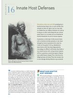
NEW CHAPTER 14: FACIAL GROWTH AND DEVELOPMENT
CHAPTER 14
FACIAL PROFILES
There are three basic types of facial profiles (Figure 14-3):
(1) the straight-jawed, or orthognathic, type; (2) the retrognathic profile, which has a retruding chin and is the most
common profile among white populations; and (3) the prognathic profile, which is characterized by a bold lower jaw
and chin.
To identify a person’s profile type, imagine a line projecting horizontally from the orbit. Drop a perpendicular line
from this just brushing the surface of the upper lip. If the
chin touches this vertical line, the profile is orthognathic; if
it falls behind or ahead, the profile is retrognathic or prognathic. For a female face, the vertical line generally passes
through the nose at a point about halfway along its upper
Facial Growth and Development
329
slope. In male faces that are long and narrow, however,
the more marked extent of the upper nasal prominence is
such that more of the nose sometimes lies forward of the
vertical line.
People with a dolichocephalic head form (a characteristic
feature of some white populations in northernmost and
southernmost Europe, North Africa, and the Middle East)
tend to have a retrognathic face. Those with a brachycephalic head form (a characteristic feature of Middle Europe
and East Asia) have a greater tendency toward prognathism.
Also, Asians commonly have a maxillary and mandibular
alveolodental protrusion characterized by labial tipping of
334
Ten Cate’s Oral Histology
A
A
B
FIGURE 14-10 Superimposed growth stages of the mandible from a child (5 years old) compared to an adult. A, Remodeling of the infant
mandible occurs by local combinations of resorption and deposition. This process relocates the ramus in posterior and superior direction
and provides for a lengthening of the corpus. B, During the growth, the whole mandible undergoes an anterior and inferior displacement.
B
FIGURE 14-1 Changes in craniofacial proportions between an
infant (2 months) and an adult. The skull at about birth has been
enlarged to match the adult skull to illustrate the differences in form
and proportions of craniofacial complex components. Note that the
neurocranium in the infant is prominent whereas the face predominates in the adult and represents a large part of the whole skull.
A
B
FIGURE 14-2 A, Dolichocephalic head form. B, Brachycephalic
head form.
C
D
FIGURE 14-3 In A, an orthognathic profile, the chin touches a vertical line along the upper lip perpendicular to the neutral orbital axis.
In B, a slightly retrognathic profile, the chin tip falls several millimeters behind this line. In C, a severely retrognathic face, the chin is
well behind the vertical line. The lower lip also is much less prominent. In D, a prognathic profile, the chin tip lies well forward of this
vertical line.
FIGURE 14-11 Perfectly balanced craniofacial composite. The occlusal plane is approximately perpendicular to the maxillary tuberosity.
It is rotated neither upward nor downward to any marked extent and is approximately parallel to the neutral orbital axis. In mo st faces, some
degree of occlusal plane rotation occurs.
xiii
This page intentionally left blank
Oral
Histology
Ten Cate’s
This page intentionally left blank
CHAPTER
1
Structure of the Oral Tissues
CHAPTER OUTLINE
The Tooth
Enamel
Dentin
Pulp
Supporting Tissues of the
Tooth
Periodontal Ligament
Cementum
Oral Mucosa
Salivary Glands
Bones of the Jaw
Temporomandibular Joint
Hard Tissue Formation
The Organic Matrix in Hard
Tissues
Mineral
Mineralization
Crystal Growth
Alkaline Phosphatase
Transport of Mineral Ions to
Mineralization Sites
Hard Tissue Degradation
Summary
T
his chapter presents an overview of the histology of the
tooth and its supporting tissues (Figure 1-1), setting the
stage for more subsequent detailed consideration. The salivary glands, the bones of the jaw, and the articulations
between the jaws (temporomandibular joints) also are
discussed.
Clinical crown
Enamel
Dentin
THE TOOTH
Teeth constitute approximately 20% of the surface area of the
mouth, the upper teeth significantly more than the lower
teeth. Teeth serve several functions. Mastication is the function most commonly associated with the human dentition,
but teeth also are essential for proper speech. In the animal
kingdom, teeth have important roles as weapons of attack
and defense. Teeth must be hard and firmly attached to the
bones of the jaws to fulfill most of these functions. In most
submammalian vertebrates the teeth are fused directly to the
jawbone. Although this construction provides a firm attachment, such teeth frequently are broken and lost during
normal function. In these cases, many successional teeth
form to compensate for tooth loss and to ensure continued
function of the dentition.
The tooth proper consists of a hard, inert, acellular
enamel formed by epithelial cells and supported by the less
mineralized, more resilient, and vital hard connective
tissue dentin, which is formed and supported by the dental
pulp, a soft connective tissue (Figures 1-1 and 1-2). In
mammals, teeth are attached to the bones of the jaw by
tooth-supporting connective tissues, consisting of the
Gingiva
Anatomical
crown
PDL
Pulp
Cementum
Bone
FIGURE 1-1 The tooth and its supporting structure. PDL, Periodontal ligament.
1
2
Ten Cate’s Oral Histology
FIGURE 1-2 Vertical Cone Beam CT slice of mandibular
molars and premolars. (Courtesy M. Schmittbuhl.)
Enamel
Crown
Dentin
Pulp
Root
Alveolar
bone
cementum, periodontal ligament (PDL), and alveolar bone,
which provide an attachment with enough flexibility to
withstand the forces of mastication. In human beings and
most mammals, a limited succession of teeth still occurs,
not to compensate for continual loss of teeth but to accommodate the growth of the face and jaws. The face and jaws
of a human child are small and consequently can carry few
teeth of smaller size. These smaller teeth constitute the
deciduous or primary dentition. A large increase in the size
of the jaws occurs with growth, necessitating not only
more teeth but also larger ones. Because the size of teeth
cannot increase after they are formed, the deciduous dentition becomes inadequate and must be replaced by a permanent or secondary dentition consisting of more and larger
teeth.
Anatomically the tooth consists of a crown and a root (see
Figures 1-1 and 1-2); the junction between the two is the
cervical margin. The term clinical crown denotes that part of
the tooth that is visible in the oral cavity. Although teeth vary
considerably in shape and size (e.g., an incisor compared
with a molar), histologically they are similar.
Rod
Interrod
ENAMEL
Enamel has evolved as an epithelially derived protective
covering for the crown of the teeth (Figures 1-1 and 1-2).
The enamel is the most highly mineralized tissue in the
body, consisting of more than 96% inorganic material in
the form of apatite crystals and traces of organic material.
The cells responsible for the formation of enamel, the ameloblasts, cover the entire surface of the layer as it forms but
are lost as the tooth emerges into the oral cavity. The loss of
these cells renders enamel a nonvital and insensitive matrix
that, when destroyed by any means (usually wear or caries),
cannot be replaced or regenerated. To compensate for this
inherent limitation, enamel has acquired a high degree of
mineralization and a complex organization. These structural and compositional features allow enamel to withstand
large masticatory forces and continual assaults by acids
Rod
FIGURE 1-3 Enamel. Electron micrography showing that enamel
consists of crystallites organized into rod and interrod enamel.
from food and bacterial sources. The apatite crystals within
enamel pack together differentially to create a structure of
enamel rods separated by an interrod enamel (Figure 1-3).
Although enamel is a dead tissue in a strict biologic sense,
it is permeable; ionic exchange can occur between the
C H A P T E R 1
Odontoblasts
process
Odontoblasts
Predentin
3
Structure of the Oral Tissues
Predentin
Dentin
Odontoblasts
A
B
Pulp
FIGURE 1-4 Dentin and pulp. A, The odontoblasts (cells that form dentin) line the pulp. B, These cells at higher magnification show processes extending into dentin.
enamel and the environment of the oral cavity, in particular
the saliva.
DENTIN
Because of its exceptionally high mineral content, enamel is
a brittle tissue, so brittle that it cannot withstand the forces
of mastication without fracture unless it has the support of
a more resilient tissue, such as dentin. Dentin forms the
bulk of the tooth, supports the enamel, and compensates for
its brittleness.
Dentin is a mineralized, elastic, yellowish-white, avascular tissue enclosing the central pulp chamber (Figure 1-4; see
also Figures 1-1 and 1-2). The mineral is also apatite, and the
organic component is mainly the fibrillar protein collagen.
A characteristic feature of dentin is its permeation by closely
packed tubules traversing its entire thickness and containing
the cytoplasmic extensions of the cells that once formed it
and later maintain it (Figure 1-4, B). These cells are called
odontoblasts; their cell bodies are aligned along the inner
edge of the dentin, where they form the peripheral boundary
of the dental pulp (Figure 1-4, A). The very existence of
odontoblasts makes dentin a vastly different tissue from
enamel. Dentin is a sensitive tissue, and more importantly,
it is capable of repair, because odontoblasts or cells in the
pulp can be stimulated to deposit more dentin as the occasion demands.
PULP
The central pulp chamber, enclosed by dentin, is filled with
a soft connective tissue called pulp (Figure 1-4, A). Histologically, it is the practice to distinguish between dentin and
pulp. Dentin is a hard tissue; the pulp is soft (and is lost in
dried teeth, leaving a clearly recognizable empty chamber;
see Figure 1-2, A). Embryologically and functionally,
however, dentin and pulp are related and should be considered together. This unity is exemplified by the classic functions of pulp: it is (1) formative, in that it produces the dentin
that surrounds it; (2) nutritive, in that it nourishes the avascular dentin; (3) protective, in that it carries nerves that give
dentin its sensitivity; and (4) reparative, in that it is capable
of producing new dentin when required.
In summary, the tooth proper consists of two hard tissues:
the acellular enamel and the supporting dentin. The latter is
a specialized connective tissue, the formative cells of which
are in the pulp. These tissues bestow on teeth the properties
of hardness and resilience. Their indestructibility also
gives teeth special importance in paleontology and forensic
science, for example, as a means of identification.
SUPPORTING TISSUES OF THE TOOTH
The tooth is attached to the jaw by a specialized supporting
apparatus that consists of the alveolar bone, the PDL, and
4
Ten Cate’s Oral Histology
FIGURE 1-5 Light microscope histologic sections of the periodontal ligament (PDL). A, Supporting apparatus of the tooth in longitudinal
section. B, At higher magnification, note
the fibrocellular nature of the periodontal
ligament.
Enamel
PDL
Dentin
Dentin
Pulp
A
B
Bone
Cementum
PDL
Collagen
the cementum, all of which are protected by the gingiva
(see Figure 1-1).
PERIODONTAL LIGAMENT
The PDL is a highly specialized connective tissue situated
between the tooth and the alveolar bone (Figure 1-5). The
principal function of the PDL is to connect the tooth to the
jaw, which it must do in such a way that the tooth will
withstand the considerable forces of mastication. This
requirement is met by the masses of collagen fiber bundles
that span the distance between the bone and the tooth and
by ground substance between them. At one extremity the
fibers of the PDL are embedded in bone; at the other
extremity the collagen fiber bundles are embedded in
cementum. Each collagen fiber bundle is much like a
spliced rope in which individual strands can be remodeled
continually without the overall fiber losing its architecture
and function. In this way the collagen fiber bundles can
adapt to the stresses placed on them. The PDL has another
important function, a sensory one. Tooth enamel is an inert
tissue and therefore insensitive, yet the moment teeth come
into contact with each other, we know it. Part of this sense
of discrimination is provided by sensory receptors within
the PDL.
CEMENTUM
Cementum covers the roots of the teeth and is interlocked
firmly with the dentin of the root (see Figures 1-1, 1-2, and
1-5, B). Cementum is a mineralized connective tissue similar
to bone except that it is avascular; the mineral is also apatite,
and the organic matrix is largely collagen. The cells that form
cementum are called cementoblasts.
The two main types of cementum are cellular and
acellular. The cementum attached to the root dentin and
covering the upper (cervical) portion of the root is acellular
and thus is called acellular, or primary, cementum. The lower
(apical) portion of the root is covered by cellular, or secondary, cementum. In this case, cementoblasts become trapped
in lacunae within their own matrix, very much like osteocytes occupy lacunae in bone; these entrapped cells are now
called cementocytes. Acellular cementum anchors PDL fiber
bundles to the tooth; cellular cementum has an adaptive role.
Bone, the PDL, and cementum together form a functional
unit of special importance when orthodontic tooth movement is undertaken.
ORAL MUCOSA
The oral cavity is lined by a mucous membrane that consists
of two layers: an epithelium and subjacent connective tissue
(the lamina propria; Figure 1-6). Although its major functions are lining and protecting, the mucosa also is modified
to serve as an exceptionally mobile tissue that permits free
movement of the lip and cheek muscles. In other locations
it serves as the organ of taste.
Histologically, the oral mucosa can be classified in three
types: (1) masticatory, (2) lining, and (3) specialized. The
masticatory mucosa covers the gingiva and hard palate. The
masticatory mucosa is bound down tightly by the lamina
propria to the underlying bone (Figure 1-6, B), and the covering epithelium is keratinized to withstand the constant
C H A P T E R 1
5
Structure of the Oral Tissues
A
Gingiva
Alveolar
mucosa
Labial mucosa
Epithelium
Epithelium
B
Loose CT
Dense CT
Bone
Submucosa
C
Salivary
gland
FIGURE 1-6 Oral mucosa. A, Note the difference between tightly bound mucosa of the gingiva (gum) and mobile mucosa of the labial
sulcus (alveolar mucosa). B, In histologic sections, the gingival epithelium is seen to be tightly bound to bone by a dense fibrous connective
tissue (CT), whereas the epithelium of the lip (C) is supported by a much looser connective tissue.
pounding of the food bolus during mastication. The lining
mucosa, by contrast, must be as flexible as possible to
perform its function of protection. The epithelium is not
keratinized; the lamina propria is structured for mobility and
is not tightly bound to underlying structures (Figure 1-6, C).
The dorsal surface of the tongue is covered by a specialized
mucosa consisting of a highly extensible masticatory mucosa
containing papillae and taste buds.
A unique feature of the oral mucosa is that the teeth perforate it. This anatomic feature has profound implications in
the initiation of periodontal disease. The teeth are the only
structures that perforate epithelium anywhere in the body.
Nails and hair are epithelial appendages around which epithelial continuity is always maintained. This perforation by
teeth means that a sealing junction must be established
between the gum and the tooth.
The mucosa immediately surrounding an erupted tooth
is known as the gingiva. In functional terms the gingiva
consists of two parts: (1) the part facing the oral cavity, which
is masticatory mucosa, and (2) the part facing the tooth,
which is involved in attaching the gingiva to the tooth and
forms part of the periodontium. The junction of the oral
mucosa and the tooth is permeable, and thus antigens can
pass easily through it and initiate inflammation in gum tissue
(marginal gingivitis).
SALIVARY GLANDS
Saliva is a complex fluid that in health almost continually
bathes the parts of the tooth exposed within the oral cavity.
Consequently, saliva represents the immediate environment
of the tooth. Saliva is produced by three paired sets of major
salivary glands—the parotid, submandibular, and sublingual
glands—and by the many minor salivary glands scattered
throughout the oral cavity. A precise account of the composition of saliva is difficult because not only are the secretions
of each of the major and minor salivary glands different, but
their volume may vary at any given time. In recognition of
this variability, the term mixed saliva has been used to
describe the fluid of the oral cavity. Regardless of its precise
composition, saliva has several functions. Saliva moistens
the mouth, facilitates speech, lubricates food, and helps with
taste by acting as a solvent for food molecules. Saliva also
contains a digestive enzyme (amylase). Saliva not only dilutes
noxious material mistakenly taken into the mouth, it also
cleanses the mouth. Furthermore, it contains antibodies and
antimicrobial substances, and by virtue of its buffering
capacity plays an important role in maintaining the pH of
the oral cavity.
The basic histologic structure of the major salivary
glands is similar. A salivary gland may be likened to a
6
Ten Cate’s Oral Histology
Lobule
Main
excretory duct
Excretory duct
Connective
tissue septum
Striated duct
Intercalated duct
Canaliculus
between cells
Tubular secretory
end piece
FIGURE 1-8 Low-power photomicrograph of a salivary gland
showing its lobular organization.
Spherical secretory
end piece
FIGURE 1-7 Diagrammatic illustration of the ductal system of a
salivary gland.
bunch of grapes. Each “grape” is the acinus or terminal
secretory unit, which is a mass of secretory cells surrounding a central space. The spaces of the acini open into ducts
running through the gland that are called successively the
intercalated, striated, and excretory ducts (Figure 1-7), analogous to the stalks and stems of a bunch of grapes. These
ducts are more than passive conduits, however; their lining
cells have a function in determining the final composition
of saliva.
The ducts and acini constitute the parenchyma of the
gland, the whole of which is invested by a connective tissue
stroma carrying blood vessels and nerves. This connective
tissue supports each individual acinus and divides the gland
into a series of lobes or lobules, finally encapsulating it
(Figure 1-8).
BONES OF THE JAW
As stated before, teeth are attached to bone by the PDL
(Figures 1-1 and 1-5, A). This bone, the alveolar bone, constitutes the alveolar process, which is in continuity with the
basal bone of the jaws. The alveolar process forms in relation
to teeth. When teeth are lost, the alveolar process is gradually
lost as well, creating the characteristic facial profile of the
edentulous person whose chin and nose approximate because
of a reduction in facial height. Although the histologic structure of the alveolar process is essentially the same as that of
the basal bone, practically it is necessary to distinguish
between the two. The position of teeth and supporting
tissues, which include the alveolar process, can be modified
easily by orthodontic therapy. However, modification of the
position of the basal bone is usually much more difficult; this
can be achieved only by influencing its growth. The way
these bones grow is thus important in determining the position of the jaws and teeth.
TEMPOROMANDIBULAR JOINT
The relationship between the bones of the upper and lower
jaws is maintained by the articulation of the condylar process
of the mandible with the glenoid fossa of the temporal bone.
This articulation, the temporomandibular joint (TMJ), is a
synovial joint with special features that permit the complex
movements associated with mastication. The specialization
of the TMJ is reflected in its histologic appearance (Figure
1-9). The TMJ cavity is formed by a fibrous capsule lined
with a synovial membrane and is separated into two compartments by an extension of the capsule to form a specialized movable disk. The articular surfaces of the bone are
covered not by hyaline cartilage but by a fibrous layer that is
a continuation of the periosteum covering the individual
bones. A simplified way to understand the function of the
C H A P T E R 1
7
example, how is mineralization initiated in the organic
matrix? Or, for that matter, how are mineral ions brought to
the mineralization site?
B
THE ORGANIC MATRIX IN HARD TISSUES
A
D
F
C
Structure of the Oral Tissues
E
FIGURE 1-9 Sagittal section through the temporomandibular
joint. The disk (dividing the joint cavity into upper and lower compartments) is apparent. A, Intra-articular disc; B, mandibular
(glenoid fossa); C, condyle of mandible; D, capsule; E, lateral pterygoid muscle; F, articular eminence. (From Berkovitz BKB, Holland
GR, Moxham BJ: Oral anatomy, histology, and embryology, ed 3,
London, 2002, Mosby.)
TMJ is to consider it as a joint with the articular disk being
a movable articular surface.
HARD TISSUE FORMATION
The hard tissues of the body—bone, cementum, dentin, and
enamel—are associated with the functioning tooth. Because
the practice of dentistry involves manipulation of these
tissues, a detailed knowledge of them is obligatory (and each
is discussed separately in later chapters). The purposes of this
section are (1) to explain that a number of common features
are associated with hard tissue formation, even though the
final products are structurally distinct; (2) to indicate that
the functional role of a number of these features is still not
understood; and (3) to describe the common mechanism of
hard tissue breakdown.
Three (i.e., bone, cementum, and dentin) of the four hard
tissues in the body have many similarities in their composition and formation. They are specialized connective tissues,
and collagen (principally type I) plays a large role in determining their structure. Although enamel is not a connective
tissue and no collagen is involved in its makeup, its formation still follows many of the principles involved in the formation of hard connective tissue. Hard tissue formation may
be summarized as the production by cells of an organic
matrix capable of accommodating mineral. This rather
simple concept, however, embraces a number of complex
events, many of which are still not fully understood. For
A hallmark of calcified tissues is the various matrix proteins that attract and organize calcium and phosphate ions
into a structured mineral phase based on carbonated
apatite. The formative blast cells of calcified tissues produce
the organic matrix constituents that interact with the
mineral phase. These cells specialize in protein synthesis
and secretion, and they exhibit a polarized organization for
vectorial secretion and appositional deposition of matrix
proteins.
Of great interest is the fact that the proteins involved in
these hard tissue, with one exception (enamel), are similar,
comprising a predominant supporting meshwork of type I
collagen with various added noncollagenous proteins functioning primarily as modulators of mineralization. Table
1-1 provides a comparative analysis of the characteristics of
the various calcified tissues. This basic similarity of constituents is consistent with the general role of collagen-based
hard tissues in providing rigid structural support and protection of soft tissues in vertebrates. Enamel has evolved to
function specifically as an abrasion-resistant, protective
coating that relies on its uniquely large mineral crystals for
function. The organic matrix of enamel consists essentially
of noncollagenous proteins which have no “scaffolding”
role. However, enamel is not the only calcified tissue
without collagen. Mineralization of cementum situated
along the cervical margin of the tooth occurs within a
matrix composed largely of noncollagenous matrix proteins
also found in bone. In invertebrates, the shell of mollusks
consists of laminae of calcium carbonate separated by
a thin layer of organic material, acidic macromolecules
among others.
MINERAL
The inorganic component of mineralized tissues consists of
hydroxyapatite, represented as Ca10(PO4)6(OH)2 and which
has undergone a number of substitutions with other ions.
This formula indicates only the atomic content of a conceptual entity known as the unit cell, which is the least number
of calcium, phosphate, and hydroxyl ions able to establish
stable relationships. The unit cell of biologic apatite is hexagonal; when stacked together, these cells form the lattice of
a crystal. The number of repetitions of this arrangement
produces crystals of various sizes. Generally the crystals are
described as needlelike or platelike and, in the case of enamel,
as long, thin ribbons. Some believe that the formation of
crystals is preceded by an unstable amorphous calcium
phosphate phase.
A layer of water, called the hydration shell, exists around
each crystal. Each apatite crystal has three compartments,