Ebook The practice of emergency and critical care neurology: Part 2
Bạn đang xem bản rút gọn của tài liệu. Xem và tải ngay bản đầy đủ của tài liệu tại đây (30.3 MB, 608 trang )
PART VII
Management of Specific
Disorders in Critical Care
Neurology
26
Aneurysmal Subarachnoid Hemorrhage
M
ajor medical institutions may admit 50–75
patients with an aneurysmal subarachnoid
hemorrhage (SAH) a year. A multidisciplinary
team is required to respond to the immediate
needs of the patient and to plan for repair of the
aneurysm.8,42,101,154,175 Expertise may prevent poor
outcome.25,47,133
After aneurysmal rupture, 10% of patients
die suddenly or within the first hours before ever
receiving adequate medical attention. Many of
these patients had marked intraventricular extension of the hemorrhage and acute pulmonary
edema, both reasons for sudden death.144 Of those
most severely affected who reach the emergency
department (ED) or neurosciences intensive care
unit (NICU), half die within 3 months. Some of
these patients may have been found pulseless and
required prolonged cardiopulmonary resuscitation. Patients who survive a major first rupture
face the immediate risk of catastrophic rebleeding, rapidly developing hydrocephalus, potentially
life-
threatening pulmonary edema, and cardiac
arrhythmias. Presentation in a poor clinical condition often indicates that the hemorrhage is not confined to the subarachnoid space but rather there is
intraventricular and intraparenchymal extension.
Many have additional large ventricles and are in
need of CSF diversion with a ventriculostomy.
The critical steps in managing SAH are to
surgically clip the aneurysm or occlude the sac
by inserting platinum coils, to treat clinical neurologic deterioration early, and to manage major
systemic complications.169
Aneurysmal subarachnoid hemorrhage is a
prime example of a neurocritical and neurosurgical disorder where outcome in the first days after
presentation cannot be judged adequately and
care of the initially comatose patient can lead to a
satisfactory outcome.
Fortunately, a considerable proportion of
patients with SAH present with severe headache and are alert with little other findings. Early
repair of the aneurysm may result in an excellent
outcome.
CLINICAL RECOGNITION
The incidence of aneurysmal SAH varies, but
overall is 10 cases per 100,000 persons per year
(doubled in Finland and Japan).112 The risk is
nearly two times higher in women (particularly
with smoking history) than in men and in blacks
than in whites. Subarachnoid hemorrhage is
more common in patients with a family history of
SAH,101 polycystic kidney disease, systemic lupus
erythematosus, or Ehlers-Danlos disease (Capsule
26.1).60,61
Aneurysmal SAH may be manifested in many
ways. Typically, an unexpected instantaneous
headache warns the patient of a very serious disorder and is often described as excruciating and
overwhelming113 (Chapter 4).
Vomiting may occur several minutes into the
ictus as a result of further distribution of blood
throughout the subarachnoid space. Profuse vomiting may override the headache and has been
mistaken for a “gastric flu” by the patient or initially consulted physician.
With an incomplete medical history and no
inquiry about acute headache, patients may be
wrongly transferred to a medical ICU (cardiac
resuscitation and pulmonary edema), gastrointestinal service (vomiting), or coronary care unit
(cardiac arrhythmias with new electrocardiographic [EKG] changes). Other unusual clinical presentations have included acute paraplegia
(anterior cerebral artery aneurysm rupture into
frontal lobes) and severe thoracic and lumbar
pain caused by meningeal irritation. These presentations may have resulted in a delay in cranial
computed tomography (CT) scan imaging.
The abruptness of the headache is not specific
for SAH; it may occur in conditions such as arterial dissection, pituitary apoplexy, hypertensive
encephalopathy, spontaneous intracranial hypotension, and cerebral venous thrombosis43,44,143
(Chapter 4). Some patients briefly lose consciousness. Inappropriate behavior and agitation or
drowsiness may follow. Localizing neurologic
findings, although transient, may indicate the
318
Part VII: Management of Specific Disorders
CAPSULE 26.1 ANEURYSMAL RUPTURE
What causes aneurysms to rupture is puzzling. Risk factors have included recently documented
enlargement (rupture of aneurysms < 4 mm is uncommon; most ruptured aneurysms are 7–8
mm, and risk of rupture increases significantly in aneurysms of ≥ 10 mm),88 hypertension, cigarette smoking, and family history of aneurysms and SAH. Aneurysmal rupture has been reported
to have occurred during weightlifting, sexual orgasm, and brawling, events that suggest acute
hypertensive stress on a thin aneurysmal wall.160 However, at least 50% of patients have SAH
while at rest. Seasonal changes have been implicated with increased rupture rate during colder
temperatures and influenza peaks. An association between a recent infection and aneurysmal
rupture has not been definitively established, but is plausible.
Intracranial pressure rises dramatically to at least the level of the diastolic blood pressure,
causing cerebral perfusion standstill. The increase in intracranial pressure decreases within 15
minutes but may persist if acute hydrocephalus or a shift from intracerebral hematoma has
occurred. Rupture stops within 3–6 minutes after ejection of up to 15–20 mL/min into the basal
cistern.
Hemodynamic variables have been tested on cadaver and computer models. Variables that
may determine rupture are wall shear stress, intra-aneurysmal flow velocity, and inflow jet and
angles of entry and vortexes. Wall sheet stress is caused by the frictional force of blood, and areas
with high forces may fragment the internal elastic lamina and cause blebs and aneurysms.56,148
Hemodynamic stress may cause morphologic changes involving the endothelial lining of the
walls, with intimal hyperplasia, and organizing thrombosis. Many ruptured vacular aneurysms
show inflammatory changes, with infiltrating leukocytes and macrophages promoting fibrosis.
Other theories focus on the multitudes of vortices or unstable flow. High inflow jets with large
impact zones may result in thrombus or daughter sac formation.20,21
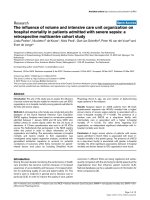
VORTEX
FORMATION
DAUGHTER
SAC
Subarachnoid hemorrhage. Left: aneurysmal rupture causing diffuse subarachnoid hemorrhage.
Right: Vortex formation in aneurysm.
site of the ruptured aneurysm. For example,
patients with a ruptured middle cerebral artery
(MCA) aneurysm may have transient or persistent aphasia. In patients with a ruptured MCA
aneurysm and intraparenchymal extension, hemiparesis often is found. Abulia most often occurs
as a complication of a rupture of an aneurysm of
the anterior cerebral artery (ACA). Generalized
Chapter 26: Aneurysmal Subarachnoid Hemorrhage
319
and vitreous hemorrhages. Top: Subhyaloid hemorrhage in subarachnoid hemorrhage. Bottom left: Red reflex is absent from vitreous hemorrhage also known as Terson syndrome. Bottom middle: Improvement in vision. Bottom right: Normal red reflex as shown by retro illumination with fundus camera.
FIGURE 26.1: Subhyaloid
tonic-clonic seizures are not quite so often seen at
the time of rupture, and it is possible that extensor
posturing or brief myoclonic jerks with syncope at
onset may be mistaken for a seizure. These clinical features in SAH are identical whether or not
an aneurysm is detected. Different presentation is
expected, however, in an established benign variant of nonaneurysmal SAH, so-called pretruncal or perimesencephalic SAH. The patients are
almost exclusively alert. Loss of consciousness is
seldom observed and seizures are absent, and the
onset of headache is less acute—in minutes rather
than a second.
Neurologic examination reveals neck stiffness
in most patients, except those seen early after the
initial event and those who are comatose. Nuchal
rigidity can be demonstrated by failure to flex the
neck in the neutral position and failure of the neck
to retroflex when both shoulders are lifted. Retinal
subhyaloid hemorrhages are present in approximately 25% of the patients (Figure 26.1). (This
syndrome is more often observed in comatose
patients and after rebleeding.) These flat-topped
hemorrhages occur when outflow in the optic
nerve venous system is suddenly obstructed by the
intracranial pressure (ICP) wave.55 Visual loss may
be severe, with perception of light or hand motion
only, if the hemorrhage expands and ruptures
into the vitreous (Terson syndrome).131 Cranial
nerve abnormalities occur infrequently in SAH
unless a giant basilar artery aneurysm (third-or
sixth-nerve palsy) or a large carotid artery aneurysm (chiasmal syndromes) directly compresses
surrounding structures. The pupil is dilated and
unreactive to light in a third-nerve palsy because
of compression of the exteriorly located fibers that
form the light reflex. However, up to 15% of posterior communicating artery aneurysms may occur
with a pupil-sparing third-nerve palsy. Aneurysm
of the basilar artery may produce unilateral or
bilateral third-or sixth-nerve palsy.87 If the basilar
artery aneurysm enlarges and progressively compresses the oculomotor nuclei of the pons, horizontal gaze paralysis, skew deviation, internuclear
ophthalmoplegia, and nystagmus occur, commonly in association with long-tract signs such
as hemiparesis and ataxia. Occlusion of the proximal posterior cerebral artery, often encased in a
giant aneurysm, may occur, causing either classic
Weber syndrome (Chapter 30) due to mesencephalon infarction (third-nerve palsy with opposite
hemiparesis) or homonymous hemianopia due to
occipital lobe infarction.
In comatose patients, a certain eye position
may be localizing. These forced gaze positions
include downward gaze and wall-eyed bilateral
internuclear ophthalmoplegia, and are characteristically seen with acute hydrocephalus
(Figure 26.2).90
Hemiparesis that usually involves the face,
arm, and leg should point to an intracranial hematoma in SAH. An anteriorly placed intracranial
320
Part VII: Management of Specific Disorders
FIGURE 26.2: Wall-
eyed bilateral internuclear ophthalmoplegia with acute hydrocephalus in patient with
aneurysmal subarachnoid hemorrhage.
hematoma in the frontal lobe may not produce
motor weakness but may be associated with agitation and bizarre behavior. Many patients are confused, and may ramble nonsensically. Korsakoff
syndrome with impaired recall and fabrications
may occur in ruptured anterior communicating
aneurysm. Abulia, a general sense of disinterest,
and lackluster attention are also features, becoming apparent days later.62 Temporal lobe hematoma in the dominant hemisphere may produce
aphasia, but often associated mass effect and
brainstem displacement decrease the level of consciousness and word output.
Generalized tonic-clonic seizures are accompanied by aneurysmal rupture in 10% of patients,
or these appear during rebleeding. These “seizures” are likely ischemic in nature and a result
of a major increase in ICP. Nonconvulsive status
epilepticus may occur, and the clinical signs are
difficult to differentiate from the effects of initial
rupture. However, in our experience, electroencephalography (EEG) has rarely documented
nonconvulsive status epilepticus. Epilepsia partialis continua is equally uncommon in aneurysmal SAH. It is more common in patients with
additional subdural hematoma and when delayed
cerebral infarction occurs.
Systemic manifestations may include respiratory failure and oxygen desaturation from aspiration, pulmonary edema, or obstruction of the
airway. Cardiac arrhythmias may involve the
entire spectrum of supraventricular and ventricular arrhythmias. Most of the time they are associated with EKG changes, which may simulate
or indicate acute myocardial infarction. Elevated
troponin I levels may occur in approximately 25%
of the cases seen on the first day. Major cardiac
arrhythmias may lead to cardiac resuscitation
after SAH and generally portend poor outcome,
but patients may improve substantially.152
When patients are comatose at presentation, it
is largely due to the initial rise in ICP with reduction of cerebral blood flow and, as a consequence,
diffuse bihemispheric ischemia.79 However, one
should try to make a distinction between the direct
effects of the initial impact and early neurologic
deterioration due to other causes. Acute hydrocephalus may have developed in the interim, and
placement of a ventricular drain could markedly
improve the level of consciousness. Patients admitted days after the ictus may have symptomatic
cerebral vasospasm, and focal signs and symptoms
may not be present. Coma may be caused by brain
tissue shift from a large expanding hematoma in
the sylvian fissure. Removal of the hematoma
and repair of the aneurysm may result in marked
improvement.
The clinical course in poor-grade aneurysmal
SAH is unpredictable in the first 24–48 hours.
Patients moribund at presentation may improve
in a matter of hours without much neurosurgical
or medical intervention, although the prognosis
may remain guarded.
Systemic metabolic factors may contribute, and each of them should be excluded.
Measurements of arterial blood gas, electrolytes,
and serum glucose must be obtained rapidly in
every patient with SAH who enters the NICU.
A simple clinical grading system proposed by
the World Federation of Neurological Surgeons
(WFNS) introduced the Glasgow Coma Scale in
SAH grading46,139 (Table 26.1), and for practical
reasons the severity is graded as good (WFNS
I–III) or poor (WFNS IV or V). A correlation
between outcome and initial grading level exists.
This rather crude scale may also guide the timing
TABLE 26.1. GRADING SYSTEM
PROPOSED BY THE WORLD FEDERATION
OF NEUROLOGICAL SURGEONS FOR THE
CLASSIFICATION OF SUBARACHNOID
HEMORRHAGE
WFNS Grade
Glasgow Coma
Scale Score
Motor Deficit
I
II
III
IV
V
15
14–13
14–13
12–7
6–3
Absent
Absent
Present
Present or absent
Present or absent
WFNS, World Federation of Neurological Surgeons.
Chapter 26: Aneurysmal Subarachnoid Hemorrhage
(a)
(b)
(c)
(d)
321
FIGURE 26.3: Subarachnoid hemorrhage. (a, b) Subarachnoid hemorrhage with complete filling of the basal cisterns and fissures (arrows), creating a “crab-like” cast. (c) Global cerebral edema. (d) Extensive low-attenuation
changes (arrows) in frontal and insular cortex.
of surgery. Some neurosurgeons defer craniotomy
for aneurysmal clipping in patients with WFNS V,
but coiling may proceed. Improvement in grade
may make the patient eligible for aneurysmal
clipping.
N E U R O I M AG I N G A N D
L A B O R AT O R Y T E S T S
Subarachnoid hemorrhage shows on CT scan
(Figure 26.3a and b). Some patients show CT
scans with massive SAH and early global edema
(Figure 26.3c and d).27 CT perfusion may show
reduced blood flow. These findings are more
common in patients who remain comatose after
cardiopulmonary resuscitation. When CT scan
is done within hours after the event, the sensitivity in aneurysmal SAH is very high and may
approach 95%. In 2%–5% of the patients, subarachnoid blood has completely “washed out” on
CT scans within 24 hours, but more likely, CT
may have missed a thin layer of blood. Repeat CT
scan in patients with initial “negative CT” and
xanthochromia often documents traces of SAH in
sulci or ventricles.54
322
Part VII: Management of Specific Disorders
Fisher developed one of the first grading systems for SAH. The Fisher scale, although deeply
ingrained in neurological practice, remains a
gross estimate of the amount of subarachnoid
blood, and it has significant inter-observer variability. This scale, currently modified (Table 26.2),
emphasizes the presence or absence of a thick clot
and the presence of intraventricular hemorrhage,
and predicts the development of delayed cerebral
ischemia.53
Another grading system was developed by
Hijdra and colleagues78 (Table 26.2). A sum score
TABLE 26.2. COMPUTED TOMOGRAPHY
FINDINGS IN THE MODIFIED FISHER
AND HIJDRA SCALE
Grade
Finding
1
2
3
4
Focal or diffuse thin SAH without IVH
Focal or diffuse thin SAH with IVH
Thick SAH present without IVH
Thick SAH present with IVH
SAH: subarachnoid hemorrhage; IVH: intraventricular hemorrhage.
Data from Kistler JP, Crowell RM, Davis KR, et al. The relation
of cerebral vasospasm to the extent and location of subarachnoid blood visualized by CT scan: a prospective study. Neurology
1983;33:424–436; and Frontera J, Claassen J, Schmidt JM, et al.
Prediction of symptomatic vasospasm after subarachnoid hemorrhage: the modified Fisher scale. Neurosurgery 2006;59:21–27.
A
B
C
C
B
D
D
E
E
F
Hijdra method of grading subarachnoid hemorrhage
identifies 10 basal cisterns and fissures: (A) frontal
interhemispheric fissure; (B) sylvian fissure, lateral
parts both sides; (C) sylvian fissure, basal parts both
sides; (D) suprasellar cistern both sides; (E) ambient
cisterns both sides; and (F) quadrigeminal cistern. The
amount of blood in each cistern and fissure is graded 0,
no blood; 1, small amount of blood; 2, moderately filled
with blood; and 3, completely filled with blood. The sum
score is 0 to 30 points.78
of greater than 20 is considered predictive of
cerebral vasospasm. In our recent study of different scales, we found the Hijdra scale superior
to other scales in prediction of cerebral vasospasm.49 Quantification of SAH and calculation of
volume may remain a useful alternative, but the
applicability of this method remains unknown.
Grading after “resuscitation” correlates better with
outcome.59
Important information can be gathered by
careful inspection of CT scans. The distribution of
the subarachnoid blood on CT scan may suggest
the location of the aneurysm, but despite subtle
differences, CT scanning often cannot reliably predict the location of the aneurysm. There is no correlation between the size of the aneurysm and the
amount of SAH.140 Generally, patients with diffuse
distribution of blood in cisterns and fissures often
have a basilar artery or ACA aneurysm. However,
patients with a concentration of blood in the interhemispheric fissure may have an aneurysm of the
anterior cerebral artery, and patients with cisternal blood surrounding the perimesencephalic cisterns most likely harbor a basilar artery aneurysm.
Likewise, sylvian fissure hemorrhages are mostly
from an aneurysm of the MCA.
The additional presence of an intracerebral
hematoma, however, has more localizing value.
Hematomas may be found in the frontal lobe
(anterior communicating artery aneurysm), in the
medial part of the temporal lobe (internal carotid
artery aneurysm), and within the sylvian fissure
extending into the temporal lobe (MCA aneurysm) (Figure 26.4).73
As alluded to earlier a benign form of SAH
has been reported in which bleeding is confined
to the cisterns in front of the brainstem without
evidence of an aneurysm in the posterior cerebral
circulation—so-called pre-truncal SAH141,142,170
(also called perimesencephalic hemorrhage)159
(Figure 26.5a). True perimesencephalic hemorrhages are purely traumatic, due to either a
P2 aneurysm or spinal dural arteriovenous fistula.141,146 Typically, in these variants, blood clots
do not extend to the lateral sylvian fissures or to
the anterior interhemispheric fissure. Some extension to the basal part of the sylvian fissure is possible when CT scanning is performed very early.
Intraventricular hemorrhage is absent except
for some sedimentation in the posterior horns.
Magnetic resonance imaging (MRI) is helpful in
localizing the blood clot in front of the brainstem,
and no cause has been found with this modality
(Figure 26.5b). MRI of the cervical spine has also
been unrevealing.170
(a)
(a1)
(b)
(c)
(a2)
(d)
(e)
(f)
(g)
(h)
FIGURE 26.4:
Computed tomographic patterns of subarachnoid hemorrhage with associated hematomas indirectly
localizing ruptured aneurysms. Temporal lobe and sylvian fissure hematoma (a, a1) (middle cerebral artery aneurysm
on CTA (a2)). Frontal hematoma (anterior cerebral artery) (b). Hematoma in cavum septum pellucidum (c). Medial
temporal lobe hematoma (d). Subdural hematoma (carotid artery, mostly ophthalmic artery) (e). Corpus callosum
(pericallosal artery) (f). Cerebellopontine angle hematoma with posterior inferior cerebellar artery aneurysm (g, h).
(a)
(b)
(c)
(d)
(e)
(f)
FIGURE 26.5: Nonaneurysmal pretruncal (perimesencephalic) subarachnoid hemorrhage. (a, b) Computed tomographic scan patterns of pretruncal nonaneurysmal subarachnoid hemorrhage in different patients. The spectrum
includes complete filling of suprasellar cisterns to more restricted clots and subtle interpeduncular hematoma. The
amount of blood is not critical in its recognition. However, the distribution of blood is limited and should not involve
the entire lateral part of the sylvian fissure or the anterior hemisphere and ventricles. (c, d) Magnetic resonance imaging patterns of pretruncal nonaneurysmal subarachnoid hemorrhage. Blood may involve all or part of the cisterns in
front of the brainstem. (e) Typical pretruncal CT pattern, but PICA aneurysm (f) proving a detailed vascular study
is needed.
Chapter 26: Aneurysmal Subarachnoid Hemorrhage
The cause of this perplexing benign form of
nonaneurysmal SAH remains unclear. Prior speculative explanations have included spinal dural
arteriovenous fistula, rupture of a dilated vein in
the prepontine cistern, and intramural dissection,72,141,171 but there is accumulating evidence
that a small blister-like aneurysm of the posterior
circulation may be implicated. Recent 3D cerebral
angiograms have been able to document these
small lesions.166
Pretruncal hemorrhage may closely mimic a
ruptured basilar artery or dissecting P1–P2 aneurysm, and therefore a four-vessel cerebral angiogram is warranted.93 In our experience and that of
others,126 we have found small (dissecting) aneurysms occasionally on repeat studies; and repeat
cerebral angiograms on CTA may remain warranted in this subset.
Localized blood in the sulci alone is unusual in
aneurysmal SAH and often indicates trauma coagulopathy or, much less common, vasculitis.104,136
Subarachnoid blood caused by trauma is most often
confined to the vertex and superficial cortical sulci
or accumulates in the ambient cisterns at the level
of the tentorium.40 Computed tomography scans
should be scrutinized for fractures on bone windows when physical examination shows other signs
of trauma, for example, skin bruising or a soft-tissue
swelling. When blood is in the sylvian fissure, a reliable distinction from a ruptured middle cerebral
aneurysm cannot be made on clinical grounds or by
CT scan, and cerebral angiography is needed.
Intraventricular blood on CT scans signals a
severe SAH. Aneurysms of the ACA have a proclivity to perforate the lamina terminals and enter
the ventricular system. Massive intraventricular
hemorrhage in patients with SAH may also suggest rerupture. Subdural hematomas are seen
in 1% of patients with SAH, often with cisternal
blood, and very rarely in isolation. Most often, a
carotid artery (ophthalmic or posterior communicating) aneurysm can be demonstrated on the
angiogram.
An important feature on CT scanning is acute
hydrocephalus. Enlargement of the lateral ventricles is often asymptomatic in acute SAH, and acute
hydrocephalus as an explanation for drowsiness is
more convincing if progression on sequential CT
scans can be demonstrated or if the ventricles are
very plump (Chapter 36).
Cerebral angiography remains the unchallenged gold standard for the diagnosis of cerebral
aneurysm. One can argue for early four-vessel
cerebral angiography in every patient, including
325
those with poor-grade SAH. These patients may
have aneurysms that can be occluded through
endovascular techniques.
Before cerebral angiography is undertaken,
serum creatinine concentration should be determined. The risk for neurologic deficit associated
with the procedure is 0.07%.29 The most important risk factor for contrast-induced nephrotoxicity is preexisting renal failure. The risk is also
increased in patients with reduced intravascular
volume and in patients using drugs that impair
renal responses, such as angiotensin-converting
enzyme inhibitors and nonsteroidal anti-
inflammatory drugs. In patients with preexisting
renal impairment, defined as a creatinine value
of more than 1.8 mg/dL, 0.45% saline should be
given intravenously at a rate of 1 mL/kg of body
weight per hour beginning 12 hours before the
scheduled angiography.29,74
Cerebral angiography may demonstrate
aneurysms at typical locations (Figure 26.6).
Standard examination should include anteroposterior and lateral views, but because overlapping is significant, oblique views are often
necessary. The neuroradiologist may be guided
by the findings on CT scan and should frequently use additional oblique views in evaluating the circle of Willis. Important additional
views are submentovertex views (particularly
useful for demonstrating the neck of an anterior
communicating aneurysm) and transorbital
projection (neck of the MCA). Towne’s projection is important to visualize the tip of the
basilar artery. Failure to demonstrate an aneurysm may be related to inadequate projection
or incomplete study (three-vessel study), and a
second angiogram at a slightly different angle
may uncover an aneurysm. Three-dimensional
image volume generated by digital fluorography
with rotational image acquisition has improved
detection. Multiple aneurysms may be found,
and it is virtually impossible to predict which
aneurysm has bled. However, additional clues
(next to CT scan patterns) may be present,
such as irregularity of the wall of the aneurysm
produced by the sealing clot, vasospasm in the
vicinity of the aneurysm, and size between 5
and 15 mm.
When an angiogram is negative, a second cerebral angiogram may demonstrate an aneurysm in
approximately 10% of cases. The second cerebral
angiogram should be particularly carefully scrutinized for a posterior circulation aneurysm, which
could have been “missed” on the first angiogram.
326
Part VII: Management of Specific Disorders
Calcarine and parietooccipital branches
Anterior cerebral
Anterior choroidal
Posterior parietal
Angular artery
Superior cerebellar
branches
Pericallosal
artery
Posterior cerebral artery
PICA
Basilar artery
Ascending frontal
Vertebral artery
Anterior temporal
Ophthalmic
Internal carotid
ACom
Internal frontoparietal branches
Callosomariginal
artery
Frontopolar
artery
Pericallosal artery
Orbital-frontal
artery
Ophthalmic
Internal carotid with MCA branches removed
FIGURE 26.6: Anterior
cerebral artery, middle cerebral artery, basilar artery tip, and posterior communicating
artery aneurysms on cerebral angiogram in their optimal projections.
ACom, anterior communicating artery; MCA, middle cerebral artery; PICA, posterior inferior cerebellar artery.
Whether exploratory craniotomy is needed in
patients with a high suspicion of an aneurysm
(presence of subarachnoid blood and intracranial
hematoma) is very unclear and this is rarely done,
even though some explorations have been successful in detecting the ruptured aneurysm.41
CT angiogram has been used in patients with
large aneurysms to better document anatomical configuration,71,176 in patients with an initial negative cerebral angiogram (as a means of
follow-up), and as the only additional diagnostic test in patients with pretruncal SAH in some
European centers.162 Its place in the diagnostic
evaluation of patients with an SAH is unclear.
Moreover, the less than perfect sensitivity of
97% (87% in aneurysms, 3 mm) and specificity
of 86% may have medicolegal implications if it is
the only study done. Moreover, anatomic bulges
of the basilar tip and pituitary stalk mistaken
for aneurysms and incorrect three-dimensional
reconstruction of overlapping MCAs are some
of the reasons for false-positive results. The role
of CT angiogram largely is as a simple and quick
(but also expensive) method to demonstrate or
exclude an aneurysm. CT angiogram, therefore, has been performed during night hours
while waiting for a more definitive study in the
morning.
Magnetic resonance imaging is usually not
sensitive for SAH but may be able to show SAH
when fluid attenuation inversion recovery (FLAIR)
sequences are used. Recirculation of bloody cerebrospinal fluid (CSF) over the convexity is commonly seen as well. MRI may be important in
Chapter 26: Aneurysmal Subarachnoid Hemorrhage
demonstrating an acute SAH in the posterior fossa,
which, as mentioned previously, may be difficult
to detect on CT scan because of beam-hardening
artifacts. Often, in retrospect, CT scans showed a
similar blood clot. Sometimes a small deposit of
blood in the sylvian fissure not visualized on CT
scans can be demonstrated on MRI.
Magnetic resonance angiography (MRA) is
equally useful in demonstrating the aneurysm,
and with three-dimensional time-of-flight MRA,
aneurysms 3 mm in diameter and larger can be
demonstrated.
FIRST STEPS
I N M A NAG E M E N T
Initial management in patients with aneurysmal SAH can be adapted to the initial grade.
Subarachnoid hemorrhage of WFNS grade I to
III should be differentiated from poor-grade SAH
(WFNS grade IV or V), assuming that the poor
clinical grade is caused by the initial impact alone.
The initial management in aneurysmal SAH is
summarized in Table 26.3. Continuous assessment
327
of alertness and performance remains important.
Experienced nurses in neurologic intensive care
usually are familiar with the peak time of cerebral ischemia and the first clinical signs of acute
hydrocephalus.
An important component of management in
SAH is the relief of pain. Severe headache is best
treated by acetaminophen with codeine. Many
patients benefit from the calming effect of these
agents, but others do not tolerate opioids and may
vomit excessively. Codeine remains effective in
many patients. Tramadol (usually only in its maximal dose of 400 mg/day) may be helpful in this
situation but should be avoided if the patient had a
seizure at onset. In patients with marked neck stiffness and severe unrelenting headache, 4 mg of dexamethasone for a few days may do wonders in some.
Respiratory care is largely supportive, and
serial chest radiographs should be reviewed for
signs of gastric aspiration or pulmonary edema.
Intubation and mechanical ventilation are
often indicated in poor-grade SAH. The ventilatory mode chosen should provide adequate
TABLE 26.3. INITIAL MANAGEMENT OF ANEURYSMAL SUBARACHNOID
HEMORRHAGE
Airway management
Mechanical ventilation
Fluid management
Blood pressure management
Nutrition
Prophylaxis
Other measures
Access
Intubation if patient has hypoxemia despite facemask with 10 L of 60%–100%
oxygen/minute, if abnormal respiratory drive or if abnormal protective
reflexes (likely with motor response of withdrawal or worse)
IMV/PS
AC with aspiration pneumonitis, ARDS or early neurogenic pulmonary edema
2–3 L of 0.9% NaCl per 24 hours
Fludrocortisone acetate, 0.2 mg b.i.d. orally, if patient has hyponatremia
Aim at SBP of < 160 mm Hg
IV labetalol 10–15 mg every 15 min if needed
Hydralazine 10–20 mg IV if bradycardia
Enteral nutrition with continuous infusion (on day 2)
Blood glucose control (goal 140–180 mg/dL)
DVT prophylaxis with pneumatic compression devices
SC heparin 5,000 U t.i.d. after clipping or coiling of aneurysm
GI prophylaxis: pantoprazole 40 mg IV daily or lansoprazole 30 mg orally
through nasogastric tube.
Nimodipine, 60 mg six times a day orally for 21 days
Tranexamic acid 1 gram IV, second dose 2 hours later, third dose 6 hours later
if delayed clipping or coiling
Codeine 30–60 mg orally every 4 hours as needed
Tramadol, 50–100 mg orally q4h, for pain management
Levetiracetam 20 mg/kg IV over 60 minutes; 1,000 mg b.i.d. maintenance
(if seizures have occurred)
Arterial catheter to monitor blood pressure (if IV antihypertensive drugs
anticipated)
Peripheral venous catheter or peripheral inserted central catheter
ARDS, acute respiratory distress syndrome; DVT, deep vein thrombosis; GI, gastrointestinal; IMV, intermittent mandatory ventilation; IV,
intravenously; MAP, mean arterial pressure; NaCl, sodium chloride; PS, pressure support; SBP, systolic blood pressure; SC, subcutaneously.
328
Part VII: Management of Specific Disorders
minute ventilation at the lowest possible airway
pressure—
in most instances, an intermittent
mandatory ventilation mode.
Stress cardiomyopathy tends to develop in
patients with poor-
grade SAH, and it can be
observed clinically and on repeat echocardiograms.
It may be a cause for the development of pulmonary
edema (Chapter 46).
To provide adequate fluid intake is an essential
part of the management of SAH. Approximately
one-third of the patients have a decrease in plasma
volume of more than 10% in the first days, often
detected by negative fluid balance.173 Initially,
most patients are probably best managed with 3
L of isotonic saline (or infusion of 125 mL/hr).
Fever (> 38.5°C) is more common in poor-grade
intubated patients, but fever is also associated,
in the absence of any infection, with the development of cerebral vasospasm after other causes
have been excluded. Fever is typically controlled
aggressively, and different methods are available3
(Chapter 21).
The management of acute hypertension after
SAH is uncertain18 (Chapter 19). When using antihypertensive treatment, a fine line separates necessity from harm. A retrospective study suggested that
the incidence of cerebral infarction is increased in
patients treated with antihypertensive drugs (largely
clonidine).172 On the other hand, earlier studies suggested that rebleeding and death from rebleeding
are increased in patients with persistently increased
systolic blood pressures (Chapter 19). Given the lack
of evidentiary data, there is insufficient guidance for
antihypertensive management soon after aneurysmal subarachnoid hemorrhage.
Most practicing neurointensivists and neurosurgeons decrease blood pressure with intravenous labetalol when a mean arterial pressure of
approximately 120 mm Hg or systolic blood pressure of 180 mm Hg persists.
Patients with SAH may be combative and may
require sedation. Agitation may be directly related
to placement of the endotracheal tube and to
inappropriate mechanical ventilator settings (e.g.,
high-frequency assist-control in an alert patient).
Not infrequently, these patients can be extubated
without any difficulty, which resolves the distress
and agitation. Combative and agitated patients
can be best treated with low-dose midazolam or
propofol infusion.
Nutrition can usually be deferred until the second day. Enteral feedings in patients with critical
neurologic illness are not always tolerated, and
poor gastric emptying may lead to aspiration.
However, placement of a nasoenteric feeding tube
into the duodenum or jejunum may overcome
these problems. Usually, concentrated commercial solutions infused at a low rate are administered (see Guidelines).
Stool softeners are prescribed, particularly for
patients who regularly require opiates. Prophylaxis
of deep vein thrombosis is provided by stockings
and pneumatic compression devices. Proton-
pump inhibitors are provided only for patients
who have a history of gastric ulcers or who have
been using nonsteroidal anti-inflammatory agents
or aspirin and in patients on the mechanical ventilator. Patients who have a decreased level of
consciousness need an indwelling bladder catheter. The use of intermittent catheterization may
decrease the incidence of urinary tract infection,
but the procedure is too stressful for patients with
acute SAH.
Nimodipine is administered in all patients
with SAH to prevent delayed cerebral ischemia.2,5,130 It can be crushed and applied through
the nasogastric tube.2 A regimen of nimodipine
(60 mg orally every 4 hours) is instituted for 21
days on the basis of significant reduction in the
incidence of delayed cerebral ischemia and mortality.2,132 A review of 90 patients treated with
nimodipine for 15 days or less did not suggest an
increase in delayed cerebral ischemia, but there
is no reason to shorten the period of administration.153 Nimodipine can be discontinued when
cerebral angiogram shows no aneurysm. No other
agents have been found to reduce cerebral ischemia.66,67 There was interest in the use of statins
following SAH. Cholesterol-lowering agents may
also prevent thrombogenesis, increase cerebral
arterial diameter, and reduce inflammation. Only
small studies have been performed, and there
were early indications of a possible benefit.116,149,155
The Simvastatin in Aneurysmal Subarachnoid
Hemorrhage (STASH) trial, however found no
benefit.98
The use of prophylactic antiepileptic medication is very questionable. The incidence of seizures
after acute SAH is low, and most seizures recur
during re-rupture. The risk of late seizures may
theoretically be increased in patients who have
a temporal lobe or frontal lobe hematoma and
large amounts of blood on CT, but again, no hard
data are available to specifically justify prophylactic antiepileptic agents. Newer studies raised the
possibility of worse cognitive outcome after the
use of phenytoin.127 The underlying mechanism
is unclear and could be related to a pharmaceutical interaction between phenytoin and nimodipine (phenytoin may reduce bioavailability of
Chapter 26: Aneurysmal Subarachnoid Hemorrhage
nimodipine through induction of the hepatic
cytochrome P450 isoenzymes).
Currently, antifibrinolytic therapy is not used
routinely. Antifibrinolytic therapy is very effective in preventing rebleeding and significantly
reduces the risk of rebleeding.83 However, when
used for prolonged periods of time, a reciprocal
increase in delayed cerebral ischemia is observed
and results in no overall benefit.163 A pilot study in
which tranexamic acid was given for only 4 days
produced the reverse of the desired result, with
no effect on the incidence of rebleeding and an
increase in the incidence of cerebral ischemia.168
Use of antifibrinolytic agents varies among
institutions and among neurosurgeons. There is
a tendency to use a few doses of antifibrinolytic
drugs in recently admitted patients while they
await the planning of surgical repair or endovascular coiling.26
Emergency or early surgery is indicated in
patients with evidence of rebleeding or intra-
cerebral hematoma in the temporal lobe and
tissue shift12 and, at the opposite end of the spectrum, any patient in good prior health with WFNS
grade I–III.13 Surgery can be temporarily withheld
in patients in WFNS grade IV or V with packed
intraventricular hemorrhage and hydrocephalus.
Ventriculostomy could produce improvement
in such patients. Surgery may also be postponed
in patients with early symptomatic vasospasm,
but the timing of surgery has always remained
contentious.
For eligible patients, cerebral angiography
should be performed as soon as feasible and should
be followed by surgical clipping of the aneurysm
(operative techniques and neuro-anesthesia are
beyond the scope of this book). A cooperative
study group found in a large survey that no major
differences existed between early and late surgery
but that outcome was worse when surgery was
performed between days 7 and 10.95
The development of detachable coils
(Guglielmi detachable platinum coils) has dramatically modified practices.14,19,38,107,123,152,165 A direct
electrical current disconnects the coil, and the
positive electrical charge increases thrombus formation. The procedure of multiple coil placement
is time-
consuming, taking several hours, and
needs general anesthesia monitoring. Coil placement has become the first consideration in most
patients with a ruptured aneurysm, irrespective of
the WFNS grade.7,11,100 It is often the first choice
of treatment in basilar artery apex aneurysms
because clipping is more complicated and risky.64
In the International Subarachnoid Aneurysm
329
Trial (ISAT) study,96,122 results found benefit from
the use of coils in good-grade patients with small
anterior circulation aneurysms, but no sufficient
proof in other patients with SAH. At 1 year, coiling was superior, with a 7.4% absolute risk reduction in mortality and major disability. At 5 years,
mortality in the endovascularly treated group was
lower than in the surgically treated patients (11%
versus 14%). There was no difference in disability
between the groups.124
Large series of patients from France reported
good outcomes in endovascularly treated patients,
many with poor-grade SAH.11 The generalizability of the ISAT trial has been questioned, most
recently by a study from the University of Toronto
that suggested worse hemorrhage-
free survival
of coiling compared with clipping.127 Long-term
outcome is not yet available, and concern about
imperfect repair with coiling remains. A review
of 509 patients with treated ruptured aneurysms
found ischemic complications in 7% and aneurysm perforation in 3%, with procedure-related
mortality of 1%.14 The estimated morbidity related
to the technique was 9%, with an overall mortality of 6%, but these numbers are now likely lower
with improved skills.14 A considerable drawback
of endovascularly treated patients is rebleeding
from a remnant aneurysm, with reported rebleeding rates of 6%–25%.
Experience with endovascular coil placement
in acute ruptured aneurysm is currently substantial, but the decision to “clip or coil” remains arbitrary. In the ISAT trial, the inclusion of patients
required that both the neurosurgeon and neurointerventionalist considered the patients eligible
for both treatments. However, in this trial122 the
involved physicians did not agree with each other
in more than two-thirds of cases. Currently it
can be estimated that more than 70% of ruptured
aneurysms are treated endovascularly in US referral centers.
Certain criteria have emerged that are based
on the width of the neck and the size and location of the aneurysm. Selection for coiling is
often determined by location of the aneurysm
in the posterior circulation, width of neck less
than 5 mm, and a dome-to-neck ratio greater
than 2 (Figure 26.7). There is a sharp reduction
in the rate of complete persistent occlusion for
aneurysms greater than 10 mm in diameter and
in aneurysms with broad necks. However, some
of these aneurysms with complex anatomy can
be treated with stent-assisted coiling or balloon-
modeling techniques in which a soft balloon
is temporarily inflated in the parent artery to
330
Part VII: Management of Specific Disorders
GOOD GEOMETRY
Dome to neck
ratio ≥ 2:1
POOR GEOMETRY
Dome to neck
ratio < 2:1
POOR GEOMETRY
Dome to neck
ratio ≥ 2:1;
wide neck
FIGURE 26.7:
Assessment of the geometry of cerebral aneurysm. Using the neck: dome ratio to assess the feasibility
of coil placement.
hold coils within the aneurysm cavity.105,167 The
endovascular techniques are evolving (hydrogel-
coated and bioactive coils106), but the rate of complete occlusion (50%–70%) remains frustratingly
low. The current coiling materials have been
recently reviewed,105,167 but a comprehensive
discussion is outside the scope of this chapter.
Many types of cerebral aneurysms can be coiled,
but middle cerebral bifurcation aneurysms often
have arterial branches arising from the sac,
making coiling hazardous. The neurosurgeon is
able to avoid these branches by carefully modeling and clipping the aneurysm (Figure 26.8).
Platinum coil placement is illustrated in Figure
26.9, and clipping is shown in Figure 26.10. More
recently, a pipeline embolization device has been
used in complex (dissecting, blister, or dysplastic) aneurysms; but to use this technology (and
its complications) in acute aneurysmal subarachnoid hemorrhage is not fully known, and
many neurointerventionalists would use it later
for secondary repair of partially occluded giant
aneurysms. Clopidogrel and aspirin are needed
to avoid occlusion of the device and massive
cerebral infarction.24,36
D E T E R I O R AT I O N : C A U S E S
A N D M A NAG E M E N T
Most often, patients with SAH are prone to deterioration from delayed cerebral ischemia,68 rebleeding, acute hydrocephalus, and enlargement of a
temporal lobe hematoma.164
Delayed cerebral ischemia or symptomatic
vasospasm is manifested by a gradual decrease
in the level of consciousness in most patients,77,80
and in some is associated with hemiparesis,
mutism, and, less frequently, apraxia. Unusual
presentations, such as paraparesis, have been
described.62 Patients with delayed cerebral ischemia may become apathetic, cut short answers to
questions, and have initial weakness of one leg or
both legs, indicating infarction in both territories of the anterior cerebral arteries.62 However,
cerebral infarcts may appear without appreciable clinical signs.147 Delayed cerebral ischemia
may cause sudden deterioration and coma, and
then often massive brain swelling, and bihemispheric infarction is detectable on a repeat CT
scan. Early recognition of the decrease in level
of consciousness remains crucial. Patients have
a fluctuating level of consciousness: days with
daytime sleep and being barely arousable, intermingled with days of appropriate behavior and
better responsiveness. Risk factors for delayed
cerebral ischemia include a large number of cisternal and ventricular clots (mostly on the first
FIGURE 26.8: Middle cerebral artery aneurysm (arrow) CT scan),15,48 poor WFNS clinical grade, hyperwith multiple branches.
glycemia, and early surgery.34 The incidence of
Chapter 26: Aneurysmal Subarachnoid Hemorrhage
FIGURE 26.9: Successful
(arrow).
331
endovascular coil placement in anterior cerebral artery (ACOM complex) aneurysm
cerebral vasospasm in patients who have endovascular treatment is not known exactly, but our
review suggests significantly less symptomatic
vasospasm than that which occurs with clipping
of the aneurysm.134 Additional laboratory testing
(e.g., transcranial Doppler ultrasonography, CT
perfusion, or cerebral angiography) may confirm
cerebral vasospasm.
Diffusion-weighted MRI can detect abnormalities and a reduction in diffusion coefficients. It is
unknown whether these abnormalities are potentially reversible with therapeutic intervention.
FIGURE 26.10: Aneurysmal clip. Clipping of aneurysm on 3D cerebral angiogram.
Currently, limited experience suggests a role in
the diagnosis of delayed cerebral ischemia. Studies
have shown scattered multiple hyperintense signals highly consistent with the diffuse nature of
cerebral vasospasm.
One study reported the use of diffusion-
weighted MRI in patients with vasospasm.
All 10 patients with Doppler-
confirmed
vasospasm had diffusion-
weighted imaging
abnormalities, whereas four control patients
without vasospasm had no such abnormalities. Interestingly, seven of the 10 patients
with vasospasm were asymptomatic, and
some of the diffusion-weighted abnormalities
were reversible.32 Another modality that may
become clinically useful is CT perfusion. 37
However, the definition of hypoperfusion,
despite use of color maps, remains unclear.
There is insufficient data to use CT perfusion
as guidance for hemodynamic augmentation.
We recognize the difficulties in the timely
acquisition of these tests.
The management of cerebral vasospasm has
been guided by a medical attempt first and then,
almost simultaneously, a cerebral angiogram and
endovascular intervention if severe vasospasm
can be demonstrated. Current published data
on the best approach are unconvincing because
systematic measurements of variables are lacking, with different methods used in each of the
cohorts. The areas of uncertainty are the timing
of hemodynamic augmentation, the variable to
be augmented (perfusion pressure or cardiac
332
Part VII: Management of Specific Disorders
TABLE 26.4. PROTOCOL FOR EUVOLEMIC HYPERTENSION IN THE TREATMENT
OF CEREBRAL VASOSPASM IN ANEURYSMAL SUBARACHNOID HEMORRHAGE
SAH, clinically asymptomatic but TCD or CT (angiogram or perfusion) evidence of diffuse cerebral vasospasm
Obtain hourly readings of fluid balance and body weight
Accomplish volume repletion with crystalloids
Avoid antihypertensive and diuretic agents
SAH, secured aneurysm, clinical evidence of cerebral vasospasm
Notify neurointerventionalist for possible cerebral angiography
Give crystalloid bolus or albumin 5%
Match fluid input with urine output
When urine output is > 250 mL/hr, start administration of fludrocortisone acetate, 0.2 mg b.i.d.
Concurrently start administration of IV phenylephrine, 10–30 μg/min, with increase in MAP 25% above
baseline or > 120 mm Hg (a central access is secured).
Start administration of IV dobutamine, 5–15 μg/kg/min if no response.
Consider replacing phenylephrine with norepinephrine if no response.
Perform cerebral angiography for angioplasty or intra-arterial infusion with verapamil.
CT, computed tomography; MAP, mean arterial pressure; SAH, subarachnoid hemorrhage; TCD, transcranial Doppler ultrasonography.
output), the management of a concomitant cerebral salt-wasting syndrome,172,173 and the timing of endovascular procedures.109–111,119 One
such protocol is outlined in Table 26.4, and we
summarize our approach with euvolemic hypertension. Maintenance of intravascular volume
expansion can be enhanced by fludrocortisone
acetate 0.2 mg orally twice a day. The fluid balance is carefully calculated every hour and scrutinized for changes in urinary output. Weight
change is essentially equivalent to change in
body water, and therefore the daily availability of
body weight is useful in adjusting fluid intake.
Commonly used hemodynamic agents are shown
in Table 26.5.
Particular care is warranted in patients with
significant EKG changes, and induced hypertension may possibly trigger cardiac arrhythmias.
When patients do not rapidly improve with
these measures, we proceed with a cerebral
angiogram. Angioplasty can be considered if
adequate volume expansion has not resulted in
marked clinical improvement. Cerebral vasospasm can be arbitrarily categorized as mild,
moderate, or severe with 50% luminal narrowing.
Focal cerebral vasospasm indicates vasospasm
in one cerebral artery; in diffuse cerebral vasospasm, multiple vessels are involved. Angioplasty
of focal spastic segments is a potentially effective
treatment for cerebral vasospasm. Neurologic
TABLE 26.5. COMMONLY USED HEMODYNAMIC AGENTS
IN SUBARACHNOID HEMORRHAGE
Agent
Action
Dose
Side Effect
Dobutamine
β1 agonist (↑CO)
β2 stimulation (↓SVR)
Low dose (0.5–3 μg/kg/min)
→renal vasodilatation
→small decrease in BP
High dose (10–20 μg/kg/min)
(↑β2 receptors)
↑increase in CO
↑increase in BP
agonist (↑SVR)
No effect on CO
5–40 μg/kg/min
Tachycardia (often
when hypovolemic)
Tachyarrhythmia
(common)
Dopamine
Phenylephrine
1–20 μg/kg/min
10–30 μg/min
Reflex bradycardia
BP, blood pressure; CO, cardiac output; SVR, systemic vascular resistance. (Also see appendix for titration schedule.)
Chapter 26: Aneurysmal Subarachnoid Hemorrhage
FIGURE 26.11:
333
Technique of angioplasty.
improvement has been reported in 60%–70% of
patients who did not have a response to hypervolemic hypertensive treatment, but these results
seem too optimistic.
Angioplasty of the major cerebral arteries is performed with a silicone balloon catheter.33,50,51,91,102,108 After proper placement, the
balloon is gently inflated to one atmosphere and
almost immediately deflated and advanced 1 cm
to the next segment. The technique most commonly used is shown in Figure 26.11. The middle
cerebral, anterior cerebral, posterior cerebral,
and vertebral arteries are eligible for angioplasty.
More distal arteries are technically accessible,
but the risk of rupture from overextension is
real. Angioplasty of a feeding artery of a recently
ruptured aneurysm is contraindicated unless the
aneurysm is secured first with coils or clips. Risk
of rupture of the artery itself is low, but rupture
may occur with overdistention or distal placement
in the artery.114 Except for this caveat, most neurointerventionalists treat all accessible vasospastic
arteries at once.178
Histopathologic studies showed that compression and expansion of the intima caused considerable stretching of the vessel to diameters larger
than original.86 Intimal damage appeared minimal. Angioplasty can be performed without major
complications. Virtually no patients have subsequent infarcts in the territory of the perforators of
the MCA, most likely because there is no intimal
damage.
Several intra-arterial agents have been used in
small groups of patients and have shown variable
success (Table 26.6). The main objective against
its use is a temporary effect (not more than 24
hours) of any of the vasodilating agents and safety
concerns, particularly papaverine,31,76 resulting in myocardial depression and suppression of
TABLE 26.6. INTRA-A RTERIAL AGENTS
TO IMPROVE CEREBRAL VASOSPASM
Agent
t1/2
Improvement
Arteries vs. Clinical
Papaverine85
Verapamil52
Nicardipine4,150
Nimodipine9
2 hours
7 hours
16 hours
9 hours
43% vs. n/a
44% vs. 33%
60% vs. 91%
43% vs. 76%
n/a = not available.
334
Part VII: Management of Specific Disorders
patients with symptomatic cerebral vasospasm. Upper row: Some improvement of cerebral
vasospasm with intra-arterial verapamil. Lower row: Marked improvement with angioplasty.
FIGURE 26.12: Two
the AV and SA node. Most institutions now use
intra-
arterial verapamil or nicardipine, either
selectively or in the carotid artery (Figure 26.12).
Some groups92 have advocated multiple papaverine infusions with a follow-up angiogram 24
hours later, followed by repeat infusions (up to
three infusions on consecutive days), but papaverine is out of favor with most interventional
neuroradiologists.6,30,94,115
Failure to reverse clinical deficits most commonly indicates cerebral infarction. Computed
tomography scanning may be helpful but, if done
early, may give only a limited view of the area that
is infarcted. Not infrequently, only a single arterial
territory appears affected, but multifocal infarction may become apparent on subsequent CT
scans or at autopsy.135 (One should be aware that
multiple small hypodensities on CT scan, particularly in the cerebellum, thalamus, and cortical
areas, may be related to complications from cerebral angiography.89) Mass effect from large hemispheric infarction may occur and often is fatal.
Temporal lobectomy may salvage the patient but
at the price of severe disability. It may be an option
only in young patients.
The risk of rebleeding after the first rupture is approximately 30% in the first month.
Larger aneurysms are at higher risk for rebleeding (possible cutoff of 10 mm).10 Early placement
of ventriculostomy in patients not treated with
antifibrinolytics157 was a major risk factor in one
study not ours.120 Many patients rebleed within
hours after the first bleeding.58,81,97 The clinical
presentation of re-rupture can be dramatic and
could involveare loss of consciousness associated
with loss of several brainstem reflexes, including pupillary light response and oculocephalic
responses. In most patients, respiratory arrest or
Chapter 26: Aneurysmal Subarachnoid Hemorrhage
(a)
(b)
(e)
(c)
(f)
335
(d)
(g)
FIGURE 26.13: Two examples of rebleeding. Initial hemorrhage (a, b). Rebleeding (c, d); note new blood in ventricles (arrows). Initial SAH with worsening hemiparesis soon after admission. (e) Contrast CTA shows contrast
leakage. (f) Cerebral angiogram shows carotid artery blister (1 mm by 2.5 mm) aneurysm (g).
gasping breathing occurs, necessitating immediate endotracheal intubation and mechanical ventilation.82 Computed tomography scanning very
often demonstrates fresh blood, more common in
the ventricular system (Figure 26.13), or less often
a new intracerebral hematoma that causes marked
brain tissue shift. Recovery from rebleeding is difficult to predict, but many patients begin to trigger
the ventilator within hours, and recovery is also
signaled by a return of brainstem reflexes. These
patients may improve rapidly, up to the point of
self-
extubation. Rebleeding can be much less
dramatic in patients presenting with acute headache alone. In some fortunate patients, rebleeding
begins with sudden emergence of fresh blood in
the collection bag of the ventricular drain, and
rapid evacuation of intraventricular blood is often
life-saving. More subtle presentation are possible
with patients complaining of a worsening headache after headache had subsided or became more
tolerable. New onset and transient focal signs
maybe observed.
Management of rebleeding is essentially supportive. Emergency clipping or coiling of the
aneurysm must be strongly considered, since
most patients will have a second rebleed, which
is associated with high mortality. The initial
mortality of rebleeding is 50%. The total mortality
from rebleeding and from complications associated with persistent coma is 80% in 3 months.81
Patients with a devastating rebleed may progress
to brain death. This clinical course is most likely
in patients with massive hydrocephalus and ventricles packed with blood clots.
The clinical presentation of acute hydrocephalus is characterized by progressive impairment of
consciousness.45,70,158 Patients become much more
drowsy, tachypneic, and may not be able to protect
the airway or cough up secretions. Most patients
cannot follow complex commands, and only vigorous pain stimuli will open the eyes and cause
localization of a pain stimulus. Pinpoint pupils
and downward deviation of the eyes may develop,
most often in patients with dramatic enlargement
of the ventricular system. The diagnosis of acute
hydrocephalus becomes clear when serial CT
scans show further enlargement of the ventricular
system.
Placement of a ventricular drain is indicated
in patients with intraventricular blood and clinical deterioration. It has been suggested that the
risk of rebleeding is increased in patients with
ventricular drainage. Our study in SAH failed to
show an increased incidence of rebleeding when
336
Part VII: Management of Specific Disorders
TABLE 26.7. CONTRAINDICATIONS FOR LUMBAR DRAIN PLACEMENT
IN ANEURYSMAL SUBARACHNOID HEMORRHAGE
Any hemispheric or extracranial hematoma with mass effect or shift of midline structures
Effacement of the basilar cisterns
Obstructive clot in third or fourth ventricle
Coagulopathy (INR > 1.4)
preoperative ventriculostomy was done within 24
hours after SAH before aneurysmal repair.120
Ventriculostomy is often performed when
enlarged hemoventricles are present in comatose
patients, but we have not often seen dramatic
improvement in patients with loss of upper brainstem reflexes. Late hydrocephalus may be more
common in patients with intraventricular casts,
and 20%–50% may need a permanent shunt.23
The external ventricular drainage (EVD) is kept
open at 10 cm above the external auditory canal
or lower if no clinical improvement is seen after
CSF drainage the first day of placement.
Increased intracranial pressure is common
in SAH from edema in severe cases or due to
acute hydrocephalus.177 Acute hydrocephalus
may also be managed with placement of a lumbar
drain.84,117 Contraindications are summarized in
Table 26.7. Placement is simple through a lumbar
FIGURE 26.14:
puncture needle, but may need fluoroscopy84
(Figure 26.14). The collection chamber is placed
at the level of the shoulder and CSF of 20 mL or
less is drained per hour. The collection chamber
can be raised to reduce CSF collection. It is unresolved whether lumbar drainage provides better
clot removal than ventriculostomies, but one
retrospective study found a dramatic threefold
reduction in cerebral vasospasm using a lumbar
drain. However, differences in cerebral vasospasm may be related to better ICP control and
not blood washout.99 A prospective study using
lumbar drain versus standard therapy reduced
ischemia but did not improve outcome.1 We
found aggressive CSF diversion improved CBF
after lumbar drainage.57 Higher complications
were found in one study.128
There are different practices of weaning of the
ventriculostomy. It can be convincingly argued
Lumbar drain in situ.
Chapter 26: Aneurysmal Subarachnoid Hemorrhage
that patients with high risk of cerebral vasospasm
should continue to drain CSF to reduce ICP and
to enhance clot removal. Acute hydrocephalus
may also reduce cerebral perfusion in the periventricular white matter and basal ganglia and
somewhat less in cortical areas.156 Therefore,
weaning should be considered in patients only
after 7–10 days in situ. Patients with CSF red
blood cell counts of less than 10,000 cells/mL,
CSF protein levels less than 40 mg/dL, and normal or improving bicaudate and third ventricle
size after 24 hours clamping can be weaned successfully. In some patients, raising the EVD to 20
cm will develop headaches and increasing ICP,
but multiple attempts in the following days may
still be successful. We have used acetazolamide
to reduce CSF production because it inhibits
carbonic anhydrase mediated CSF production
and this can be substantial, up to 50% of normal
CSF production. Rapid or slow weaning does
not predict ventricular peritoneal shunt placement. Some studies found a higher incidence
of shunt dependency in coiled patients versus
clipped patients, a finding tentatively explained
by clot removal during surgery.39,161 Shunt valves
maintain an instant flow of CSF. Flow control
valves with low settings may cause overdrainage,
in particular if it lowers CSF below the physiologic limits (<5 cm H2O). Valve settings can be
programmed (from 3–20 cm H2O pressures). In
some patients normal pressure hydrocephalus
may occur weeks after subarachnoid hemorrhage and low ventriculostomy levels are needed
to maintain drainage. Low valve settings or no
valve may be needed to avoid post ventriculoperitoneal hydrocephalus.
Subarachnoid hemorrhage in a patient admitted with a temporal lobe hematoma, almost
invariably associated with an MCA aneurysm, is
relatively unusual but potentially life-threatening.
The hematoma usually is large, and virtually no
blood is present in the cisterns other than the
suprasellar cistern.
Acute deterioration with massive enlargement of the hematoma may occur with rebleeding, most often diagnosed when additional
intraventricular hemorrhage is found. Early neurosurgical intervention is indicated and, in addition to evacuation of the clot, includes repair of
the aneurysm.75 It is difficult to decide whether
patients with drowsiness alone should have
emergency neurosurgical evacuation, but one
may opt for emergency angiography in this situation and proceed with clipping of the aneurysm
soon after presentation. A study of intracerebral
337
hematoma in aneurysmal SAH showed that
intracranial hemorrhage on CT scan alone was
more often associated with a poor outcome. In
another study, rebleeding occurred statistically
more often in patients with SAH-
associated
intracranial hematomas. Therefore, patients with
intracerebral hemorrhage should be scheduled
for early angiographic study and emergency surgery. The management of temporal lobe hematoma in the current endovascular era has become
more difficult. Patients may have the aneurysm
secured, but clinical improvement may stall
due to mass effect. In some of these patients,
later evacuation of the temporal hematoma is
performed.
Subarachnoid hemorrhage may be the first
manifestation of a ruptured giant aneurysm.
Sudden deterioration in a patient with a giant
aneurysm may indicate thrombus formation,
and extension to the parent vessel may cause
infarction.97 Timing of surgery and planning of
techniques, including hypothermic cardiopulmonary bypass, may take additional days after
admission. The management mortality has been
estimated to be about 21%, with perioperative
mortality reaching 10%. Temporary occlusion
of a patent vessel is needed in two-thirds of
the cases.
A particularly difficult problem arises when a
patient’s condition deteriorates in the days after
clipping of the aneurysm. Drowsiness is common in patients who have had early surgery, and
whether lifting and retraction causing swelling of
the brain or vasospasm is the cause of neurologic
deterioration is clinically difficult to determine.
Transcranial Doppler ultrasonography or perfusion CT scan may distinguish between the two
possibilities. In patients with postoperative swelling, transcranial Doppler ultrasonography findings are within normal limits, and most of these
patients improve over days.
Of all possible systemic complications, hyponatremia is the most common but is seldom a
cause of deterioration. It is more common in
patients with hydrocephalus, particularly enlargement of the third ventricle. A mild degree of hyponatremia (125–134 mmol/L) is asymptomatic and
self-limiting. Severe hyponatremia (< 120 mmol/
L) requires urgent treatment with 3% saline but is
very rare after SAH. If hyponatremia is persistent,
fludrocortisone can be added (Chapter 57).68,174
Pituitary dysfunction is more common than
appreciated.63
An unusual but well-
documented cause of
sudden deterioration is acute cardiac arrhythmia
338
Part VII: Management of Specific Disorders
with a significant decrease in blood pressure.118
Well-known life-threatening cardiac arrhythmias
are brief ventricular tachycardia, asystole, and torsades de pointes (Chapter 56).
Seizures may cause sudden deterioration, but
most are observed at the initial rupture or during
rebleeding.69 Failure to fully awaken after a generalized tonic-clonic seizure may point to nonconvulsive status epilepticus, but again, this cause of
deterioration is very unusual.17
An uncommon cause of sudden deterioration
is pulmonary embolism. The risk is increased
after craniotomy and in patients who have leg
paralysis predisposing to deep vein thrombosis (often after clipping of the ACA aneurysm).
Sudden death from pulmonary embolism may
occur in the first 2 weeks after successful clipping
of the aneurysm.
In summary, acute, often transient, deterioration in SAH remains unexplained in 20%–30% of
patients. It is certainly possible that unwitnessed
seizures, drug effects (e.g., from large doses of opioids for pain management), or swelling surrounding a parenchymal hematoma can be implicated in
some instances, but the cause often remains elusive.
(Figure 26.15).22,132,145 Failure to improve in neurologic grade within 48 hours in poor-grade SAH (IV
or V) despite ventriculostomy is associated with
a high likelihood of poor outcome, particularly
in patients with intraventricular hemorrhage and
ventriculomegaly. Many of these patients die from
systemic complications if they do not awaken from
coma 2–3 weeks after admission.35 Many other factors also contribute, such as amount of blood on
CT scan, aneurysm site (particularly the posterior
circulation), and size, age, and further neurologic
deterioration, all of which determine a less satisfactory outcome. Poor outcome is likely in patients
with early or delayed cerebral edema, but reasonably good outcome is found in approximately 40%
of patients.27 In several studies, seizures at onset
emerged as an independent risk factor for late seizures and poor outcome.16,28
Lower hemoglobin concentrations may be
associated with worse outcome. This association
can be explained by more blood samples in poor-
grade SAH patients and possibly more aggressive
fluid management. However, microdialysis and
brain tissue oxygen tension data suggest increased
brain tissue hypoxemia and a higher lactate/pyruvate ratio (indicative of cell energy dysfunction)
in patients already with hemoglobin levels of less
than 9 gr/dL.103
Good clinical grade at presentation, no cerebral hematoma on CT or later cerebral infarction,
OUTCOME
Several outcome studies have shown that, in
patients with SAH who reach the hospital, the initial
grade and coma on admission determine outcome
SAH
Stupor or coma
Alert
Cerebral
vasospasm
Yes
No
Improved
consciousness
after
ventriculostomy
or hematoma
removal
No
Functional
independence
Indeterminate
or good
outcome
Poor outcome
FIGURE 26.15:
Outcome algorithm. Functional independence: No assistance needed, minor handicap may remain.
Indeterminate: Any statement would be a premature conclusion. Poor outcome: Severe disability, persistent vegetative state, or death.
SAH, subarachnoid hemorrhage.
Chapter 26: Aneurysmal Subarachnoid Hemorrhage
and absence of severe anemia requiring blood
transfusion all increased the likelihood of excellent functional outcome.129 Patients who have
a supposedly good outcome after SAH could
have neuropsychologic deficits characterized
by disturbed concentration, disturbed mood,
short-term memory lapses, and difficulty with
information processing.65 This condition may
be more prevalent in patients with surgery for
anterior circulation aneurysms. In many of these
patients, extensive neuropsychologic battery tests
are needed to demonstrate these findings. Mood
changes may remain at 1 year after SAH.
Patients with normal angiograms have a
much better outcome, but only if they have a
pretruncal pattern on CT scan.138,159 One study
found that patients with normal angiograms and
so-called aneurysmal patterns on CT scan (diffuse localized blood in all cisterns rather than
more focal perimesencephalic hemorrhage) did
as poorly as patients with aneurysmal hemorrhage, whereas patients with pretruncal nonaneurysmal hemorrhage did not have any major
cognitive deficits, rebleeding, or delayed cerebral ischemia.137
Recent follow-
up data of the ISAT trial
revealed after 1 year higher mortality in coiling (10% coiling vs. 8% clipping) and more disability in the surgical treated patients (21%
clipping vs. 15% coiled).125 Rebleeding rates were
substantial—and unacceptable for some critics—
with 2.9% for coiling and 0.9% for surgery. A statistical model using the ISAT data also projected
that the lifetime rebleeding rate may be unacceptably high in young patients (< 40 years).121 Better
coiling techniques and less “redos” may change
these projections.
Recurrence of SAH after satisfactory obliteration of the aneurysm by surgical clipping is low.
In a large study from Japan with a median follow-
up of 11 years, recurrence approximated 3%. The
risk of regrowth of a previously clipped aneurysm
was 0.26% annually. De novo formation of aneurysms after clipping was 0.89% annually and, as
expected, was more common in patients with
prior multiple aneurysms.
CONCLUSIONS
• Basic management in SAH consists of
(a) endotracheal intubation if patients
cannot protect their airway, have aspirated,
or have acquired neurogenic pulmonary
edema; (b) adequate fluid management with
2 or 3 L of 0.9% sodium chloride; (c) no
antihypertensive agents unless mean arterial
339
pressure is more than 120 mm Hg or 160 mm
Hg systolic; (d) nimodipine, 60 mg every
4 hours; and (e) pneumatic compression
devices and pain management with codeine.
• The management of rebleeding consists
of mechanical ventilation, antiepileptic
agents if seizures occurred and emergency
angiography on recovery, and early clipping
or coiling.
• Delayed cerebral ischemia is managed
by hemodynamic augmentation and, if
this is unsuccessful, angioplasty or intra-
arterial administration of verapamil or
nicardipine.
• Ventriculostomy is indicated in acute
hydrocephalus and hemoventricles.
• Lumbar drain placement may decrease
subarachnoid blood and control ICP.
REFERENCES
1. Al-Tamimi YZ, Bhargava D, Feltbower RG, et al.
Lumbar drainage of cerebrospinal fluid after
aneurysmal subarachnoid hemorrhage: a prospective, randomized, controlled trial (LUMAS).
Stroke 2012;43:677–682.
2. Allen GS, Ahn HS, Preziosi TJ, et al. Cerebral
arterial spasm: a controlled trial of nimodipine in
patients with subarachnoid hemorrhage. N Engl J
Med 1983;308:619–624.
3. Badjatia N, O’Donnell J, Baker JR, et al. Achieving
normothermia in patients with febrile subarachnoid hemorrhage: feasibility and safety of a novel
intravascular cooling catheter. Neurocrit Care
2004;1:145–156.
4.Badjatia N, Topcuoglu MA, Pryor JC, et al.
Preliminary experience with intra-arterial nicardipine as a treatment for cerebral vasospasm.
AJNR Am J Neuroradiol 2004;25:819–826.
5. Barker FG, 2nd, Ogilvy CS. Efficacy of prophylactic nimodipine for delayed ischemic deficit
after subarachnoid hemorrhage: a metaanalysis. J
Neurosurg 1996;84:405–414.
6. Barr JD, Mathis JM, Horton JA. Transient severe
brain stem depression during intraarterial papaverine infusion for cerebral vasospasm. AJNR Am
J Neuroradiol 1994;15:719–723.
7. Bavinzski G, Killer M, Gruber A, et al. Treatment
of basilar artery bifurcation aneurysms by using
Guglielmi detachable coils: a 6-year experience. J
Neurosurg 1999;90:843–852.
8. Bederson JB, Connolly ES, Jr., Batjer HH, et al.
Guidelines for the management of aneurysmal subarachnoid hemorrhage: a statement for
healthcare professionals from a special writing
group of the Stroke Council, American Heart
Association. Stroke 2009;40:994–1025.