Ebook Color atlas of human anatomy Vol.2 - Internal organs (5th edition): Part 2
Bạn đang xem bản rút gọn của tài liệu. Xem và tải ngay bản đầy đủ của tài liệu tại đây (37.8 MB, 208 trang )
230
Urinary
System: Overview
Overview
Retroperitoneal
The organs of the urinary and genital systems have tradiuonally been grouped (0gether as the "urogenital system: a term
that reflects their common embryological
origin but is less suitable lor describing
morphological
and funcnonal aspects of
mature organ systems. This book therefore
presents the organs of the urinary system
and the male and female genital systems
III separate consecutive chapters, followed
by a chapter comparing the topographtcal
anatomy of the male and female pelves
which house most of the organs of the un
nary and genual systems,
Ihe retroperitoneal
space :Cllies in front of
the vertebral column and behind the penioneal cavity. On either side of the vertebral
column Me muscles underlying each kidney.
i.e., the quudratus IUlllborum ((6) and psoas
mujor ((7). Near these muscle, IS ,111 mdentauon alongsrde ell her side of the vertebral
column referred to as the lumbar gutter The
retroperitoneal
space IS bounded superiorly
by the diaphragm and IS connnuous inferiorly WIth the subperuoneal
space of the
lesser pelvis, lnllarnmation involving the
retroperitoneal
space can spread via the
muscular span' along the psoas major to the
Organization and Position of the
Urinary Organs
Organs in the retropentoneal
space, In adduion to the organs of the urinary system. the
retropenroueal
Sp.lCC
also contains
the
adrenal glands ((8). the great vessels, i.e., the
aorta ((9) and inferior vena cava ((10). and the
5Ympathtlic trunk ((11). Retroperitoneal
organs are surrounded
by loose conll!'ccive
nssue and adipos(' 115511(,.
The organs of the urinary system consist of
the paired kidneys (A-Ct). the paired renal
pelves (8(2), the paired ureters (A-ClJ. the
unpaired
urinary bladder (A84" and the
urethra (AS J.
Functional arrangement.
Ihe organs of the
urinary system can be diVided into those
that are involved III urine formation and
those mvolved in us excretion. Unne IS produced and concentrated
ill the kidney from
an ultraliltrate
of blood plasma. It is collected by the renal pelvis and transported
into the ureter, which empties into the unnary bladder. There it IS briefly stored before
being excreted via the urethra.
Regional arrangement.
The organs of the
urinary system lie outside of the pentoneurn lining the abdorrunal cavity. Ihey
Me situated either III the retroperitoneal
space or III the connective
ussue of the
lesser pelvis known as the subpentoneal
space (see p. 2). Ihc kidneys and the larger.
proximal parI of the ureter are situated In the
retroperitoneal space while the distal part of
tile ureter, the urinary bladder, and the
felllale uretllra Me located in the subperitoneal space. The mall' urethra leaves the
lesser pelvts after a short distance and then
continues in the male sex organ, the penis.
Space
ttugh.
For topograptucal anat'1my of the renopentonea:
space. see p l4t
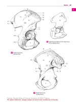
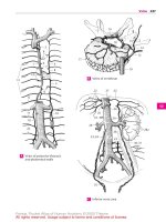
Organization and Position of Organs of Urinary System
231
I
I
A Anterior
aspect
A. B Organsof t'le
unnary system
I
C Retropentoneal space
'I
f
232
Urinary
System:
Kidney
Kidney
Gross Anatomy
External
I
i
Features
The kidney may be divided Into two surfaces, an anterior surface A' ;;nd d posterior
surface B l. JS well as 01 wide superior pole
'AB1) and corucal inferior pole (A82l. The
ante I tor and postenor surfaces are hounded
by the convex lateral border (AB3l. which IS
connnuous with the supenor and Inf"ll.or
poll'S. and d COHCdVl'medial border A4' On
the rned 1..1I border IS a depression Called the
hilum of kidney (AS: whuh allows passage of
vessels Into and out of the organ and atso
houses till' renal pelvis 1he hilum of the
ktdney ' C) leads to the renal sinus {(G). a (JV
Ity SUI rounded on all "des by the paren
chyma,
An adult kidney IS to 12[111 long • .., b cm
Wide and 4(m thick. [-aeh kidney weighs
120· 300g. and the nght kidney I. usually
srnalte: than the left.
Renal sinus. The renal SJI1UScan be vlsudl ..
izcd after removma the vessels. IIClVl". fat.
and renal pelvis. The boundary around It,
entrance IS formed bv 01 lip-hke indentation
on thr- medial border Projecnng mID the
renal Sinus are pyrarrudal etevanons called
renal papillae (C7l. Ihe human kidney hilS
male than one p.lplll,l (I) 121; It IS multiple
because II Is developed from multiple kId·
ney lones that later merge Traces Ilf the
structure of the multiple kidney lobes can
stili be idennfred (lobulated kidncy' Ill! the
kldn('y of 01 newborn,
Surface. In the adult. the surface of the kid·
neys IS usually smooth. It IS covered !)y a
tough fibrous capsule :08) that contams co!
Idgen fibers Mid is attached to rbe kicnev by
loose connective trssue.
Internal
Structure
A (ross, section or long.tudinal
secnon (If
the kldney reveals two drsunct
regions
forming Its internal
structure:
the renal
medulla I 09) and the outer renal cortex (010),
Ihe rnacroscopu
appearance
of the sec
troned kidnev IS produced by the orgamzanon of unniterous tubules and vessels 'see
pp.234 117l.
Renal medulla. The renal medulla IS com,
posed of corneal renal pyramids (011, thaI
appear pale and striated III cross section.
The bas('s of tit£' renal p'yramlds (012) are'
directed toward the surface of the kidncv,
The' rounded apices form the renal papil/ar
,OBI whr •.h project (award tile huum and
rnto the renal cahces of the renal pelvrs, On
I(S surface, each renal papllIJ bears a m
bri/onn area of numerous perforations pro
duced by the openings of papd/ar)' ducts. the
open-rigs of the uriniferous
tubules. all
closer rnspecuon, 01 renal pyramid can be
funher subdivided Into a reddish outer lone
and .I lighter inner zone
Renal cortex. Ihe rena: cortex lies Immediately beneath the fibrous capsule lt rs about
Il m wide and III the unmounted speurnen
has a reddish brown color It nverlies the
pyramid; of the rena: medulla like a capsule
between rhe lateral aspects of the renal
pyrarruds scnd.ng extensions (all:"d renal
columns (014) mto thl' mtenor of the organ
Ihe renal cortex IS permeated 11Vlonguudinoll stnanons known .1S medullary rays DIS}
wh It hare conunuauons of the medullary
substance rdchattng from tile bases of the
pvra.mds toward the capsule lht' corneal
part coruarrnng rho medullarv rays IS known
as the cortex corticis. a lid the cortical substance between the rru-dullarv reys IS the
cortical labyrinth,
Kidney lobes. hlCh kidnev lobe •.onusts of a
renal pyramid and It< surrounding cortex (see
abovcj.Indivrdual kidney lobes arc bounded
by the renal co' UI11 [IS.
Gross Anatomy of Kidney
233
I
i
A Riqht kidnev, antenor dWl'CI
~ll
11
8
14
11
o
Frontal section throuqn r.qht kidney
234
Urinary
System: Kidney
Microscopic
Anatomy
The macroscopically distinct portions of the
parenchyma
of the kidney (see p. 232; are
produced by a characrerisnc
pattern of distnbution of different structural units of till'
organ. These structural
units mcludc the
numerous, densely packed uriniferous tubules, as well ,IS blood vessels and connective
tissue containing nerves and lymphatic vessels.
Uriniferous Tubules
I
i
The uriniferous tubules consist of two com
ponent ••, a nephron and collecting ducts,
which have different embryological
origins.
l-ach nephron.
or basic functional Unit 01
the kidney, consists of J renal corpuscle and
an associated renal tubule which IS J segment of the uriniferous tubules.
Renal corpuscle (A 1). E.1Ch renal corpuscle
consists of a cluster of capillanes called ,1
glomerulus (Al) and ,1 surrounding
glomerular capsule (A3 J.
Renal tubule. Connected to the renal corpuscle IS ,1 continuous
system of renal
tubules that mav be divided into various
segments.
The renal tubules begm with a
preximal tubule which has a twisted pan
known as the proximal convolwed wbule
(A4; and a straight part called the proxi1lJui
seraigllt cobule (AS). Following the proximal
tubule IS the intermediate tubule. or thin
tubule (A6), which can be divided IOta the
descending rilln IlIlIb (AS a) and ascending
ellln limb (AS b). The mtermediate tubule tS
continuous
with the distal tubule, consisung
of a distal stralgll! cobu/e (A7) followed by
the dISCO/(Ollvo/utl'd tubule (A8).
The tortuous segment of the distal I ubule IS
connected by a junctional tubule (A9; With
cerves fluid from approximately
10 nephrons and empties into a papillary duct
(A 11) which opens on the tip of the papilla.
Intra renal Blood Vessels
The functions of the kidney rely closely on
the mtcracnon
between nephrons, collect
ing ducts, and intrarenal blood vessels,
The It'nal artery carries waste laden blood
[0 [he kidneys.
Its branches. the interlobar
arteries or kidney AI2). pass between the
renal pyranuds toward [he cortex, becommg continuous with the arcuate arteries
of kidney (A1l1 at the rorricornedullary
border. Spnngrng from the arcuate arteries
are numerous
interlobular arteries of kidney
(A 14). These radiate toward the fibrous capsule and give off afferent glomerular arterioles
(A 1S) that feed the capillary tuns (glomeruli:
I All
of the renal corpuscles.
Blood flows
from the glomeruli via the efferent glomerular arterioles (A1S) into tilt! capillary network
of the renal cortex and via the interlobular
veins (A17 J, arcuate veins (A 18 j, and interlobar
veins (A 19110 the renal vein. The straight arterioles (A20) are branches of the etlerent
auenoles that radiate from the glorneruh
near t he renal cortex down into the renal
medulla. Ascending parallel to these are the
straight venules (A21) wluch transport blood
vra the nrmarr I'I';IIS to the i1iCer/obur \'cim.
Microanatomy of Kidney
2
1
4
17
14
14
5
/
i
I
!
I
I
!
6b
/
A Uriniferous tubules and
blood vessels on renal cortex and medulla
11
/
!
i
235
236
Urinary System: Kidney
Microscopic Anatomy of the
Kidney, cont.
Renal Corpuscles
J
j
Glomerulus
(Al). The glomerulus
forming
the lena I corpuscle consists of30-40capillary
loops and IS situated between an afferent
glol1ll'rular arteriole (Al). leading to it. and
,10 ('fferclI! glol1lerular arreriole (AJ) draining it The afferent and efferent artenoles he
In close proximity to one another. fornung
the vascular pole (A4) of the renal corpuscle.
Each glomerulus
is surrounded by a dU,II·
layered glomerular capsule. The imemal part
(AS) lies adjacent to the capillary loops and
the external parr or Bowman capsule (A6)
separates Ihe glomerulus from its surround
mgs, The space bel ween the two layers. the
capsular space. collects glomerular
filtrate
and conveys it via the urinary pole into the
tubule system.
Glomerular
capillaries
(8). The glomerular
capillaries are composed of an endothelium
(87). with evenly distributed fenestrations
between the endothelial cells. and a continuo
ous, triple·layer
basement membrane.
the
middle layer of which acts as a rnerhamcal
filter lhe outer layer. faring the capsular
space, IS covered by podocytes (AS). branching cells with numerous processes. The long
primary processes (A9; of the podocyrcs give
rise to secondary or foot processes that II1tNdigitate like fingers with those of ,ldJ,KCIl!
podorytes. leaving narrow gaps, or /ill ra non
slits. between them.
Special connective
115SUl' cells known
,IS
mesangial
cells (intragtomerular
mesangiat
cells) (810; he between the adjacent capillanes of a glomerulus.
MesJnglal cells also
lie at the' vascular pole between the afferent
arteriole and efferent arteriole (extragtomerular mesangial cells) (811). The mesangial
(ells arc pan of the juxtaglomerular apparatus
01 the kidney which also includes the mac
ula densa (AB12) and polar cushion ( A8B).
Ihe macula densa refers to specialized
epithelial cells lying along the distal convoluted tubule in places of contact with the
vascular pole. The polar cushion refers to tilt'
(granular)
myoepithelial
(ells of the jux
taglomcrular
apparatus III the prcglornerular P,Ht of the afferent arteriole, Renin and
angrotensinase
A haw been detected
in
polar cushion cells.
Renal Tubules
and Collecting
Ducts (e)
The walls of the renal tubules are lined by
simple epithelium which vanes by region.
The proximaltubule
(C14) ISlined by cuboidal
eprthehal cells with a high brush border as
well as infoldmgs of till' (ell membrane at
the base otthe cell and abundant mitochondria.
The intermediate tubule (CIS) is lined by nattened epithehal eel Is with short nucrovilli,
The distal tubule (C16) has tall low cuboidal
cells with l>.lsdl struuons.
lhe cells are
somewhat flatter than those ofthe proximal
tubule and have only short minovilli projecung from them.
lhe collecting ducts (C17) are composed of
about I of pale-staining epithelial cells
with distinct c('11 borders and I of darkstaining
intercalated
cells, The cpithehal
cells lining the collecnng ducts become progrl'sslwly
flatter as the duct progresses
toward the papitlae,
Function or the kidneys .. Ihe renal corpuscles
lorm the filter that d.lIly "squeezes" ISO liters of
ullrdfillrdle (primdry urine) out of the blood. Of
these, 178htrrs JI~ reabsorbed
In the tubule sysrem, and I.S 2 Iuers of findl urine (secondary
urine' are formed per d.lY. UW1l' rs cxcrered by tbe
excretory organs. lhe juxtaglomerular
apparatus
funcuons as part of the rerun .1IlglOlenslIl system
mvotved in blood pressure regul.1tion
Microanatomy of Kidney. cont.
237
8
9
I
j
A RpnJIcorpusct«,
orqamzaton
B Section through renal corpuscle
of components
>
C Renal tubules in crovs-secuon, appearance in light micrograph
cellutar cornponems, appearance in electron micrograph
•••
_-
238
Urinary
System:
Neurovascular
Drainage
Kidney
Supply and lymphatic
Arteries.
Wastl' substances are carried to
the kidneys by the renal artery A 1). The ri!(llI
rellol orrery springs trorn the abdominal
aorta (Al) at the level of 1.1. In most people
the left renal orrery anses ilt a short distance
above It. The left renal artery is usually
shorter than the right renal artery. Ihc pri
mary intrarenal
branches of the two rnarn
artenes are end arteries and supply specific
regions of the parenchyma.
These regions
may be classified as renal segments: the superior seglllelll. anterior superior segment.
r
onrenor mIerior segment. inicnor segf1leJil.
and the posterior se,!!f1Ielll. GIVen the (0111,
plex nature of kidney development these
segments
may vary considerably: ,1110111.1
lies In the course or the renal artery also
occur
Veins. Venous dramage from the kidney is
via the renal vein (AC3). The nght renal win
is short and has ,1 straight course while the
path of the left renal win is longer and curving. During its course U receives the left 511prarelwl vein and the lefl teslicular win or
left overinn vei".
Nerves. Autonomic
fibers to the kidneys
anse from the renal nerve plexus whrch ,1~"
companies
the renal artery and is mainlv
formed by fibers from t h~ adjacent ce/i(l~'
plexus.
Regional
lymph nodes. Lymph from
kidneys drams to the lateral aortic nodes
the
Topography of the Kidneys
Position. The kidneys lie on either Side of
rhe vertebral column in the lumbar groove.
Their long axes are directed upward and
backward
so that If .m imagmary
lme IS
drawn as a cont inuauon from each ,1XIS.
these lines would intersect. The superior pole
lies at the level ofn2. and the inferior pole ,11
the level of the L3. The hilum of kidney is located ,11 the level of U. The nght kidney
usually lies about half a vertebra lower than
the left kidney. The posruon of the kidneys
varies with respiration
and posture Peste-
rior to the kidney. the 12th rill (A4l passes
diagonally over the boundary between the
upper ,1I1d middle thirds ot the organ. CrossII1g over the kidney Ill'arly parallel to the
12th rib 111.1 cramocaudal
direction arc the
subcostal nen'e (AS). iliollypogastric nerve
(A6). and
iliomglllno/llt'rw
Adjacent organs and vessels. lymg anteriorly on the superior poll's ofthe kidneys are
the sliprarenal/adrenal glands (A7l. The
anterior surface ofthe right kidney IS 111cont,lt! with the lil't'r and right colic flexure:
near rhe hilum of the right kidney arc the inJaior vena COWl (A8) ,111d dllodt'num. The
anterior surface of (he left kidney is 111COI1tart with the stomarh. pancreas. and le/t
colic flexure: (he (wr/a runs near the hilurrof the left kidney.
A9lJreter
Capsules of the Kidney
The capsutc-, enclosing the krdnev
(Bl0) and d perirenal f~lt capsule (Bell). The
fascial pouch IS composed of a Ihlll olllerior
layer and ,I tough posterior la.vn. The two
layers are connected with each other at
their superior and lateral borders and surround t he kidney. adrenal gland. and per1rl'l1.11f,1I capsule. The medral side of the tas(1,11 pouch IS open, and its intcrio! side IS
only closed by adipose tissue, The volume of
the perirenal fat capsule vanes depending on
the mdividual nutriuorul
status: with extrcme emaciation I( may even be absent.
Loss of the perirenal fat capsule can result In
mobility of the kidney which may descend
toward till' pelvis. an abnormal condinon
known JS floating kidney.
Clinical note. Anatorntc varialion\ and renal
anomalies Me common. Common abnormatrnes Include the presence ,)f extra kidneys. kid-
ney drsplacernem.
shoe kidneys
kidney fUSion. and horse-
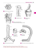
Kidney Topography
239
I
I
A vcssets. nerves, and topography
of kidneys
10
8 Kidney capsules, cross-section
C F<1S0.11 pouch of krdney
240
Urinary
System: Excretory
Organs
Excretory Organs
renal pelvis wuh me ureter The U[('IN possesses
an l'SIll'ClJlly strong muscular layer (C14). As It
proceed,' toward the unnarv bladder. II " aug
menred hy ,I IIlInl ou/('r lon811udwol larer of
muscle. The loose conn,'Cllve tissue of lhl' adventitia
Renal Pelvis and Ureter
Gross Anatomy
I
i
Renai pelvis and calices (A). The renal pelvis
(AB1) is a reservoir
for the collection uf
unne formed by the uruon uf the 8 10renal
calices (A2) that empty mto It. Minor calices
(A2 a) are small, trumpet-shaped
renal calICes that surround one (or occasionally two
or three) renal papilla. They give usc to the
2 3 major calices (A2 b) which open i11l0 the
renal pelvis.
The shape of the renal pelvis vanes (AI according to
the branching pattern of the renal calices. If the
minor (.Ilict's cnnsisteruly open into major c,11
«cs, the renal pelvis is of the br~oching type: If the
mmor calircs also open directly into Ih,' rendl pelvis. formmg a Widened sarhke renal pelvis, U IS
considered an ampullary type. Ihe volume of rhe
ren.,1 pelvis is ·1·8011.
Ureter (83). The ureter is J slightly flat
rencd, thick-walled tube that connects the
renal pelvis with the urinary bladder. It IS
25 JOcmlong and is divided into two pans
based un its course: an abdominal part (83 a)
and a pelvic part (83 b). Its terminal PMt Iollows .111 oblique course in the wall of the un
nary bladder and is knuwn as the intramural
part.
B4 Kidney. 85 Hrlurn of kidnev, 86 Renal artery,
87 Renal vern, 88 Anna. 891nfN,or vena cava. 810
Ovarian artery. 811 lnternal iliac artery. B12
Uterine arterv
Microanatomy.
The w,11I of the renal pelvrs ~
thm, while 11],11 of the ureter IS very II1Ilk In
cross-sec lion the ureter h,IS .1 sur shaped lumen
(C), lhe walls ,1" both organs are composed 01
three lavers, the mucosa ((13: consists of rhe transuional epithelium. or urothelillm. that .; (para(
tcnstc of Ihe unnary excretory ducts and a layer
of loose conllre five tissue The urothelium lOnSISIS
of 5 7 layers of Cf/lS and can adapr 10 the ,1111OUI1l
01" distcnuon of rhc ureter by alrenng the height
and number of rell layers. the thickelled apical
membrolle in the top layer of I'll' (ells Ih.1I are visible in rght rrnc OS(OPY protects the epuhehal
surface from hypertonic urrr.e In the renal pelvis
Ihe muscular layer consrsts of ap IfIllfr IIllIgitlUlinal
lawr and an outer orcll/m lowr. The muscle tiber,
are rruerwoven
IU [nrm sirur/llrf'
resemb/lllg
'pllillrters
In
the calices and
11 11]"
u.ncnrn
01" the
,CIS) embeds the renal pelvis and ureter in their
surroundings, The ronnecuve us-uc of the renal
pelvis, which cont.uns abundant blood vessels
•.md nerves, .lIsa (OntlllllS smooth muscle cells
that rcntrot 'Is drstennon.
Neurovascular
Drainage
Supply and lymphatic
The vessels of till' renal pelvis (B) anse from
the renal artery and vein (86. B7). tymphatic
drarnage corresponds to that of the kidneys,
The renal pelvis rl'(l'IVI'S sensory mnervanon and hence Its distcnuon rs painful.
The ureter i~ supplied by branches from the
large surrounding arteries the rcnal artery
(86). teslicular ortery or ovanan emery
I Bl0), ;/11I'r1wl pUc/I'lIdalurlery. and superior
vesical ertery. The artencs are accompanied
by veins of the same name.Lymph drains (0
the lumbar nodes Autonormr
Innervation is
by t he splanchnic nerves.
Topography of the Renal Pelvis and the
Abdominal Part of the Ureter
Till' greater part of the renal pelvis (A) lies
hidden 111till' r(,I1.11 sinus,
Ihe abdominal part of the ureter begins at lIS
eXI! from the renal pelvis with the first point
of constriction of the ureter. The ureter t hen
proceeds caudally to the medial Side 01 the
pSO.1S major :816) where it hI'S between the
muscle f.1(ia (posterior to ill and the peritoneum (covering Its antcnor
aspect).
During its course. the path uf the ureter is
crossed over by the rcsucular or ovarian
vein (B10). and the ureter IIsell crosses over
the gcnitofemoral
uerve.It enters the lesser
pelvis at the level uf the common ih.tc vessels or external iliac vessels. ThiS IS the sue
of t he second point of constriction of the ureter
(see also Topography of the Pelvic Part of the
Ureter. p. 244).
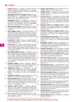
Renal Pelvis and Ureter
I
2a
2b
•...~J
/
i
t
:0
A Renal pelvis:
branching type (above),
ampullary type (below)
12
3b
15
13
241
14
B Ureter, posinon
and topoqr aphy
C Cross secuon throuqh ureter,
light, nicrogr aph
242
Urinary
System:
Excretory
Organ>
Urinary Bladder
The urinary
bladder
(AI) IS a hollow.
muscular organ whose size varies with the
amount of contained urine. It IS located behmd the pubis (A2) 10 the subpentoneal
connective tissue of the lesser pelvis.
Parts of urinary bladder. The body of bladder
lAB3) constitutes
the largest part of the
organ, It IS continuous
anterosuperiorly
with the ap['x of bladder rAB4:. The apex
gives attachment to the obhrcratcd urachus
which passes in the median umbilical ligament (ABS) (see p. 188) to the navel. OpenII1g uno the lateral and posterior aspects of
the fundus of bladder (A6). which empties
posteriorly
and rnfcnorty, are the ureters
(87). Ihe neck of bladder (88) is continuous
anteriorly with the urethra :A89).
As the ~rindry bladder crnpues, the apex ul t~e
bladder and upper portion of the wall descend
and the organ becomes bowl-shaped, As II Llls,
[he apex and w.ll1 are drawn forward and upward
to form an OVOId SIMpe Depcndmg 011 ure
amuunt of ItS contents,
the unnary bladder can
extend .1$ far as the superior borner of [he pubic
svmphysrs. ",c c.p.lcity 01 the urinary bladder is
normally about o;OOrrol the urge 10 void occurs at
about 300 mi. It is possible, however, voluntarily
10 rciam larger a mou 111, of urine.
Internal
surface (C). The inner surface of
the urinary bladder is a pall' red in color
Two parts can be identified
throughout
most of the urinary bladder the mucosa
contains folds due to its mobility against the
underlying muscular ~ayl'r when the bladder is very full. the folds disappear. The tri
angular region formed on the tundus of the
bladder. wluch is bounded by the two openings of the ureters known as the ureteric orifices (COlO) and the exit of the urethra
called the internal urethral orifice (Ctt), IS
known as the trigone of bladder (C012 I. The
mucosa of the trigone of the bladder is flat:
It IS firmly attached
to the underlying
muscular
layer and thus does not contain
folds. In the malt'. the uvula ofblodder(013).
a conical elevation produced by thl' under
lying prostate.
projects into the internal
urethral orifice
Microanatomy.
The walls of the urinary
bladder are made up of three layers. The
mUCO>d consists of !rClIIsicioliUI epillielium
(urothehunu
overlying
loose connective
tissue (IarlllrlCl pmprm) which is absent at
till' trigone of t he bladder. Most of the
muscular layer IS made up 01 three disunct
1,)YNs that are collccuvcly
known as the
detrusor musr/c. At the trigone of the bladder. the muscular layer constitutes
,) conunuauon of the muscular layer of the ureter
and thus consists of only two I,WNS. At the
openings of the ureters mro the bladder, the
smooth muscle IS org.nuzed In a complex
circular ammgrml'lIt. Til!' serosa. which IS
acromparucd by connecnve tissue of the
subserosa. rovers the supenor surface of the
unnary bladder and the portion of the postenor surface above the tngone of the bladder
Neurovascular
Drainage
Supply and lymphatic
Arteries. The unnary bladder IS nourished
by branches from the internal iliac artery. r.e,
the 5l1paior wsic(I/ (//'(['I)' I- urnbrhcal
artery)
and
illferior vesical
nrrerv,
Veins. The vesical venous plexus. whirh surrounds the fundus 01 rhe bladder. collects
blood from' he urm,lry bladder. and usually
empties drrectlv mto the 11IIl'1"/I(l1 ili(l( vems,
Nerves. Similar to tho.' intestine, mnervarion
of the urinary bladder is divided Into extrinsic and intrinsic nervous systems (r.e .. inside
and outside of the wall of the unnary bladder). Parasymp.lthetic fibers of the extrinsic
system arise from 52 S4 and act to constrict
the detrusor
(mirtunuon),
Sympathetic fi·
bel'S supply the Sl1100t h muscle of the vessel
wallv and presumably
C,lUSl' contracuon of
the muscle around the neck of the bladder
and the upper portion of the urethra.
Regional
lymph nodes. Lymph flows in
VMIOUSdirernons from the urinary bladder:
the external iliac nodes collect lymph from
the upper and lateral portions of the wall.
internal iliac nodes collect lymph from the
fundus and the tngone (If the bladder.
Lymph from the anterior wall of the urinary
bladder also ulumately drains to the internal IIloK nodes,
Urinary Bladder
243
4
5
2
9
E
•••
1
i
A ..,edian sdgittdl sectton
through mall' petvrs
B Urinary bladder
male, ant e nor aspect
o
Trigone of bladder. male
C Opened urrnarv bladder, Iernale, anterior aspect
244
Urinary
System:
Excretory
Organs
Female Urethra
TIll' female urethra (A I) IS wry short, only
3-5 em .• 1I1d lies behind t he pubic symphysis (A2). It begins ,11the internal urethral orifice (A3) and passes upward in an anteriorly
concave curvature
in close proxrrnuy
to the
anterior wall of the vagina (A4).lt ends at a
longitudinal
slit, i.e. the external urethral orifice(A5) in the vcstibuleofvaglllU
2 ](111 behind the glans of eli tons (A6).
Microscopic Anatomy
The walls of till' urethra consist 01 a mucosa
tl1<11lies in longitudinal
folds and IS lined by
transitional
epitheliulIl resung on a highly
vascularized
lam ilia propria or spongy laver
that coruams abundant
veins and glands
(urethral glands): and a muscular layer that is
derived from the muscular layer of the w,llh
of the urinary bladder and is arranged in an
inlier IOllgitudillal layer and an outer circular
la.\'l'r.
The urethra IS surrounded
by the external
urethral sphincter, a crrrular arrangement
of
striated muscle that forms a type 01 loop 01
fibers that is open posteriorly and extends
as far .1S the neck of the bladder.
The male urethra is discussed on p.1.61..
Function or the excretory organs. Urine expelled
Irom rne renal papillae IS ""I collected in Ihe
ren.ll calices and then conveyed In the renal pelvi~
After reaching a certam volume, the urine rs
ejected into the ureter by rapid movements. Once
m the ureter pertst.iluc waves transport the "nne
distally and empty il III poruons 11110the urinary
bladder When the unnary bladder IS filled to (111dividual)
capacitv. snrnuh
mecrated by the
nervous system inmate Its emptying. or micturition (urination).
Topography of the Excretory
Organs
Female pelvis. After exiting the renal pelvis
: first point of constriction of the ureter) and
completing
Its intra abdominal course (see
p. 241 8). t he ureter enters the lesser pclvrs
111 front of the sacroiliac
joint. the fight ureter at the level of t he bifurcauon of the common iliac artery (87) and the left ureter at
the level 01 the external iliar artery, This is
the sitl' of the second point of constriction of
the ureter. In Ihe female lesser pelvis, the
ureter runs superficially
along the lateral
wall 01 t he pelvis immediately underneath
t he peritoneum.
At about t he level 01 the
rschtal spine it leaves the lateral wall 01 the
pelvis .lIld runs in the base of the broad ligament of the UI!'rUS (88), coursing medially
and antcnorlv. It crosses under the uterine
artery (89) a;ld .• It d variable distance from
the vagina. reaches the posterolateral wall
of the urinary bladder which II penetrates
diagonallv from posrerol.ueral to anreromedial. This irnrarnural
part of the ureter is
approximately
2 em long and forms the third
point of constriction of the ureter.
Ihe urinary bladder lA810) lies III the subperitoneal connective tissue behmd the
pubic wmphysis. The retropubic space (A 11),
.J region of loose ronnccuve
tissue, 1i('S 111
front of It. Ihe retropubic Sp.lC1.'extends between the antenor abdonunal wall and the
peritoneum
as far .15 the navel and permits
movement
of the unrury
bladder as it
swells upward dunng filling. The superior
part of the unnarv bladder IS covered by
peritoneum:
Its mferopostcnor
surface IS
firmly attached
to the surrounding
structures.
The female urethra lies between the pubic
symphyxis .1I1d 1I11' amenor wall of the
vagll1.l (A4).
Male pelvis. In the lesser pelvis of the male
(see p.l55 B) the ureter also passes immedr.uety beneath the peruoneurn
along the
lateral wall of the pclvrs.It reaches the POS(crotatcral wall of the unnarv bladder at a
point above the seminal vesicle. crossing
below the ductus deferens.
Cllnk ••1 not e. Kidney stones CJn get Murk near
the constnctcd parts of the ureter.
A duphcauon of ureters OCClifS III about 2:1:of
the populauon: ureter duplex. double ureter:
ureter flsSlIs. bifid ureter.
Urethra and Topography of Excretory Organs
245
J
A Median sagittal section
through female pelvis
8 Female pelvic organs,
viewed from above
i
244
Urinary
System: Excretory
Organs
Female Urethra
The female urethra (Al) IS very short, only
3 5cI11, and lies behmd [he pubic symphySIS (Al). I[ begins at [he internal urethral orifice(Al) and passes upward in an .mtertorly
concave curvature In close proximity to [he
anterior wall of the vagin.t (A4). I[ ends at ,1
longitudinal
slit. i.e. the external urethral orifice (AS; 111 the vesrrbuleoJl'Ogino 2- 3cm behind the glans of clitoris (AG).
Microscopic Anatomy
I
i
The walls of the urethra consist of d mucosa
that lies in longitudinal
folds and IS lined by
transitional epitheliulll resung on .1 highly
vascularized
lamina propria or .Ipollgy la}w
that contains abundant
veins and glands
(urethral glands), and a muscular layer that IS
derived from the muscular layer of the walls
of the urinary bladder and IS arranged In .111
inlier longitudinal/ay('" and an outer circular
layer,
The urethra is surrounded
by the external
urethral sphincter, a circular arrangement of
striated muscle that forms a type of loop of
fibers that is open posteriorly and extends
as far as the neck of the bladder
The mate urethra is discussed on p,262.
Function of the excretory organs. Unne expelled
from Ire renal papillae is (irst collected in the
renal catices ,1Odthen conveyed to the renal ptlvis
After reachmg a ccrtam volume. the urine IS
ejected into the ureter by raprd movements, Once
III the Url'IN. peristaltic waves transport the unne
d,stally .1Odempty U In portions mto t re urinary
bladder. When the unnary bladder IS rolled 10 (ondrvrdual] caparuy, surnulr mediated Ily the
nervous system mruote Us ernptvrr-g, or rnieturilion (urination),
Topography
Organs
of the Excretory
Female pelvis. After exiting the renal pelvis
(first poinl of constriction of the ureter) a lid
compleung
its mtra-abdommal
course (SCl'
p.241 6), the ureter enters the lesser pelvis
111 front of the sacroiliac JOint, the right ureter at the level 01 the bifurcauon of [he rommon ihac artery (87) and the Il'f[ ureter at
the level of the external ilr.lC artery, This is
the ~Il('01 the second poinl of constriction of
the ureter In the 1",'m,lll' lesser pelvis, the
ureter funs supcrftcially
along the 1,1tNa!
wall 01 the pelvis irnmcdratcly underneath
the peruoneum. At about the level of [he
isrhi.ll spine it leaves the lateral wall of [he
pelvis and runs III the base of the broad ligamenr 01 the uterus (88), coursing medially
and anteriorly,
It crosses under t he uterine
artery (69) and, dt ,1 vanable distance from
the vagma, reaches [he posterolateral wall
of till' urinary bladder which if penetrates
diagouallv from posterol.neral to anreromedial. TIllS intramural
part of the ureter is
approximately
z cm long and forms till' third
point of constriction of the ureter,
The urinary bladder CABJO) lies In the sub
peruoneal
connecnve !lS511l' behind the
pubic symphysis, The retropubic space (A 11),
.l Il'gllJn of loose connecuve
1ISSU(', lies in
front of II, rill' rcuopubrc Sp.lCC extends betWl'('11 the antcnor abdommal wall and the
Pl'flIOI1l'UOl as far ,IS the navel and pcrrmts
movement
of the unnary bladder as it
swells upward duung filling. The superior
part of the UlltJ.1IY bladder IS covered by
peritoneum: I[S mfcropostcnor surface IS
firmly attached to the surroundmg
strurturcs,
TIll' female urethra lies between the pubic
syrnphysiv
and t he antenor
wall of the
vagina (A4),
Male pelvis. In the !l'ssl'r Pl'lVIS of the male
(see p, 2558) the ureter also passes irnrnediatelv beneath
the pcruoneum
along the
lateral wall otthe pelvis. It INches the posterolatcral w.11I of tIll' unnarv bladder at a
pornt above the serumal veside. crossing
below the ductus deferens.
Oink ••1 note, Kidneystones can get stuck near
the constnctcd parts of the ureter
A dupucanon of ureters occurs ui ahoul 2% 01
rhe population. ureter duplex • double ureter:
t.reter flssus • bill" ureter
Urethra and Topography of Excretory Organs
A Medi,lI1 sagittal
7
\ v ,
B r ornale pelvic organs.
viewed from above
245
248
Male Genital System: Overview
Overview
Male Reproductive
Organs
The organs of the male genital system can
be divided topographically
and developmentally mto Internal and external geru
taha.
The internal genitalia consist of the resns
(Al). epididymis (All. ducrus deferens (A3l.
and accessory sex glands. i.e.. the prostate
(M). seminal v!'side I seminal gland (AS).
and bulbo-urelilral
gland (Cowper's glands)
(A6).
The external male genitalia mclude the penis
(A7). scrotum (AS). and tunics ofllle tesrcs,
The internal genitalia arise above the pelvic
floor from the urogenital ridge. while the
external genitalia are derived from the urogenital sinus below the pelvic floor,
Function.
The male germ cells. or spermatoIn the testis and trans
ported through a system of small canals to
the epididymis where they mature. Mature
spermatozoa Me conveyed by the spermatic
cord to the male urethra through which they
can leave the body cavity. As they travel
through the sernmal duct the germ cells ML'
mixed with secretions from the accessory sex
glands.
zoa. are produced
Peritoneal
Relations of the Male Pelvis
1he peritoneal cavity extends over the linea
terminalis into the pelvic cavity. The parietal
peritoneum continues along till' wall of IIll'
lesser pelvis. covering the pelvrc viscera
projecting from it. it reflects from the nnreriot abdominal wall onto the ape« oj bladder
(AB9) and covers the entire superior sutjece
(ABlO) of the urinary bladder. Extending
caudally
and laterally
the peritoneum
passes to the level of the union of the ureters with the urinary bladder, The upper
portions of Ihl' seminal vrsic/rs extend along
the posterior surface of the urinary bladder
up to the level of the openings of the ureters
or higher and are usually covered by parietal
pentoneurn. Ihe ductus dejerens is likewise
covered by peritoneum
lip to its terminal
portion. the ampulla of ductus deferens. Oc
casronally,
the peritoneum
passes even
deeper to cover a part of the prostate. It does
not cover the fundus of the unnary bladder
but rather forms the rectovesical pouch (Bllt
" peritoneal reflection from the posrenor
wall oj chI"urinary bladder onto the nnrenor
wall ojche rccwmlBl2).ln
the male. the reotovesrcal pouch IS the lowesr poinl in the
abdominal
cavily.
On either side it is
bounded by a Iold known as the rectovesical
jold. The subserosa! connecuve
tissue of the
rectovesical
lold contains the autonomic
nerves of the inferior hypogastric
nerve
plexus. When the urinary bladder is full. a
peruoneal fold IS also produced between
the antcnor abdominal wall and the apex of
the bladder
813 P<'fiton~al fold produced by ureter
Clinical note, In patients wuh urin.lry retention
the distended unnary bladder can be punclured JUst above the border of the pelvic svm-
phySlS wuhout injurmg the peritoneum
operung th e abdornmal cavlly.
or
Male Reproductive Organs
A Mal~ genitalia, schematic
drawing
5
4
6
IJ
8 Mal~ pelvl( organs,
viewed from above
249
250
Male Genital
System:
Testis and Epididymis
Testis and Epididymis
Gross Anatomy
Testes. The paired male gonads Me the sill'
of sperm produC/lon and are located outside
of the body cavity in the SCrotUIII. Each testis
is an egg-shaped
organ with a firm. elastic
consistency. rneasunng 4-·5cm in length
and 3cm across, 1111'left testis IS usually
somewhat larger than the right. Each testis
11
IMs a lateral surface (A3: and a medial surface
~A4) which are continuous
,11 the narrow,
anterior border (A85) and the Wide. posterior
border (A6). nil' testes Ill' obhquely in the
scrotum With their superior poles directed
antcrolarerally
and their infertor poles post
eromedially, Investing each testis IS ,1 rhick,
white connective
nssue capsule ,",1III'd the
tunica albuginea. At the superior poll' IS a
remnant
of the ernbrvomc
miillerwll
ducl
known as the appendix of testis (87).
Epididymis
(A8S). Resting like ,1 tail 011 the
posterior surface of each of the testes is the
epididymis.
It consists of three parts. the
head of epididymis (AS a) is tholt part thai projeers above the superior pole of the testis
while the body of tpididymis (AS b) and the
tail of epididymis (AS c) are cornplerely
in
contact With the testis, Eitch epididymis has
its own connecnve
tISSU,' capsule. which is
distinct from that of the tunica albuginea of
the testis and surrounds
the roughly 5 m
long. lightly COiled duct of epididymis (A89,.
Near the head or the epididymis
is the appendix of epididymis (C10;. a remnant or the
lII(>soll('pliros.
Coverings
of testis
and epididymis.
The
testes tim develop in the abdonunal cavity
and later descend dunng fetal development
into the scrotum ldesc('llsus restis), As it
travels from the abdorninal cavity through
the inguinal canal. the tesus penetrates
the
layers of the abdominal
wall (see Vol. 1. p,
96). formmg the processus vaginalis testis. ,I
perilolleal diwrtiwlulIl
which guides It into
the scrotum. After birth. most of the proccssus vaginalis testis IS obluerated.
Only its
caudal end rernams, tonrung the tunica vagi-
nalis of testis \ C11). J dosed serous s/Iearh that
envelops
till' tl'SUS and epididymis.
The
visceral layer (epiorchium) lies 011 top of Ihe
tUIlICd albuginea and (overs those parts of
the testis that ate not covered by the epididymis. It also covers most of the epididymis and rcflert-, onto the paricldllayer (ptriorchium] .It the exit Site of the sperrnauc
cord. Between the (('sUS and epididymis is a
narrow spare called the sillus oJ epididymis
:C12) which
IS bounded
cranially
and
l,lUdJlly by peruoncal folds known as the
supt'riol and illfcrior h~r"'II'lIIs of l'pidldytnlS
(A 13). The epionluum Jnd periorchium are
separated
by ,I fluid-filled serous pocket,
lying on the external surface of the parietal
laver 01 the !Unic,l vaginalis is the internal
spermatic fascia (C14). ,1 conunuauon of tr.e
rrall.wrrsalis fasoCl. 111('Internal spermatic
fam,1 IS covered
by Iibers from the
cremaster 'C15) that make up the cremasteric fascia. an expansion of the inrrrnal abliqul' IIlllsril' oj' rill' abdollll'lI. lhe external
spermatic fascia (C16) IS derived from an
outer layer of fascia of till' abdonunal wall.
I C., the fascia of the external oblique muscle
of the abdomen, and lonns 111l'outer fJsClal
sheath enclovmg the testis. epididymis. and
spermatic cord.
The testis. epididymis.
,111d their coverings
are contamcd m the scrotum (C17). The thin
skill of the scrotum IS COIII;1I110IlSwirli rhr
of rill' ubdOlllt'1I and IS heavily pigmerited. covered wuh hair. and contains sebdCl'OOS glands.
the subcutaneous tissue is
devoid of [,ll. Consisting of connective tissue
and smooth muscle cells, it is thus known as
the dartos fascia. The scrotum is divided into
two parts by the connect IV,' tissue septum of
scrotum Its outer surface is marked by the
raphe of scrotum. ,I Hill' III the skin that extends to the perineum.
skill
Clinical note. The testes should be fully descended II>!(>the scrotum at the lime of birth
(,ign of maturity IIIthe male newborn I.
Gross Anatomy of Testis and Epididymis
6
9
A RighIIC~ls.IJleral
view
B Rig~l testts,
C 1 uruca of tesns
'nedial view
251
252
Male Genital System:
Iestis and Epididymis
Microscopic Anatomy
E
i
OIl
j
j
Tissue framework of the testis and epididymis. The tunica albuginea sends numerous septa testis (A8l 1 into the interior of the
organ. dividing the parenchyma into 200
300 conical lobules of testis (Al) and con
verging to form the mediastinum testis (AJl.
Each lobule contains several seminiferous
tubules. or COllvo/tlled seminiferous tubules
(84). These continue Into the straigllt
tubules (85) which in turn are continuous
with a network of tubules in the medi
astinum testis known as the rete testis (86).
The rete testis is connected by efferent duetules (A87) with the duct of the epididymis
(88). Each efferent ductule is about 20cm
long and is coiled to form a conical. 2cm
long lobule of epididymis whose apex is
directed toward the rete testis and whose
base faces the duct of epididymis.
Seminferous tubules (C). The seminiferous
tubules are surrounded by loose connective
tissue called interstitial tissue ((9) which
contains testosterone-producing
interstilIal cells known as Leydig celts (see p, 356). A
thin layer of myofibroblasts ,1Od fibroblasts
((10) immediately surround the seminiferous tubules, The tubules are lined by germinal epithelium which is composed of spermatogenic cells and support ing Senoii cells.
Spermatogenesis.
Spermatozoa develop 10 the
gerrmnal epuheliurn (oJ in a rnulnsrage process.
amlOg from stem cells called spermatogonia.
Spermatogonia. which lie along rhe basement
membrane can be classitred Into two types. Type
A spennorogoruo are stem cells thar are ell her
resting or undergoing muouc division 10 form
more stem cells, Jypr B spennalOgonio 1011) can
be considered precursor cells of the spermatozoa.
i.e, they are involved In meioSIS and subsequent
duferentlauon processes, throughout which they
remarn connected by bridges of cytoplasm.
Mitotic divrsion of rypc B spermatogonia gives
rise to primary spermatocyth IDI2~ After duplicat109 their DNAcontent (to become 4n DNA). they
enter the various stages of prophase of the first
rneionc division. The meiotic prophase lasts up to
24 days and results III the recombmauon of
geneuc matenal.tn lustological preparanons, pri
mary spermatocytes can be identified by their
large SIll'. The rernarmng stages of the first me.
one division occur rapidly. ,11 the conctusron rI
wluch two 5t<ondary spermalocyte> (013) (2n DN!.
alt.' lormed. In the second meiouc dIVision thr
secondary sperrnarocytes divide to form 'per.
matids (014). Sperrnatids are the smallest cella
{he germinal epithelium. They contam only,
smgle SCI of chromosomes (22 aurosornes and :
Sl'X chromosome. In DNA). They lie in bunches on
the ups of the Sertoli cells (015) from where the)
are secreted lOW the adlummal compartment of
the seminiferous tubule (sec below). After a 10!li
process of maturauon ccnsrsting of nuclear condensauon and acrosome and flagella formatlO1l.
the spermauds give nse to spermatozoa [ap.i~ of
(ertiliution I016: which are released from the tC'
nunal epithelium In the final phase of sperm»
genesis (E).
Spermatozoa. The mature spermatozoon
(F) ISdbOUI60 urn long and consists ora head
(F17) and a tail (F18). The tail can be further
divided into a neck (F18 a). a middle piece
(F18 b). a pnncipal piece (F18 c). and an ('lid
piece. The head is characterized by the presence of a dense nudeus (F19) surrounded by
a cap called an acrosome (F20) which contarns important substances for penetrating
the egg cell.
Sertoti cells (015). The Sertoh cells rest
on the basement membrane with theu
processes projecting into the lumen of the
seminiferous tubules. Their basal pornons
are interconnected by numerous cell junctions, forming the blood-testis barrier which
divides the germinal epithelium into a basal
compartment and an adluminal compartment
The germ cells travel through the interrellular spaces between the cell Junctions of the
Sertoli cells as they slowly move toward the
lumen of the seminiferous tubule. They are
nourished by the Sertoli cells which also
secrete a fluid that transports the spermatozoa into the epididyrrus.
Microscopic Anatomy of Testis and Epididymis
3
A Section through testis with intact epididymis
B Seminiferous tubules
and epididymis
ISa
c..
~
E Sperrnloqenesrs
o
Seminiferous tubules.
magnification. detail Irorn C
253
254
Male Genital
System:
Testis and Epididymis
Microscopic
Anatomy,
cont.
Rete testis. efferent ductules, and duct of
epididymis.
In histological sections of the
testis and epididymis
(AI. the rete testis
(Al) can be Identified by Its location m the
mediastinum
testis. The rete testis (B) is a
system of canals lined by simp/l' squamous
or cuboidal epilhl.'l;um from which 12 20
efferent ductules (A21 lead to the duct of the
epididymis (Al I. The efferent ducrules (C) an'
lined by pseudostraliJied epillielium with
cells of variab/(' height. Their star-shaped
IUIll~11 IS lined by alternatmg
segments 01
columnar cells and flattened cells, The flat
epithelial
cells are absorpuve. while the
columnar cells possess kinoci/Ia for transporting sperm. Throughout
the duct of epididymis (D; the epithelium
IS characterized
by pseudoslranfied tal/ columnar eprt/lclia/
c('l/s that have srereocilia. The epithelium of
the duct of the epididymis produces a Sl'W"
tion that assists in maturation
of the spermatozca. Ihe walls of the duct of the eprdidymis are formed by J few layers of
smooth muscle cells,
Function
of testis and epididymis.
The
production of spermatozoa in the seminiferous tubules ofthe testis lasts about 74days.
Movement through the epididymis takes an
additional
8-17 days. Ihere the sperrnatolQ,1 undergo
,1 maturation process at the end
of which they are capable of tertrhzation.
fhe epididymis also serves as a storage site
for mature spermatozoa.
The cndocnne and
paracnne
processes
necessary
for spermatogenesis are discussed 1I1tl1e chapter on
the endocrine system (see p. 356).
Hormonal regulation and suuable temperature.
at least 2 (below body temperature,
are essential
to the development
of mature
sperm.
The size otthe testes steadily increases during
childhood, reaching its maximum between
the ages of 20 and 30. In older age, the testes
shrink. In the male child, the sermniterous
tubules of the testis consist of cords of
epithelial cells without lumen, contaming
only Sertoh cells and spcrmatogoma.
Spcrmatogenevis,
which commences
during
puberty,
age.
normally
continues
into advanced
CUnkal not e. Ihe higher temper aturcs in in·
guin.t testes, compared to testes that have descended lOW the scrotum. prevent sperm production
Neurovascular
Drainage
Supply and lymphatic
Arteries.
Till' r('.I(l'S are supplied by the
testicular artery which anses directly from
the aorta and also sends ,1 br anch to the
('pididvrnis. I he tesurular
artery anastomoses With the artery to ductus deferens (see
p, 25(;; and the cremasteric artery (~ Inferior
epigastnc artery) wluch supplies the tuniq
of IIII' tesles. The sCJ'OruJJl IS nourished b'l
branches from the internal pudendal artery.
Veins. Blood trom the testes and epididymis
drains IIltO the pampiniJorm vel/ous plexus
wluch in turn cmpues via the right testicular
vein into the inferior vena cava and via the
left testicular vein into the left renal veto.
Drainage from the turnrs of the testes and
the scrotum IS to the grea( sap/lerrous vein,
interior epl.gastrir v!'il/. and rnrema/ puden·
da/witl.
Nerves. Sympathetic
fibers from the celi.lc
plexus accompany till' supplving arteries to
the testes and epididvnudes.The
scrotum IS
mnervatcd bv till' scrotal nerves ariSing from
the ilioil/gu;lIa( lien'" and pudenda/ nerve
Nerve supply to the cremaster
muscle IS
provided bv the genual branch ofthe genitofemoral nerve.
Regional lymph nodes. l.ymph from the
testes and epididymides
drains to the lumbar nodes. that from the tunics of the testes
and scrotum dr.uns to the inguinal nodes.
Clinical note. \I~ricoctlt' is a condition of unknown etiology lilJt involves abnormal dilalion of the wide- •.aliber, valveless veins of the
parnpinifo: m venous plexus. Ihe lett rcsus IS
more often ,,(fecled than the nght.