Nghiên cứu sự biểu hiện mRNA của gen CIZ1b, VEGF và đột biến EGFR với nhiễm virus merkel cell ở bệnh nhân ung thư phổi không tế bào nhỏ tt tiếng anh
Bạn đang xem bản rút gọn của tài liệu. Xem và tải ngay bản đầy đủ của tài liệu tại đây (266.6 KB, 28 trang )
MINISTRY OF EDUCATION AND TRAINING
MINISTRY OF DEFENSE
VIETNAM MILITARY MEDICAL UNIVERSITY
HO VAN SON
STUDYING THE mRNA EXPRESSION OF CIZ1b, VEGF
GENES AND EGFR MUTATIONS WITH MERKEL CELL
VIRUS INFECTION IN PATIENTS WITH NON- SMALL
CELL LUNG CANCER
Major: Biomedical Science
Code: 9 72 01 01
SUMMARY OF DOCTORAL THESIS IN MEDICINE
HA NOI – 2020
THE THESIS IS COMPLETED AT THE
VIETNAM MILITARY MEDICAL UNIVESITY
Supervisors:
1. Prof. Dr. NGUYỄN LĨNH TOÀN
2. Dr. NGÔ TẤT TRUNG
Reviewer 1: Prof. Dr. VAN DINH HOA
Reviewer 2: Prof. Dr. PHAN THU PHUONG
Reviewer 3: Prof. Dr. TA BA THANG
The thesis is defended in front of the scientific committee at the
Vietnam Military Medical University at
on
2020.
The thesis can be found at:
- National Library
- Library of the Vietnam Military Medical University
1
INTRODUCTION
Primary lung cancer (PLC) is one of the most common
malignancies today and is the leading cause of death in cancer. In
Vietnam, lung cancer (LC) has increased rapidly. According to the
Global Cancer Research Organization (GLOBOCAN) in 2018, there
are 23,667 new people in the country with LC. About 90% of
patients with PLC die in the first year. Recently, CIZ1, a gene coding
for intracellular protein involved in the initiation of DNA replication,
has been studied. CIZ1b, a variant of CIZ1, manifests itself in nonsmall cell lung cancer (NSCLC). Besides, Vascular endothelial
growth factor (VEGF) plays a very important role in tumour growth.
EGFR mutation is a valuable molecular target in the use of Tyrosine
Kinase inhibitors - TKIs in treatment of NSCLC. Recently, the role
of Virus Merkel cell (Merkel cell polyomavirus, MCV) in LC has
been mentioned and researched. We conducted the project:
"Studying the mRNA expression of CIZ1b, VEGF and EGFR
mutations with Merkel cell virus infection in patients with nonsmall cell lung cancer" with these objectives:
1. Assessed the level of mRNA expression of CIZ1b, VEGF, EGFR
mutations and the rate of Merkel cell infection in patients with nonsmall cell lung cancer.
2. Analysed the relationship between the level of mRNA expression
of CIZ1b, VEGF, EGFR mutations with Merkel cell infection and
some clinical and subclinical symptoms in patients with non-small
cell lung cancer.
Rationale of the Study
Study on mRNA expression of CIZ1b, VEGF, EGFR mutation and
Merkel cell virus (MCV) infection contribute to early diagnosis,
identify the cause of disease and its application in monitoring and
treatment of NSCLC, contribute to improving the quality of life,
reduce mortality for patients with NSCLC.
2
Significance of the Study
This thesis is the first study to evaluate the mRNA expression of
CIZ1b, VEGF and MCV infection rates in patients with NSCLC in
Vietnam. The thesis also raised the diagnostic value of two mRNA
markers of CIZ1b, VEGF in NSCLC and the relationship of MCV
with EGFR mutation as well as the risk of LC when being infected
with MCV.
Layout
The thesis has 120 pages, including: Introduction (2 pages),
Chapter 1: Literature Review (31 pages), Chapter 2: Research
subjects and Methodology (28 pages), Chapter 3: Results (33 pages),
Chapter 4: Discussion (22 pages), Conclusion (2 pages),
Recommendations (1 page).
The thesis has 150 references (English: 146).
CHAPTER 1: LITERATURE REVIEW
1.1. Overview of lung cancer
Lung cancer is currently the most common type of cancer in both
incidence and mortality. In 2018, there were about 2.1 million new
cases of lung cancer detected and an estimated 1.8 million deaths,
accounting for more than 18% of all cancer deaths. In Vietnam, LC
accounted for 15.48% of all new cancers. About 90% died in the first
5 years. Prevalence of male is higher than female (The ratio of male
to female is approximately 2.5:1)
The main risk factors associated with lung cancer include tobacco
use, environmental pollution, Amian infection, Randon gas,
infectious agents, interactions between the sensitive genome and the
environment and.
The oncogene in NSCLC often involves four main functional
pathways: cell proliferation, programmed resistance to death,
invasion or metastasis and angiogenesis.
3
1.2. Expression of CIZ1b in non-small cell lung cancer.
CIZ1b is a variant of the CIZ1 gene, due to the deficiency of eight
amino acids at the beginning of the C end. CIZ1 gene encodes for an
intracellular protein that plays a role in controlling cell proliferation:
regulate cell cycle, regulate transcription. CIZ1 is an intermediate
molecule that connects cyclin E and A to cyclin dependent kinase
proteins such as CDK2 and p21, to accelerate mammalian DNA
replication. This protein also plays an indirect role in DNA
replication by regulating the expression of genes. It can bind directly
to DNA molecules or act as a co-activator of transcription factors.
In LC, CIZ1b manifested much in NSCLC samples when
compared to adjacent tissue samples. By quantifying the
concentration of CIZ1b in plasma (using Western blot technique),
Higgins et al. were able to distinguish 98% of LC cases from normal
people. In addition, compared patients with stage 1 NSCLC to the
smoking control group of the same age or the group of benign lung
tumours, this marker can also classify 95% of cases. Thus, CIZ1b
shows the potential of a molecular marker that can detect NSCLC
early with high clinical accuracy.
CIZ1b affected the DNA replication function of lung cancer cells.
Using RNAi technique to reduce the expression of CIZ1b (without
affecting other forms of CIZ1) in the SBC5 lung cancer cell line can
inhibit the proliferation of this cell line. The same result was
obtained with in vivo experiments, when implanting this cancer cell
line in mice, CIZ1b depletion could reduce tumour growth. These
experiments have shown that CIZ1b variant plays a role in
controlling proliferation of cancer cells.
1.3. Manifestations of VEGF in non-small cell lung cancer.
VEGF, which has been shown to play an important role in lung
cancer, is strongly correlated with abnormal blood vessel formation
and promotes tumour growth. This factor has been applied to
differential diagnosis in patients with chronic obstructive pulmonary
4
disease and lung cancer, benign and malignant pleural effusion.
Notably, for patients whose tumours are detected by a chest X-ray or
CT scan, the level of VEGF expression in bronchial lavage is a
marker that can be used to diagnose primary lung cancer.
1.4. EGFR mutation in non-small cell lung cancer
Epidermal growth factor receptor (EGFR) has a molecular weight
of 170 kiloDaltons (kDa).
When the epithelial growth factor (EGF) binds to the receptor
(EGFR), the two EGFR molecules combine with each other
(dimerization) to activate phosphorylation of the tyrosine kinase
region, which activates specific tyrosins and EGFR receptordependent intracellular signal proteins that induce transcription of
target genes to promote proliferation, programmed resistance,
invasion, metastasis and neovascularization.
EGFR is especially important in the pathogenesis of non-small
cell lung cancer, especially adenocarcinoma. In people who do not
smoke or rarely smoke, women and Asian people, EGFR
manifestations occur in more than 50% of cases of lung
adenocarcinoma. However, they also make malignant cells
susceptible to TKIs, and even predict response to broad-spectrum
TKI drugs like erlotinib and gefitinib. In addition to the TKIsensitive mutation, EGFR also has a mutation that helps cancer cells
resist this drug, such as the T790M mutation that occurs in exon 20.
About 50–60% of recurrent patients have a T790M mutation. This
mutation reduces the effectiveness of the first generation of TKI
drug, but so far the third generation TKI can help treat drug-resistant
patients.
1.5. Merkel cell Carcinoma Virus (MCV) in non-small cell lung
cancer
1.5.1. Merkel cell virus and carcinogenic mechanism
MCV belongs to the Polyomaviridae family and is listed on the
5
list of class 2A carcinogens by the world cancer research fund
international. The MCV genome is a double-stranded circular DNA
molecule, about 5 kb in size, divided into three main regions: a noncoding control region (NCRR) containing the replication centre of
virus. Transcription factors are located between two regions: the
early coding region and the late coding region. Early coding region,
coding for antigens: LT (Large T), ST (Small T). The late coding
region encodes for the capsid proteins, VP1 and VP2.
The LT and ST antigens play an important role in tumour
formation caused by MCV. LT carries domain J (associated with
thermal shock protein), retinoblastoma binding motif (RB) (inhibits
RB family members), domain affixes replication centre at C end and
domain helicase / ATPase (necessary for DNA replication of virus).
Much of the carcinogenic function of LT antigen is due to its high
affinity for RB, causing isolation and inactivation of this tumour
suppressor gene. The RB binding function of MCV LT is essential
for the sustainable growth of MCC-positive MCV tumours both in
vitro and in experimental models.
1.5.2. Relationship between Merkel virus and lung cancer
Based on the histological similarities between MCC and small
cell lung cancer (SCLC), the relationship between MCV and SCLC
has been studied. Two studies in Germany showed the presence of
MCV DNA by PCR with detection rates of 6.7% (2/30) and 38.9%
(7/18). For NSCLC, epidemiological studies conducted in the US
and Europe detected MCV DNA at 16.7% (5/30), 4.7% (4/86) and
9.1% (10/110) patients with NSCLC. Recently, Hashida et al. first
published the incidence of MCV in Asian lung cancer and detected
MCV DNA in 17.9% (20/112) of NSCLC patients in Japan. In
particular, the Japanese team found two cases of NSCLC infection
with MCV bearing the characteristic mark of the tumour. These cases
have the LT antigen expressed in cancer cells and integrate the viral
genome into the cell chromosome and cause cancer modification.
Thus, integrated or mutated forms of MCV have been shown in a
6
specific cancer other than MCC, suggesting that MCV is associated
with the onset of an NSCLC group. However, no studies have been
conducted in Vietnam to detect MCV in patients with NSCLC.
CHAPTER 2: RSEARCH SUBJECTS AND METHODOLOGY
2.1. Research subjects
2.1.1. Patient group: 100 patients with non-small cell lung cancer,
identified by histopathology, aged 27- 83.
2.1.2. Healthy people (control group): 51 healthy people (without
cancer), ages 48-60, are similar in age and gender to the patient
group.
2.1.3. Exclusion: SCLC, treated NSCLC, metastatic LC, COPD,
asthma, acute and chronic bronchitis, patients receiving
corticosteroids, pregnant women, patients with autoimmune disease
and non-cooperative participants in the study.
2.2. Research Methods
2.2.1. Research design
- Cross-sectional descriptive and controlled study, combined with
clinical examination and laboratory analysis.
2.2.2. Location and study time
- Research location: Department of Pathophysiology - Military
Medical Academy, Military Hospital 175 and Central Military
Hospital 108.
- Research period: From October 2014 to February 2018.
2.2.3. Sampling method
- Sample selection method: Convenience sampling.
7
2.2.4. Data collection
- Selected patients, established research medical records in the
unified form, coded the patient symptom information, extracted
information, collected registration data, and entered the records.
2.2.5. The implementation techniques
2.2.5.1. Diagnosis of non-small cell lung cancer
Based on the guidelines: the diagnosis and treatment of non-small
cell lung cancer of the Ministry of Health of Vietnam applies to
medical examination and treatment facilities nationwide.
2.2.5.2. Diagnosis of stage of disease
WHO TNM classification for lung cancer 2015
2.2.5.3. Histopathological diagnosis, differentiation
Based on WHO classification of lung cancer 2015
2.2.5.4. Using real-time PCR technique, PCR technique, direct
sequencing technique to determine:
- The level of mRNA expression of CIZ1b and VEGF in peripheral
blood.
- Identify EGFR mutations.
- Determine the presence of Merkel cell virus in peripheral blood of
healthy people, in blood and cancer tissue of patients.
2.2.6. Evaluation criteria
- Level of expression of mRNA VEGF and mRNA CIZ1b in LC
group and the control group.
- EGFR mutation rate.
- The prevalence of Merkel cell virus infection in the LC group and
the control group.
8
- The relationship between the expression level of mRNA VEGF,
mRNA CIZ1b and some subclinical factors: renal function, liver
function, blood sugar, CEA, CYFRA21-1, EGFR mutation, disease
stage and tissue type pathology, differentiation and associated factors
with MCV infection.
2.3. Data processing
- Processed data using SPSS 20.0 software.
CHAPTER 3: RESEARCH RESULTS
3.1. General characteristics of the research team
3.1.1. Age and gender
Table 3.1. Comparing age and gender of the two groups
Character
istic
NSCLC group
(n = 100)
± SD Median
Control group
(n = 51)
± SD
P
Median
Age 60,0 ± 10,5 61,0
53,9 ± 2,9
53,0
0,158
Gender
(Male/Fem
73/27
35/16
0,47
al)
There is no difference in age and gender in the two groups (disease
and control).
3.1.2. Histopathological results of the treatment group
Table 3.2. Histopathology of the NSCLC group
No.
Histopathology results (n=100) Quantity
Percentage
(%)
1
Adeno carcinoma
91
91
2
Squamus cell carcinoma
9
9
9
Gland carcinoma makes up the majority (91%), barbed carcinoma
accounts for only 9%. No large and mixed cell carcinoma.
3.1.3. Results of differentiation evaluation in the treatment group
Table 3.3. Differentiation of tumour cells of NSCLC patients
No.
Differentiation (n=100)
Quantity
Percentage
%
1
No rating
37
37
Degree 1
6/63
9,52
Degree 2
35/63
55,56
2
To be
evaluated
(n=63)
Degree 3
22/63
34,92
Average differentiation is the most common, followed by high
differentiation, low differentiation only accounts for 9%.
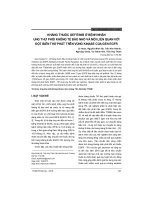
IV; 7 5.82%
IA; 2.20%
IIA; 4 .4 0 %
IIIA; 2.20%
IIIB; 15.38%
Figure 3.1. Distribution of desease stage if NSCLC patients
3.1.4. Results of the phase evaluation of NSCLC group
Stage IV had the highest rate (76%), followed by stage IIIB, early
stages encountered less.
3.2. mRNA expression of CIZ1b and VEGF, EGFR mutation and
prevalence of MCV infection in patients with KTBN
10
3.2.1. mRNA expression of CIZ1b and VEGF in two study groups
Table 3.4. mRNA expression of CIZ1b and VEGF in two study groups
VEGF
expression
(2
-∆Ct
)
NSCLC group
Control group
(n = 100)
±
SD
(n = 51)
Median
p
± SD
Median
CIZ1b
expression 10,5 ± 6,3
(2-∆Ct)
8,9
7,8
± 2,6
7,0
0,008
VEGF
expression 2,3 ± 2,3
(2-∆Ct)
1,7
1,5
± 1,0
1,3
0,007
The level of mRNA expression of CIZ1b and VEGF in the
treatment group was significantly higher that that of healthy people
(p<0,05).
3.2.2. EGFR mutation in NSCLC group
Table 3.5. EGFR mutation of the NSCLC patients group
No.
Mutant position
Number of patients
(n=51)
Percentage %
1
Exon 19
11
21,57
2
Exon 20
2
3,92
3
Exon 21
13
25,49
4
No mutation
25
49,02
Mutations in exon 21 accounted for the highest percentage, followed
by mutations in exon 19, mutations in exon 20 was only 4%.
3.2.3. Prevalence of MCV infection in NSCLC group
11
Table 3.6. The prevalence of MCV in patients compared to healthy
people group
Result
Treatment
group(%)
Blood Tissue
MCV
MCV
(%)
(%)
Control
group (%)
(Blood)
Positive
25 (25) 25 (25)
3 (5,9)
Negative
75 (75) 75 (75)
48 (94,1)
OR
(95% CI)
P
5,33
0,004
(1,49-28,83)
100
100
51 (100)
(100)
(100)
The rate of MCV infection in patients with NSCLC (25%) was
higher than that of normal people (5.9%), people infected with MCV
were 5.3 times more likely to be infected with UV than those without
MCV infection.
Total
3.3 Relationship between the level of mRNA expression of CIZ1b,
VEGF, EGFR mutations and Merkel cell infection and some
clinical and subclinical symptoms in patients with NSCLC
3.3.1. Relationship between mRNA expression of CIZ1b and VEGF
and stage of disease
Table 3.7. mRNA expression of CIZ1b and VEGF, stages of disease
Stages of
disease
Gene
Stage I+II+III
(n = 22)
± SD Median
Stage IV
(n = 69)
p
± SD Median
CIZ1b
9,6 ± 5,1
8,1
10,7 ± 6,7
9,4
> 0,05
VEGF
2,2 ± 1,3
2,0
1,5 ± 1,3
1,6
> 0,05
No association between mRNA expression of CIZ1b and VEGF was
observed.
12
3.3.2. Relationship between mRNA espression of CIZ1b and VEGF
with tumour histopathology
Table 3.8. mRNA expression of CIZ1b and plasma VEGF and
tumour histopathology
Histopathology
Adeno carcinoma
(n=91)
Squamus cell
Carcinoma (n=9)
± SD Median
Gene
p
± SD Median
CIZ1b
10,1 ± 6,1
8,6
14,5 ± 9,5
13,8
> 0,05
VEGF
2,3 ± 2,4
1,7
1,9 ± 1,2
1,2
> 0,05
No association between mRNA expression of CIZ1b and VEGF
with histopathology was observed.
3.3.3. Relationship between mRNA expression of CIZ1b and VEGF
with tumour cell differentiation
Table 3.9. mRNA expression of CIZ1b, VEGF and tumour cell
differentiation
Differenti
ation
Gene
Grad 1
(n = 6)
± SD
Grad 2
(n = 35)
Median
Grad 3
(n = 22)
P
± SD Median ± SD Median
CIZ1b
11,8±5,8
10,9 11,5±6,7
9,9
9,3±6,9
7,7
> 0,05
VEGF
6,1±6,5
3,8
1,7
1,9±0,9
1,7
> 0,05
1,9±1,3
No association between mRNA expression of CIZ1b and VEGF
with tumour cell differentiation was observed.
3.3.4. Relationship between mRNA expression of CIZ1b and VEGF
with EGFR gene mutation
13
Table 3.10. mRNA espression of CIZ1b and VEGF and EGFR gene
mutation
EGFR gene
mutation
Gene
Mutations
(n = 26)
No mutations
(n = 25)
± SD Median
p
± SD Median
CIZ1b
10,3 ± 4,9
8,3
10,4 ± 7,7
9,0
> 0,05
VEGF
1,6 ± 0,8
1,4
2,3 ± 1,8
1,9
> 0,05
No association between mRNA expression of CIZ1b and VEGF
with EGFR mutation was observed.
3.3.5. Relationship between MCV infection and the stages of
NSCLC
Table 3.11. Relationship between stages of NSCLC and MCV
infection
MCV
Stage
Positive n(%)
Negative n(%)
IA
0
2 (3)
IIA
0
4 (6)
IIIA
0
2 (3)
IIIB
3 (12)
11 (16,7)
IV
22 (88)
47 (71,3)
p
>0,05
Total
25 (100)
66 (100)
No association between MCV infection and stages of NSCLC was
observed.
3.3.6. Relationship between tumour cell differentiation and MCV
infection
14
Table 3.12. Relationship between tumour cell differentiation and MCV
infection
MCV
Differentiation
Positive n(%)
Negative
n(%)
Grad 1
1 (4)
5 (6,7)
Grad 2
8 (32)
27 (36)
Grad 3
7 (28)
15 (20)
Undefined
9 (36)
28 (37,3)
Total
25 (100)
75 (100)
p
>0,05
No association was found between MCV infection and tumour
cell differentiation.
3.3.7. Relationship between tumour cell differentiation and MCV
infection
Table 3.13. Relationship between MCV infection and cancer marker
Characteristics
Positive MCV
Negative MCV
(n = 25)
(n = 75)
± SD
p
± SD
Median
Median
CEA (ng/mL)
121,7±250,2
6,9
95,2±273,8
6,02
>0,05
CYFRA 21.1
(ng/mL)
24,7±70,5
2,69
8,98±21,5
3,8
>0,05
No association was found between MCV infection and expression
of cancer markers.
3.3.8. Relationship between MCV infection with EGFR mutation
Table 3.14. Relationship between MCV infection with EGFR
15
mutation
EGFR mutation
Positive
MCV
Negative
MCV
OR (95% CI)
Mutation
17 (85)
9 (29)
13,8 (2,8-86,5)
No mutation
3 (15)
22 (71)
Chi2(1): 15,24
Total number of
patients
analysed
20 (100)
31 (100)
p
0,0001
The percentage of EGFR mutations in the group of NSCLC
infected with MCV was significantly higher than the group without
the mutation, in patients with NSCLC infected with MCV, the risk of
EGFR mutation increased by 13.8 times.
3.3.9. Relationship between MCV infection with the level of gene
expression in VEGF and CIZ1b
Table 3.15. Relationship between MCV infection with gene
expression level of VEGF and CIZ1b
Characteristics
Positive MCV
Negative MCV
(n = 25)
(n = 75)
± SD
Median
± SD
p
Median
VEGF (2-∆Ct)
2,07±1,1
1,89
2,33±2,5
1,66
>0,05
CIZ1b (2-∆Ct)
12,15±7,0
9,45
9,77±5,9
8,34
>0,05
No association between MCV infection and mARN of CIZ1b and
VEGF expression was found.
CHAPTER 4: DISCUSSION
16
4.1. Some common characteristics of the NSCLC group
4.1.1. Age of illness
According to the majority of domestic and foreign authors, the
disease is usually occur with people aged between 50 and 70. In this
study, the average age of disease was 60 ± 10.5, of which there were
61 patients from 55 to 74 years old, accounted for 61% of the total
number of patients studied. We found that the age of the disease in
this study is lower than that of Patricia M.de Goot, which may be
because our statistics are not big enough. It may also be because
people with lung cancer in Vietnam are getting younger.
4.1.2. Gender
Lung cancer is more common in men than women, in this study
the ratio of male to female was 3.7:1. With this ratio, the amount of
women having lung cancer is increasing. In Vietnam, the incidence
of lung cancer is also tending to balance between men and women,
according to Globocan 2018 the rate of men over women is 2.5:1.
The difference in the incidence between men and women in this
study and Globocan's overall rate was probably due to the fact that
our sample was not large enough, moreover we only got data from
one cancer treatment center, this also affected the proportional
distribution between men and women.
4.2. The level of mRNA expression of CIZ1b, VEGF, EGFR
mutations and the rate of Merkel Cell virus infection in patients
with non-small cell lung cancer.
4.2.1. Levels of mRNA expression of CIZ1b gene in patients with
non-small cell lung cancer.
In previous research conducted by Higgins et al. in 2012, CIZ1b
was proved to be a valuable biomarker in the diagnosis of lung
cancer. CIZ1b was only found in lung cancer patients but not in
normal people. Therefore, CIZ1b was proposed as a suitable marker
for early stage lung cancer.
17
Studies showed that the CIZ1b variant was sensitive enough to
allow accurate identification of patients with stage 1 cancer, in highrisk groups, including patients with benign lymphadenopathy,
pneumonia, asthma, chronic obstructive pulmonary disease) and
smokers. It is also found that cancer patients have much higher levels
of CIZ1b than normal people without cancer. When analyzed by
stage of cancer, CIZ1b concentration also increased by stage in
patients with non-small cell lung cancer.
In this study, specific primers had been designed to amplify and
quantify mRNA expression of the CIZ1b gene in the blood by realtime PCR. The method was based on the PCR technique, which
results in fast and accurate expression of CIZ1b mRNA in the blood.
Our research results showed that the level of mRNA expression of
CIZ1b gene in plasma in patients with non-small cell lung cancer
was significantly higher than that of control group. These results
confirm that CIZ1b gene expression is affected by lung cancer
development and CIZ1b gene play an important role in lung cancer
development. From this result, the CIZ1b gene has great potential to
be used as a Biomarker to diagnose, to monitor lung cancer
development as well as to monitor the prognosis of lung cancer
treatment.
4.2.2. Levels of mRNA expression of VEGF gene in patients with
non-small cell lung cancer.
One study assessed the mRNA expression of VEGF in three
groups of patients including squamous cell carcinoma,
adenocarcinoma, and undifferentiated cell carcinoma. The results
showed that in 65% of cases, VEGF mRNA expression was higher in
cancer tissue than normal tissue. mRNA expression of VEGF was
higher in non-squamous cell carcinoma and higher in tumors with
lymph node metastases. Similarly, this study also showed that mRNA
expression of VEGF in peripheral blood was also significantly higher
in lung cancer patients compared to the control group.
18
In this study, we did not observe the association of VEGF mRNA
expression with clinical features such as liver dysfunction, Glucose
disorder, tumor histopathology, stage of disease. We also did not see
the difference in mRNA expression of VEGF between differentiation
stages of the tumor (p> 0.05). This shows that the level of VEGF
expression increased significantly between lung cancer group and
healthy people (p <0.05) but, there was no difference with other
clinical and subclinical factors.
In our study, analysis of diagnostic efficacy for identifying lung
cancer patients showed that VEGF mRNA expression was able to
identify lung cancer patients with AUC value = 0.615.
Therefore, this study, together with previous studies, indicates that
assessing the mRNA expression of VEGF in plasma is a nonintervention measure that can be used in combination with other
diagnostic methods to diagnose and screen lung cancer in high-risk
groups such as those with lung disease, a family history of lung
cancer, or in groups that are frequently exposed to toxic agents such
as those who smoke or group of workers in the mines. However, this
factor can be used as an effective marker to monitor and evaluate the
effectiveness of treatment in non-small cell lung cancer.
4.2.3. EGFR mutation in non-small cell lung cancer patients in the
study
EGFR mutation plays a crucial role in the pathogenesis of nonsmall cell lung cancer. Moreover, this is a molecular target for the
use of small molecule inhibitors (TKIs) for treatment.
Previous studies have shown that over 50% of non-small cell lung
cancers with adenocarcinoma have EGFR mutations. In our study,
there were 51 cases of adenocarcinoma identified with EGFR
mutation and 26 cases with mutation, accounting for 51%. Although
the number of patients who have an EGFR mutation was small, the
percentage of patients with EGFR mutations in our study s also
consistent with previous studies.
19
4.2.4. The prevalence of MCV in patients with non-small cell lung
cancer in the study
We proceeded to determine the presence of MCV virus in lung
cancer tissues and in the peripheral blood of lung cancer patients.
Results showed that the rate of MCV infection in patients with lung
cancer (25%) was significantly higher than that in the control group
(5.9%). The results of the correlation analysis showed that MCV
infection was related to lung cancer. People who were infected with
MCV had 5.33 times higher risk of lung cancer than those without
MCV (OR = 5.33). Our research results are similar to some previous
studies in the world. A previous study showed that 30 of 163 patients
with adenocarcinoma cancer and 2 of 8 patients with squamous cell
carcinoma were positive with MCV.
NSCLC epidemiological studies conducted in the US and Europe
had detected MCV DNA at 16.7% (5/30), 4.7% (4/86) and 9.1%
(10/110) patients with NSCLC. Recently, Hashida et al. first
published the incidence of MCV in Asian lung cancer and detected
MCV DNA in 17.9% (20/112) of NSCLC patients in Japan. In
particular, the Japanese team found two cases of NSCLC infection
with MCV bearing the characteristic mark of the tumor. These cases
have LT antigen manifested in cancer cells and integrated with the
cancer cell's chromosome genome. In our study, the MCV infection
rate in healthy people was 7.8% and in the lung cancer patients group
was 25%, much higher than the studies of other authors in the world.
4.3. Relationship between the level of mRNA expression of
CIZ1b, VEGF, EGFR mutations with Markel cell virus infection
and some clinical and subclinical symptoms in patients with nonsmall cell lung cancer.
4.3.1. Relationship between mRNA expression of CIZ1b and VEGF
genes with clinical and subclinical in non-small cell lung cancer
20
In this study, we did not find a correlation between mRNA
expression of CIZ1b and VEGF with hematological index, ionic
graph, abnormal indicators of liver function, renal function and
glucose concentration. This suggests that mRNA expression of
CIZ1b and VEGF has little to do with subclinical manifestations in
patients with lung cancer.
The association between mRNA expression of CIZ1b and VEGF
with other subclinical characteristics such as lung cancer
classification, differentiation of lung cancer tumors and stage of
disease development was also not noted. The analysis showed no
statistically significant correlation between the expression of CIZ1b
and VEGF mRNA with the different forms of lung cancer, cell
differentiation, and disease progression.
The results of CIZ1b and VEGF mRNA expression between
mutated lung cancer patients and the group of lung cancer patients
without mutations on the EGFR gene were compared. However, the
comparison results showed that the levels of mRNA expression of
CIZ1b and VEGF in plasma of these two patient groups were not
significantly different. This suggests that mRNA expression of
CIZ1b and VEGF is not related to the mutation in EGFR gene.
We also compared the mRNA expression of CIZ1b and VEGF
among patients with NSCLC and MCV-free. However, the
comparison results showed that the levels of mRNA expression of
CIZ1b and VEGF in peripheral blood of these two patient groups
were not different. This suggests that mRNA expression of CIZ1b
and VEGF is not related to MCV factor.
4.3.2. Relationship between MCV infection and EGFR mutation
in non-small cell lung cancer
In this study, MCV infection was linked to the EGFR mutation
rate. The percentage of EGGFR mutants (85%) was higher among
NSCLC patients who had MCV infection than those without MCV
(29%), and the difference was statistically significant (P = 0.0001).
21
There are two hypotheses that explain this correlation. First of all,
the virus' genetic material mutations often occur against the host's
protection system or anti-virus agents, so viral infections create
genome instability, leading to the occurrence of somatic mutations
that cause cancer. It can be said that MCV infection increases the risk
of EGFR mutation, which in turn leads to lung cancer. Xu et al.
support this by showing that the risk of EGFR mutations was 5.71
times higher in the anatomical form (95% CI: 2.03–16.04, P = 0.001)
and 7.84 times in the adenocarcinoma form ( 95% CI: 2.54–24.25, P
= 0.0004). Our study showed that patients with MCV infection were
13.8 times more likely to have an EGFR mutation than patients
without MCV infection (p = 0.0001) with a smaller sample size than
the study mentioned above. If this association is further proved, then
MCV infection may be used as one of the early or prognostic factors
for NSCLC, especially in people without a history of smoking. The
second hypothesis is that activation of the EGFR/PI3K/AKT
pathway or other EGFR-related signaling pathways such as Wnt/catenin may facilitate certain steps in MCV infection or increase the
reproduction process of this virus. To prove this hypothesis, more
studies are needed to find the mechanism of infection of MCV host.
Recent research indicated that MCV DNA is detected in 30
patients (18.0%) out of 167 patients and EGFR mutations were found
in 31 of 127 patients (24.4%). EGFR mutations are detected more
frequently in MCV-positive patients than in MCV-negative patients.
The presence of MCV DNA was significantly correlated with cancer
prognosis in groups of lung cancer patients. These results suggest
that MCV may be partly related to the pathogenesis and prognosis in
some cases of lung cancer.
Our study is the first to assess the association between MCV
infection and the expression of VEGF and CIZ1b genes in non-small
cell lung cancer patients. However, the results showed that the status
of MCV infection was not significantly related to the level of
expression of the two genes VEGF and CIZ1b in peripheral blood.
22
CONCLUSION
1. The level of mRNA expression of CIZ1b, VEGF, EGFR
mutations and the rate of Merkel cell infection in patients with
non-small cell lung cancer.
- There was a difference in the level of mRNA expression of
CIZ1b and VEGF genes in patients with KTBN UTP and without
lung cancer.
+ The level of mRNA expression of CIZ1b gene in patients with
ARB is 10.5 ± 6.3 whicH was significantly higher than that of the
control group without lung cancer: 7., 8 ± 2.6 (p <0, 05).
+ The level of mRNA expression of VEGF gene in patients with
ARB was 2.3 ± 2.3 significantly higher than that of the control group
without lung cancer: 1.5 ± 1.0 (p <0.05).
- The rate of EGFR mutation in patients with adenocarcinoma
was 51%, of which the most common was mutation in exon 21,
accounting for 25.49%, exon 19 accounting for 21.57%, the rest was
mutation in exon 19.
- The prevalence of Merkel cell virus infection in patients with
LC was 25%, while the normal group infected with MCV was 5.9%.
The difference was statistically significant between the two groups
with p <0.05. People who were infected with MCV had 5.33 times
higher risk of lung cancer than those without MCV (OR = 5.33).
2. The relationship between the level of mRNA expression of
CIZ1b, VEGF, EGFR mutations with Markel cell virus infection
and some clinical and subclinical symptoms in patients with nonsmall cell lung cancer.
There was no statistically significant difference in the level of
mRNA expression of CIZ1b and VEGF genes between lung cancer
group with hematological disorders, blood glucose, liver function
and lung cancer group with normal tests. (p> 0.05).
23
No association was found for the level of mRNA expression of
CIZ1b and VEGF genes with tumor histopathological characteristics,
degree of differentiation, stage of disease and EGFR mutation in
patients with non-small cell lung cancer of the treatment group (p>
0.05).
The relationship between EGFR mutation and disease stage, cell
differentiation and Hematology, Biochemical indicators and lung
cancer markers has not been found.
The relationship of Merkel cell virus infection has not been seen
in patients with ARV with stage of disease, cell differentiation,
Hematological indicators, Blood biochemistry, lung cancer markers.
Among patients with MCV infection, the percentage of EGFR
mutation (85%) was significantly higher than that of the non-MCV
patients (29%), the difference was statistically significant (p =
0.0001). NSCLC patients with MCV infection who had MCV
infection were 13.8 times more likely to have an EGFR mutation
than patients without MCV infection.
SUGGESTION
1. Need to study the mRNA expression of CIZ1b and VEGF genes