Altered expression of Arabidopsis genes in response to a multifunctional geminivirus pathogenicity protein
Bạn đang xem bản rút gọn của tài liệu. Xem và tải ngay bản đầy đủ của tài liệu tại đây (1.05 MB, 16 trang )
Liu et al. BMC Plant Biology 2014, 14:302
/>
RESEARCH ARTICLE
Open Access
Altered expression of Arabidopsis genes in
response to a multifunctional geminivirus
pathogenicity protein
Lu Liu1, Ho Yong Chung2, Gabriela Lacatus3, Surendranath Baliji4, Jianhua Ruan1* and Garry Sunter2*
Abstract
Background: Geminivirus AC2 is a multifunctional protein that acts as a pathogenicity factor. Transcriptional
regulation by AC2 appears to be mediated through interaction with a plant specific DNA binding protein, PEAPOD2
(PPD2), that specifically binds to sequences known to mediate activation of the CP promoter of Cabbage leaf curl
virus (CaLCuV) and Tomato golden mosaic virus (TGMV). Suppression of both basal and innate immune responses
by AC2 in plants is mediated through inactivation of SnRK1.2, an Arabidopsis SNF1 related protein kinase, and
adenosine kinase (ADK). An indirect promoter targeting strategy, via AC2-host dsDNA binding protein interactions,
and inactivation of SnRK1.2-mediated defense responses could provide the opportunity for geminiviruses to alter
host gene expression and in turn, reprogram the host to support virus infection. The goal of this study was to
identify changes in the transcriptome of Arabidopsis induced by the transcription activation function of AC2 and
the inactivation of SnRK1.2.
Results: Using full-length and truncated AC2 proteins, microarray analyses identified 834 genes differentially
expressed in response to the transcriptional regulatory function of the AC2 protein at one and two days post
treatment. We also identified 499 genes differentially expressed in response to inactivation of SnRK1.2 by the AC2
protein at one and two days post treatment. Network analysis of these two sets of differentially regulated genes
identified several networks consisting of between four and eight highly connected genes. Quantitative real-time
PCR analysis validated the microarray expression results for 10 out of 11 genes tested.
Conclusions: It is becoming increasingly apparent that geminiviruses manipulate the host in several ways to
facilitate an environment conducive to infection, predominantly through the use of multifunctional proteins. Our
approach of identifying networks of highly connected genes that are potentially co-regulated by geminiviruses
during infection will allow us to identify novel pathways of co-regulated genes that are stimulated in response to
pathogen infection in general, and virus infection in particular.
Keywords: Geminiviruses, Microarray, Pathogenesis, Expression, Regulatory networks
Background
The Geminiviridae family comprises a large and diverse
group of viruses that infect a wide range of important
monocotyledonous and dicotyledonous crop species and
cause significant yield losses [1,2]. Viral pathogenesis
depends on a series of interactions between virus, host
* Correspondence: ;
1
Department of Computer Science, The University of Texas at San Antonio,
One UTSA Circle, San Antonio, TX, USA
2
Department of Biology, The University of Texas at San Antonio, One UTSA
Circle, San Antonio, TX, USA
Full list of author information is available at the end of the article
and insect vector. As very few viral proteins are encoded
by geminiviruses, they rely, in large part, on the replication
and transcription machinery of the host. One consequence
of this host dependence is that geminiviruses are useful
models for providing novel insights into the control of
both plant and animal DNA replication and transcription.
The circular single-stranded DNA (ssDNA) genome of
geminiviruses is amplified in the nuclei of infected cells by
rolling circle (RCR) and recombination-dependent (RDR)
replication using cellular DNA polymerases [3,4]. The
resulting double-stranded DNA replicative forms (RF) are
used as template for generation of viral transcripts by host
© 2014 Liu et al.; licensee BioMed Central Ltd. This is an Open Access article distributed under the terms of the Creative
Commons Attribution License ( which permits unrestricted use, distribution, and
reproduction in any medium, provided the original work is properly credited. The Creative Commons Public Domain
Dedication waiver ( applies to the data made available in this article,
unless otherwise stated.
Liu et al. BMC Plant Biology 2014, 14:302
/>
RNA polymerase II. Geminiviruses produce small multifunctional proteins to compensate for a limited coding
capacity. For example, begomoviruses including Cabbage
leaf curl (CaLCuV) and Tomato golden mosaic (TGMV)
virus, code for a pathogenicity protein, AC2 (Figure 1A),
that modulates metabolism [5,6], regulates transcription
[7,8] and suppresses RNA silencing [9-11].
AC2 (also known as AL2 and TrAP) is required for
expression of the coat protein (CP) and BR1 movement
protein genes of both CaLCuV and TGMV [12-15]. It
has been shown that AC2 is capable of inducing CP
expression through two distinct and independent mechanisms. In mesophyll cells AC2 activates the CP promoter,
but in vascular tissue AC2 acts to derepress the promoter
[7,12]. Distinct sequences mediate activation and derepression by AC2. Sequences required for activation are
located within the common region upstream of the CP
transcription start site [8,12], whereas sequences required
for repression are located 1.2 to 1.5 kbp upstream of CP
transcription start site [7,12]. Among begomoviruses, the
transcription function of AC2 is not virus specific as both
CaLCuV or TGMV AC2 proteins can transactivate the
TGMV coat protein (CP) promoter [12,16].
AC2 does not appear to be a canonical transcription
factor as it does not bind dsDNA efficiently and appears
to be targeted to responsive promoters via proteinprotein interactions with cellular factors. A recent study
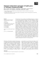
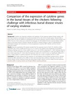
Figure 1 Diagram of CaLCuV AC2 and SCTV C2 proteins used in
over-expression studies. (A) The linear drawing represents functional
domains (span of amino acids indicated) present within the full-length
CaLCuV AC2 protein. The N-terminal region contains a basic region of
four arginine residues and a potential nuclear localization sequence.
The C-terminus contains a minimal transcription activation domain
within an acidic region. A region containing conserved cysteine and
histidine residues forms a putative zinc finger domain, with a high
degree of homology with the SCTV C2 protein. (B) Truncated form
of the CaLCuV AC2 protein lacking the C-terminal 29 amino acids
containing the acidic activation domain. (C) Full-length SCTV C2
protein, which lacks an acidic activation domain, but has homology to
the putative zinc finger domain in CaLCuV AC2.
Page 2 of 16
has identified a plant specific DNA binding protein,
PEAPOD2 (PPD2), that specifically binds to sequences
known to mediate activation of the CP promoter of
CaLCuV and TGMV in mesophyll cells [17]. If AC2 is
targeted to responsive promoters via protein:protein
interactions, we would predict that these interactions
will in turn lead to activation of host genes important
for pathogenesis. An indirect promoter targeting strategy,
via AC2-host dsDNA binding protein interactions, might
provide the opportunity for geminiviruses to alter host
gene expression and in turn, reprogram the host to support virus infection. One finding that supports this idea is
that AC2 can transactivate CP promoter-reporter transgenes integrated into cellular chromosomes [7,12], indicating that AC2 can gain access to the host chromosome.
The transcription function of AC2 is dependent on the
C-terminal 29 amino acids [18], which contains an acidic
activation domain (Figure 1A). AC2 also exhibits transcription-independent functions involving interactions
with different cellular proteins involved in RNA silencing suppression and modulation of metabolism, mediated through sequences lacking the activation domain
(Figure 1B). The L2/C2 homolog of curtoviruses (Figure 1C),
including Beet curly top (BCTV) and Spinach curly top
(SCTV) virus, share limited sequence homology with
CaLCuV AC2 and lack any semblance of a transcriptional
activation domain [19]. Despite the limited homology,
curtovirus C2 protein does suppress RNA silencing and
modulate metabolism, but does not regulate transcription
[16]. The TGMV AC2, BCTV C2 and SCTV C2 proteins
have been shown to interact with SnRK1.2; an Arabidopsis
SNF1 related protein kinase (AKIN11) [5,19]. The consequence of this interaction is inhibition of kinase activity.
Expression of an antisense SnRK1.2 transgene in Nicotiana benthamiana plants leads to increased susceptibility
to infection [5]. The SnRK1 protein kinases play an important role in regulating energy balance in eukayotes and are
members of a conserved family of protein kinases [5].
Related to this interaction, AC2 and C2 [6,19,20] also
interact with and inactivate adenosine kinase (ADK).
Evidence that adenosine kinase activity is reduced in
virus-infected tissue and in transgenic plants expressing
AC2/C2 [6,20], and that ADK-deficient plants display
silencing defects [21], supports a link between silencing
suppression by AC2/C2, ADK and methylation. Recent
evidence indicates that the silencing suppression activity
of geminivirus AC2/C2 proteins is a consequence of ADK
inactivation. This is supported by results demonstrating
that the ability of these proteins to suppress transcriptional gene silencing is accomplished by inhibition of
ADK, which results in interference with methylation [22].
A link between ADK and SnRK1.2 is provided by evidence that SnRK1 kinases are known to be activated
upon binding of 5′-AMP [23], and ADK phosphorylates
Liu et al. BMC Plant Biology 2014, 14:302
/>
adenosine producing 5′-AMP [6]. Thus, AC2 and C2
may interact with and inactivate both SnRK1.2 and ADK
to prevent SnRK1-mediated metabolic (stress) responses
that could enhance resistance to geminivirus infection
[5]. This underscores the importance of SnRK1-mediated
responses to host defense, but exactly how suppression of
these responses leads to suppression of host defenses, specifically the consequence for host gene expression, has not
been examined. The complex interactions and functions
of geminivirus AC2 in regulating transcription and suppressing host defense mechanisms warrants the need to
further investigate the host genes that respond to geminivirus AC2 protein during an infection.
Some microarray profiling of genome-wide changes in
the transcriptome in response to geminivirus infection
has been performed [24]. However, the asynchronous
nature of an infection causes significant difficulties in
determining host genes responsive to a single viral gene
product. To overcome these difficulties we chose to
analyze global changes in gene expression in response to
the effects of a single gene, AC2. A previous study has
been performed using Mungbean yellow mosaic virus and
African cassava mosaic virus AC2 proteins [25]. In these
studies, RNA profiling was performed in Arabidopsis protoplasts and so we chose to use a whole plant infusion
assay for Arabidopsis [26]. The focus of this study was to
identify changes in host gene expression induced by the
transcription-dependent function of the viral AC2 protein,
and induced by the interaction of AC2 with SnRK1. We
identified large-scale changes in host gene expression in
both cases. Further, computational analysis identified
potential regulatory networks that respond to the two
functions of AC2. Lastly, we validated the response of
the top hits within these networks.
Page 3 of 16
DNA gel blot hybridization analysis using specific probes.
In all cases specific cDNA products of the predicted size
were detected in samples at one, two and three days, postinfusion (data not shown). As it was expected that protein
and subsequent changes in host gene expression would
be detectable at these time points, we used RNA isolated one and two days dpi. In addition, at these time
points no phenotypic effects were observed in the
Arabidopsis plants. Thus, these time points could be
more representative of early events rather than late
time points where a phenotype, such as senescence,
represents the end of a signaling response. For the
microarray analysis, Arabidopsis plants were vacuum
infiltrated with Agrobacterium capable of expressing
each of the constructs along with a vector control
(pMON530) to eliminate effects due to Agrobacterium
infection. Total RNA was isolated from four individual
plants, one and two dpi, for three independent sets of
plants infused with the different constructs. This results in
three independent samples per treatment per time point.
Total RNA from the samples was converted into cRNA,
hybridized to the Arabidopsis ATH1 Genome Array, processed and scanned in parallel. Raw intensity data was preprocessed and normalized using the Robust Multi-array
Average (RMA) procedure in MATLAB Bioinformatics
Toolbox. Differentially expressed genes between experimental samples and controls were detected using twosample t-tests with a p-value of 0.05 as the cutoff. Overall,
the variability of the assay is within reasonable range and
expected. The average Pearson correlation coefficient
(PCC) between biological replicates is 0.971 and the
average PCC between the vector controls is slightly
smaller, 0.956.
Differential expression of genes responding to CaLCuV AC2
Results and discussion
Expression profiling of CaLCuV AC2, AC21-100, SCTV C2
and asSnRK1.2 in infiltrated Arabidopsis plants
For these experiments we used full length and truncated
versions of the AC2 gene from CaLCuV, and the fulllength C2 gene from SCTV (Figure 1), as both viruses
are known to cause an infection in Arabidopsis. SnRK1.2
is an endogenous Arabidopsis gene, which interacts with
both AC2 and C2, and expression of antisense (as)
SnRK1.2 increases the susceptibility of plants to infection [5]. We monitored the expression of CaLCuV AC2,
AC21-100, SCTV C2, asSnRK1.2 and an empty plasmid
vector control (pMON530) over three days to determine
the time at which RNA capable of expressing each gene
could be detected. Total RNA was isolated from whole
Arabidopsis plants at one to three days post-infusion
(dpi) with Agrobacterium cultures containing each DNA.
Transcription directed by each construct was confirmed by
RT-PCR analysis and resulting cDNA products subjected to
One of the main goals of this study was to identify genes
that are differentially expressed in response to the trancriptional activation function of AC2. To do this we compared the transcriptome in Arabidopsis leaves expressing
full-length AC2 (FL) or a truncated AC2 (DEL), lacking
the C-terminal 29 amino acids containing the acidic activation domain (AC21-100) at one and two dpi (Additional
file 1: Table S1 and Additional file 2: Table S2). We observed 214 genes that were specifically up-regulated by
full length AC2 protein at one dpi and 269 at two dpi
(Figure 2). For genes that were down-regulated, a total of
158 genes specifically responded to full length AC2 protein at one dpi, and 193 at two dpi. As the difference between the two proteins is the presence of the C-terminal
activation domain in the full length protein we conclude
that these potentially represent genes differentially regulated in response to the transcription function of AC2.
In samples over expressing a truncated AC2 protein
we detected 116 and 195 genes specifically up-regulated
Liu et al. BMC Plant Biology 2014, 14:302
/>
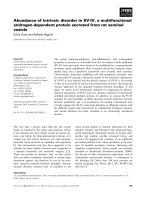
Figure 2 Numbers of genes differentially expressed in response
to geminivirus pathogenicity factors. Venn diagrams illustrating
the intersection between up- and down-regulated genes in Arabidopsis
leaves expressing full-length (FL) or truncated (Δ) versions of CaLCuV
AC2 for one and two dpi respectively.
at one dpi and two dpi respectively. For genes specifically down regulated by the truncated AC2 protein, 156
were detected at one dpi and 219 at two dpi. Given that
the truncated AC2 protein lacks the C-terminal activation
domain, we conclude that these may represent genes differentially regulated in response to the known interactions
of AC2 with the cellular proteins SnRK1.2 and/or ADK
[5,6]. It is of course possible that there are additional, hitherto unknown, functions within the AC2 protein that
could result in differential gene expression.
Interestingly, we observed that 41 and 29 genes were
up-regulated in Arabidopsis leaves expressing both full
length and truncated AC2 protein at one dpi and two
dpi respectively. In addition, 33 and 22 genes were
down-regulated in leaves expressing both full length and
truncated AC2 protein at one and two dpi respectively
(Figure 2). We would expect these genes to be differentially
regulated in response to the interaction with SnRK1.2 and/
or ADK, given that these are functions common to both
full-length and truncated AC2 protein.
To further analyze the genes where expression was
differentially regulated in response to the transcription
function of AC2, we made a comparison to microarray
data from Arabidopsis plants infected with CaLCuV
[24]. We observed a number of genes in our study that
were also detected during CaLCuV infection (Additional
file 3: Table S3). Of the genes up-regulated by full-length
AC2 and CaLCuV-infection at two dpi, several that had
functions related to RNA metabolism, including a DEA
(D/H)-box RNA helicase (At3g58510) and Argonaute 2
(AGO2) (At1g31280). It is interesting that AGO2, which
binds viral siRNAs and regulates innate immunity against
viral infection, is up-regulated in response to AC2 and that
AC2 suppresses RNA silencing. We also detected an
RNA-dependent RNA polymerse gene (RdRp) (At2g19930),
Page 4 of 16
which functions in amplification of the RNA silencing signal,
that was down-regulated in response to both AC2 and
CaLCuV-infection at one dpi. Thus, it is possible that AC2
acts as an effector that is recognized by the plant, activating
the innate immune response, and then acts to overcome
RNA silencing. The number of genes shared between both
experimental data sets were realtively small and no statistical
significance was measured. However, we observed that the
number of genes shared between the two data sets increased
three to four-fold at two dpi (Additional file 3: Table S3).
Differences observed between the two experimental data
sets may be reflective of the different time scales being used
in each experiment. The profiling study for CaLCuV was
performed at 12 days post infection, in comparison to this
study where profiling was performed one and two days after
infusion. In addition this study used agroinfiltration where
AC2 would be expressed in all cell types, in comparison to a
systemic infection where a small number of phloem cells
actually contain virus [24]. Despite this, the observation that
some AC2-responsive genes are differentially regulated
during virus infection, gives added confidence that we
are analyzing genes relavant to viral infection.
Functional categorization of genes differentially regulated
in response to the transcription function of CaLCuV AC2
We have focused our analysis on those genes that were
differentially regulated specifically in response to fulllength AC2. This is interpreted to represent, at least in
part, those genes differentially regulated in response to
the transcriptional activation domain of full length AC2
protein. To categorize these genes by biological process
we used the DAVID Bioinformatics Resource (http://david.
abcc.ncifcrf.gov/summary.jsp). Most of the GO biological
process categories were represented among the significant
genes, but several categories were significantly enriched as
compared to the Arabidopsis genome as a whole. Specifically, genes in the categories of DNA/RNA Metabolism, Transcription, Response to Stress, Protein Metabolism,
Signal transduction, Cell organization and Biogenesis,
Transport and Electron transport or Energy pathways
were enriched at day one and day two (Additional file 4:
Table S4 and Additional file 5: Table S5 respectively).
Network analysis of genes differentially regulated in
response to full length AC2
To allow us to more specifically focus on genes coregulated in response to the transcription function of
the AC2 protein we performed a network analysis. To
this end, we overlayed these genes to a whole-genome
co-expression network derived from more than 1000
Arabidopsis Affymetrix microarray experiments, where
two genes are connected by an edge if their expression
levels are highly correlated across all experimental conditions (see Methods). Our previous results showed that
Liu et al. BMC Plant Biology 2014, 14:302
/>
the connections between genes indeed suggest functional
associations, and that the whole network contains many
relatively independent, densely connected, sub-networks
that contain co-regulated functional gene modules [27].
Interestingly, while most of the full length AC2-specific
genes do not have direct connections to other AC2
responsive genes, indicating that AC2 regulates diverse
functional processes, a small fraction of them are tightly
linked to each other, resulting in dense sub-networks that
may represent the core functional modules regulated by
the transcription function of full length AC2.
Of the 214 unique genes that were up regulated in
response to full length AC2 at one dpi, five sub-networks
consisting of between four and eight highly connected
genes were identified (Additional file 6: Figure S1A). Within
these, it is interesting to note that two sub-networks
(Additional file 6: Figure S1A; I and V) contained genes
having functions associated with the chloroplast (Figure 3A, B).
Alterations of the chloroplast transcriptome may be of
interest to geminivirus infections given that chloroplasts
contain components of the salicylic acid and jasmonic acid
biosynthetic pathways, which elicit defense responses to
viral and bacterial pathogens [28]. For example, two highly
linked genes in sub-network I, Translocon at the Inner
envelope membrane of Chloroplasts 110 (TIC110) and
Translocon at the Outer envelope membrane of Chloroplasts 75-III (TOC75-III), are associated with complexes
involved in protein import into chloroplasts. There
appears to be two systems driving protein import into the
chloroplast stroma, both of which utilize heat shock
proteins as the motor [29]. One system utilizes heat
shock cognate 70 kDa protein (cpHSC70-1), as part
of the chloroplast translocon for general import, and
Page 5 of 16
is of potential relevance for geminivirus infections. It has
been recently determined that stromules (thin projections
from plastids) containing cpHSC70-1 are induced in
plants infected with Abutilon mosaic virus (AbMV) [30].
Alteration of plastid structures and stromule biogenesis is
known to occur during viral infection, and also relevant to
RNA-virus infections [30]. Thus, it has been suggested
that this may be important for intra- and intercellular
movement of geminiviruses, given the interaction between
cpHSC70-1 and the AbMV movement protein [30]. It is
also worth noting that stromule formation is strongly
induced in plants responding to pathogen infection, and
that chloroplast structure may undergo alterations following pathogen recognition [31].
Another sub-network (Additional file 6: Figure S1A; IV),
consists of genes encoding proteins associated with the
cell wall and/or cytoskeleton (Figure 3C). There has been
substantial work on the involvement of cytoskeletal and
membrane components on plant virus movement, with
many viruses encoding proteins that interact with the
cytoskeleton [32]. The possibility that viruses can utilize
host membranes for movement has increased based on
observations that there are numerous diverse viruses that
replicate in association with membranes [32]. Geminiviruses including Bean dwarf mosaic virus, encode a
movement protein (MP) that alters the size exclusion limit
of plasmodesmata to promote movement of the viral genome to adjacent cells [33]. In contrast, the Squash leaf curl
virus MP induces the formation of ER-derived tubules,
which mediate transport of a viral protein–DNA complex
to adjacent cells [34]. While the relationship of genes in
these sub-networks to viral pathogenesis is currently unknown, it is interesting to speculate that AC2 may induce
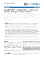
Figure 3 Sub-networks of genes up-regulated in the Arabidopsis genome in response to full-length CaLCuV AC2 protein. The diagrams
illustrate sub-networks of genes that may be co-regulated in Arabidopsis, in response to the transcription activation domain of AC2. Sub-networks I
(A), V (B) and IV (C) were up-regulated at one dpi. Highly linked genes in sub-network IV (D) were up-regulated at two dpi. The sub-networks were
selected from the network analysis presented in (Additional file 6: Figure S1).
Liu et al. BMC Plant Biology 2014, 14:302
/>
host genes that are important for cell-to-cell and longdistance movement of the virus. This would support the
known role of AC2 in activating transcription of the
BR1 nuclear shuttle protein in begomoviruses to facilitate
movement of the virus [14].
Of the six sub-networks identified within the 269
genes that were up-regulated in response to full length
AC2 protein at two dpi (Additional file 6: Figure S1B),
one may be of particular interest. The highly linked
genes within sub-network IV (Figure 3D), all appear to
have functions related to the cell cycle. One gene encodes the MYB domain protein 3R-4 (At5g11510), which
is a transcription factor that positively regulates cytokinesis
[35]. However, activation appears to require phosphorylation of the C-terminal domain of the protein, since unphosphorylated MYB3R4 acts as a repressor of mitosis [36]. In
fact, a functional MYB3R4 protein appears to be required
for establishment of the endocycle, which is induced in
response to powdery mildew infection [36]. This may be
extremely relevant to geminiviruses, especially as ploidy
increases during CaLCuV infection [24], and Maize
streak virus RepA protein induces endoreduplication
[37]. Alterations in expression of cell cycle-associated
and core cell cycle genes in response to CaLCuV infection suggests specific activation of S phase and inhibition of M phase, as a possible mechanism to induce the
endocycle [24]. A second gene, Cyclin A2;4 (At1g80370),
also up regulated in response to full-length AC2, plays a
role in determining the balance between mitosis and the
endocycle. However, it has been suggested that an absence
or reduction in CYCA2 levels controls endoreduplication,
and that expression of CYCA2 is achieved through the protein, Increased Level of Polyploidy1 (ILP1) [38]. Interestingly, ILP1 levels were elevated in CaLCuV infected leaves,
although no change in the expression of CYCA2 genes was
detected [24]. In contrast, an increase in the expression of
CYCA2;4 was detected in transgenic Arabidopsis plants
expressing BCTV L2 [39].
For the 158 unique genes that were down regulated in
response to full length AC2 at one dpi (Additional file 7:
Figure S2A), five of these were highly connected in a
network of genes that are co-regulated, and all five appear
to be involved in the defense response to pathogen infection (Figure 4A). MAP Kinase Substrate 1 (MKS1) is a
substrate for MAP kinase 4 (MPK4), which in Arabidopsis
regulates pathogen defense responses. Overexpression of
MKS1 appears to be sufficient to activate SA-dependent
resistance, and MKS1 interacts with WRKY transcription
factors, including WRKY33, which is an in vitro substrate
of MPK4 [40]. As different domains of MKS1 interact with
MPK4 and WRKY it has been suggested that these proteins play a role in transcription or chromatin remodeling
complexes, contributing to MPK4-regulated defense activation [40]. The fact that steady state mRNA levels for
Page 6 of 16
MKS1 and WRKY33 are down-regulated by AC2, could
be interpreted as a strategy to circumvent SA-dependent
responses to virus infection. Two other genes connected
to MKS1 and WRKY33 are E3 ubiquitin ligases. PUB24 is
a U-box-type E3 ubiquitin ligase, which acts to negatively
regulate PAMP-triggered immunity (PTI) [41]. Pathogen
infection leads to an increase in expression of PUB24,
but decreased expression results in an impaired ability
to down-regulate responses triggered by PAMPs [41].
Toxicos En Levadura 2 (ATL2), a RING-H2 Ubiquitin
E3-Ligase, is rapidly induced in response to elicitors, including chitin, and may function to mediate ubiquitination
of negative regulators of defense response [42]. Thus,
down-regulation of this gene by AC2 would prevent
degradation of proteins involved in turning off defense
responses, thus preventing the host from initiating a
response to infection. Interestingly, WRKY33, ATL2 and
Embryo Sac Development Arrest 39 (EDA39), a calmodulin binding protein in this regulatory network, are also
induced in response to chitooctaose, an elicitor of plant
defense responses against pathogens [43]. Therefore, it
appears as though this network of genes could be a high
value target for geminiviruses.
At two dpi, 193 genes were down-regulated in response
to the full length AC2 protein, and two sub-networks were
detected consisting of highly connected genes (Additional
file 7: Figure S2B). Within sub-network II (Figure 4B), two
genes are of potential relevance for geminivirus pathogenicity. Expression of full length AC2 down-regulated
cytokinin-hypersensitive 2 (CKH2; At2g25170), which
encodes PICKLE, a protein similar to the CHD3 class of
SWI/SNF chromatin remodeling factors [44]. Mutations
within this gene result in rapidly growing green calli,
which is attributed to hypersensitivity to cytokinins, where
cytokinin-responsive genes respond to much lower levels
of cytokinin [44]. Down regulation of CKH2 by CaLCuV
AC2 could be interpreted as a mechanism to induce cytokinin responses in order to promote cell proliferation and
therefore viral replication. Some evidence for this conclusion is provided by data demonstrating that begomovirus
AC2, and curtovirus C2, proteins increase cytokininresponsive promoter activity and that application of exogenous cytokinin increases susceptibility to geminivirus
infection [26].
A second gene within this sub-network that is downregulated by AC2 is Hobbit (HBT; At2g20000), which
encodes a homolog of the CDC27/Nuc2/BimA/APC3
subunit of the anaphase-promoting complex (APC) [45].
The HBT protein regulates M-phase progression. HBT
transcripts mainly accumulate around the G2/M phase
in dividing cells, and mutations in the HBT gene interfere with post-embryonic cell division and differentiation
of different cell types [45]. This gene may therefore be a
valuable target for geminiviruses as down-regulation
Liu et al. BMC Plant Biology 2014, 14:302
/>
Page 7 of 16
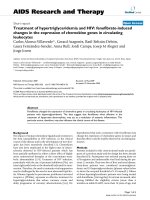
Figure 4 Sub-networks of genes down-regulated in the Arabidopsis genome in response to full-length CaLCuV AC2 protein. The
diagrams illustrate sub-networks of genes that may be co-regulated in Arabidopsis, in response to the transcription activation domain of AC2.
Genes within sub-network I (A) and sub-network IV (B) were down-regulated at one and two dpi respectively. The sub-networks were selected
from the network analysis presented in (Additional File 7: Figure S2).
would presumably interfere with progression of cell differentiation shifting the balance in favor of cell proliferation,
possibly in conjunction with down-regulation of CKH2 to
promote cell proliferation.
Validation of microarray results by quantitative real-time
PCR
For this analysis we focused on a single network that
contained five down-regulated genes associated with
plant defense, that were found to be highly connected at
one dpi after expression of full-length AC2 (Figure 4A).
Even though these five genes were only differentially regulated at one dpi in the microarray analysis, total RNA
was isolated at both one and two dpi from Arabidopsis
leaves infused with Agrobacterium containing DNA capable of expressing full-length AC2 or a vector control.
After generation of cDNA, quantitative real time PCR
(qPCR) analysis was performed using gene-specific primers
(Additional file 8: Table S6) to verify differential regulation. As can be seen (Figure 5), at one dpi expression of
AtPUB24, AtWRKY33, AtATL2 and AtEDA39 were all
significantly down regulated up to two fold in samples
from leaves infused with AC2 relative to samples from
leaves treated with empty vector (pMON530). However, at
two dpi no significant difference in expression was detectable for any of the four genes, although expression was
still lower than that in samples from leaves treated with
empty vector (Figure 5). These results are consistent with
the microarray data, where these genes were significantly
down regulated at one dpi but not at two dpi (Additional
file 1: Table S1 and Additional file 2: Table S2 respectively). Interestingly, expression of AtMKS1 was not significantly altered at one dpi (Figure 5) in samples from leaves
infused with AC2 relative to samples from leaves treated
with empty vector (pMON530). The reasons for this are
not clear but may be a consequence of differences between the two methods, including but not limited to, the
utilization of vastly different normalization procedures,
different strategies in probe design and sensitivity limits of
PCR vs. hybridization-based approaches [46].
Differential expression of genes responding to
inactivation of SnRK1 by SCTV C2 or asSnRK1.2
A second goal of this study was to examine the consequence(s) of the interaction between SCTV C2 and
SnRK1.2. To do this we compared the transcriptomes in
Arabidopsis leaves expressing full-length SCTV C2 or an
antisense construct of SnRK1.2 (asSnRK1.2) at one and
Figure 5 Quantitative (q)PCR analysis of genes differentially
regulated in response to full length CaLCuV AC2 protein. Values
were determined by qPCR analysis of total RNA isolated from
Arabidopsis leaves infused with Agrobacterium containing DNA
capable of expressing full-length Cabbage leaf curl virus AC2, or an
empty plasmid vector (pMON530). The columns represent relative
mRNA levels in CaLCuV AC2-infused leaves as compared to levels
present in leaves infused with Agrobacterium containing empty
plasmid vector (pMON530), which was arbitrarily assigned a value of
1 at each time point. The fold change was calculated from the mean
ΔΔCt values from three independent experiments using RNA isolated
one and two days post-infusion (dpi). Error bars represent the Standard
Error of the mean and asterisks indicate significant differences in
expression as determined using the Student’s t-test (P < 0.05) on
ΔCt values.
Liu et al. BMC Plant Biology 2014, 14:302
/>
two dpi (Additional file 9: Table S7 and Additional file 10:
Table S8). The rationale for this approach is that interaction between geminvirus AC2 and C2 proteins results
in inactivation of the kinase [5,19], and asSnRK1.2 is
expected to result in degradation of sense mRNA through
the siRNA pathway and lead to loss of SnRK1.2 activity.
Thus, genes found to be differentially regulated in response
to both treatments is presumed to be a consequence of
reduced SnRK1.2 activity. Of those genes up-regulated
in response to C2 or asSnRK1, 49 were common to both
treatments at one dpi and 210 at two dpi (Figure 6). For
genes down-regulated in response to C2 or asSnRK1.2
at one or two dpi, we observed 37 and 203 respectively,
that were common to both treatments (Figure 6). These
genes are therefore interpreted to represent genes
responding to inhibition of SnRK1 activity by geminvirus
C2 protein. It is important to note here that the total
number of genes differentially regulated in response to
both C2 and asSnRK1 was ~ five-fold higher at day two
(Figure 6).
Some differentially regulated genes were specific to
each individual treatment. Of those genes specifically
up-regulated by SCTV C2, we detected 235 at one dpi
and 401 at two dpi (Figure 6). 144 and 342 genes were
specifically down-regulated by SCTV C2, at one and two
dpi respectively. Presumably, these genes are differentially regulated in response to additional functions of
SCTV C2, which would include interaction with and
inactivation of ADK [6], and possibly additional unknown
functions. There were also many genes whose expression
changed specifically in response to expression of asSnRK1.2.
At day one and two dpi, we detected 377 and 489 genes
respectively, up-regulated in response to asSnRK1 alone
(Figure 6). For genes down-regulated in response to
asSnRK1 alone, 228 and 591 were detected at one and two
Figure 6 Numbers of genes differentially expressed in response
to SCTV C2 and antisense SnRK1.2. Venn diagrams illustrating the
intersection between up- and down-regulated genes in Arabidopsis
leaves expressing SCTV C2 or antisense SnRK1.2, for one and two
dpi respectively.
Page 8 of 16
dpi respectively (Figure 6). As these genes were not differentially regulated in response to SCTV C2, we conclude that this may be a consequence specific to SnRK1.2
activity.
Functional categorization of genes differentially regulated
in response to asSnRK1.2
The focus of this analysis was to characterize genes
found to be differentially regulated in response to both
SCTV C2 and asSNRK1.2. We categorized these genes
by biological process using the DAVID Bioinformatics
Resource. Most of the GO biological process categories
were represented among the significant genes, but several categories were significantly enriched as compared
to the Arabidopsis genome as a whole. In this case,
genes associated with Transcription, Protein Metabolism
and Transport, and Electron transport or Energy pathways were over-represented (Additional file 11: Table S9
and Additional file 12: Table S10).
Network analysis of genes differentially regulated in
response to inactivation of SnRK1.2
We overlayed the asSnRK1.2 responsive genes to the
Arabidopsis co-expression network, and extracted dense
subnetworks for further investigation. Given the small
number of genes that were up- (Additional file 13: Figure
S3A) or down- (Additional file 14: Figure S4A) regulated
in response to both SCTV C2 and asSnRK1.2 at one dpi,
no networks consisting of highly connected genes were
identified. However, at two dpi a large increase in the
number of genes that were up- (Additional file 13: Figure
S3B) and down- (Additional file 14: Figure S4B) regulated
revealed complex networks (Additional file 15: Table S11).
Of the 209 genes that were up regulated in response to
SCTV C2 and asSnRK1.2 at two dpi, a large complex network was identified (Figure 7A), within which several
genes have functions associated with autophagy. This is a
process by which cytoplasmic contents, including proteins
and organelles, are sequestered within the autophagosome, a double-membrane vesicle, which can deliver the
contents to lysosomes or vacuoles through fusion for
degradation [47]. Autophagy is involved in both the responses to biotic stresses, including viral infection, and
in regulating senescence, and many autophagy genes
have been identified and functionally analyzed in plants.
Of the three genes within this network found to be upregulated in response to C2 and asSnRK1.2, the role of
the APG9 (At2g31260) complex is unclear. However,
APG7 (At5g45900) is an E1 ubiquitin-activating enzyme
that conjugates phosphatidylethanolamine to ATG8H
(AT3G06420) [48]. More evidence is being provided that
autophagy may function either to facilitate or prevent viral
pathogenesis [49,50]. As a defense against pathogen infection, autophagy has been shown to play an important role
Liu et al. BMC Plant Biology 2014, 14:302
/>
Page 9 of 16
Figure 7 Sub-networks of genes differentially regulated in response to full-length CaLCuV AC2 protein. The diagrams illustrate sub-networks of
genes that may be co-regulated in response to to both SCTV C2 and asSnRK1.2 at two dpi. (A) Network of genes up-regulated at two dpi. (B) and (C)
Networks of genes down-regulated at two dpi. The sub-networks were selected from the network analysis presented in (Additional file 13: Figure S3 and
Additional file 14: S4). A list of the connections between genes in the networks (edges) is given in (Additional file 15: Table S11).
in both pathogen-induced hypersensitive cell death (HR),
and the plant antiviral immune response. Rapid immune
responses, including HR, are induced in tobacco plants
carrying the N-resistance gene when infected by Tobacco
mosaic virus (TMV). The result of this is limitation on
the replication and systemic spread of the virus [51]. Silencing of BECLIN1/ATG6, ATG3, or APG7 resulted in
the spread of cell death, suggesting that autophagy plays
an anti-death role during pathogen infection to limit the
spread of HR beyond initially infected cells [52]. A suppressor of programmed cell death in tomato (Adi3) has
been shown to interact with tomato ATG8H although it
is not clear at this time whether Adi3 is targeted by
autophagy [53]. Since autophagy is an emerging antiviral
process employed by the host immune system, certain
viruses have successfully evolved to either avoid, subvert
or even actively induce autophagy to ensure a productive
infection [54]. Interestingly, autophagy-related transcripts,
including ATG8H and ATG9, were up regulated during
infection of tomato with Tomato yellow leaf curl Sardinia
virus (TYLCSV) [55] and in Arabidopsis infected with
CaLCuV [24].
Of particular relevance to geminiviruses are recent
studies that have shown a role for autophagy in RNA
silencing [50]. This is an antiviral response that results
in dsRNA-mediated degradation of viral RNAs. As a
counter-defense, viruses encode RNA silencing suppressors (RSSs) that act to suppress the RNA silencing
machinery [9]. A recent study indicates that a tobacco
regulator of gene silencing calmodulin-like protein
(Nt-rgsCaM) binds to an arginine-rich region within a
number of viral RSSs, resulting in degradation through
autophagosomes [56]. This supports the idea that autophagy can provide a secondary antiviral mechanism by
targeting viral RSSs for degradation. However, we have
recently demonstrated that in the case of geminiviruses,
there appears to be a different mechanism where AC2,
the begomovirus RSS, induces rgsCaM and may in fact
Liu et al. BMC Plant Biology 2014, 14:302
/>
sequester rgsCaM in the nucleus to prevent targeting of
AC2 for degradation via the autophagy pathway [57].
While we cannot explain this apparent discrepancy, it
could reflect a difference between the RNA viruses used
in one study [56] and geminiviruses in our study [57].
Recently, it has been shown that the polerovirus P0 RSS
targets Argonaute 1 (AGO1) for degradation via the
autophagy pathway [58]. At this time it is unknown
whether AC2 specifically targets genes in the autophagy
pathway to facilitate pathogenesis.
Of further interest to geminivirus pathogenesis is
the observation that under conditions of stress, including pathogen infection, AMPK appears to regulate
the autophagy pathway through two mechanisms.
First, AMPK directly interacts with Ulk1, an autophagy
initiator, through phosphorylation [59]. AMPK can indirectly induce autophagy through phosphorylation of raptor,
which inhibits the mTORC1 complex [60]. Thus, phosphorylation of Ulk1 by mTORC1 and/or AMPK results
in either negative or positive regulation of autophagy
respectively [61]. The geminvirus AC2/C2 proteins have
been shown to interact with and inactivate SnRK1, the
plant homolog of AMPK [5]. Under the stress of viral
infection, this would prevent phosphorylation of raptor
maintaining an active mTORC1 complex. This would
ensure that the autophagy pathway is inhibited. Secondly,
inhibition of SnRK1 by AC2/C2 would prevent direct
phosphorylation of Ulk1, again preventing activation of
the authophagy pathway. However, there is an apparent
paradox given that we detect up-regulation of autophagy
genes in response to both full length SCTV C2 and
asSnRK1.2. This can be partially explained by observations
that the autophagosome marker ATG8 is rapidly up regulated under starvation conditions in yeast, and that most
of the autophagy genes are regulated at a transcriptional
level [62]. This reiterates the importance of SnRK1 as a
high value target for geminiviruses [5,6,20,26], by preventing activation of autophagy in the event of up-regulation
of genes in that pathway.
For the 203 common genes that were down regulated
at two dpi, a large complex network containing highly
connected genes that appear to be co-regulated was
identified (Additional file 14: Figure S4B). Two smaller
clusters of genes within this network (Figure 7B and C)
have functions associated with the ribosome and translation. Although the genes identified have not been specifically reported to play roles in viral pathogenesis, there
are examples of ribosomal proteins that play a role in
antiviral defense, and so it may not be surprising that
geminiviruses down-regulate these genes to facilitate
infection. With respect to geminiviruses, the nuclear
shuttle protein (BR1) has been shown to target the NSPinteracting kinases (NIKs), which are leucine-rich-repeat
(LRR) receptor-like-kinases (RLKs) involved in antiviral
Page 10 of 16
defense [63]. NIK1 phosphorylates the ribosomal protein,
rpL10A, which functions as an immediate downstream
effector of the NIK1-mediated response and binding of
NSP to NIK1 inhibits its kinase activity preventing the
antiviral defense pathway from impacting geminvirus infection [63,64].
Validation of microarray data by quantitative real-time
RT-PCR
We chose to analyze six genes with functions associated
with autophagy and senescence (Figure 7A) that were
up-regulated in response to both C2 and asSnRK1.2.
Total RNA was isolated at both one and two dpi from
Arabidopsis leaves infused with Agrobacterium containing
DNA capable of expressing full-length C2, asSnRK1.2 or
the vector control (pMON530). In addition, we also used
an inverted repeat construct designed to express dsRNA
(dsSnRK1.2) that is known to reduce target mRNA levels
in infused N.benthamiana leaves [20]. After generation of
cDNA, qPCR analysis was performed using gene-specific
primers (Additional file 8: Table S6) to verify differential
regulation. As shown (Figure 8), significant increases in
expression were observed in response to SCTV C2,
asSnRK1.2 and dsSnRK1.2 at two dpi for all six genes
tested. No significant changes in expression were detectable at one dpi (data not shown). This is consistent with
the microarray data where expression of these genes
Figure 8 Quantitative (q)PCR analysis of genes differentially
regulated in response to inactivation of SnRK1. Values were
determined by qPCR analysis of total RNA isolated from Arabidopsis
leaves infused with Agrobacterium containing DNA capable of
expressing full-length Spinach curly top virus C2, antisense (as)SnRK1.2,
an inverted repeat construct designed to express dsRNA (dsSnRK1.2) or
an empty plasmid vector (pMON530). The columns represent relative
mRNA levels in C2, asSnRK1, or dsSnRK1-infused leaves as compared to
levels present in leaves infused with Agrobacterium containing empty
plasmid vector (pMON530), which was arbitrarily assigned a value of 1
at each time point. The fold change was calculated from the mean
ΔΔCt values from three independent experiments using RNA isolated
two days post-infusion (dpi). Error bars represent the Standard Error of
the mean and asterisks indicate significant differences in expression as
determined using the Student’s t-test (P < 0.05) on ΔCt values.
Liu et al. BMC Plant Biology 2014, 14:302
/>
increased in response to both SCTV C2 and asSnRK1.2
(Additional file 10: Table S8). Given that we also observed
up-regulation of these genes in response to silencing of
SnRK1.2 with an inverted repeat construct (dsSnRK1) we
interpret this to be a consequence of the inactivation/inhibition of SnRK1.2.
Conclusion
It is becoming increasingly apparent that geminiviruses
manipulate the host in several ways to facilitate an environment conducive to infection, predominantly through
the use of multifunctional proteins. As one example,
TGMV AL1 protein is necessary for origin recognition
and initation of RCR [65,66]. TGMV AL1 also binds to a
plant retinoblastoma (pRb) protein [67,68], and is sufficient for PCNA accumulation [69]. This is analogous to
small DNA tumor viruses, where adenovirus and SV40
deregulate the cell cycle via interaction with the pRb and
p53 pathways [70-73]. In addition, infection by CaLCuV
has been shown to influence the host transcriptome
[24], again demonstrating the ability of geminiviruses to
manupulate the host to ensure efficient infection. A second multifunctional protein encoded by geminviruses that
influences the host response to infection, is the AC2/C2
protein. We have recently shown that the CaLCuV CP
promoter is regulated by AC2 through an interaction
with PPD2, a plant specific DNA binding protein, that
specifically binds sequences known to mediate activation of the CP promoter of CaLCuV and TGMV [17].
An indirect promoter targeting mechanism could provide
an opportunity for the virus (via AC2) to alter host gene
expression. This may in turn reprogram the host to support virus infection and/or evade host defense responses.
Additional interactions between AC2/C2 and SnRK1.2
and ADK lead to suppression of host defenses [5,6], which
could also lead to alterations in host transcriptome. In
support of this, our study along with others using either
whole virus infections [24] or over-expression of AC2
from ACMV or MMYMV [25,74], identified large scale
changes in the host transcriptome. The other studies were
performed either in whole Arabidopsis plants [24], transient assays using Arabidopsis protoplasts [25] or transgenic
Nictotiana tabacum constitutively expressing AC2 [74].
The complexity of possible effects of AC2 makes it desirable to extend this type of analysis under different
conditions to identify key host factors independent of
laboratories and host plant-virus interactions. Thus, the
current study is complementary to the others and provides completely novel aspects for the functional analysis. As with the other studies, we identified several
categories of genes that were significantly enriched as
compared to the Arabidopsis genome as a whole, including genes for DNA/RNA Metabolism, Transcription, Response to Stress, Protein Metabolism, Signal transduction,
Page 11 of 16
Cell organization and Biogenesis, Transport and Electron
transport or Energy. Our analysis enabled us to identify networks containing highly connected genes that could reflect
co-regulated functional gene modules. Two of these highlight the significance of our approach in uncovering novel
clusters of genes targeted by geminiviral RSSs. As an
example, sub-networks containing genes having functions
associated with the chloroplast and the cell wall and/or
cytoskeleton, could reflect a direct role for AC2 in inducing the expression of genes important for virus movement. The latter may have uncovered an explanation for
the observation that mutations within the TGMV AC2
gene lead to loss of infectivity [75]. This is due, primarily,
to the fact that AC2 is required for the transcriptional
activation of the BR1 nuclear shuttle protein which is
necessary for movement of the virus [14]. Thus, alteration of genes associated with the chloroplast and cell
wall and/or cytoskeleton could reflect a direct role for
AC2 in inducing the expression of genes important for
virus movement. It will be interesting to determine
whether the promoters of the genes identified have any
cis-acting elements in common with the BR1 genes of
begomoviruses.
In a second example, our network-based approach has
identified a potential link between RNA silencing suppressors, SnRK1.2 and autophagy (Figure 7). This is supported
by recent evidence demonstrating that autophagy plays
a role in directing degradation of DICER and AGO2,
important proteins in miRNA processing and in posttranscriptional regulation of DICER mRNA [76]. Therefore, it has been proposed that autophagy may represent
a checkpoint for maintaining homeostasis of miRNA
populations [76], and so it interesting to speculate that
inhibition of SnRK1.2 by the geminivirus AC2/C2 proteins may have wide-reaching effects on both RNA
silencing and autophagy. However, many unresolved
questions remain regarding the role of autophagy in viral
pathogenesis, but targeting of this pathway underscores
the likely importance of autophagy as a component of
antiviral immunity.
Our approach to identifying highly connected genes
that are differentially regulated by AC2 has revealed coregulated gene networks that are potentially targeted by
geminiviruses during infection. Many of these genes
would not have been thought of as functioning in a network, but this approach allows us to assess them as a
functioning unit and determine the importance of the
network as a whole in viral pathogenesis. We can now
identify novel pathways of co-regulated genes that are
stimulated in response to pathogen infection in general,
and virus infection in particular. We are currently confirming the differential expression of genes in all the
sub-networks and are investigating the role each subnetwork plays in viral pathogenesis.
Liu et al. BMC Plant Biology 2014, 14:302
/>
Methods
DNA constructs
Cloned DNAs capable of constitutively expressing CaLCuV AC2 (p35S-CaLCuVAC2) or SCTV C2 (p35s-SCTV
C2) from the CaMV 35S promoter have been described
previously [12,26] . A DNA construct capable of constitutively expressing a truncated CaLCuV AC2 protein lacking
the C-terminal activation domain (CaLCuV AC21-100) was
generated by PCR. A 300 bp fragment was amplified with
primers CaLCVAC2F (5′-gcgagatctatgcaaaattcatcactcttg-3′)
and CaLCVAC2Rdel (5′-gcgctcgagctacgtaggttgtggttgaac-3′)
using CaLCuV DNA A as a template. Following restriction with XhoI-BglII the fragment was cloned into similarly cut pMON530 to generate p35S-CaLCuVAC21-100.
To generate a DNA construct capable of constitutively
expressing an antisense RNA to Arabidopsis SnRK1.2
(AKIN11) from the CaMV 35S promoter, pAS2-AKIN11
DNA [19] was restricted with NcoI and treated with
Klenow to generate a blunt end. Following restriction
with BamHI, the resulting 1.5 kbp fragment was cloned
into the plant binary vector pMON530 [76] at the BglII
and SmaI sites, to generate DNA containing the SnRK1.2
coding region in the antisense orientation (asSnRK1.2).
The presence of each ORF in the correct orientation was
confirmed by DNA sequencing. The resulting Ti plasmid
constructs were mobilized into Agrobacterium strain
GV3111SE by triparental mating [77] and used for agroinfiltration. As a control, vector DNA containing the CaMV
35S promoter alone (pMON530) was introduced into
Agrobacterium.
Agrobacterium infusion assays and RNA isolation
Vacuum infiltration of Arabidopsis thaliana plants with
Agrobacterium cultures was performed essentially as described [26]. Arabidopsis Col-0 plants were sprinkled with
water prior to infiltration and whole plants submerged in
the Agrobacterium culture ensuring all rosette leaves were
submerged in the solution. Vacuum was drawn for 2030 min at a pressure of approximately 0.05 Bar. Plants were
removed from the beaker, replanted into moist soil, covered
and placed in a growth chamber under long day conditions
(16 h light and 8 h dark) and incubated at 21°C. Infiltrations
were performed in the afternoon and infiltrated leaf tissue
from four different plants harvested in the afternoon one
to three days post-inoculation, depending on the experiment. Total RNA was isolated from infiltrated leaves of
Arabidopsis using Plant RNA Reagent as described by
the manufacturer (Invitrogen, Carlsbad, CA), treated with
DNaseI (Ambion, Austin, TX) and purified through RNeasy
MiniElute clean up kit (Qiagen, Valencia, CA).
GeneChip hybridization and microarray data analysis
Affymetrix ATH1 GeneChips (Affymetrix P/N 510690),
containing more than 22,500 probe sets representing ~
Page 12 of 16
24,000 genes, were used through out the experiment and
all procedures were carried out according to the manufacturers instructions (Affymetrix, Santa Clara, CA). For one
comparison, Arabidopsis plants were infused with Agrobacterium cultures containing CaLCuV AC2, CaLCuV
AC21-100, or empty plasmid vector (pMON530). In a second comparison, Arabidopsis plants were infused with
Agrobacterium cultures containing SCTV C2, asSnRK1.2
or empty plasmid vector (pMON530). Three independent
experiments were performed for each comparison, at different times, and total RNA isolated from infused plants
at one and two days post-infiltration. This resulted in a
total of nine samples for each comparison at one and two
dpi. Comparison 1: Nine arrays for samples 530 × 3,
CaLCuV AC2 × 3, CaLCuV AC21-100 × 3 at day one and
two = 18 total. Comparison 2: Nine arrays for samples 530 × 3, SCTV C2 × 3, asSnRK1.2 × 3 at day one
and two = 18 total. Total RNA (10 μg) was processed by a
one-step labeling protocol (Affymetrix), and fragmented
cRNA (15 μg) hybridized to the Arabidopsis ATH1
Genome using the recommended standard procedures
(45°C for 16 h). Washing and staining were performed in
a fluidics station 400, using the standard protocol
EUkGEWS2v4 and scanned using an Agilent GeneArray
Scanner. Array quality was assessed following the parameters recommended by Affymetrix (GeneChip Expression
Analysis, Technical Manual, 701021 rev 1). Raw intensity
data was processed using The Robust Multi-array Average
(RMA) procedure in MATLAB Bioinformatics Toolbox,
which first performs background adjustment and quantile
normalization on the probe level, and then summarizes
the intensity levels from each probe set to gene-level
expression values in logarithmic scale [78]. Fold changes,
while not used for selecting differentially expressed genes,
were computed by first taking the arithmetic mean of the
log2(gene expression) of the three biological replicates,
and then calculating the ratio of the mean expression
values in linear scale. From a total of 22810 genes represented on the array, genes differentially expressed between
experimental samples and controls were detected using
two-sample t-tests with a multiple-testing corrected
p-value of 0.05 used as the cutoff. Permutation test with
1000 permutations was performed to correct for multiple
testing [79].
There are many different methods for defining/selecting differentially expressed (DE) genes and each could
result in a different set of genes. In general, fold change,
while simple and intuitive, is not a preferred criterion in
selecting DE genes, because of lack of indication in the
level of confidence and reproducibility [78]. It is important
to note that fold change is not necessarily a biologically
more meaningful measure than statistical significance, as
some genes can have their effects at very low level of fold
changes while some other genes need to function at a
Liu et al. BMC Plant Biology 2014, 14:302
/>
much higher level. In addition, the fold change approach
is usually subject to bias as it tends to select low-intensity
genes whose fold change values have a larger variance
than the fold change values of high-intensity genes. Last
but not least, raw intensity data from microarray experiments often need to be preprocessed and normalized,
which could dramatically impact the fold change estimation, depending on the procedure used, leaving the definition of fold change obscure. We choose our approach
based on a study that shows, with Affymetrix arrays in
particular, the t-test usually results in more accurate discovery of DE genes, especially when combined with RMA
for preprocessing and normalization [80]. At the same
time, the study also showed that RMA often produces a
biased estimation of fold change, which is probably the
reason that the observed fold changes for the DE genes in
our experiment are relatively small. Simulations in their
study showed that RMA can reduce the fold change by as
much as 2 fold (e.g. a 4-fold change could be reduced to
2-fold after RMA).
Statistical and network-based analysis
Over-representation of Gene Ontology (GO) terms within
each gene list was performed using the hypogeometric test
implemented on the DAVID Bioinformatics Resource
[81]. To identify sub-networks for a list of genes, we
overlayed these input genes to an Arabidopsis gene
co-expression network [27] using gene expression data
from >1300 microarray experiments, and retrieved subnetworks that consists of only the input genes and their
connections. For genes down-regulated at two dpi by
asSnRK1.2, as the returned network is very large, we iteratively removed genes with less than four connections in
the sub-network and the remaining sub-network is used
for further analysis.
Quantitative real-time PCR
Quantitative real-time PCR (qPCR) was used to assess
differences in the steady state mRNA levels of genes in
response to the proteins of interest by comparison to a
plasmid vector treated control. Total RNA (1 μg) isolated
from Arabidopsis leaf tissue was treated with DNase I and
reverse transcribed using a high-capacity cDNA archive
kit (Applied Biosystems, Foster city, CA). qPCR analysis
was performed with SYBR Green using gene specific
probes (Additional file 8: Table S6), with a 7500 Real-time
PCR system (Applied Biosystems, Foster city, CA) as described previously [26], or with the Biomark HD System
(Fluidigm Corporation). Primer sequences were designed
using Primer Express 2.0 software (Applied Biosystems).
For each experiment, target samples were normalized to
EF1α, which was used as an reference. In each experiment,
samples from three independent biological samples were
used for the analysis. Ct values for each well position were
Page 13 of 16
examined prior to data analysis. Differences in gene
expression (ΔΔCt) were calculated using the 7500 System
SDS software package (Applied Biosystems, Foster city,
CA), which measured differences in expression of the
target gene and the endogenous control (ΔCt) in each
replicate.
Availability of supporting data
The microarray dataset used in this manuscript has been
deposited with the Gene Expression Omnibus (GEO) and
assigned the following GEO accession number: GSE62180.
All of the data can be accessed through the following
link: />GSE62180.
Additional files
Additional file 1: Table S1. Genes differentially expressed in response
to CaLCuV AC2. Genes that are up- or down regulated at one dpi, in
response to full-length AC2 (FL) or a deletion derivative lacking the
transcription activation domain (DEL), are shown.
Additional file 2: Table S2. Genes differentially expressed in response
to CaLCuV AC2. A list of genes that are up- or down-regulated at two
dpi, in response to a full-length AC2 (FL) or a deletion derivative lacking
the transcription activation domain (DEL), are shown.
Additional file 3: Table S3. A comparison of differentially regulated
genes found in CaLCuV-infected Arabidopsis with those found in
response to full-length (FL) AC2. The first two worksheets list genes
up- or down-regulated in Arabidopsis plants infected with CaLCuV at
12 days post infection [24] and indicates whether the same genes were
also differentially regulated in response to FL AC2. A hypergeometric
distribution is shown in the third worksheet.
Additional file 4: Table S4. GO biological process categories
represented among the genes differentially expressed in response to
CaLCuV AC2 at one dpi.
Additional file 5: Table S5. GO biological process categories
represented among the genes differentially expressed in response to
CaLCuV AC2 at two dpi.
Additional file 6: Figure S1. Network analysis using genes that were
up-regulated specifically in response to full length AC2. Sub-networks
(red boxes) containing highly connected genes that were up-regulated in
response to full length AC2 at one (A) or two (B) dpi.
Additional file 7: Figure S2. Network analysis using genes that were
down-regulated specifically in response to full length AC2. Sub-networks
(red boxes) containing highly connected genes that were up-regulated in
response to full length AC2 at one (A) or two (B) dpi.
Additional file 8: Table S6. Primer sets used for qPCR analysis in this
study.
Additional file 9: Table S7. Genes differentially expressed in response
to inactivation of Arabidopsis SnRK1.2. Genes that are up- or
down-regulated at one dpi, in response to SCTV C2 or antisense
SnRK1.2, are shown.
Additional file 10: Table S8. Genes differentially expressed in
response to inactivation of Arabidopsis SnRK1.2. Genes that are up- or
down-regulated at two dpi, in response to SCTV C2 or antisense SnRK1.2,
are shown.
Additional file 11: Table S9. GO biological process categories
represented among the genes differentially expressed in response to
inactivation of Arabidopsis SnRK1.2 at one dpi.
Liu et al. BMC Plant Biology 2014, 14:302
/>
Additional file 12: Table S10. GO biological process categories
represented among the genes differentially expressed in response to
inactivation of Arabidopsis SnRK1.2 at two dpi.
Additional file 13: Figure S3. Network analysis using genes that were
up-regulated specifically in response to full length AC2. Sub-networks
(red boxes) containing highly connected genes that were up-regulated in
response to SCTV C2 and antisense SnRK1.2 at one (A) or two (B) dpi.
Additional file 14: Figure S4. Network analysis using genes that were
down-regulated specifically in response to full length AC2. Sub-networks
(red boxes) containing highly connected genes that were up-regulated in
response to SCTV C2 and antisense SnRK1.2 at one (A) or two (B) dpi.
Additional file 15: Table S11. Network Edges: A list of connections
between two genes in the networks shown in Figure 7.
Abbreviations
35S: CaMV promoter; 5′-AMP: 5′ adenosine monophosphateAbMV,
Abutilon mosaic virus; AC2: Begomovirus transcriptional activator protein;
ADK: Adenosine kinase; AKIN11: Arabidopsis SNF1 related protein kinase 1.2;
asSnRK1.2: Antisense version ofArabidopsis SnRK1.2; Atg: autophagy related
genes; ATL2: RING-H2 Ubiquitin E3-Ligase; BCTV: Beet curly top virus;
BR1: Begomovirus nuclear shuttle protein; CaLCuV: Cabbage leaf curl virus;
CaMV: Cauliflower mosaic virus; CP: Coat protein; cpHSC70-1: Chloroplast heat
shock cognate 70 kDa protein; cRNA: Complementary RNA; dpi: Days
post-infusion; DNA: Deoxyribonucleic acid; dsDNA: Double stranded DNA;
EDA39: Embryo Sac Development Arrest 39; EF1α: Eukaryotic elongation
factor 1α; eIF: Eukaryotic initiation factor; ER: endoplasmic reticulum;
GO: gene ontology; HBT: Hobbit; ILP1: Increased level of polyploidy1;
L2/C2: Curtovirus pathogenicity protein; LRR: Leucine rich repeat; MKS1: MAP
kinase substrate 1; MP: Movement protein; MPK4: MAP kinase 4;
MYB3R4: MYB domain protein 3R-4; NIK: NSP-interacting kinases;
NSP: Nuclear shuttle protein; p53: Tumor suppressor protein;
PAMP: Pathogen associated molecular pattern; PCNA: Proliferating cell
nuclear antigen; PCR: Polymerase chain reaction; PPD2: Arabidopsis peapod2
protein; pRb: Plant retinoblastoma protein; PTI: PAMP-triggered immunity;
PUB24: U-box-type E3 ubiquitin ligase; qPCR: Quantitative real time PCR;
RCR: Rolling circle replication; RDR: Recombination dependent replication;
RF: Replicative form; RLK: LRR receptor-like kinase; RMA: Robust multi-array
averagerpL, ribosomal protein L; RNA: Ribonucleic acid; RSS: RNA silencing
suppressor; RT-PCR: Reverse transcriptase PCR; SCTV: Spinach curly top virus;
SNF1: Sucrose non-fermenting 1 protein; SnRK1: SNF1 related protein kinase;
ssDNA: Single stranded DNA; SV40: Simian virus 40; TGMV: Tomato golden
mosaic virus; TIC110: Translocon at the inner envelope membrane of
chloroplasts 110; TMV: Tobacco mosaic virus; TOC75-III: Translocon at the
outer envelope membrane of chloroplasts 75-III; WRKY33: Plant-specific
transcription factor.
Competing interests
The authors declare that they have no competing interests.
Authors’ contributions
GS conceived and coordinated the study, designed the experiments and
helped to draft the manuscript, HC designed and performed the qPCR
confirmation experiments and provided input on the manuscript, JR
coordinated the statistical analysis and helped draft the manuscript, SB and
GL participated in the design of the experiments and performed the
microarray experiments, LL performed the statistical analysis and provide
input on the manuscript. All authors read and approved the final manuscript.
Acknowledgements
This material is based upon work supported by the National Science
Foundation under Grant Number IOS-0948669 to GS and Grant Number
IIS-1218201 to JR. Any opinions, findings, and conclusions or recommendations
expressed in this material are those of the authors and do not necessarily reflect
the views of the National Science Foundation.
Author details
1
Department of Computer Science, The University of Texas at San Antonio,
One UTSA Circle, San Antonio, TX, USA. 2Department of Biology, The
University of Texas at San Antonio, One UTSA Circle, San Antonio, TX, USA.
Page 14 of 16
3
Current address: Scripps Health/Hematology/Oncology Division, 15004
Innovation Drive, San Diego, CA 92128, USA. 4Current address: Bayer
CropScience Vegetable Seeds, 7087 East Peltier Road, Acampo, California
95220, USA.
Received: 1 July 2014 Accepted: 23 October 2014
References
1. Varma A, Malathi VG: Emerging geminivirus problems: a serious threat to
crop production. Annals Appl Biol 2003, 142(2):145–164.
2. Navas-Castillo J, Fiallo-Olive E, Sanchez-Campos S: Emerging virus diseases
transmitted by whiteflies. Ann Rev Phytopathol 2011, 49:1–30.
3. Preiss W, Jeske H: Multitasking in replication is common among
geminiviruses. J Virol 2003, 77:2972–2980.
4. Stenger DC, Revington GN, Stevenson MC, Bisaro DM: Replicational release
of geminivirus genomes from tandemly repeated copies: evidence for
rolling circle replication of a plant viral DNA. Proc Natl Acad Sci U S A
1991, 88:8029–8033.
5. Hao L, Wang H, Sunter G, Bisaro DM: Geminivirus AL2 and L2 proteins
interact with and inactivate SNF1 kinase. Plant Cell 2003, 15:1034–1048.
6. Wang H, Hao L, Shung C-Y, Sunter G, Bisaro DM: Adenosine kinase is
inactivated by geminivirus AL2 and L2 proteins. Plant Cell 2003,
15:3020–3032.
7. Sunter G, Bisaro DM: Regulation of a geminivirus coat protein promoter
by AL2 protein (TrAP): evidence for activation and derepression
mechanisms. Virology 1997, 232:269–280.
8. Sunter G, Bisaro DM: Identification of a minimal sequence required for
activation of the Tomato golden mosaic virus coat protein promoter in
protoplasts. Virology 2003, 305:452–462.
9. Voinnet O, Pinto YM, Baulcombe DC: Suppression of gene silencing: a
general strategy used by diverse DNA and RNA viruses of plants.
Proc Natl Acad Sci U S A 1999, 96:14147–14152.
10. Carrington JC, Kasschau KD, Johansen LK: Activation and suppression of
RNA silencing by plant viruses. Virology 2001, 281:1–5.
11. van Wezel R, Liu H, Tien P, Stanley J, Hong Y: Mutation of three cysteine
residues in Tomato yellow leaf curl virus-China C2 protein causes
dysfunction in pathogenesis and posttranscriptional gene silencingsuppression. Mol Plant Microbe Interact 2002, 15:203–208.
12. Lacatus G, Sunter G: Functional analysis of bipartite begomovirus coat
protein promoter sequences. Virology 2008, 376:79–89.
13. Sunter G, Bisaro DM: Transactivation in a geminivirus: AL2 gene product
is needed for coat protein expression. Virology 1991, 180:416–419.
14. Sunter G, Bisaro DM: Transactivation of geminivirus AR1 and BR1 gene
expression by the viral AL2 gene product occurs at the level of
transcription. Plant Cell 1992, 4:1321–1331.
15. Berger MR, Sunter G: Identification of sequences required for AL2mediated activation of the Tomato golden mosaic virus-yellow vein BR1
promoter. J Gen Virol 2013, 94:1398–1406.
16. Sunter G, Stenger DC, Bisaro DM: Heterologous complementation by
geminivirus AL2 and AL3 genes. Virology 1994, 203:203–210.
17. Lacatus G, Sunter G: The Arabidopsis PEAPOD2 transcription factor
interacts with geminivirus AL2 protein and the coat protein promoter.
Virology 2009, 392:196–202.
18. Hartitz MD, Sunter G, Bisaro DM: The geminivirus transactivator (TrAP)
is a zinc-binding phosphoprotein with an acidic activation domain.
Virology 1999, 263:1–14.
19. Baliji S, Sunter J, Sunter G: Transcriptional analysis of complementary
sense genes in Spinach curly top virus and the functional role of C2 in
pathogenesis. Mol Plant Microbe Interact 2007, 20(2):194–206.
20. Wang H, Buckley KJ, Yang X, Buchmann RC, Bisaro DM: Adenosine kinase
inhibition and suppression of RNA silencing by geminivirus AL2 and L2
proteins. J Virol 2005, 79:7410–7418.
21. Moffatt BA, Stevens YY, Allen MS, Snider JD, Pereira LA, Todorova MI,
Summers PS, Weretilnyk EA, Martin-McCaffrey L, Wagner C: Adenosine
kinase deficiency is associated with developmental abnormalities and
reduced transmethylation. Plant Physiol 2002, 128:812–821.
22. Buchmann RC, Asad S, Wolf JN, Mohannath G, Bisaro DM: Geminivirus AL2
and L2 proteins suppress transcriptional gene silencing and cause
genome-wide reductions in cytosine methylation. J Virol 2009,
83:5005–5013.
Liu et al. BMC Plant Biology 2014, 14:302
/>
23. Sugden C, Crawford RM, Halford NG, Hardie DG: Regulation of spinach
SNF1-related (SnRK1) kinases by protein kinases and phosphatases is
associated with phosphorylation of the T loop and is regulated by
5’-AMP. Plant J 1999, 19:433–439.
24. Ascencio-Ibañez JT, Sozzani R, Lee T-J, Chu T-M, Wolfinger RD, Cellab R,
Hanley-Bowdoin L: Global analysis of Arabidopsis gene expression
uncovers a complex array of changes impacting pathogen response and
cell cycle during geminivirus infection. Plant Physiol 2008, 148:436–454.
25. Trinks D, Rajeswaran R, Shivaprasad PV, Akbergenov R, Oakeley EJ,
Veluthambi K, Hohn T, Pooggin MM: Suppression of RNA silencing by a
geminivirus nuclear protein, AC2, correlates with transactivation of host
genes. J Virol 2005, 79:2517–2527.
26. Baliji S, Lacatus G, Sunter G: The interaction between geminivirus
pathogenicity proteins and adenosine kinase leads to increased
expression of primary cytokinin responsive genes. Virology 2010,
402:238–247.
27. Ruan J, Perez J, Hernandez B, Lei C, Sunter G, Sponsel V: Systematic
identification of functional modules and cis-regulatory elements in
Arabidopsis thaliana. BMC Bioinformatics 2011, 12:52.
28. Jones AME, Thomas V, Bennett MH, Mansfield J, Grant M: Modifications to
the Arabidopsis defense proteome occur prior to significant
transcriptional change in response to inoculation with Pseudomonas
syringae. Plant Physiol 2006, 142:1603–1620.
29. Su P-H, Li H-M: Stromal Hsp70 is important for protein translocation into
pea and Arabidopsis chloroplasts. Plant Cell 2010, 22:1516–1531.
30. Krenz B, Jeske H, Kleinow T: The induction of stromule formation by a
plant DNA-virus in epidermal leaf tissues suggests a novel intra- and
intercellular macromolecular trafficking route. Front Plant Sci 2012, 3:291.
31. Caplan JL, Mamillapalli P, Burch-Smith TM, Czymmek K, Dinesh-Kumar SP:
Chloroplastic protein NRIP1 mediates innate immune receptor recognition of a viral effector. Cell 2008, 132:449–462.
32. Schoelz JE, Harries PA, Nelson RS: Intracellular transport of plant viruses:
finding the door out of the cell. Mol Plant 2011, 4:813–831.
33. Noueiry AO, Lucas WJ, Gilbertson RL: Two proteins of a plant DNA virus
coordinate nuclear and plasmodesmal transport. Cell 1994, 76:925–932.
34. Lazarowitz SG, Beachy RN: Viral movement proteins as probes for
intracellular and intercellular trafficking in plants. Plant Cell 1999,
11:535–548.
35. Haga N, Kato K, Murase M, Araki S, Kubo M, Demura T, Suzuki K, Müller I,
Voß U, Jürgens G, Ito M: R1R2R3-Myb proteins positively regulate
cytokinesis through activation of KNOLLE transcription in Arabidopsis
thaliana. Development 2007, 134:1101–1110.
36. Chandran D, Inada N, Hather G, Kleindt CK, Wildermuth MC: Laser
microdissection of Arabidopsis cells at the powdery mildew infection site
reveals site-specific processes and regulators. Proc Natl Acad Sci U S A
2010, 107:460–465.
37. Desvoyes B, Ramirez-Parra E, Xie Q, Chua NH, Gutierrez C: Cell type-specific
role of the retinoblastoma/E2F pathway during Arabidopsis leaf
development. Plant Physiol 2006, 140:67–80.
38. Yoshizumi T, Tsumoto Y, Takiguchi T, Nagata N, Yamamoto YY, Kawashima
M, Ichikawa T, Nakazawa M, Yamamoto N, Matsuia M: INCREASED LEVEL OF
POLYPLOIDY1, a conserved repressor of CYCLINA2 transcription, controls
endoreduplication in Arabidopsis. Plant Cell 2006, 18:2452–2468.
39. Caracuel Z, Lozano-Durán R, Huguet S, Arroyo-Mateos M, Rodríguez-Negrete
EA, Bejarano ER: C2 from Beet curly top virus promotes a cell environment
suitable for efficient replication of geminiviruses, providing a novel
mechanism of viral synergism. New Phytologist 2012, 194:846–858.
40. Andreasson E, Jenkins T, Brodersen P, Thorgrimsen S, Petersen NHT,
Zhu S, Qiu J-L, Micheelsen P, Rocher A, Petersen M, Newman M-A,
Bjorn Nielsen H, Hirt H, Somssich I, Mattsson O, Mundy J: The MAP kinase
substrate MKS1 is a regulator of plant defense responses. EMBO J 2005,
24:2579–2589.
41. Trujillo M, Ichimura K, Casais C, Shirasu K: Negative regulation of
PAMP-triggered immunity by an E3 ubiquitin ligase triplet in arabidopsis.
Curr Biology 2008, 18:1396–1401.
42. Serrano M, Guzmán P: Isolation and gene expression analysis of
Arabidopsis thaliana mutants with constitutive expression of ATL2, an
early elicitor-response RING-H2 Ziznc-finger gene. Genetics 2004,
167:919–929.
43. Libault M, Wan J, Czechowski T, Udvardi M, Stacey G: Identification of
118 Arabidopsis transcription factor and 30 ubiquitin-ligase genes
Page 15 of 16
44.
45.
46.
47.
48.
49.
50.
51.
52.
53.
54.
55.
56.
57.
58.
59.
60.
61.
62.
63.
64.
responding to chitin, a plant-defense elicitor. Mol Plant Microbe Interact
2007, 20:900–911.
Furuta K, Kubo M, Sano K, Demura T, Fukuda H, Liu Y-G, Shibata D,
Kakimoto T: The CKH2/PKL chromatin remodeling factor negatively
regulates cytokinin responses in Arabidopsis calli. Plant Cell Physiol 2011,
52:618–628.
Blilou I, Frugier F, Folmer S, Serralbo O, Willemsen V, Wolkenfelt H, Eloy NB,
Ferreira PCG, Weisbeek P, Scheres B: The Arabidopsis HOBBIT gene
encodes a CDC27 homolog that links the plant cell cycle to progression
of cell differentiation. Genes Develop 2002, 16:2566–2575.
Wang Y, Barbacioru C, Hyland F, Xiao W, Hunkapiller KL, Blake J, Chan F,
Gonzalez C, Zhang L, Samaha RR: Large scale real-time PCR validation on
gene expression measurements from two commercial longoligonucleotide microarrays. BMC Genomics 2006, 7:59–75.
Klionsky DJ: The molecular machinery of autophagy: unanswered
questions. J Cell Sci 2005, 118:7–18.
Kim SH, Kwon C, Lee JH, Chung T: Genes for plant autophagy: functions
and interactions. Mol Cells 2012, 34:413–423.
Shoji-Kawata S, Levine B: Autophagy, antiviral immunity, and viral
countermeasures. Biochim Biophys Acta 2009, 1793:1478–1484.
Zhou J, Yu J-Q, Chen Z: The perplexing role of autophagy in plant innate
immune responses. Mol Plant Pathol 2014. doi:10.1111/mpp.12118.
Marathe R, Anandalakshmi R, Liu Y, Dinesh-Kumar SP: The Tobacco mosaic
virus resistance gene, N. Mol Plant Pathol 2002, 3:167–172.
Liu Y, Schiff M, Czymmek K, Tallóczy Z, Levine B, Dinesh-Kumar SP:
Autophagy regulates programmed cell death during the plant innate
immune response. Cell 2005, 121:567–577.
Devarenne TP: The plant cell death suppressor Adi3 interacts with the
autophagic protein Atg8h. Biochem Biophys Res Commun 2011,
412:699–703.
Kim HJ, Lee S, Jung JU: When autophagy meets viruses: a double-edged
sword with functions in defense and offense. Semin Immunopathol 2010,
32:323–341.
Miozzi L, Napoli C, Sardo L, Accotto GP: Transcriptomics of the interaction
between the monopartite phloem-limited geminivirus Tomato yellow
leaf curl Sardinia virus and Solanum lycopersicum highlights a role for
plant hormones, autophagy and plant immune system fine tuning
during infection. PLoS One 2014, 9:e89951.
Nakahara KS, Masuta C, Yamada S, Shimura H, Kashihara Y, Wada TS, Meguro
A, Goto K, Tadamura K, Sueda K, Sekiguchi T, Shao J, Itchoda N, Matsumura T,
Igarashi M, Ito K, Carthew RW, Uyeda I: Tobacco calmodulin-like protein
provides secondary defense by binding to and directing degradation
of virus RNA silencing suppressors. Proc Natl Acad Sci U S A 2012,
109:10113–10118.
Chung HY, Lacatus G, Sunter G: Geminivirus AL2 protein induces
expression of, and interacts with, a calmodulin-like gene, an
endogenous regulator of gene silencing. Virology 2014,
460–461:108–118.
Derrien B, Baumberger N, Schepetilnikov M, Viotti C, De Cillia J, Ziegler-Graff V,
Isono E, Schumacher K, Genschik P: Degradation of the antiviral component
ARGONAUTE1 by the autophagy pathway. Proc Natl Acad Sci U S A 2012,
109:15942–15946.
Kim J, Kundu M, Viollet B, Guan KL: AMPK and mTOR regulate
autophagy through direct phosphorylation of Ulk1. Nature Cell Bio 2011,
13:132–141.
Gwinn DM, Shackelford DB, Egan DF, Mihaylova MM, Mery A, Vasquez DS,
Turk BE, Shaw RJ: AMPK phosphorylation of raptor mediates a metabolic
checkpoint. Mol Cell 2008, 30:214–226.
Alers S, Loffler AS, Wesselborg S, Stork B: Role of AMPK-mTOR-Ulk1/2 in
the regulation of autophagy: cross talk, shortcuts, and feedbacks. Mol
Cell Biol 2012, 32:2–11.
He C, Klionsky DJ: Regulation mechanisms and signaling pathways of
autophagy. Ann Rev Gen 2009, 43:67–93.
Fontes EPB, Santos AA, Luz DF, Waclawovsky AJ, Chory J: The geminivirus
NSP acts as virulence factor to suppress an innate transmembrane
receptor kinase-mediated defense signaling. Genes Develop 2004,
18:2545–2556.
Carvalho CM, Santos AA, Pires SR, Rocha CS, Saraiva DI, Machado JPB,
Mattos EC, Fietto LG, Fontes EPB: Regulated nuclear trafficking of rpL10A
mediated by NIK1 represents a defense strategy of plant cells against
virus. PLoS Pathog 2008, 4:1–13.
Liu et al. BMC Plant Biology 2014, 14:302
/>
Page 16 of 16
65. Fontes EPB, Eagle PA, Sipe PS, Luckow VA, Hanley-Bowdoin L: Interaction
between a geminivirus replication protein and origin DNA is essential
for viral replication. J Biol Chem 1994, 269:8459–8465.
66. Orozco BM, Hanley-Bowdoin L: A DNA structure is required for
geminivirus origin function. J Virol 1996, 70:148–158.
67. Ach RA, Durfee T, Miller AB, Taranto P, Hanley-Bowdoin L, Zambriski PC,
Gruissem W: An alternatively-spliced, multigene family in maize encodes
retinoblastoma-related proteins which can interact with a plant D-type
cyclin and a geminivirus replication protein. Mol Cell Biol 1997,
17:5077–5086.
68. Settlage SB, Miller AB, Gruissem W, Hanley-Bowdoin L: Dual interaction of a
geminivirus replication accessory protein and a plant cell cycle regulator.
Virology 2001, 279:570–576.
69. Nagar S, Pedersen TJ, Carrick K, Hanley-Bowdoin L, Robertson D: A
geminivirus induces expression of host DNA synthesis protein in
terminally differentiated plant cells. Plant Cell 1995, 7:705–719.
70. Bargonetti J, Reynisdottir I, Friedman PN, Prives C: Site-specific binding of
wild-type p53 to cellular DNA is inhibited by SV40 T antigen and mutant
p53. Genes Dev 1992, 6:1886–1898.
71. de Stanchina E, McCurrach ME, Zindy F, Shieh S-Y, Ferbeyre G, Samuelson
AV, Prives C, Roussel MF, Sherr CJ, Lowe SW: E1A signaling to p53 involves
the p19ARF tumor suppressor. Genes Dev 1998, 12:2434–2442.
72. Dobbelstein M, Arthur AK, Dehde S, van Zee K, Dickmanns A, Fanning E:
Intracistronic complementation reveals a new function of SV40 T
antigen that co-operates with Rb and p53 binding to stimulate DNA
synthesis in quiescent cells. Oncogene 1992, 7:837–847.
73. Whyte P, Buchkovich KJ, Horowitz JM, Friend SH, Raybuck M, Weinberg RA,
Harlow E: Association between an oncogene and an antioncogene: the
adenovirus E1A proteins bind to the retinoblastoma gene product.
Nature 1988, 334:124–129.
74. Soitamo AJ, Jada B, Lehto K: Expression of geminiviral AC2 RNA silencing
suppressor changes sugar and jasmonate responsive gene expression in
transgenic tobacco plants. BMC Plant Biol 2012, 12:204.
75. Sunter G, Hartitz MD, Hormuzdi SG, Brough CL, Bisaro DM: Genetic analysis
of Tomato golden mosaic virus. ORF AL2 is required for coat protein
accumulation while ORF AL3 is necessary for efficient DNA replication.
Virology 1990, 179:69–77.
76. Gibbings D, Mostowy S, Jay F, Schwab Y, Cossart P, Voinnet O: Selective
autophagy degrades DICER and AGO2 and regulates miRNA activity.
Nat Cell Biol 2012, 14:1314–1321.
77. Rogers SG, Klee HJ, Horsch RB, Fraley RT: Improved vectors for plant
transformation: expression cassette vectors and new selectable markers.
Meth Enzymol 1987, 153:253–277.
78. Irizarry RA, Bolstad BM, Collin F, Cope LM, Hobbs B, Speed TP: Summaries
of affymetrix GeneChip probe level data. Nuc Acids Res 2003, 31(4):e15.
79. Dudoit S, Yang Y, Matthew J, Speed TP: Statistical methods for identifying
differentially expressed genes in replicated cDNA microarray
experiments. Statistica Sinica 2002, 12:111–139.
80. Cui X, Churchill GA: Statistical tests for differential expression in cDNA
microarray experiments. Genome Biol 2003, 4:210.
81. Huang DW, Sherman BT, Lempicki RA: Systematic and integrative analysis
of large gene lists using DAVID Bioinformatics Resources. Nature Protoc
2009, 4(1):44–57.
doi:10.1186/s12870-014-0302-7
Cite this article as: Liu et al.: Altered expression of Arabidopsis genes in
response to a multifunctional geminivirus pathogenicity protein. BMC
Plant Biology 2014 14:302.
Submit your next manuscript to BioMed Central
and take full advantage of:
• Convenient online submission
• Thorough peer review
• No space constraints or color figure charges
• Immediate publication on acceptance
• Inclusion in PubMed, CAS, Scopus and Google Scholar
• Research which is freely available for redistribution
Submit your manuscript at
www.biomedcentral.com/submit